Introduction
IFN-induced protein 16 (IFI16) is a potent innate
immune response evoker that amplifies DNA damage-mediated innate
immune response in inflammatory disease (1–3). It
also contributes to cell senescence (3). However, previous studies revealed the
IFI16, which belongs to the pyrin and HIN domain family, serves a
key role as an innate immune sensor and sensitizes foreign or
intracellular double-stranded (ds)DNA (4,5).
Although IFI16 predominantly sensitizes cytosolic DNA, another
study showed that it can also translocate into nucleus to detect
dsDNA (5,6).
The alternate splicing of mRNA yields three
structural isoforms when IFI16 is transcribed: A, B and C (7,8).
IFI16 gene comprises 200-amino acid repeats known as HIN-200
domain, which is classified as HIN-200-A or HIN-200-B. Both A and B
domains are separated by the serine-threonine-proline-rich spacer
region (9–11). Moreover, the basic region of 1–159
residues binds dsDNA at the N-terminus of the HIN-200-A subunit as
well as the N-terminus of the HIN-200-A subunit to form a peptide
sequence of amino acids with PYRIN domain that mediates the signal
for nuclear localization of IFI16 from cytosol to nucleus (11).
IFI16 exerts its key innate immune responses in
diverse manners. For example, upon recognition of cytosolic dsDNA
by cyclic GMP-AMP synthase, it activates the downstream
STING/TANK-binding kinase 1/IFN regulatory factor 3 pathway to
induce transcription of IFNβ (12). Moreover, IFI16 binds with dsDNA and
induces IFNβ transcription, which, in turn, induces IFN-stimulated
gene 15 (ISG15) transcription via the common STING axis (13). Another study showed that IFI16
induces IFNβ expression while knockdown of IFI16 abrogates the IFNβ
response (14). Transcriptional
cross-talk between IFI16 and IFNβ has been proposed in a previous
study (15). In addition to acting
as a dsDNA sensor, IFI16 also serves as an ISG (16–18).
Previous studies have reported downregulated
expression of IFI16 in cancer cells. For example, a study on the
cytoplasm of prostate cancer cell lines demonstrated that IFI16
gene was not expressed or expressed in variant forms; the study
also showed that functional overexpression of IFI16 halted colony
formation (19). Another study
showed that in certain cancer cells, loss of IFI16 expression
facilitates cell survival even in the presence of low glucose
(20). Moreover, IFI16 acts as a
tumor suppressor gene, inhibits proliferation of hepatocellular
carcinoma and triggers apoptosis (21). However, controversial findings have
shown an oncogenic role of IFI16 in certain types of cancer cell,
such as oral squamous and renal cell carcinoma and pancreatic
adenocarcinoma (22–24).
Natural compounds, such as like vitamin C and green
tea extract polyphenol Epigallocatechin gallate (EGCG), exert
multiple effects on lipid antioxidation (25) and transcriptional machinery to
modulate gene expression by targeting signaling pathways or
transcription factors or epigenetically modulating gene expression
(26). Moreover, EGCG has been
shown to block DNA methyltransferase (DNMT) activity and
reactivates methylation-mediated silenced genes, such as p16, in
cancer cell lines (27).
Additionally, EGCG and vitamin C increase DNA demethylation by
inhibiting DNMT1/DNMT3b and modulating ten-eleven translocation
enzymes, respectively (28). EGCG
exhibits modulatory effects on innate and adaptive immune response
stimuli in murine and human models (29,30).
In addition, vitamin C also showed innate immune response booster
through viral mimicry (pseudo-infection) in breast, colon, leukemia
and hepatocellular cancer cell lines (31).
To the best of our knowledge, few studies have been
conducted on epigenetic drug-mediated IFI16 expression. For
example, IFI16 is acetylated in herpes simplex virus-1 (HSV-1)
(32). Acetylation and
deacetylation regulate nuclear and cytoplasmic localization of
IFI16 (32). Another study showed
that histone deacetylase inhibitor trichostatin A and CGK1026
induce IFI16 gene expression in prostate cancer cells (33). In addition, at a single-cell level,
low dose treatment with DNA demethylating agents induces IFI16 gene
expression (34). On the other
hand, EGCG is a promoter demethylation-mediated epigenetic drug
that induces expression of tumor suppressor genes in colon cancer
cells (35). To the best of our
knowledge, no previous study has investigated whether EGCG induces
IFI16 expression by decreasing methylation of the IFI16 promoter.
Therefore, the present study aimed to investigate this
hypothesis.
Materials and methods
Preliminary screening of mRNA
correlation, expression and immune responsiveness of targeted
protein
IFI16 and DNMTs gene (Database of Genotypes and
Phenotypes accession no. phs000424.v8.p2) expression was screened
out using The Cancer Genome Atlas (TCGA) datasets through the
UALCAN web server (ualcan.path.uab.edu/). The correlation between
IFI16 and DNMTs (accession no. ENSG00000163565.17) were screened
from the web server and validated by data retrieved from
xenabrowser.net/. The immune responsiveness study of IFI16 was
assessed by TIMER2.0 web server (timer.cistrome.org/). Finally, cell lines were
screened from the Human Protein Atlas database where the targeted
protein IFI16 gene expression was absent or downregulated. Based on
the mRNA expression, SHSY5Y (neuroblastoma), T47D (ductal breast
carcinoma), HepG2 (hepatocellular carcinoma), MCF-7 (breast cancer
cell) and HeLa (cervical cancer cell) cell lines were
identified.
Cell culture and treatment
MCF-7 cell line was procured from American Type
Culture Collection and sub-cultured in DMEM supplemented with 10%
FBS (both UFC Biotech) and 1% penicillin and incubated at 37°C in a
5% CO2 incubator. Upon 60–80% confluence, cells were
trypsinized, seeded (3,000 cells/well) in 6-well plates and
incubated at 37°C overnight to ensure that cells were healthy
without any contamination. Based on previous studies, half maximal
inhibitory concentration doses for EGCG were added to MCF-7 and
T47D cell lines at 40 and 20 µM for 48 h, respectively and
incubated at 37°C (36,37). At the same time, 5-azacytadine
(5-aza-dc) and vitamin C were added at 60 and 240 µM, respectively,
and incubated at 37°C for 48 h.
cDNA synthesis and quantitative
(q)PCR
Total RNA was extracted using PureLink™ RNA Mini kit
(cat. no. 1944999; Thermo Fisher Scientific, Inc.). A total of 100
ng/µl RNA from untreated and treated MCF-7 and T47D cell lines was
transcribed into cDNA using High Capacity cDNA Synthesis kit (cat.
no. 00656567; Applied Biosystems; Thermo Fisher Scientific, Inc.)
according to the manufacturer's protocol. IFI16, IFNβ, ISG15 primer
sequences were used as previously described (31,38,39)
(Table I). DNMTs primers were
designed using the UCSC genome browser (genome.ucsc.edu/; Table I). Reverse transcription (RT)-qPCR
was performed using PowerUp SYBR Green Master Mix (cat. no.
1805029; Applied Biosystems; Thermo Fisher Scientific, Inc.).
Thermocycling conditions were as follows: 50°C for 2 min, 95°C for
2 min, 95°C for 15 sec and 60°C for 1 min. RT-qPCR was performed
using ABI 7300 Prism. The numbers of transcripts were normalized to
RPLP0 (forward Primer; 5′ATGTGGGCTTTGTGTTCACC3′ and Reverse Primer;
5′TCCAGTCTTGATCAGCTGCA3′) and calculated via the 2−ΔΔCq
method (40).
 | Table I.Reverse transcription-quantitative
PCR primers. |
Table I.
Reverse transcription-quantitative
PCR primers.
| Gene | Forward primer,
5′-3′ | Reverse primer,
5′-3′ |
|---|
| IFI16 |
CTCGGAGAGCTCGGACAG |
TACCTATGACGACGCTGCTG |
| ISG15 |
GCCTCAGCTCTGACACC |
CGAACTCATCTTTGCCAGTACA |
| IFNB1 |
TCTGGCACAACAGGTAGTAGGC |
GAGAAGCACAACAGGAGAGCAA |
| DNMT1 |
CAGCAACGGGCAGATGTTTC |
CGGAGGGTGCTTTGTAGATG |
| DNMT3a |
CTACGCACCACCTCCACCAG |
CAATGTTCCGGCACTTCTGC |
| DNMT3b |
GAGTCCATTGCTGTTGGAACCG |
ATGTCCCTTTGTCGCCAACCT |
| Methylated
IFI16 |
TTCGAGTAGTTGGGATTATAGGC |
TAATACAAAATTAACTAAACGCGAT |
| Unmethylated
IFI16 |
TTTTTTGAGTAGTTGGGATTATAGGT |
AAATAATACAAAATTAACTAAACACAAT |
Extraction of genomic DNA
Genomic DNA was extracted from untreated and treated
MCF-7 and T47D cell lines using a DNAbler kit (havensci.com/; cat.
no. DE95050). A total of 200 µl digestion buffer was added per
sample followed by 20 µl Proteinase K and RNase A. The sample was
vortexed and centrifuged at 1,000 × g for 10 min in 25°C followed
by 5 min incubation in a heat block (55-60°C). A total of 200 µl
lysis buffer was added, vortexed and centrifuged at 10,000 × g for
5 min in 25°C. Then, 99% ethanol was added, followed by short
vortex and centrifugation at 10,000 × g for 15 sec in 25°C The
ethanol-lysis buffer content was transferred into the nuclease-free
spin column and centrifuged at 10,000 × g for 1 min in 25°C. Then,
500 µl wash buffer was added according to the manufacturer's
instructions and, 50 µl elution buffer was added to the spin column
and centrifuged at 8,000 × g for 2 min at 25°C to elute genomic
DNA.
Determination of global methylation
(5mC) levels
MethylFlash™ 5mC ELISA Easy kit (cat. no. P-1030,
EpiGentek) was used to assess 5mC level in MCF-7 and T47D cell
lines in untreated and treated conditions. According to
manufacturer's protocol, binding solution was added to 200 ng
genomic DNA/sample in 8 wells followed by addition of 5 mC antibody
and developer and stop solution. Finally, optical density was
measured at 450 nm using BioTek ELISA microplate reader.
Protein, ligand retrieval and
molecular docking simulation
The crystallographic protein structure of DNMT1,
DNMT3a and DNMT3b (ID nos. 4wxx, 6brr, 6kdl) was retrieved from the
Protein Database Bank web database (rcsb.org/). The 2D structure of
5-aza-dc (PubChem ID 9444), vitamin C (PubChem ID 54670067), EGCG
PubChem ID 65064), S-adenosyl methionine (SAM) (PubChem ID 34755)
and S-adenosyl homocysteine (SAH) (PubChem ID 439155) were
retrieved from the PubChem (https://pubchem.ncbi.nlm.nih.gov/) database as sdf
format. The 5-aza-dc was selected as a positive control for DNMT
proteins. The proteins were preprocessed using Biovia Discovery
Studio visualizer
(3ds.com/products-services/biovia/products/molecular-modeling-simulation/biovia-discovery-studio/visualization/)
version (16.1.0) (41). For
molecular docking, PyRX software version 0.8 (https://pyrx.sourceforge.io/) (42) was used via integrated Open Bable to
optimize ligands. Autodock wizards were used to create maximum grid
box dimensions. After running docking via Run Autodock in Vina
wizard, the ligand in a complex with the protein was visualized
using Biovia Discovery Studio visualizer version (16.1.0) (41).
Bisulfite modification and
methylation-specific qPCR
Using EpiJET DNA Bisulfite Conversion kit (cat. no.
00596381; Thermo Fisher Scientific, Inc.), bisulfite conversion of
genomic DNA was performed according to the manufacturer's protocol.
The bisulfite-modified DNA template was used for qPCR using kit
(Hi-Tech Green 2X qPCR Universal Mix, cat. no. GQM-M-001-10,
Molequle-On). Whole gene sequences were selected for IFI16 and
submitted to Metprimer online software (urogene.org/methprimer/)
v1.1 beta for methylated and unmethylated primer design (Table I). During primer design, CpG
islands were selected. Bisulfite-converted DNA was used for qPCR
using Methylation specific PCR primers and thermocycling conditions
as follows: 50°C for 2 min, 95°C for 2 min, 95°C for 15 sec and
60°C for 1 min. For gel electrophoresis, 1.7% (w/v) agarose gel was
prepared in 1X Tris acetate EDTA with ethidium bromide staining.
qPCR product was mixed with 6X DNA gel loading dye for separation
via electrophoresis. Amplified products were visualized using
GelDoc Biorad imaging system.
Statistical analysis
Unpaired t-test was used to compare two groups;
>2 groups were compared using one-way ANOVA followed by post hoc
Tukey's post hoc test. Data were analyzed using GraphPad Prism
software (version 9; GraphPad Software, Inc.). P≤0.05 was
considered to indicate a statistically significant difference. The
data are presented as the mean ± SD of one independent experiment
performed in triplicate.
Results
IFI16 is differentially expressed in
different tumor cell lines, inversely correlated with differential
DNMT mRNA expression and correlated with immune responsiveness
IFI16 expression was screened in various tumor and
normal cell lines. IFI16 expression was not found in MCF-7, T4D,
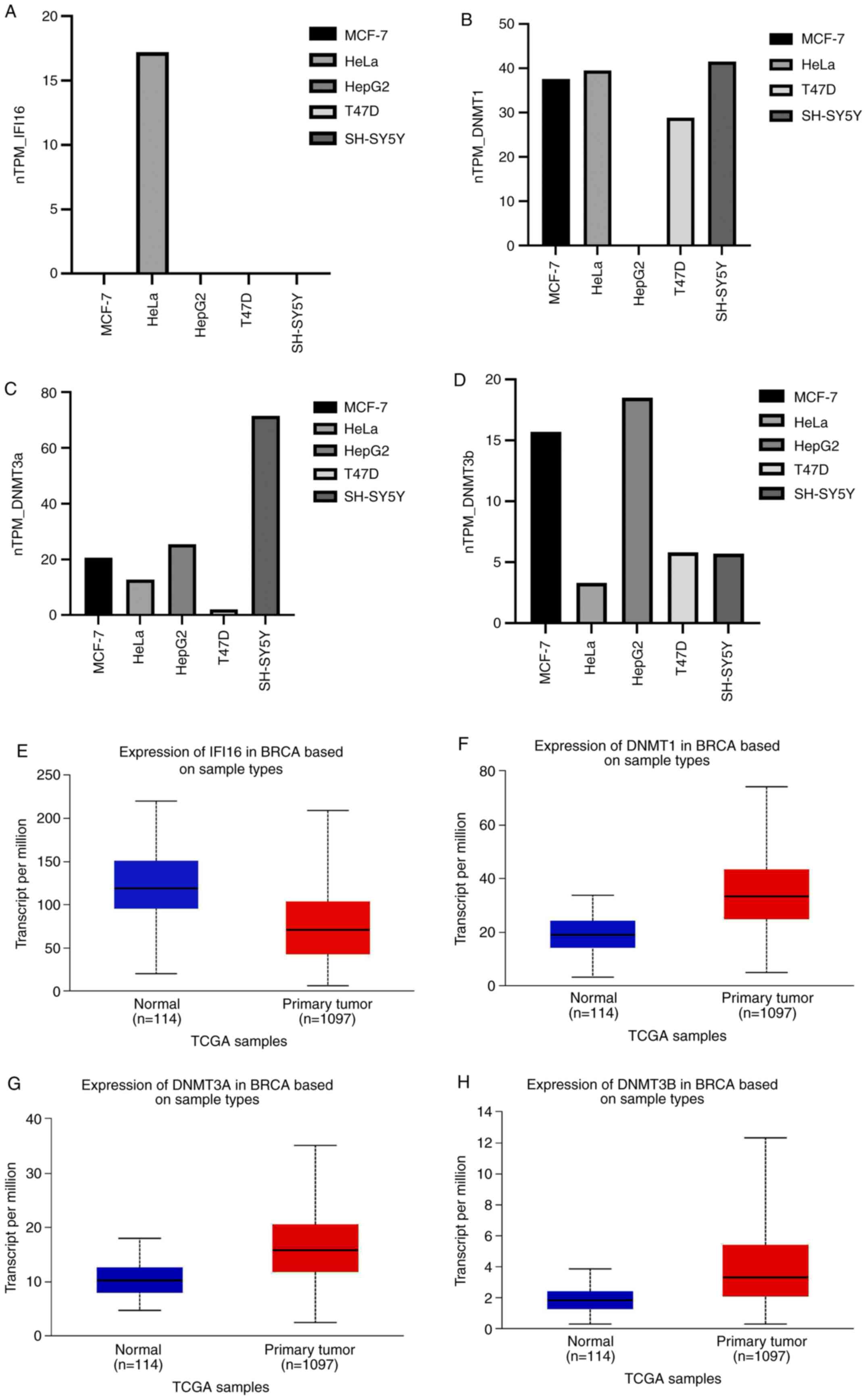
SHSY5, HepG2 cell lines but was observed in HeLa (Fig. 1A). DNMT mRNA expression was
inversely associated with IFI16 mRNA expression in most cell lines,
except HeLa (Fig. 1A-D). Moreover,
TGCA omics data showed that the breast cancer (BRCA) has a
significantly lower expression of IFI16 (Fig. 1E) but higher expression of DNMT1
(Fig. 1F), DNMT3 (Fig. 1G) and DNMT3B (Fig. 1H). An inverse correlation between
expression of IFI16 and DNMTs was observed in the breast cancer
cell line however this was not statistically significant (Fig. S1A). The immune responsiveness
study of IFI16 showed a moderate correlation with different immune
infiltrating cells such as CD8+/4+ T cells, macrophages, dendritic
cells and neutrophils (Fig. S1B).
Hence, the screening results indicated IFI16 had impact on immune
infiltrating cells.
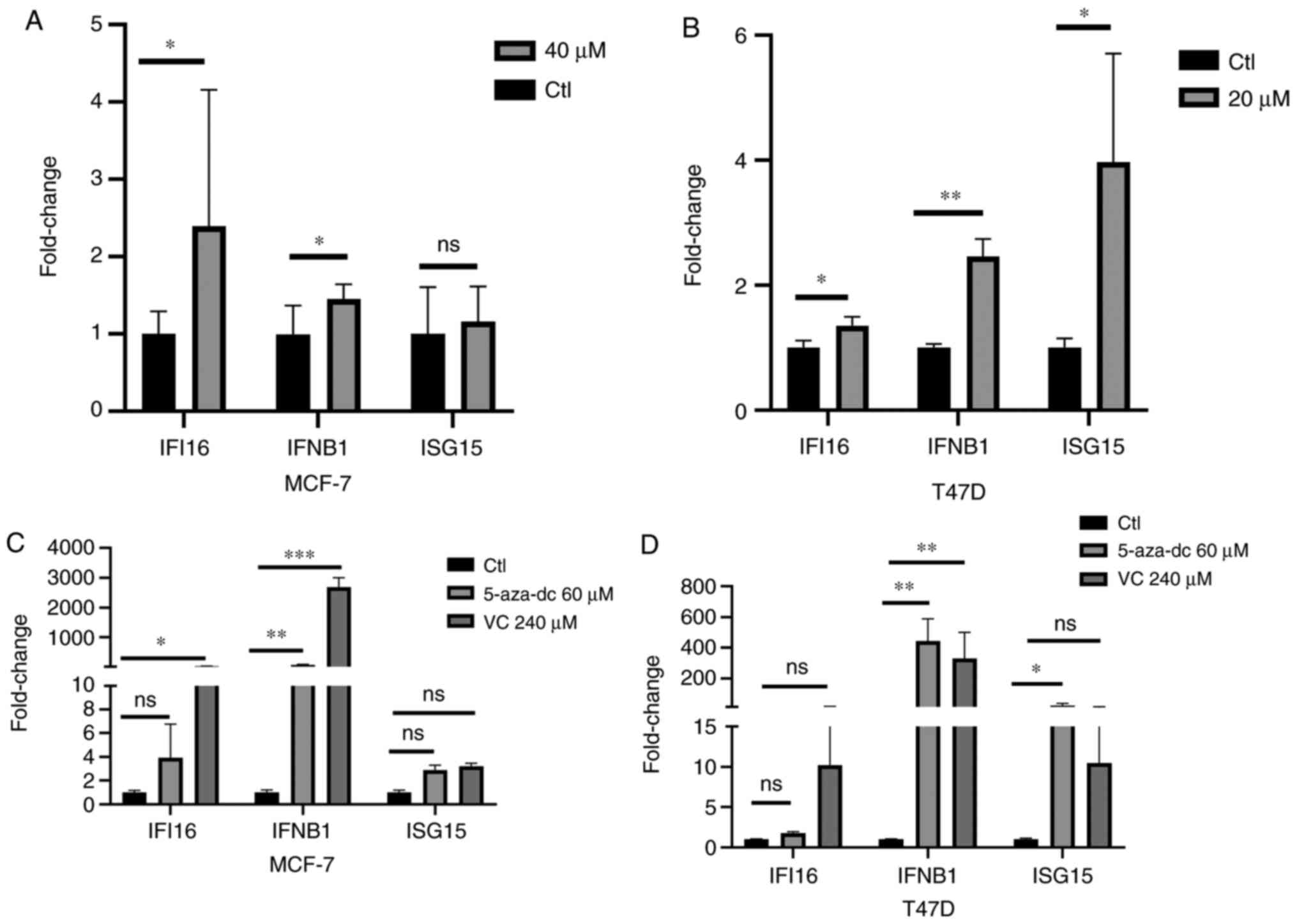
DNMTi analog EGCG induces IFI16 and
IFN-associated gene expression in breast cancer cell lines
The extracted RNAs from EGCG-treated cell lines were
used to investigate IFI16 expression. The results showed that 40
µM EGCG induced expression of IFI16 in the MCF-7 cell line
and 20 µM EGCG treatment induced expression in the T47D cell
line (Fig. 2A and B). As IFI16
acts as an upstream target for transcribing IFNβ1 and ISG15
(43), expression of IFNβ1 and
ISG15 was assessed in both cell lines. In MCF-7 cells, 40 µM
treatment induced IFNβ1 expression but did not affect ISG15 gene
expression (Fig. 2A). By contrast,
20 µM EGCG induced IFNβ1 and ISG15 gene expression in the
T47D cell line (Fig. 2B). These
data suggested that EGCG treatment induced expression of IFI16 and
its downstream targeted genes expression.
Gene expression levels in both cell lines were also
assessed following treatment with conventional DNMTis, such as
5-aza-dc and vitamin C. IFI16, IFNβ1 and ISG15 gene expression in
MCF-7 cell line was induced following treatment with 60 µM
5-aza-dc or 240 µM vitamin C (Fig. 2C) although the slight induction of
ISG15 gene expression results were not statistically significant.
Similar results were also observed in the T47D cell line in which
IFI16 gene expression was induced following treatment with 5-aza-dc
and vitamin C but this was not statistically significant (Fig. 2D). Moreover, both treatments
significantly induced the IFNβ1 gene expression in T47D cell line,
while ISG15 gene expression was significantly induced with 5-aza-dc
treatment (Fig. 2D). Hence, DNMTi
or vitamin C may induce the IFI16 gene and its downstream targeted
genes IFNβ1 and ISG15.
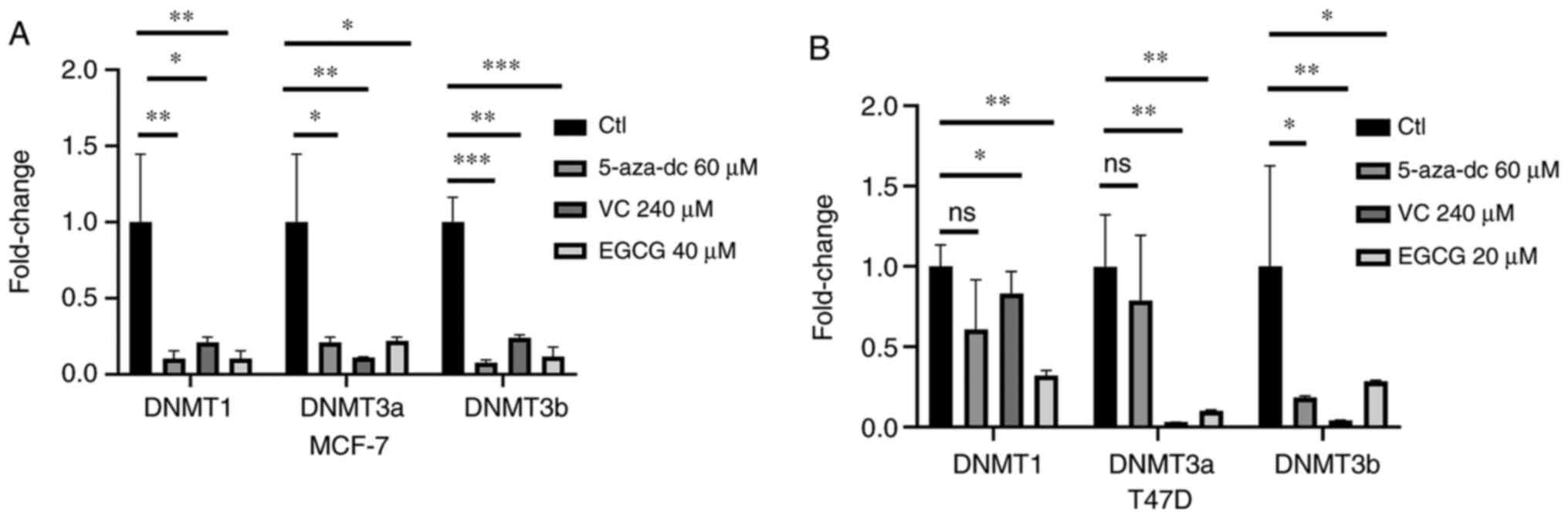
DNMTi analog EGCG decreases DNMT
expression in breast cancer cell lines
5-aza-dc and vitamin C are potent DNMTis that modify
the epigenome (31,44). Hence, DNMT gene expression was
assessed following treatment with EGCG, 5-aza-dc or vitamin C in
MCF-7 and T47D cell lines. DNMT1, DNMT3a and DNMT3b gene expression
levels were notably decreased following treatment with 60 µM
5-aza-dc or 240 µM vitamin C in both cell lines (Fig. 3A and B). Additionally, 40 and 20
µM EGCG treatment decreased expression of all DNMTs in both
MCF-7 and T47D cell lines (Fig. 3A and
B). These data indicated that DNMTi, vitamin C and EGCG may
affect DNMT gene expression.
Receptor grid box generation and
molecular docking simulation: In silico interaction of EGCG with
DNMT
Molecular docking-based computational techniques
have been considered as a mechanistic tool (45) for in silico drug design. For
DNMT1_A, grid box comprised X=205.34, Y=88.76 and Z=145.45 points
and spaced dimension was centered on DNMT1_A protein at X=−23.06,
Y=47.81 and Z=−23.78. The binding energy of SAM, SAH, vitamin C,
EGCG and 5-aza-dc with DNMT1_A was −8.5, −7.6, −5.8, −10.4 and −6.9
kJ/mol, respectively (Fig. 4). For
DNMT1_B, grid box comprised X=120.97, Y=89.06 and Z=95.42 points
and spaced dimension was centered on the DNMT1_B protein at
X=17.63, Y=21.36 and Z=−51.18. The binding energy of SAM, SAH,
vitamin C, EGCG and 5-aza-dc with DNMT1_B was −7.7, −7.4, −6.4,
−10.0 and −7.1 kJ/mol, respectively (Fig. 4). For DNMT3a, grid box comprised of
X=83.85, Y=87.88 and Z=60.33 points and spaced dimension was
centered on X=−20.31, Y=−56.03 and Z=20.19. The binding energy of
SAM, SAH, vitamin C, EGCG and 5-aza-dc with DNMT3a was −7.2, −6.6,
−5.8, −9.5 and −7.2 kJ/mol, respectively (Fig. 4). For DNMT3b, the grid box
comprised X=83.85, Y=87.88 and Z=60.33 points and spaced dimension
was centered on the DNMT3b protein at X=−20.31, Y=−9.01 and
Z=−3.88. The binding energy of SAM, SAH, vitamin C, EGCG and
5-aza-dc with DNMT3b was −8.7, −8.4, −5.8, −9.6 and −7.3 kJ/mol,
respectively (Fig. 4).
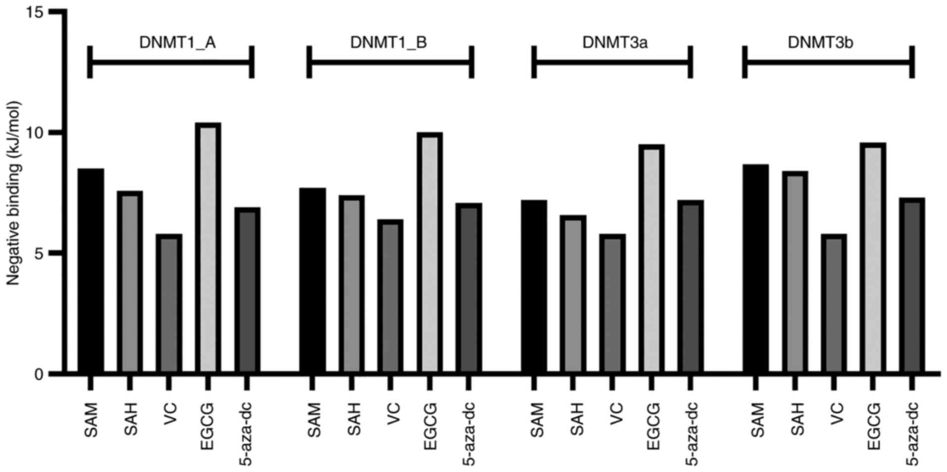 | Figure 4.In silico docking simulation
of different DNMTs with SAM, SAH, 5-aza-dc, VC and EGCG. Docking
score as binding energy of all compounds with DNMT1_A chain,
DNMT1_B chain, DNMT3a and DNMT3b. DNMT, DNA methyltransferase;
5-aza-dc, 5-Azacytadine; VC, vitamin C; EGCG, Epigallocatechin
gallate; SAM, S-adenosyl methionine; SAH, S-adenosyl
homocysteine. |
In silico molecular docking simulation
predicted that SAM and EGCG compounds bind in similar positions.
SAM was bound at Glu:1168, Phe:1145, Glu:1266, Arg:1310, Thr:1526,
Thr:1528 and Asn:1578 residue (Fig.
S2), whereas EGCG bound at Met:1169, Pro:1225, Glu:1168,
Glu:1266 and Arg:1310 residue (Fig.
S2). Though more conventional H bond interactions were found in
the case of SAM, other molecular bonding interactions, such as
π-anion and π-alkyl, in addition to four conventional H bonds, were
also observed in the EGCG-DNMT1_A complex (Fig. S2). Furthermore, 5-aza-dc was found
to interact with DNMT1_A by interacting with Glu:562, Glu:566,
Asp:565, Ser:570 and Gln:687 residue with 3 conventional H bonds
(Fig. S2). Vitamin C bound at
Gln:369, Cys:409, Ser:436 and Glu:494 residue with 4 conventional H
bonds (Fig. S2). SAH bound at
Glu:562, ASP:565, Glu:566, Pro:574 and Arg:690 with conventional H
bond and alkyl interactions (Fig.
S2). The predicted site of SAM and EGCG interaction is
presented in Fig. 5A.
 | Figure 5.In silico interaction of EGCG,
SAM, SAH, 5-aza-dc and vitamin C with DNMTs. (A) Predicted binding
sites for SAM (yellow) and EGCG (green) in DNMT1_A chain. (B)
Predicted binding sites for 5-aza-dc (orange), SAM (blue) and EGCG
(green) in DNMT1_B chain. (C) Predicted binding sites for SAM
(blue), SAH (pink), EGCG (green) and vitamin C (white) in DNMT3a.
(D) Predicted binding sites for SAM (blue), SAH (pink), EGCG
(green) and 5-aza-dc (orange) in DNMT3b. DNMT, DNA
methyltransferase; 5-aza-dc, 5-Azacytadine; VC, vitamin C; EGCG,
Epigallocatechin gallate; SAM, S-adenosyl methionine; SAH,
S-adenosyl homocysteine. |
For DNMT1_B, 5-aza-dc interacted with Phe:1145,
Ser:1146, Leu:1151 and Asn:1578 with four conventional H bonds and
three C-H bonds. SAM interacted with Phe:1145, Glu:1168, Glu:1266,
Arg:1310, Gln:1536, Arg:1574 and Asn:1578 with eight conventional H
bonds. EGCG interacted with DNMT1_B at Ser:1146, Glu:1168,
Pro:1224/1225, Glu:1266 and Ala:1579 with four conventional H
bonds, and one each of π-anion, π-alkyl and C-H bonds (Fig. S3). SAH and vitamin C interacted
with DNMT1_B at Glu:562, Asp:565, Ser:570, Asp:571, Glu:572,
Gln:594, Arg:595 Val:658 and Met:1232, Arg:1276, Ser:1277, Val:1344
residues, respectively. Nine conventional H and one alkyl bond were
found in the case of SAH, while vitamin C exhibited four
conventional H bonds (Fig. S3).
The predicted site of 5-aza-dc, SAM and EGCG interaction is
presented in Fig. 5B.
Docking study of DNMT3a showed that SAM, SAH, EGCG
and vitamin C interacted with DNMT3a as follows: SAM, Gly:707,
Cys:710, Arg:792, Arg:891 with four conventional H bonds; SAH,
Phe:640, Pro:709, Glu:765, Arg:891 with four conventional H bonds
and one each π-sulfur and alkyl bond; EGCG, Phe:640, Gly:642,
Val:665, Ser:663, Gly:685, Asp:686, Gly:707, Pro:709, Asn:711,
Arg:891 with five H, one C-H, one π-σ, one π-alkyl, one π-cation,
two π-π T-shaped bond and vitamin C, Phe:640, Pro:709, Glu:765 and
Arg:891 (Fig. S4). 5-aza-dc
interacted with Phe:640, Gly:707, Glu:765, Arg:891, Trp:893
residues with five H and one π-σ bond (Fig. S4). The predicted site of SAM SAH,
EGCG and vitamin C interaction is presented in Fig. 5C.
Docking study of DNMT3b showed that 5-aza-dc, SAM,
SAH and EGCG interacted with DNMT3b as follows: 5-aza-dc, Asp:582,
Ser:610, Arg:832, Trp:834 with two H and one π-σ bond; SAM,
Glu:605, Val:606/628, Ser:649, Glu:697 residues with eight H bonds
and one attractive charge interaction; SAH, Phe:581, Gly:583,
Thr:586, Glu:605, Cys:607, Asp:627, Val:628, Arg:832, Trp:834 with
seven H and one each C-H and alkyl bond and EGCG, Phe:581, Ser:610,
Val:606/628, Pro:650, Cys:651, Arg:832 with four H and one each
π-σ, π-sulfur, π-cation, π-alkyl and π-π T-shaped interactions
(Fig. S5). DNMT3b and vitamin C
interacted with Asp:582, Ile:584, Thr:586, Gly:648, Arg:832 with
five H and one π-donor H bond (Fig.
S5). The predicted site of SAM SAH, 5-aza-dc and EGCG
interaction is presented in Fig.
5D.
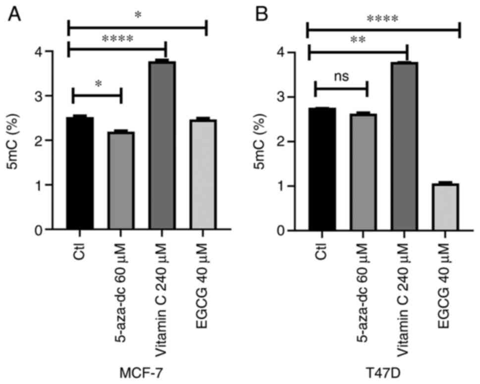
EGCG, but not vitamin C, decreases 5mC
level
As EGCG is a potent DNMTi (46), its effect on 5mC level in both cell
lines. In both cell lines, 60 µM 5-aza-dc decreased 5mC level but
this was only significant in MCF-7 cells (Fig. 6A and B). Additionally, 40 µM EGCG
significantly decreased 5mC level in MCF-7 cell line (Fig. 6A). Furthermore, 20 µM EGCG induced
a greater 5mC decrease in the T47D cell line (Fig. 6B). Vitamin C treatment
significantly increased 5mc level in both cell lines (Fig. 6A and B). These data suggested that
EGCG decreased 5mC level in breast cancer cell lines.
EGCG induces IFI16 gene expression by
decreasing DNA methylation
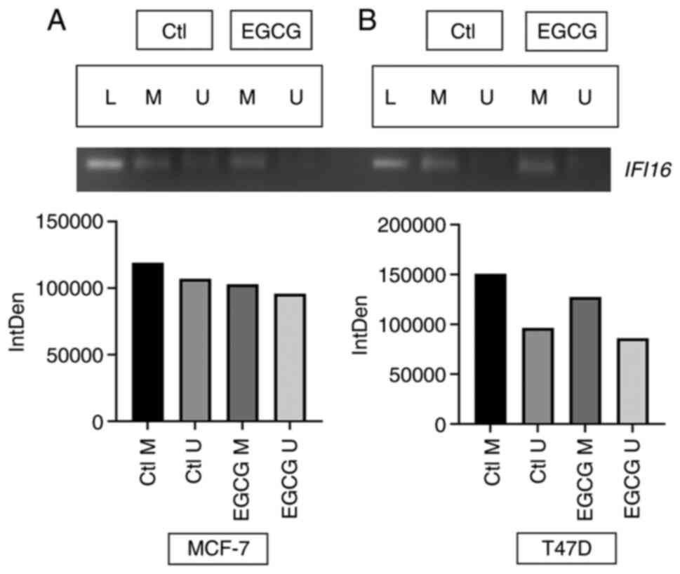
Since EGCG decreased 5mC level in both cell lines,
promoter methylation status was assessed in IFI16 promoter. EGCG
treatment decreased methylation signaling in the promoter of the
IFI16 gene in MCF-7 cell line (Fig.
7A). The decrease in methylation signaling in IFI16 was greater
in the T47D cell line than the MCF-7 cell line (Fig. 7B). The present data suggesting that
EGCG decreases methylation level in the IFI16 gene promoter.
Discussion
Nucleic acid sensors, such as IFI16 DNA sensor,
generate an innate immune response in tumor cells (47). IFI16 senses ds nucleic acid ligands
and relays molecular signaling to induce inflammatory innate immune
response via IFN stimulation. Previous epigenetic studies of these
sensors in various cancer cell lines have shown that DNMTi and
vitamin C induce their expression (31,38,48).
EGCG-mediated epi-transcriptomic based regulation of IFI16 sensor
is still not well studied. Hence, the aim of the present study was
to investigate this in breast cancer lines.
IFI16 gene expression was screened in cancer cell
lines, including MCF-7, T47D, SHSY5, HepG2 and HeLa, from the Human
Protein Atlas. The present study aimed to investigate epigenetic
mechanisms, such as DNA methylation-mediated epigenetic
transcriptional regulation of IFI16 gene; therefore, DNMT mRNA
expression was assessed in cancer cell lines. The TCGA dataset for
IFI16 and DNMT genes was validated in breast cancer cell line from
online data sources (UALCAN and UCSC Xena browser). The present
study found a negative association between IFI16 and DNMT gene
expression. Hence, MCF-7 and T47D cell lines were treated with
DNMTis, like EGCG, 5-aza-dc and vitamin C and expression of IFI16
and DNMTs was assessed. EGCG treatment induced IFI16 gene
expression in both cell lines and this was greater in the MCF-7
cell line. As IFI16 stimulates IFN and ISG response, IFNβ1 and
ISG15 expression was assessed; EGCG upregulated IFNβ1 gene
expression in both cell lines and this was greatest in MCF-7 cell
line. To the best of our knowledge, the effect of EGCG on
expression of these genes has not been reported previously.
Expression of these genes was measured following treatment with 60
µM 5-aza-dc or 240 µM vitamin C, which resulted in significant
upregulation of mRNA expression for these genes. Previously, it has
been shown that IFI16 and ISG15 gene expression is upregulated
following treatment with vitamin C and 5-aza-dc alone or
combination in HCT116, SNU398 and HL60 cancer cell lines (31). Additionally, 5-aza-dc treatment
induces IFI16, ISG15 and IFNβ1 expression in ovarian cancer cell
lines (48).
In addition, 5-aza-dc, vitamin C and EGCG treatment
decreased DNMT1, DNMT3a and DNMT3b mRNA expression. A previous
study showed that 20 µM EGCG and 5µM 5-aza-dc treatment decrease
DNMTs mRNA expression and DNMT activity and reactivates tumor
suppressor genes by decreasing promoter methylation levels in
various breast cancer lines (49).
As 5-aza-dc, vitamin C and EGCG decreased DNMTs mRNA
expression, 5mC levels were assessed post-treatment. In both MCF-7
and T47D cell lines, 5-aza-dc slightly decreased 5mC% level.
Moreover, EGCG decreased 5mC levels in MCF-7 cells and a
significant decrease in 5mC level in T47D cells was observed. A
previous study on skin cancer cells revealed EGCG mediated
declination of 5mC level and suggested that EGCG-mediated
methylation is slow and might be more effective as a treatment when
used for long periods (>72 h) (50). In the present study, no effect of
vitamin C on the 5mC level was observed in any cell line. Binding
of vitamin C outside of the binding pocket for DNMT1 and DNMT3b
might be the reasons or any other factors that might be involved in
the regulation of vitamin C-mediated 5mC regulation. A previous
study showed the effect of vitamin C on methylation is reversible
(51). DNMT3A activity regulation
is governed by 5mC-binding protein MeCP2 and histone 3 tail
modification (52). Ubiquitin-like
With PHD and Ring Finger Domains 1 can regulate DNMT1 (53). Vitamin C may not functionally
affect all these factors.
In silico molecular docking simulation showed
that EGCG exhibited the greatest binding energy with DNMT proteins.
5-aza-dc showed the second highest binding energy with DNMTs. SAM
and SAH were used as a control for docking simulation. The lowest
binding energy was observed with vitamin C for all DNMTs.
Additionally, docking interaction and predicted binding sites for
all compounds showed that SAM and EGCG bound in a similar position
across all DNMTs. Binding pocket was not specified during docking
simulation to avoid bias docking; docking simulation demonstrated
that compounds bind with certain amino acid residues in the binding
pocket site. Similar amino acid residues interactions were also
found in several previous molecular docking simulations of EGCG and
DNMT (27,54,55).
As EGCG mediated a decrease in 5mC level and
induction of IFI16 gene expression, the effect of EGCG on status of
IFI16 gene promoter was assessed. Following EGCG treatment,
methylation signaling was decreased in both cell lines. MCF-7 cell
lines showed low decrease in methylation signaling following EGCG
treatment, whereas T47D showed a greater decrease in methylation
signaling following EGCG treatment. This may be due to low decrease
in 5mC level in EGCG-treated MCF-7 cell line and greater 5mC
decrease in EGCG-treated T47D cell line. However, a recent study
revealed that IFI16 is hypomethylated in glioblastoma (56). To the best of our knowledge,
EGCG-mediated reactivation of the IFI16 gene by decreasing promoter
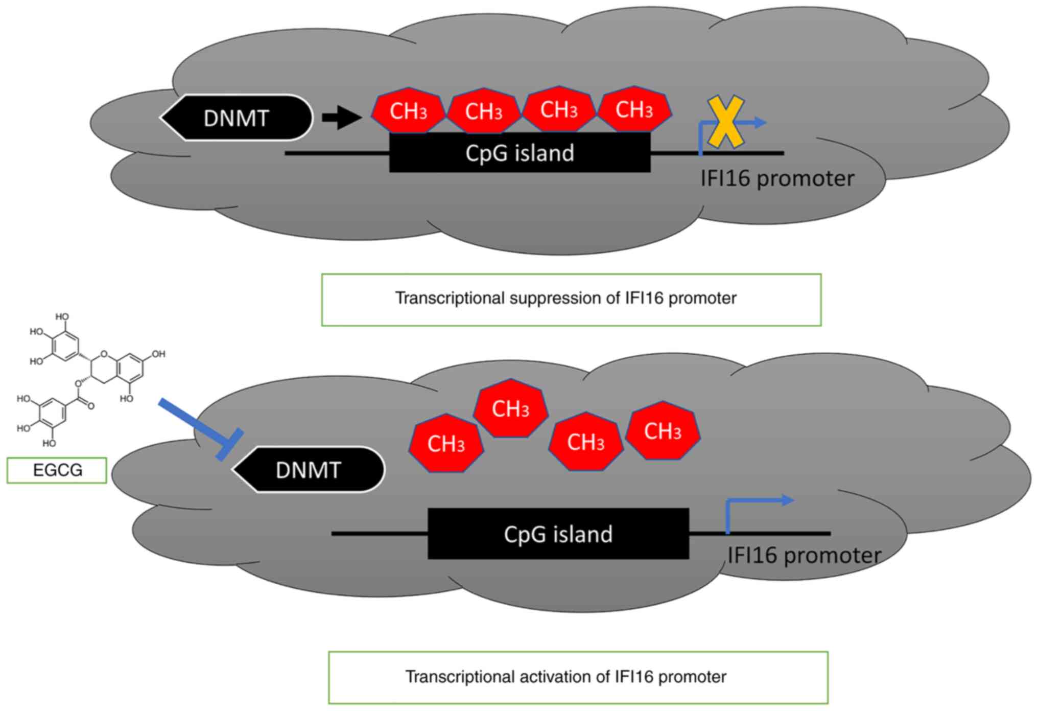
methylation has not previously been reported. IFI16 gene promoter
methylation is DNMT-mediated, whereas EGCG treatment can block the
DNMTs activity and reduce the methylation of IFI16 gene promoter,
thus activating transcription of IFI16 gene (Fig. 8).
As a potent natural DNMTi, EGCG induces expression
of innate immune sensor IFI16 by decreasing promoter methylation in
breast cancer cells. The present findings provide a basis to
investigate whether natural DNMTis (such as EGCG) exert beneficial
effects by inducing immune response.
Supplementary Material
Supporting Data
Acknowledgements
Not applicable.
Funding
The present study was supported by the Deanship Scientific
Research at King Abdulaziz University, Jeddah (grant no.
G:257-130-1441).
Availability of data and materials
The datasets used and/or analyzed during the current
study are available from the corresponding author on reasonable
request.
Authors' contributions
MIK designed and conceptualized the study. SMN and
WHA performed experiments, analyzed the data and wrote the
manuscript. MIK and WHA confirm the authenticity of all the raw
data. All authors have read and approved the final manuscript.
Ethics approval and consent to
participate
Not applicable.
Patient consent for publication
Not applicable.
Competing interests
The authors declare that they have no competing
interests.
References
|
1
|
Choubey D and Panchanathan R: IFI16, an
amplifier of DNA-damage response: Role in cellular senescence and
aging-associated inflammatory diseases. Ageing Res Rev. 28:27–36.
2016. View Article : Google Scholar : PubMed/NCBI
|
|
2
|
Ouchi M and Ouchi T: Role of IFI16 in DNA
damage and checkpoint. Front Biosci. 13:236–239. 2008. View Article : Google Scholar
|
|
3
|
Duan X, Ponomareva L, Veeranki S,
Panchanathan R, Dickerson E and Choubey D: Differential roles for
the interferon-inducible IFI16 and AIM2 innate immune sensors for
cytosolic DNA in cellular senescence of human fibroblasts. Mol
Cancer Res. 9:589–602. 2011. View Article : Google Scholar : PubMed/NCBI
|
|
4
|
Stratmann SA, Morrone SR, van Oijen AM and
Sohn J: The innate immune sensor IFI16 recognizes foreign DNA in
the nucleus by scanning along the duplex. ELife. 4:e117212015.
View Article : Google Scholar : PubMed/NCBI
|
|
5
|
Unterholzner L, Keating SE, Baran M, Horan
KA, Jensen SB, Sharma S, Sirois CM, Jin T, Latz E, Xiao TS, et al:
IFI16 is an innate immune sensor for intracellular DNA. Nat
Immunol. 11:997–1004. 2010. View
Article : Google Scholar
|
|
6
|
Orzalli MH, DeLuca NA and Knipe DM:
Nuclear IFI16 induction of IRF-3 signaling during herpesviral
infection and degradation of IFI16 by the viral ICP0 protein. Proc
Natl Acad Sci USA. 109:E3008–E3017. 2012. View Article : Google Scholar : PubMed/NCBI
|
|
7
|
Choubey D, Deka R and Ho SM:
Interferon-inducible IFI16 protein in human cancers and autoimmune
diseases. Front Biosci. 13:598–608. 2008. View Article : Google Scholar
|
|
8
|
Johnstone RW and Trapani JA: Transcription
and growth regulatory functions of the HIN-200 family of proteins.
Mol Cell Biol. 19:5833–5838. 1999. View Article : Google Scholar
|
|
9
|
Ludlow LE, Johnstone RW and Clarke CJ: The
HIN-200 family: More than interferon-inducible genes? Exp Cell Res.
308:1–17. 2005. View Article : Google Scholar : PubMed/NCBI
|
|
10
|
Choubey D, Duan X, Dickerson E, Ponomareva
L, Panchanathan R, Shen H and Srivastava R: Interferon-inducible
p200-family proteins as novel sensors of cytoplasmic DNA: Role in
inflammation and autoimmunity. J Interferon Cytokine Res.
30:371–380. 2010. View Article : Google Scholar : PubMed/NCBI
|
|
11
|
Veeranki S and Choubey D:
Interferon-inducible p200-family protein IFI16, an innate immune
sensor for cytosolic and nuclear double-stranded DNA: Regulation of
subcellular localization. Mol Immunol. 49:567–571. 2012. View Article : Google Scholar
|
|
12
|
Decout A, Katz JD, Venkatraman S and
Ablasser A: The cGAS-STING pathway as a therapeutic target in
inflammatory diseases. Nat Rev Immunol. 21:548–569. 2021.
View Article : Google Scholar
|
|
13
|
Bhat N and Fitzgerald KA: Recognition of
cytosolic DNA by cGAS and other STING-dependent sensors. Eur J
Immunol. 44:634–640. 2014. View Article : Google Scholar
|
|
14
|
Thompson MR, Sharma S, Atianand M, Jensen
SB, Carpenter S, Knipe DM, Fitzgerald KA and Kurt-Jones EA:
Interferon γ-inducible Protein (IFI) 16 transcriptionally regulates
Type I interferons and other interferon-stimulated genes and
controls the interferon response to both DNA and RNA viruses. J
Biol Chem. 289:235682014. View Article : Google Scholar : PubMed/NCBI
|
|
15
|
Kopitar-Jerala N: The role of interferons
in inflammation and inflammasome activation. Front Immunol.
8:8732017. View Article : Google Scholar
|
|
16
|
Itsui Y, Sakamoto N, Kurosaki M, Kanazawa
N, Tanabe Y, Koyama T, Takeda Y, Nakagawa M, Kakinuma S and Sekine
Y: Expressional screening of interferon-stimulated genes for
antiviral activity against hepatitis C virus replication. J Viral
Hepat. 13:690–700. 2006. View Article : Google Scholar
|
|
17
|
Jiang D, Guo H, Xu C, Chang J, Gu B, Wang
L, Block TM and Guo JT: Identification of three
interferon-inducible cellular enzymes that inhibit the replication
of hepatitis C virus. J Virol. 82:1665–1678. 2008. View Article : Google Scholar
|
|
18
|
Schoggins JW and Rice CM:
Interferon-stimulated genes and their antiviral effector functions.
Curr Opin Virol. 1:5192011. View Article : Google Scholar
|
|
19
|
Xin H, Curry J, Johnstone RW, Nickoloff BJ
and Choubey D: Role of IFI 16, a member of the interferon-inducible
p200-protein family, in prostate epithelial cellular senescence.
Oncogene. 22:4831–4840. 2003. View Article : Google Scholar : PubMed/NCBI
|
|
20
|
Duan X, Ponomareva L, Veeranki S and
Choubey D: IFI16 induction by glucose restriction in human
fibroblasts contributes to autophagy through activation of the
ATM/AMPK/p53 pathway. PLoS One. 6:e1953220122 View Article : Google Scholar : PubMed/NCBI
|
|
21
|
Lin W, Zhao Z, Ni Z, Zhao Y, Du W and Chen
S: IFI16 restoration in hepatocellular carcinoma induces tumour
inhibition via activation of p53 signals and inflammasome. Cell
Prolif. 50:e123922017. View Article : Google Scholar
|
|
22
|
Kondo Y, Nagai K, Nakahata S, Saito Y,
Ichikawa T, Suekane A, Taki T, Iwakawa R, Enari M, Taniwaki M, et
al: Overexpression of the DNA sensor proteins, absent in melanoma 2
and interferon-inducible 16, contributes to tumorigenesis of oral
squamous cell carcinoma with p53 inactivation. Cancer Sci.
103:782–790. 2012. View Article : Google Scholar
|
|
23
|
Yu B, Zheng X, Sun Z, Cao P, Zhang J and
Wang W: IFI16 can be used as a biomarker for diagnosis of renal
cell carcinoma and prediction of patient survival. Front Genet.
12:5999522012. View Article : Google Scholar
|
|
24
|
Chen JX, Cheng CS, Gao HF, Chen ZJ, Lv LL,
Xu JY, Shen XH, Xie J and Zheng L: Overexpression of
interferon-inducible Protein 16 promotes progression of human
pancreatic adenocarcinoma through interleukin-1β-induced
tumor-associated macrophage infiltration in the tumor
microenvironment. Front Cell Dev Biol. 9:6407862021. View Article : Google Scholar
|
|
25
|
Intra J and Kuo SM: Physiological levels
of tea catechins increase cellular lipid antioxidant activity of
vitamin C and vitamin E in human intestinal Caco-2 cells. Chem Biol
Interact. 169:91–99. 2007. View Article : Google Scholar
|
|
26
|
Cione E, La Torre C, Cannataro R, Caroleo
MC, Plastina P and Gallelli L: Quercetin, epigallocatechin gallate,
curcumin, and resveratrol: From dietary sources to human MicroRNA
modulation. Molecules. 25:632019. View Article : Google Scholar
|
|
27
|
Fang MZ, Wang Y, Ai N, Hou Z, Sun Y, Lu H,
Welsh W and Yang CS: Tea polyphenol (−)-epigallocatechin-3-gallate
inhibits DNA methyltransferase and reactivates methylation-silenced
genes in cancer cell lines. Cancer Res. 63:7563–7570.
2003.PubMed/NCBI
|
|
28
|
Beetch M, Harandi-Zadeh S, Shen K, Lubecka
K, Kitts DD, O'Hagan HM and Stefanska B: Dietary antioxidants
remodel DNA methylation patterns in chronic disease. Br J
Pharmacol. 177:1382–1408. 2020. View Article : Google Scholar
|
|
29
|
Kuo CL, Chen TS, Liou SY and Hsieh CC:
Immunomodulatory effects of EGCG fraction of green tea extract in
innate and adaptive immunity via T regulatory cells in murine
model. Immunopharmacol Immunotoxicol. 36:364–370. 2014. View Article : Google Scholar
|
|
30
|
Nance Cl, Mata M, McMullen A, McMullen A
and Shearer WT: Regulation of innate immune recognition of viral
infection by epigallocatechin gallate. J Allergy Clin Immunol.
133:AB2462014. View Article : Google Scholar
|
|
31
|
Liu M, Ohtani H, Zhou W, Ørskov AD,
Charlet J, Zhang YW, Shen H, Baylin SB, Liang G, Grønbæk K and
Jones PA: Vitamin C increases viral mimicry induced by
5-aza-2′-deoxycytidine. Proc Natl Acad Sci USA. 113:10238–10244.
2016. View Article : Google Scholar : PubMed/NCBI
|
|
32
|
Li T, Diner BA, Chen J and Cristea IM:
Acetylation modulates cellular distribution and DNA sensing ability
of interferon-inducible protein IFI16. Proc Natl Acad Sci.
109:10558–10563. 2012. View Article : Google Scholar
|
|
33
|
Alimirah F, Chen J, Davis FJ and Choubey
D: IFI16 in human prostate cancer. Mol Cancer Res. 5:251–259. 2007.
View Article : Google Scholar : PubMed/NCBI
|
|
34
|
Takeshima H, Yoda Y, Wakabayashi M,
Hattori N, Yamashita S and Ushijima T: Low-dose DNA demethylating
therapy induces reprogramming of diverse cancer-related pathways at
the single-cell level. Clin Epigenetics. 12:1422020. View Article : Google Scholar : PubMed/NCBI
|
|
35
|
Morris J, Moseley VR, Cabang AB, Coleman
K, Wei W, Garrett-Mayer E and Wargovich MJ: Reduction in promotor
methylation utilizing EGCG (epigallocatechin-3-gallate) restores
RXRα expression in human colon cancer cells. Oncotarget.
7:35313–35326. 2016. View Article : Google Scholar
|
|
36
|
Huang CY, Han Z, Li X, Xie HH and Zhu SS:
Mechanism of EGCG promoting apoptosis of MCF-7 cell line in human
breast cancer. Oncol Lett. 14:3623–3627. 2017. View Article : Google Scholar
|
|
37
|
Moradzadeh M, Hosseini A, Erfanian S and
Rezaei H: Epigallocatechin-3-gallate promotes apoptosis in human
breast cancer T47D cells through down-regulation of PI3K/AKT and
Telomerase. Pharmacol Rep. 69:924–928. 2017. View Article : Google Scholar : PubMed/NCBI
|
|
38
|
Roulois D, Loo Yau H, Singhania R, Wang Y,
Danesh A, Shen SY, Han H, Liang G, Jones PA, Pugh TJ, et al:
DNA-demethylating agents target colorectal cancer cells by inducing
viral mimicry by endogenous transcripts. Cell. 162:961–973. 2015.
View Article : Google Scholar
|
|
39
|
Rialdi A, Campisi L, Zhao N, Lagda AC,
Pietzsch C, Ho JSY, Martinez-Gil L, Fenouil R, Chen X, Edwards M,
et al: Topoisomerase 1 inhibition suppresses inflammatory genes and
protects from death by inflammation. Science. 352:aad79932016.
View Article : Google Scholar : PubMed/NCBI
|
|
40
|
Livak KJ and Schmittgen TD: Analysis of
relative gene expression data using real-time quantitative PCR and
the 2(−Delta Delta C(T)) method. Methods. 25:402–408. 2001.
View Article : Google Scholar : PubMed/NCBI
|
|
41
|
Islam MR, Awal MA, Khames A, Abourehab
MAS, Samad A, Hassan WMI, Alam R, Osman OI, Nur SM, Molla MHR, et
al: Computational identification of druggable bioactive compounds
from catharanthus roseus and avicennia marina against colorectal
cancer by targeting thymidylate synthase. Mol. 27:20892022.
View Article : Google Scholar
|
|
42
|
Ikwu FA, Shallangwa GA, Mamza PA and
Uzairu A: In silico studies of piperazine derivatives as potent
anti-proliferative agents against PC-3 prostate cancer cell lines.
Heliyon. 6:e032732020. View Article : Google Scholar : PubMed/NCBI
|
|
43
|
Zevini A, Olagnier D and Hiscott J:
Crosstalk between cytoplasmic RIG-I and STING sensing pathways.
Trends Immunol. 38:194–205. 2017. View Article : Google Scholar
|
|
44
|
Nur SM, Rath S, Ahmad V, Ahmad A, Ateeq B
and Khan MI: Nutritive vitamins as epidrugs. Crit Rev Food Sci
Nutr. 61:1–13. 2021. View Article : Google Scholar : PubMed/NCBI
|
|
45
|
de Ruyck J, Brysbaert G, Blossey R and
Lensink MF: Molecular docking as a popular tool in drug design, an
in silico travel. Adv Appl Bioinform Chem. 9:1–11. 2016.PubMed/NCBI
|
|
46
|
Won JL, Shim JY and Zhu BT: Mechanisms for
the inhibition of DNA methyltransferases by tea catechins and
bioflavonoids. Mol Pharmacol. 68:1018–1030. 2005. View Article : Google Scholar
|
|
47
|
Okude H, Ori D and Kawai T: Signaling
through nucleic acid sensors and their roles in inflammatory
diseases. Front Immunol. 11:6258332021. View Article : Google Scholar
|
|
48
|
Chiappinelli KB, Strissel PL, Desrichard
A, Li H, Henke C, Akman B, Hein A, Rote NS, Cope LM, Snyder A, et
al: Erratum: Inhibiting DNA methylation causes an interferon
response in cancer via dsRNA including endogenous retroviruses.
Cell. 152:974–986. 2015. View Article : Google Scholar
|
|
49
|
Sheng J, Shi W, Guo H, Long W, Wang Y, Qi
J, Liu J and Xu Y: The inhibitory effect of
(−)-Epigallocatechin-3-Gallate on breast cancer progression via
reducing SCUBE2 methylation and DNMT activity. Molecules.
24:28992019. View Article : Google Scholar
|
|
50
|
Nandakumar V, Vaid M and Katiyar SK:
(−)-Epigallocatechin-3-gallate reactivates silenced tumor
suppressor genes, Cip1/p21 and p 16 INK4a, by reducing DNA
methylation and increasing histones acetylation in human skin
cancer cells. Carcinogenesis. 32:537–544. 2011. View Article : Google Scholar : PubMed/NCBI
|
|
51
|
Blaschke K, Ebata KT, Karimi MM,
Zepeda-Martínez JA, Goyal P, Mahapatra S, Tam A, Laird DJ, Hirst M,
Rao A, et al: Vitamin C induces Tet-dependent DNA demethylation and
a blastocyst-like state in ES cells. Nature. 500:222–226. 2013.
View Article : Google Scholar : PubMed/NCBI
|
|
52
|
Rajavelu A, Lungu C, Emperle M, Dukatz M,
Bröhm A, Broche J, Hanelt I, Parsa E, Schiffers S, Karnik R, et al:
Chromatin-dependent allosteric regulation of DNMT3A activity by
MeCP2. Nucleic Acids Res. 46:9044–9056. 2018. View Article : Google Scholar : PubMed/NCBI
|
|
53
|
Bostick M, Jong KK, Estève PO, Clark A,
Pradhan S and Jacobsen SE: UHRF1 plays a role in maintaining DNA
methylation in mammalian cells. Science. 317:1760–1764. 2007.
View Article : Google Scholar : PubMed/NCBI
|
|
54
|
Khan MA, Hussain A, Sundaram MK, Alalami
U, Gunasekera D, Ramesh L, Hamza A and Quraishi U:
(−)-Epigallocatechin-3-gallate reverses the expression of various
tumor-suppressor genes by inhibiting DNA methyltransferases and
histone deacetylases in human cervical cancer cells. Oncol Rep.
33:1976–1984. 2015. View Article : Google Scholar : PubMed/NCBI
|
|
55
|
Yiannakopoulou EC: Targeting DNA
methylation with green tea catechins. Pharmacology. 95:111–116.
2015. View Article : Google Scholar : PubMed/NCBI
|
|
56
|
Alivand MR, Najafi S, Esmaeili S,
Rahmanpour D, Zhaleh H and Rahmati Y: Integrative analysis of DNA
methylation and gene expression profiles to identify biomarkers of
glioblastoma. Cancer Genet. 258–259. 135–150. 2021.
|