Introduction
Lysophosphatidic acid (LPA) is a simple glycerol
phospholipid that serves an important role in numerous
physiological and pathophysiological processes, including the
stimulation of cell migration, tumor cell invasion, neurite
retraction, as well as proliferation stimulation of a variety of
normal and tumorigenic cells (1).
LPA is also increased in the plasma of patients with cervical
cancer (2). Elevated plasma LPA
levels represent a potential biomarker for certain types of
gynecological cancer (3,4). As a bioactive lipid medium, LPA is an
endogenous lysophospholipid with signaling properties outside of
the cell. Moreover, LPA signals via specific G-protein-coupled
receptors, known as LPA1-6 (5,6).
Cervical cancer is one of the most common types of
gynecological tumors (7). An
estimated 500,000 new cases of cervical cancer are diagnosed each
year worldwide, which leads to 280,000 cancer-related deaths
annually (8). Recurrence and
metastasis events are often associated with a poor prognosis and
the five-year survival rate of patients with locally advanced
cervical cancer is as high as 75–85% following treatment via
surgical resection, radiotherapy and chemotherapy (9).
Doxorubicin hydrochloride (DOX) induces the
apoptosis of cancer cells via numerous mechanisms and serves an
anticancer role as a first-line drug in cancer treatment (10–12).
However, the traditional DOX-based chemotherapy regimen has
received numerous negative evaluations, which includes the
development of drug resistance and the development of the
epithelial-mesenchymal transformation progression (13). DOX induces oxidative stress, which
is characterized by the accumulation of reactive oxygen species
(ROS) and a reduction in antioxidant defenses. Oxidative stress
injury is closely related to apoptosis, an important antitumor
mechanism, in which ROS are involved (14). Other studies have reported that ROS
have an important role in tumorigenesis. The high level of ROS in
cancer cells is the product of a high metabolism, which can
maintain cancer cell proliferation and also stimulates cell
invasion and metastasis (15,16).
The molecular mechanism of LPA on DOX-induced
apoptosis in cervical cancer cells remains unclear. The hypothesis
of the present study, is that as a bioactive lipid medium, LPA may
protect cervical cancer cells from apoptosis via inhibiting
caspase-3 expression and reducing oxidative stress injury in cancer
cells. Therefore, the aim of the present study was to investigate
this interaction to potentially identify a novel therapeutic target
and approach for the treatment of cervical cancer.
Materials and methods
Reagents
The human cervical cancer HeLa (cat. no. CL0134),
C33A (cat. no. CL0385) and SiHa (cat. no. CL0280) cell lines were
purchased from Hunan Fenghui Biotechnology Co., Ltd. RPMI-1640
culture medium was purchased from Zhejiang Senrui Biotechnology
Co., Ltd. FBS was purchased from Omnimabs. Penicillin-streptomycin
mixed solution, the ROS detection kit (cat. no. CA1410) and DOX
were purchased from Solarbio Science & Technology Co., Ltd. The
TRIzol® RNA isolation kit was purchased from Jiangsu
Cowin Biotech Co., Ltd. LPA was purchased from Sigma-Aldrich (Merck
KGaA). The reverse transcription (RT) kit and SYBR
Green® Real-Time PCR Master Mix were purchased from
Toyobo Life Science. Primers were synthesized by Shanghai Personal
Biotechnology Co., Ltd. The BCA kit was purchased from Beyotime
Institute of Biotechnology. The rabbit polyclonal anti-β-actin
primary antibody was purchased from Biosharp Life Sciences (cat.
no. BL005B). The rabbit polyclonal caspase-3 primary antibody (cat.
no. ab13847) and the donkey anti-rabbit IgG heavy chain and light
chain Alexa Fluor 680-conjugated secondary antibody (cat. no.
ab175772) were purchased from Abcam. The Annexin V-FITC Apoptosis
Detection Kit (cat. no. 556547) was purchased from Becton Dickinson
and Company. Superoxide dismutase (SOD; cat. no. A001-3) and
malondialdehyde (MDA; cat. no. A003-2) detection kits were
purchased from Nanjing Jiancheng Bioengineering Institute. ImageJ
software (V1.48) for densitometry reading of bands of the western
blot analysis was provided by the National Institutes of
Health.
Cell culture and treatment
HeLa, C33A and SiHa cells were cultured in RPMI-1640
medium containing 10% FBS and 1% penicillin-streptomycin in an
incubator with 5% CO2 at 37°C. Cells were inoculated
into 6-well plates, according to the required number of cells for
the experiment, at 37°C with 5% CO2 for 24 h. Cells were
subsequently incubated with LPA (10 µmol/l), DOX (4 µmol/l) or LPA
(10 µmol/l) + DOX (4 µmol/l) at 37°C with 5% CO2 for 24
h.
Cell morphology evaluation
To detect cell morphology changes of HeLa cells
after LPA and DOX treatment, HeLa cells (5×10⁵
cells/well) were treated at 37°C for 24 h with LPA (10 µmol/l), DOX
(4 µmol/l) or LPA (10 µmol/l) + DOX (4 µmol/l). Following
centrifugation at 200 × g for 10 min at room temperature, the
supernatant was discarded and the cells were washed once with PBS.
The morphology of HeLa cells in the different treatment groups was
observed and the cells were imaged using an optical microscope
(magnification ×100; BX53 and DP80 models; Olympus
Corporation).
Detection of apoptosis via
transmission electron microscopy
To detect ultrastructural morphology of HeLa cells
after LPA and DOX treatment by transmission electron microscope,
HeLa cells in each treatment group were washed with PBS, digested
into a single cell suspension using trypsin and centrifuged at 200
× g for 10 min at room temperature. The supernatant was discarded
following the termination of digestion. A total of 3×10⁶
cells were fixed using 3% glutaraldehyde for 30 min at room
temperature and the supernatant was discarded. Subsequently the
cells were incubated with a fresh fixation solution at 4°C for 24
h. The cells were then washed three times with precooled PBS and
incubated with 1% osmic acid for further fixation at 4°C for 2 h.
Following gradient dehydration, immersion and embedding, 70-nm
ultra-thin sections of the treated HeLa cells were produced. The
sections were collected using a copper net and were stained using
2% uranyl acetate for 30 min at room temperature and subsequently
lead citrate (0.665 g lead nitrate and 0.88 g sodium citrate was
dissolved in 15 ml distilled water. Next, 4 ml 1N sodium hydroxide
was added and a final volume of 25 ml was created with distilled
water) for 15 min at room temperature. After drying, images of the
cells were obtained using a transmission electron microscope
(magnification, ×2,000; HT7700 model; Hitachi. Ltd.).
RT-quantitative PCR (RT-qPCR)
To determine caspase-3 and caspase-8 at the mRNA
level as a result of apoptosis in HeLa cells treated with LPA and
DOX by qPCR, total cellular RNA was extracted from HeLa cells using
the TRIzol method according to the manufacturer's protocol.
Subsequently RNA was reverse transcribed into complementary DNA
using a RT kit according to the manufacturer's protocol and the
thermocycling conditions were as follows: 37°C for 15 min and 98°C
for 5 min. SYBR Green® Real-Time PCR Master Mix was used
for qPCR. The following qPCR primers were used: GAPDH forward (F),
5′-AGAAGGCTGGGGCTCATTTG-3′ and reverse (R),
5′-AGGGGCCATCCACAGTCTTC-3′; caspase-3 F,
5′-GTGGAGGCCGACTTCTTGTATGC-3′ and R, 5′-TGGCACAAAGCGACTGGATGAAC-3′;
and caspase-8 F, 5′-CGGATGAGGCTGACTTTCTGCTG-3′ and R,
5′-GGCTCTGGCAAAGTGACTGGATG-3′. The reaction system was prepared
according to the manufacturer's protocol. The thermocycling
conditions for qPCR were as follows: Initial denaturation at 95°C
for 30 sec; and 40 cycles of denaturation at 95°C for 5 sec,
annealing at 55°C for 10 sec and elongation at 72°C for 15 sec.
GAPDH was used as the internal reference gene and the relative mRNA
expression levels were determined using the 2−ΔΔCq
method (17).
Western blotting
To determine caspase-3 and cleaved caspase-3 at the
protein level as a result of apoptosis in HeLa cells treated with
LPA and DOX by western blotting, HeLa, C33A and SiHa cells were
collected following treatment. Total protein was extracted using
RIPA buffer purchased from Beijing Solarbio Science &
Technology Co., Ltd., (cat. no. R0020; RIPA:PMSF, 100:1) and the
protein concentration was determined using a BCA kit. Total protein
(20 µg per lane) was separated on a 12% gel using SDS-PAGE.
Separated proteins were electrically transferred onto a PVDF
membrane, which was then blocked using 5% skimmed milk at room
temperature for 1 h. Subsequently the membranes were incubated with
primary antibodies, rabbit polyclonal to caspase-3 (1:1,000; cat.
no. ab13847; Abcam) for 12 h at 4°C and the membranes were then
washed with TBS with 0.05% Tween-20 three times. Following the
primary incubation the membranes were incubated with the
HRP-conjugated secondary antibody, donkey anti-rabbit IgG H&L
Alexa Fluor® 680 (1:10,000 dilution; cat. no. ab175772;
Abcam) at room temperature for 1 h. Protein bands were visualized
using ECL reagent and images were acquired using the Amersham
Imager 600 (Cytiva).
Detection of apoptosis via flow
cytometry
Flow cytometry was used to detect apoptosis of HeLa
cells after treatment using Annexin V staining. Following treatment
with LPA (10 µmol/l), DOX (4 µmol/l) or LPA (10 µmol/l) + DOX (4
µmol/l), HeLa cells were collected, washed with precooled PBS and
centrifuged at 200 × g for 10 min at 4°C. Subsequently, the
supernatant was discarded and the cells were resuspended in 100 µl
1X buffer solution with 5 µl FITC-Annexin V and 5 µl PI. The cells
were incubated at room temperature for 15 min in the dark and
subsequently 400 µl 1X buffer solution was added. After passing the
cells through a 400-mesh filter, apoptotic cells were detected via
flow cytometry (Beckman Coulter, Inc.). The plotted cells in Q1, Q2
and Q4 of the graph were used to calculate the apoptosis rate.
Detection of active oxygen levels
To detect the level of ROS of HeLa cells treated
with LPA and DOX using Rosup detection kit, HeLa cells were
inoculated into 96-well plates at a density of 2×10⁴
cells/well and were treated with the different combinations of
drugs at 37°C for 24 h. Subsequently, a ROS detection kit was used
according to the manufacturer's protocol. The positive control cell
group was stimulated using the ROS positive control Rosup (1:1,000)
for 30 min at room temperature and the supernatant was discarded.
Rosup was provided as a component of the ROS detection kit. To each
group dichloro-dihydro-fluorescein diacetate (1:1,000) was added
and the cells were incubated at 37°C for 20 min. Then the cells
were washed with PBS three times. The fluorescence intensity was
directly measured without image capture using a multifunctional
spectrophotometer (SpectraMax M5; Molecular Devices, LLC) at an
excitation wavelength of 488 nm and an emission wavelength of 525
nm.
SOD detection
As SOD balances ROS by scavenging free radicals and
prevents cell damage caused by superoxide anion radicals (18), SOD levels in HeLa cells were
detected using a SOD detection kit according to the manufacturer's
instructions. The cells were inoculated at 5×10⁵
cells/ml in 6-well plates and were cultured at 37°C with 5%
CO2 for 24 h. Following centrifugation at 200 × g for 10
min at room temperature, the supernatant was discarded and the
cells were treated as the control group, or with LPA, DOX or LPA +
DOX for 24 h. The cells were digested with trypsin, collected and
centrifuged at 200 × g for 10 min at room temperature. The
supernatant was discarded and the cells were washed using 1 ml PBS.
Subsequently the cells were collected into 1.5 ml Eppendorf tubes.
Following centrifugation at 200 × g for 10 min at room temperature,
the supernatant was discarded and 300 µl 0.5% Triton X-100 lysis
buffer was added to each tube. The cells were incubated for 30 min
on ice and were then centrifuged at 1,200 × g for 8 min at 4°C. The
supernatant was collected and the cellular protein concentration
was detected using a BCA kit. The extracted protein was added to
distilled water, enzyme working solution and substrate application
solution according to the SOD kit manufacturer's protocol.
Following the reaction, the cells were incubated at 37°C for 20
min. SOD levels were quantified using an ELISA microplate reader to
detect the absorbance value at 450 nm. SOD activity was calculated
according to the formula provided by the manufacturer (SOD
detection kit; Nanjing Jiancheng Bioengineering Institute.
MDA detection
As MDA is a widely detected marker of oxidative
damage and its elevated level is the result of lipid peroxidation
(19), MDA levels in HeLa cells
were determined using the thiobarbituric acid method. Cells were
inoculated at a density of 5×10⁵ cells/ml in 6-well
plates and were cultured in an incubator at 37°C with 5%
CO2 for 24 h. Following centrifugation at 200 × g for 10
min at room temperature, the supernatant was discarded and the
cells were treated as the control group or with LPA, DOX or LPA +
DOX for 24 h at 37°C. Subsequently, cells were digested with
trypsin, collected and centrifuged at 200 × g for 10 min at room
temperature. The supernatant was discarded and the cells were
washed with 1 ml PBS prior to being collected in a 1.5 ml Eppendorf
tube. Following centrifugation at 200 × g for 5 min at 4°C, the
supernatant was discarded and 300 µl 0.5% Triton X-100 lysis buffer
was added to each tube. The cells were incubated for 30 min on ice
and then centrifuged at 1,200 × g for 8 min at 4°C. The supernatant
was collected and the cell protein concentration was determined
using a BCA kit. The extracted protein was used for the measurement
of MDA level using an MDA detection kit (cat. no. A003-2) according
to the manufacturer's protocol. The absorbance value of MDA was
determined using a UV spectrophotometer at 532 nm. MDA levels were
quantified according to the formula provided by the manufacturer
(MDA detection kit; Nanjing Jiancheng Bioengineering
Institute).
Statistical analysis
Data are presented as the mean ± SD. All data were
statistically analyzed using GraphPad Prism 5.0 (GraphPad Software,
Inc.). One-way ANOVA was used to statistically compare more than
two groups and Tukey's post hoc test was used to compare the
different groups following ANOVA. P<0.05 was considered to
indicate a statistically significant difference.
Results
LPA protects HeLa cells from
DOX-induced apoptosis
The morphology of HeLa cells in the different
treatment groups was determined using a light microscope. The
results demonstrated that there was no significant change in cell
morphology in the control and LPA-treated groups. However, cell
shrinkage and abscission in the DOX-treated group were observed and
the degree of cell abscission and shrinkage in the LPA + DOX
treated group was reduced compared with the DOX group (Fig. 1A). Using transmission electron
microscopy it was determined that the cell morphology and nuclear
condensation of the control and LPA groups remained unchanged.
However, in the DOX group, chromatin gathered at the edge of the
nuclear membrane and broken nuclear membrane and cells were
observed. Moreover, in the LPA + DOX group, nuclear chromatin
gathered at the edge of the nuclear membrane and an increased
vacuole was observed; however, the percentage of apoptotic cells
was less compared with the DOX group (Fig. 1B). Moreover, the results indicated
that LPA may have protected HeLa cells from DOX-induced apoptosis.
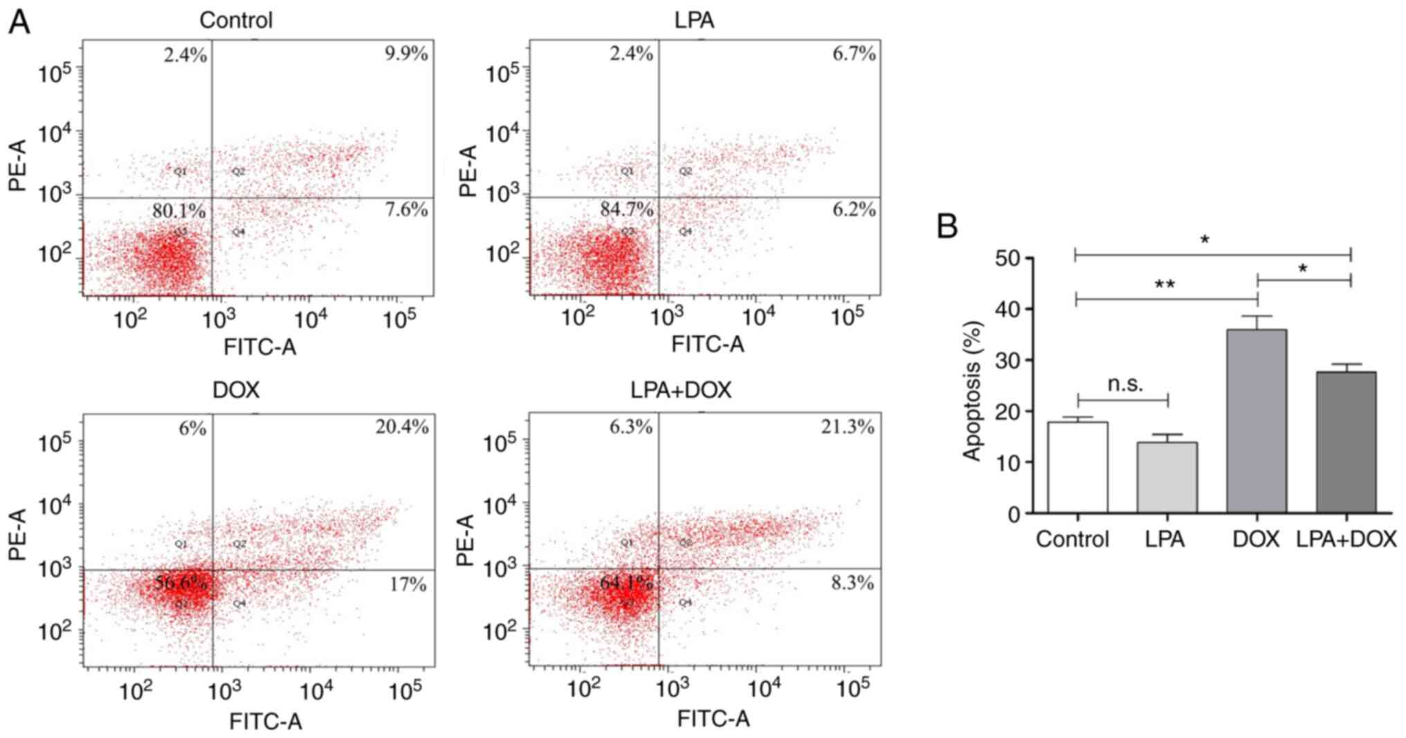
Subsequently, apoptosis was detected via flow cytometry. The
results demonstrated that there was no significant change in the
apoptotic rate in the control and LPA groups. However, the
apoptotic rate of the DOX group increased significantly, whereas
the apoptotic rate of the LPA + DOX group decreased by 23.24%
compared with the DOX group (P<0.05) (Fig. 2A and B). These results indicated
that LPA potentially protected HeLa cells from DOX-induced
apoptosis.
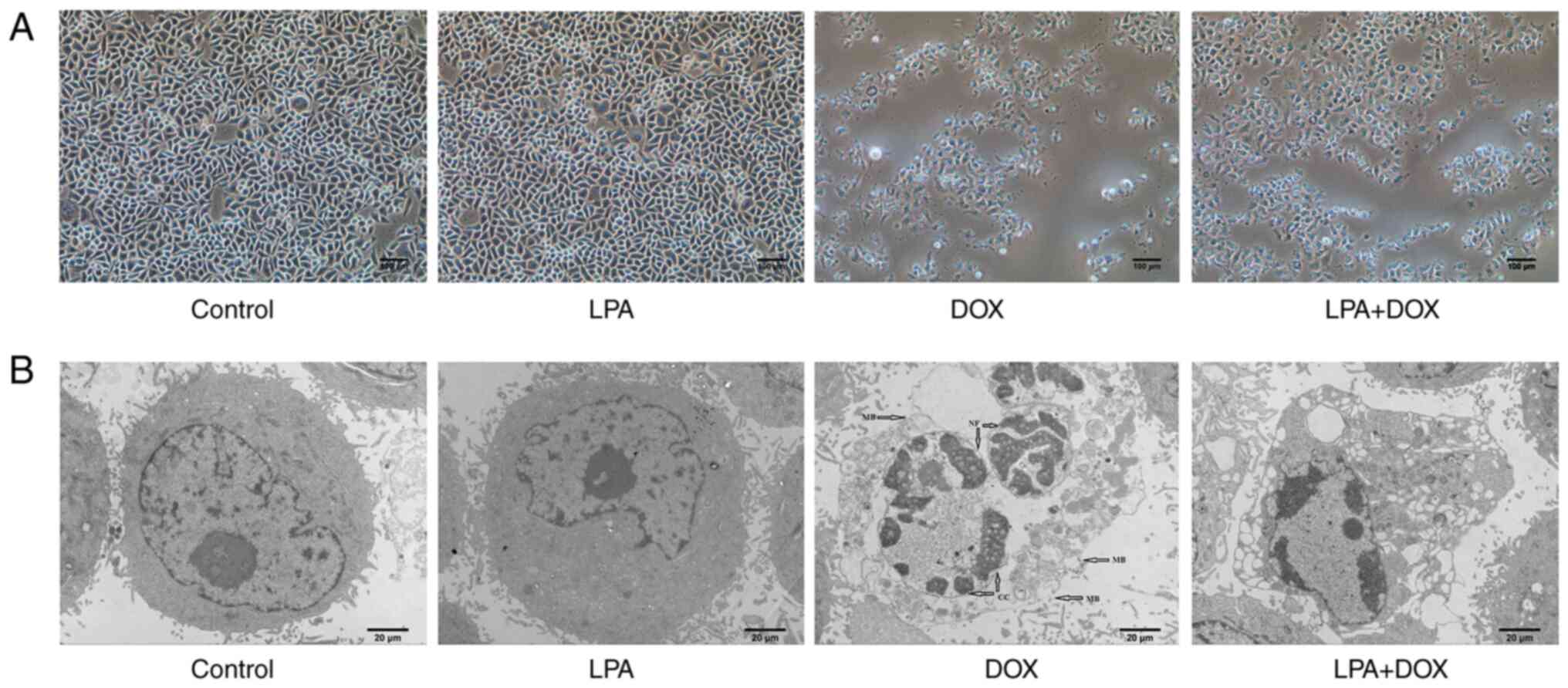 | Figure 1.Morphology of HeLa cells treated with
DOX and LPA. (A) Images were captured using an optical microscope.
Magnification, ×100. Scale bar, 100 µm. (B) Images were captured
using a transmission electron microscope. Magnification, ×2,000.
Scale bar, 20 µm. Arrows indicate membrane broken, nuclear
fragmentation, and chromatin condensation. DOX, doxorubicin
hydrochloride; LPA, lysophosphatidic acid; MB, membrane broken, NF,
nuclear fragmentation; CC, chromatin condensation. |
LPA downregulates the mRNA expression
levels of caspase-3 and caspase-8 in DOX-induced HeLa cells
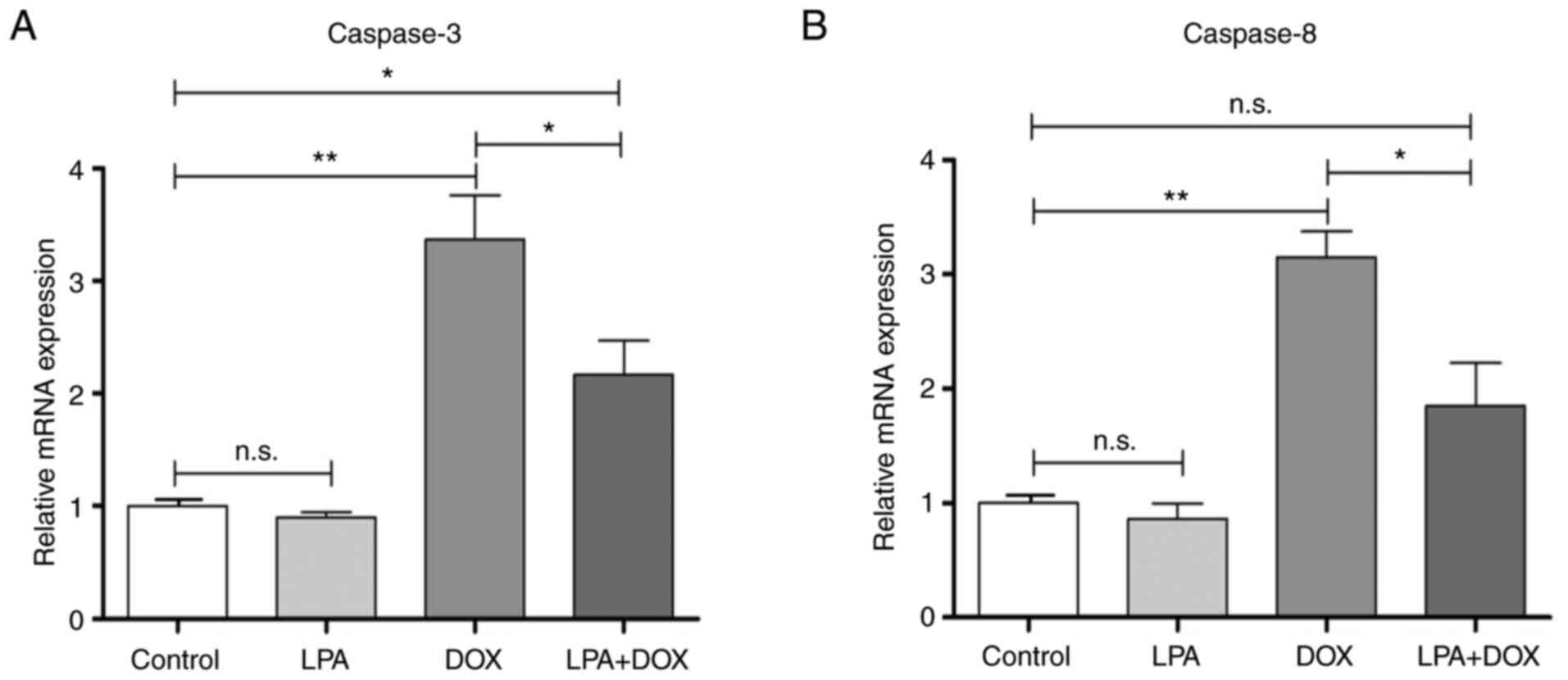
RT-qPCR was used to determine the mRNA expression
levels of caspase-3 and caspase-8 in HeLa cells in the different
treatment groups. The results demonstrated that there were no
significant changes in the mRNA expression levels of caspase-3 and
caspase-8 in the control and LPA-treated groups. However, the
caspase-3 mRNA expression levels significantly increased in the DOX
group (P<0.01), whereas they decreased in the LPA + DOX group by
35.61% compared with the DOX group (P<0.05) (Fig. 3A). The mRNA expression levels of
caspase-8 in the DOX group significantly increased (P<0.01),
whereas they significantly decreased in the LPA + DOX by 41.38%
compared with the DOX group (P<0.05) (Fig. 3B). These results indicated that LPA
potentially downregulated the mRNA expression levels of caspase-3
and caspase-8, which suggested that LPA may protect HeLa cells from
DOX-induced apoptosis via the downregulation of these proapoptotic
genes.
LPA downregulates the protein
expression levels of caspase-3 in DOX-induced cervical cancer
cells
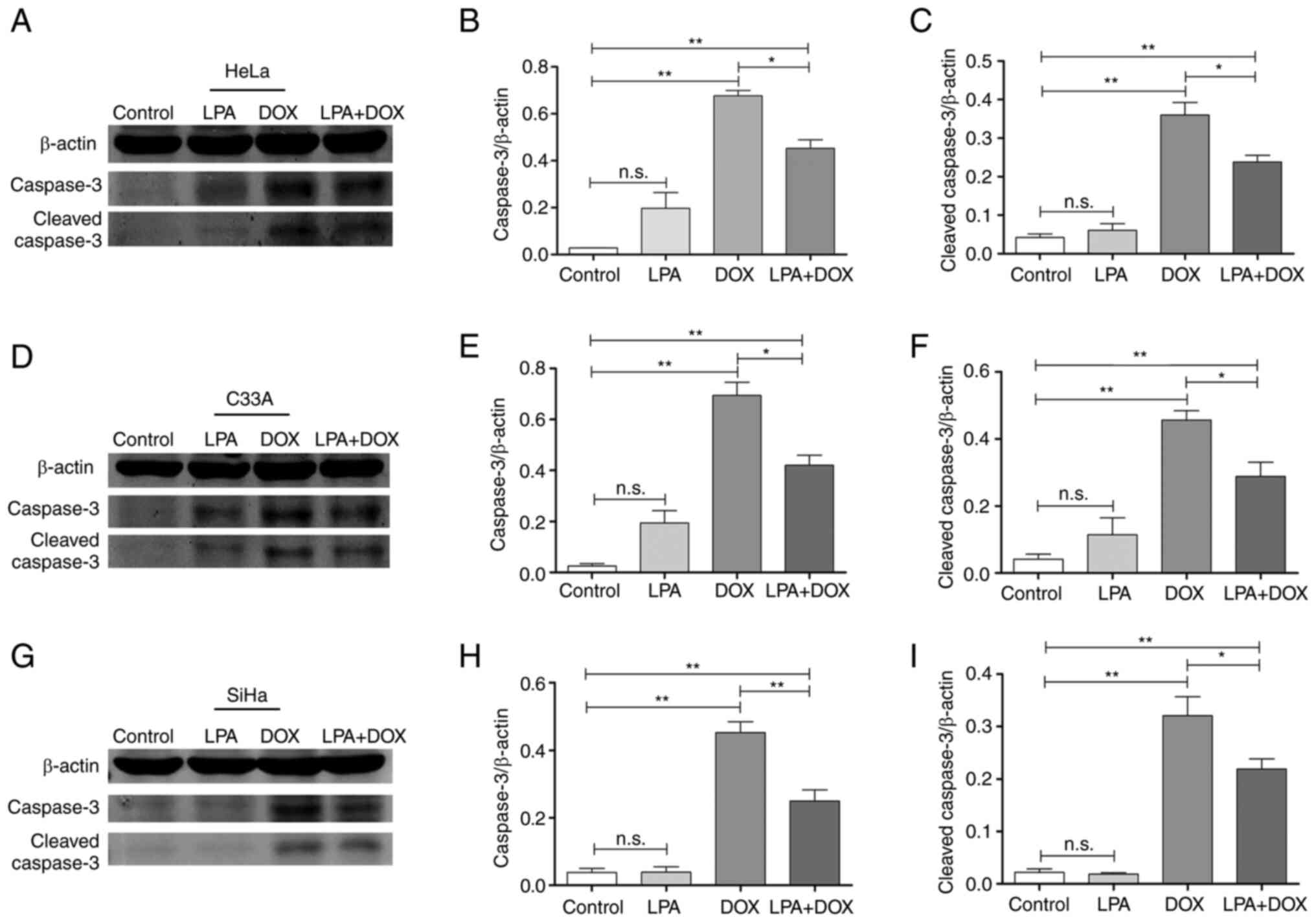
Western blotting demonstrated that there were no
significant changes in the protein expression levels of caspase-3
and cleaved caspase-3 in the control and LPA groups. However, the
protein expression levels of caspase-3 and cleaved caspase-3 in the
DOX group were significantly increased in HeLa, C33A and SiHa cells
(P<0.01). Moreover, the protein expression levels of caspase-3
and cleaved caspase-3 in the LPA + DOX group decreased compared
with the DOX group in HeLa (Fig.
4A), C33A (Fig. 4D) and SiHa
cells (Fig. 4G). Densitometric
analysis of the western blotting results demonstrated that
caspase-3 protein expression levels in HeLa cells significantly
decreased by 33.13% (P<0.05; Fig.
4B) and cleaved caspase-3 protein expression levels
significantly decreased by 33.88% (P<0.05; Fig. 4C) in the LPA + DOX group compared
with the DOX group. Moreover, caspase-3 protein expression levels
in C33A cells significantly decreased by 39.38% (P<0.05;
Fig. 4E) and cleaved caspase-3
protein expression levels significantly decreased by 36.89%
(P<0.05; Fig. 4F) in the LPA +
DOX group compared with the DOX group. Furthermore, caspase-3
protein expression levels in SiHa cells significantly decreased by
44.73% (P<0.01; Fig. 4H) and
cleaved caspase-3 protein expression levels significantly decreased
by 31.78% (P<0.05; Fig. 4I) in
the LPA + DOX group compared with the DOX group. These results
indicated that LPA potentially downregulated the protein expression
levels of apoptosis-related proteins in DOX-induced cervical cancer
HeLa, C33A and SiHa cells.
LPA reduces oxidative stress injury in
DOX-induced HeLa cells
The results of the ROS detection assay demonstrated
that there was no significant change in ROS levels in the control
and LPA groups. However, ROS levels in the DOX group significantly
increased (P<0.01), whereas ROS levels in the LPA + DOX group
significantly decreased by 24.71% (P<0.05) compared with the DOX
group (Fig. 5A). The SOD detection
assay demonstrated that there was no significant change in SOD
levels in the control and LPA groups. However, the SOD level in the
DOX group significantly decreased (P<0.01), whereas the SOD
level in the LPA + DOX group significantly increased by 24.94%
(P<0.05) compared with the DOX group (Fig. 5B). The MDA detection assay
demonstrated that there was no significant change in MDA levels in
the control and LPA groups. However, MDA levels in the DOX group
significantly increased (P<0.01), whereas MDA levels in the LPA
+ DOX group significantly decreased by 28.81% (P<0.01) compared
with the DOX group (Fig. 5C).
These results indicated that LPA potentially reduced the oxidative
stress injury of DOX-induced HeLa cells.
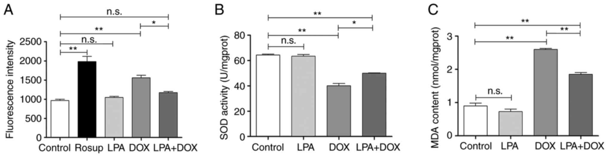 | Figure 5.LPA reduces oxidative stress injury
of DOX-induced HeLa cells. (A) Reactive oxygen species fluorescence
intensity levels of the control, LPA, DOX and LPA + DOX HeLa cell
groups. (B) SOD levels of the control, LPA, DOX and LPA + DOX HeLa
cell groups. (C) MDA levels of the control, LPA, DOX and LPA + DOX
HeLa cell groups. Data are presented as the mean ± SD (n=3).
*P<0.05 and **P<0.01 vs. the control. DOX, doxorubicin
hydrochloride; LPA, lysophosphatidic acid; SOD, superoxide
dismutase; MDA, malondialdehyde; n.s., no significance. |
Discussion
A loss of apoptosis is considered to be a
characteristic of cancer cells. Apoptosis can quickly and
selectively remove harmful, defective or damaged cells from the
body (20). Therefore, molecules
or mechanisms that are proapoptotic may be potential therapeutic
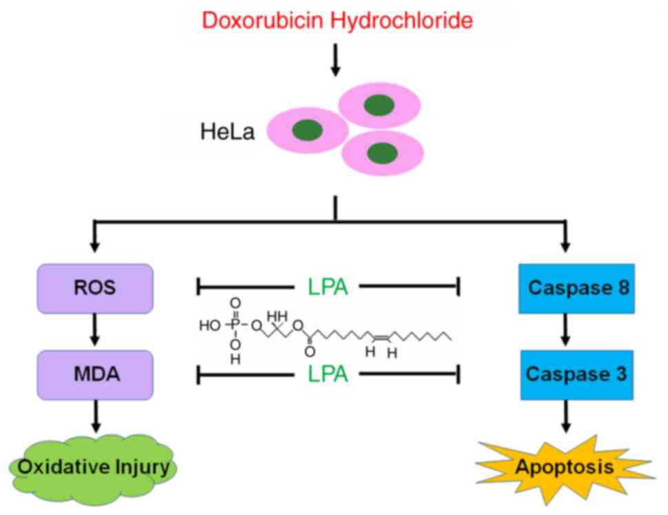
targets in cancer treatment. In the present study, the effect of
LPA on DOX-induced apoptosis in cervical cancer cells was
investigated. The results demonstrated that LPA was an
anti-apoptotic factor that potentially protected cervical cancer
HeLa cells from DOX-induced apoptosis by reducing oxygen stress
injury and inhibiting caspase-3 expression. The associated
mechanisms by which LPA potentially protects HeLa cells from
DOX-induced apoptosis are presented in Fig. 6.
Previous studies have reported that cell pyknosis is
the result of chromatin agglutination in the nucleus and is the
most significant feature of apoptosis (21). Furthermore, the decrease or
disappearance of cell surface villi, contraction of the nuclear
membrane, reduction of nuclear volume, chromatin aggregation and
vacuoles in the cytoplasm, are also typical ultrastructural changes
of apoptotic cells (22). The
present study demonstrated that the control and LPA groups
displayed normal cellular morphology. However, DOX treatment of the
cells resulted in significant changes in cell morphology, including
cell shrinkage, roundness, a reduced volume and severe shedding.
Compared with the DOX group, the LPA + DOX group exhibited a
significant improvement in cell morphology and cell availability.
Transmission electron microscopy demonstrated that the nucleus,
nuclear membrane and chromatin state remained unchanged in the
control and LPA groups. However, chromatin aggregation and cell
fragmentation were observed in the cells of the DOX group, which
suggested that these cells were undergoing apoptosis. The flow
cytometric analysis confirmed the aforementioned result. The degree
of apoptosis in the LPA + DOX group was significantly lower
compared with the DOX group, which suggested that LPA potentially
reduced the DOX-induced apoptosis of cervical cancer cells.
Caspase-3 is an important regulatory factor of
cancer cells during apoptosis (23). Both endogenous and exogenous
apoptotic signaling pathways can lead to the activation of
caspase-3 (24). In order to
further understand the mechanism by which LPA protected cervical
cancer cells from DOX-induced apoptosis, the expression levels of
caspase-3 were analyzed. The results demonstrated that DOX
treatment of HeLa cells resulted in a significant increase in the
caspase-3 mRNA expression levels. Compared with the DOX group, the
mRNA expression levels of caspase-3 gene in the LPA + DOX group
were significantly decreased. The activated form of caspase-3 is
cleaved caspase-3 and once activated it will cleave a large number
of cytoskeleton-related proteins, cell cycle regulatory proteins
and DNA repair and DNA degradation-related molecules (25,26),
which results in cell death. Therefore, the effect of DOX on
caspase-3 and cleaved caspase-3 protein expression levels was
further investigated in cervical cancer cells treated with LPA. The
results demonstrated that the protein expression levels of
caspase-3 and cleaved caspase-3 protein significantly increased and
the degree of apoptosis was severe in the DOX group. However, the
protein expression levels of caspase-3 and cleaved caspase-3
significantly decreased in the LPA + DOX group compared with the
DOX group. Furthermore, the results of the present study
demonstrated that at both the mRNA and protein expression levels,
DOX-induced apoptosis was inhibited by LPA via decreasing caspase-3
expression. It was also previously reported that LPA inhibits
cisplatin-induced apoptosis of cervical cancer cells (8,16),
which is consistent with the results of the present study.
Therefore, further investigation into the role of LPA in
DOX-induced cell death and the downstream signaling pathway of LPA
will potentially provide novel insights into the clinical treatment
of cervical cancer.
Numerous studies have reported that oxidative stress
is strongly associated with arthritis, cancer, autoimmune diseases,
aging and cardiovascular and neurodegenerative diseases (15,27,28).
High ROS levels can disrupt the structure of the mitochondrial
membrane, which leads to a shift in membrane permeability and
ultimately to apoptosis (29). ROS
regulates the tumor suppressor p53 signal, which releases
cytochrome c that activates caspase-3 (30). Therefore, as DOX can induce
oxidative stress, several oxidative stress indicators were detected
in the present study. This was to determine if LPA affected
DOX-induced apoptosis in cervical cancer cells via the inhibition
of oxidative stress. The results demonstrated that LPA inhibited
DOX-induced apoptosis by significantly decreasing ROS levels in
cervical cancer cells. ROS-mediated oxidative stress is a complex
reaction between free radical production and the antioxidant
defense of the body (31). SOD
balances ROS by scavenging free radicals and prevents cell damage
caused by superoxide anion radicals (18). Moreover, MDA is a widely detected
oxidative damage marker and elevated MDA levels are the result of
lipid peroxidation (19). SOD
levels are negatively correlated with MDA levels and the
quantification of SOD and MDA levels can infer the metabolism of
free radicals, which therefore reflects the degree of oxidative
stress in the cells (32).
Subsequently, SOD and MDA levels were determined in HeLa cells in
the present study. The results demonstrated that there was a
significant decrease in SOD levels in the DOX group compared with
an increase of 24.94% in the LPA + DOX group. Moreover, MDA levels
were significantly increased in the DOX group, whereas there was a
significant decrease of 28.81% in the LPA + DOX group. These
results suggested that LPA potentially reduced DOX-induced
oxidative stress damage in HeLa cells.
In conclusion, the present study indicated that LPA
potentially protected HeLa cells from DOX-induced apoptosis via a
reduction in caspase-3 expression levels. Furthermore, LPA was
demonstrated to reduce intracellular ROS levels, which were induced
by DOX, to prevent HeLa cells from oxygen stress damage. These
results indicated that LPA could be a novel therapeutic target in
cervical cancer treatment. Based on the finding that LPA
potentially protects HeLa cells from DOX-induced apoptosis, further
research may be extended to the directions such as discovering an
antagonist to LPA receptor, using an animal model to investigate if
combined use of an antagonist to LPA receptor and DOX could improve
the effect of anticancer drugs, developing LPA receptor inhibitors
and targeting LPA as a new strategy for cervical cancer
therapy.
Acknowledgements
Not applicable.
Funding
The present study was supported by the National Key Technology
R&D Program of the Ministry of Science and Technology (grant
no. 2013GA740103), and the project of Shandong Province Higher
Educational Science and Technology Program (grant no. J14LK15), the
Shandong Medical and Health Science and Technology Development
Project (grant no. 2018WS063), the Shandong Provincial Natural
Science Foundation of China (grant no. ZR2020KC016) and the Weifang
Science and Technology Bureau (grant No. 2020YQFK013).
Availability of data and materials
The datasets used during the present study are
available from the corresponding authors upon reasonable
request.
Authors' contributions
XW, HW and XM performed the majority of the
experiments. YX, WH, AH, YL and HJ performed the cell culture,
western blotting and analyzed the data. XW, XY and ZH designed,
supervised the study, wrote and finalized the manuscript. XW, XM
and XY confirm the authenticity of all the raw data. All authors
read and approved the final manuscript and agree to be accountable
for all aspects of the research in ensuring that the accuracy or
integrity of any part of the work are appropriately investigated
and resolved.
Ethics approval and consent to
participate
Not applicable.
Patient consent for publication
Not applicable.
Competing interests
The authors declare that they have no competing
interests.
References
|
1
|
Murph MM, Scaccia LA, Volpicelli LA and
Radhakrishna H: Agonist-induced endocytosis of lysophosphatidic
acid-coupled LPA1/EDG-2 receptors via a dynamin2- and
Rab5-dependent pathway. J Cell Sci. 116:1969–1980. 2003. View Article : Google Scholar : PubMed/NCBI
|
|
2
|
Minis E, Holcomb K, Sisti G, Nasioudis D,
Kanninen TT, Athanasiou A, Frey MK, Chapman-Davis E, Caputo TA and
Witkin SS: Evaluation of lysophosphatidic acid in vaginal fluid as
a biomarker for ovarian cancer: A pilot study. Eur J Obstet Gynecol
Reprod Biol X. 2:1000122019. View Article : Google Scholar : PubMed/NCBI
|
|
3
|
Ramachandran S, Ramaswamy S, Cho CH and
Parthasarathy S: Lysophosphatidic acid induces glycodelin gene
expression in cancer cells. Cancer Lett. 177:197–202. 2002.
View Article : Google Scholar : PubMed/NCBI
|
|
4
|
Kim NH, Sadra A, Park HY, Oh SM, Chun J,
Yoon JK and Huh SO: HeLa E-Box Binding protein, HEB, inhibits
promoter activity of the lysophosphatidic acid receptor gene Lpar1
in neocortical neuroblast cells. Mol Cells. 42:123–134.
2019.PubMed/NCBI
|
|
5
|
Liu S, Jiang H, Min L, Ning T, Xu J, Wang
T, Wang X, Zhang Q, Cao R, Zhang S and Zhu S: Lysophosphatidic acid
mediated PI3K/AKT activation contributed to esophageal squamous
cell cancer progression. Carcinogenesis. 42:611–620. 2021.
View Article : Google Scholar : PubMed/NCBI
|
|
6
|
Feng Y, Xiao M and Zhang Z, Cui R, Jiang
X, Wang S, Bai H, Liu C and Zhang Z: Potential interaction between
lysophosphatidic acid and tumor-associated macrophages in ovarian
carcinoma. J Inflamm (Lond). 17:232020. View Article : Google Scholar : PubMed/NCBI
|
|
7
|
Obol JH, Lin S, Obwolo MJ, Harrison R and
Richmond R: Knowledge, attitudes, and practice of cervical cancer
prevention among health workers in rural health centres of Northern
Uganda. BMC Cancer. 21:1102021. View Article : Google Scholar : PubMed/NCBI
|
|
8
|
Sui Y, Yang Y, Wang J, Li Y, Ma H, Cai H,
Liu X, Zhang Y, Wang S, Li Z, et al: Lysophosphatidic acid inhibits
apoptosis induced by cisplatin in cervical cancer cells. Biomed Res
Int. 2015:5983862015. View Article : Google Scholar : PubMed/NCBI
|
|
9
|
Nie H, Bu F, Xu J, Li T and Huang J: 29
immune-related genes pairs signature predict the prognosis of
cervical cancer patients. Sci Rep. 10:141522020. View Article : Google Scholar : PubMed/NCBI
|
|
10
|
Abdoul-Azize S, Buquet C, Li H, Picquenot
JM and Vannier JP: Integration of Ca2+ signaling
regulates the breast tumor cell response to simvastatin and
doxorubicin. Oncogene. 37:4979–4993. 2018. View Article : Google Scholar : PubMed/NCBI
|
|
11
|
Wei T, Xiaojun X and Peilong C:
Magnoflorine improves sensitivity to doxorubicin (DOX) of breast
cancer cells via inducing apoptosis and autophagy through AKT/mTOR
and p38 signaling pathways. Biomed Pharmacother. 121:1091392020.
View Article : Google Scholar : PubMed/NCBI
|
|
12
|
Jawad B, Poudel L, Podgornik R, Steinmetz
NF and Ching WY: Molecular mechanism and binding free energy of
doxorubicin intercalation in DNA. Phys Chem Chem Phys.
21:3877–3893. 2019. View Article : Google Scholar : PubMed/NCBI
|
|
13
|
Xu J, Liu D, Niu H, Zhu G, Xu Y, Ye D, Li
J and Zhang Q: Resveratrol reverses Doxorubicin resistance by
inhibiting epithelial-mesenchymal transition (EMT) through
modulating PTEN/Akt signaling pathway in gastric cancer. J Exp Clin
Cancer Res. 36:192017. View Article : Google Scholar : PubMed/NCBI
|
|
14
|
Pilco-Ferreto N and Calaf GM: Influence of
doxorubicin on apoptosis and oxidative stress in breast cancer cell
lines. Int J Oncol. 49:753–762. 2016. View Article : Google Scholar : PubMed/NCBI
|
|
15
|
Islam MT: Oxidative stress and
mitochondrial dysfunction-linked neurodegenerative disorders.
Neurol Res. 39:73–82. 2017. View Article : Google Scholar : PubMed/NCBI
|
|
16
|
Prasad S, Gupta SC and Tyagi AK: Reactive
oxygen species (ROS) and cancer: Role of antioxidative
nutraceuticals. Cancer Lett. 387:95–105. 2017. View Article : Google Scholar : PubMed/NCBI
|
|
17
|
Livak KJ and Schmittgen TD: Analysis of
relative gene expression data using real-time quantitative PCR and
the 2(−Delta Delta C(T)) method. Methods. 25:402–408. 2001.
View Article : Google Scholar : PubMed/NCBI
|
|
18
|
Liu R, Gang L, Shen X, Xu H, Wu F and
Sheng L: Binding characteristics and superimposed antioxidant
properties of caffeine combined with superoxide dismutase. ACS
Omega. 4:17417–17424. 2019. View Article : Google Scholar : PubMed/NCBI
|
|
19
|
Saribal D, Hocaoglu-Emre FS, Karaman F,
Mırsal H and Akyolcu MC: Trace element levels and
oxidant/antioxidant status in patients with alcohol abuse. Biol
Trace Elem Res. 193:7–13. 2020. View Article : Google Scholar : PubMed/NCBI
|
|
20
|
Xu X, Lai Y and Hua ZC: Apoptosis and
apoptotic body: Disease message and therapeutic target potentials.
Biosci Rep. 39:BSR201809922019. View Article : Google Scholar : PubMed/NCBI
|
|
21
|
Elmore S: Apoptosis: A review of
programmed cell death. Toxicol Pathol. 35:495–516. 2007. View Article : Google Scholar : PubMed/NCBI
|
|
22
|
Wang X and Li Y, Tang X, Shang X, Zhao Z,
Jiang Y and Li Y: Stenotrophomonas maltophilia outer membrane
protein A induces epithelial cell apoptosis via mitochondrial
pathways. J Microbiol. 58:868–877. 2020. View Article : Google Scholar : PubMed/NCBI
|
|
23
|
Jiang M, Qi L, Li L and Li Y: The
caspase-3/GSDME signal pathway as a switch between apoptosis and
pyroptosis in cancer. Cell Death Discov. 6:1122020. View Article : Google Scholar : PubMed/NCBI
|
|
24
|
Ma M, Wang X, Liu N, Shan F and Feng Y:
Low-dose naltrexone inhibits colorectal cancer progression and
promotes apoptosis by increasing M1-type macrophages and activating
the Bax/Bcl-2/caspase-3/PARP pathway. Int Immunopharmacol.
83:1063882020. View Article : Google Scholar : PubMed/NCBI
|
|
25
|
Kim JM, Ghosh SR, Weil AC and Zirkin BR:
Caspase-3 and caspase-activated deoxyribonuclease are associated
with testicular germ cell apoptosis resulting from reduced
intratesticular testosterone. Endocrinology. 142:3809–3816. 2001.
View Article : Google Scholar : PubMed/NCBI
|
|
26
|
Zhang C, Feng X, He L, Zhang Y and Shao L:
The interrupted effect of autophagic flux and lysosomal function
induced by graphene oxide in p62-dependent apoptosis of F98 cells.
J Nanobiotechnology. 18:522020. View Article : Google Scholar : PubMed/NCBI
|
|
27
|
Wan T, Wang Z, Luo Y, Zhang Y, He W, Mei
Y, Xue J, Li M, Pan H, Li W, et al: FA-97, a New synthetic caffeic
acid phenethyl ester derivative, protects against oxidative
stress-mediated neuronal cell apoptosis and scopolamine-induced
cognitive impairment by activating Nrf2/HO-1 signaling. Oxid Med
Cell Longev. 2019:82396422019. View Article : Google Scholar : PubMed/NCBI
|
|
28
|
Nogueira V and Hay N: Molecular pathways:
Reactive oxygen species homeostasis in cancer cells and
implications for cancer therapy. Clin Cancer Res. 19:4309–4314.
2013. View Article : Google Scholar : PubMed/NCBI
|
|
29
|
Peng N, Jin L, He A, Deng C and Wang X:
Effect of sulphoraphane on newborn mouse cardiomyocytes undergoing
ischaemia/reperfusion injury. Pharm Biol. 57:753–759. 2019.
View Article : Google Scholar : PubMed/NCBI
|
|
30
|
Song S, Chu L, Liang H, Chen J, Liang J,
Huang Z, Zhang B and Chen X: Protective effects of dioscin against
doxorubicin-induced hepatotoxicity via regulation of
Sirt1/FOXO1/NF-κb Signal. Front Pharmacol. 10:10302019. View Article : Google Scholar : PubMed/NCBI
|
|
31
|
Rasool M, Malik A, Basit Ashraf MA,
Parveen G, Iqbal S, Ali I, Qazi MH, Asif M, Kamran K, Iqbal A, et
al: Evaluation of matrix metalloproteinases, cytokines and their
potential role in the development of ovarian cancer. PLoS One.
11:e01671492016. View Article : Google Scholar : PubMed/NCBI
|
|
32
|
Wei LF, Zhang HM, Wang SS, Jing JJ, Zheng
ZC, Gao JX, Liu Z and Tian J: Changes of MDA and SOD in brain
tissue after secondary brain injury with seawater immersion in
rats. Turk Neurosurg. 26:384–288. 2016.PubMed/NCBI
|