Introduction
Contemporaneous primary cancers of the endometrium
and ovary comprise the most common synchronous gynecologic
malignancies; they are detected in 3–5% of all patients with
endometrial cancer and in 3–10% of all patients with ovarian cancer
(1). Endometrioid endometrial
carcinoma (EEC) is the most common histological subtype of
synchronous endometrial and ovarian carcinoma, while other
comorbidities are rare (2). In
addition, the coexistence of ovarian Brenner tumor (BT) and
mucinous neoplasm is not uncommon, accounting for ~16% (3). However, to the best of our knowledge,
no previous study exists on the coexistence of ovarian malignant
(MBT) and borderline mucinous cystadenoma combined with primary
uterine corpus endometrioid carcinoma (UEC). Therefore, the present
study reported on this current rare case of synchronous primary
ovarian MBT with borderline mucinous cystadenoma complicated with
primary UEC encountered at our hospital. Furthermore, a literature
review was performed and the overall clinicopathologic features,
immunohistochemical phenotypes, differential diagnosis, molecular
changes and the prognosis and treatment of the disease were
summarized. The purpose of this case report was to alert clinicians
and pathologists that they may encounter similar cases in clinical
practice. In order to reach an accurate pathologic diagnosis of
this type of synchronous endometrial-ovarian primary cancer, it is
recommended that the pathologist aggregates and closely examines
the patient's clinical history, imaging data, morphologic
characteristics and immunohistochemical and molecular testing
results, and ultimately performs a comprehensive analysis.
Case report
Case presentation
A 50-year-old female patient presented at the Women
and Children's Hospital of Chongqing Medical University (Chongqing,
China) in May 2021 with an eight-month history of abnormal uterine
bleeding, lower-abdominal-distension pain with lumbar pain and
swelling for three months, as well as increased vaginal discharge
for four days. The patient had been physically healthy in the past
and medical history was unremarkable. Tumor-marker levels were as
follows: CA19-9, >12,000.00 U/ml; CA-125, 256.3 U/ml; and CEA,
19.7 ng/ml. Magnetic resonance imaging of the abdomen and pelvis
indicated that the uterine cavity was dilated and exhibited a lumpy
mass with slightly long T1 and T2 signals. The mass size was
~8.4×3.5 cm, with slight enhancement upon enhanced-scan imaging.
Multilocular cystic-solid occupying masses were uncovered in both
left and right appendages, with sizes of 6.2×3.2×5.7 and
15.1×15.2×9.1 cm, respectively. The solid portion of the masses was
located in their lower aspects and protruded into the respective
cavity in papillary- and cauliflower-like shapes. Enhanced scanning
indicated that the solid components, capsule wall and septum were
also significantly enhanced. Hysteroscopy indicated that the
uterine cavity was filled with abnormal endometrium with fragile
tissue structures and a large number of abnormal blood vessels were
also observed in the endometrium, with the neoplasm having invaded
the cervical canal. Curettage was performed under hysteroscopy and
the specimen was sent for pathologic examination. Microscopically,
the atypical endometrioid glands fused with each other in the
tissues collected from the cervix and uterine cavity, and the
cancer cell atypia ranged from mild to moderate, with pathologic
mitoses noted. The pathologic diagnosis based on cervical and
uterine cavity curettage was endometrioid carcinoma [Federation of
Gynecology and Obstetrics (FIGO) grade I]. Due to the pathological
diagnosis and imaging anomalies, the patient underwent total
abdominal hysterectomy, bilateral salpingo-oophorectomy,
omentectomy and bilateral pelvic lymph node dissection.
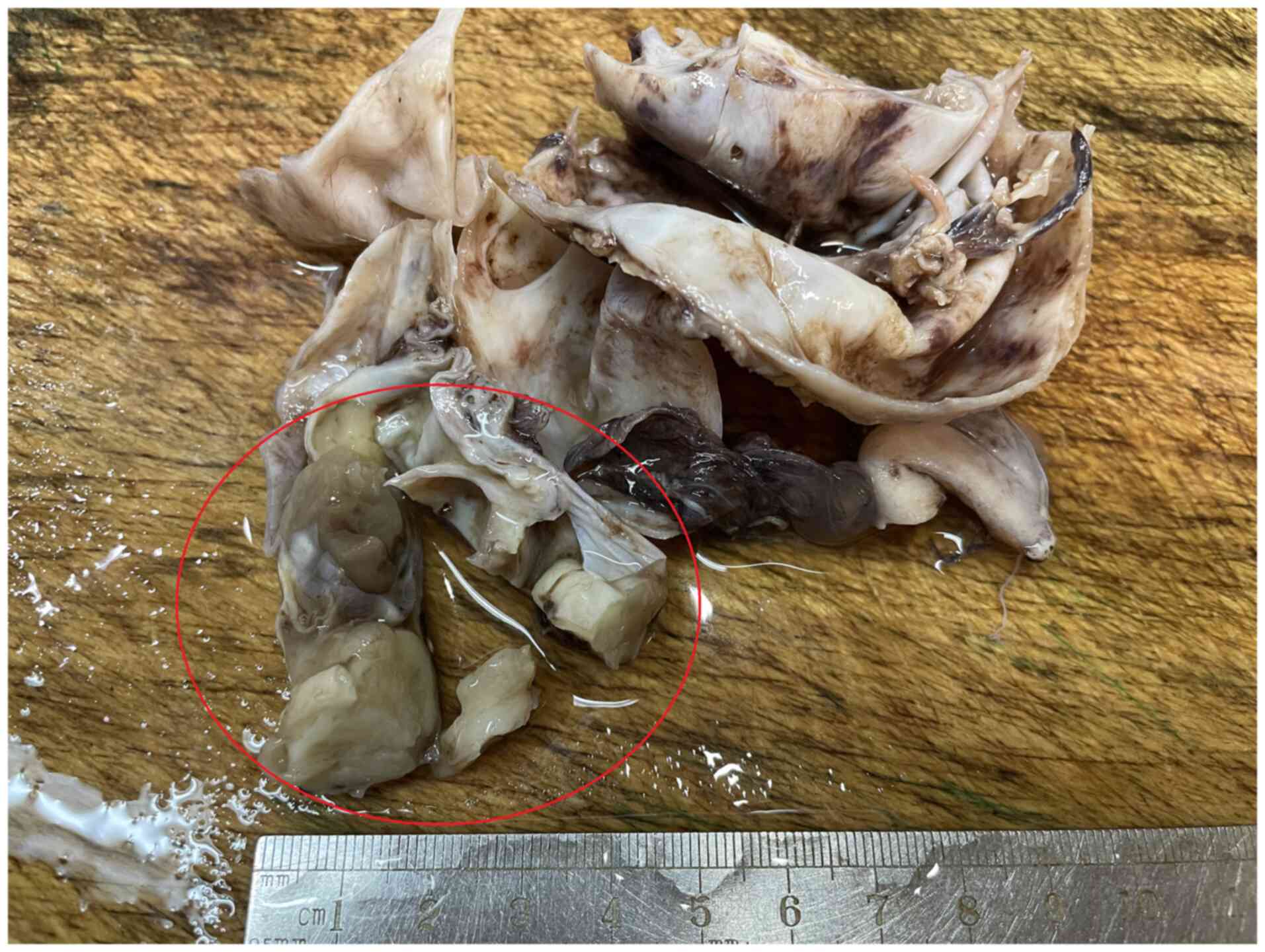
Gross specimen characteristics
The samples included the whole uterus, bilateral
adnexa, parauterine tissue and resected lymph nodes. In the left
adnexa (Fig. 1), a cystic-solid
mass of 9.0×8.0×4.5 cm with a smooth surface was noted; the mass
had been cut open and the cystic content was lost. The wall
thickness of the cyst was 0.2-0.5 cm and the size of the solid
component was 5.0×3.0×3.0 cm. The cut surface of the solid portion
was gray-white, adhesive and delicate in texture. A fallopian tube
with a length of 6.0 cm and a diameter of 1.0 cm was attached to
the surface of the mass and no oviductal umbrella was observed. A
cystic-solid mass of 12.0×8.0×4.0 cm in size in the right adnexa
was also noted; the mass had been incised and the cavity's content
was also missing. The wall thickness of the cystic component was
0.2-0.5 cm and the size of the solid component was ~9.0×6.0×3.0 cm.
The cut surface of the solid portion was gray-white, adhesive and
soft and delicate in texture, and the papilla protruded into the
cyst. The uterine cavity was filled with grayish-white and fragile
tumorous tissue and the endometrium was diffusely thickened to
~0.5-1.0 cm. The tumors infiltrated half-way into the muscle wall
layer and extended into the cervical canal. The section of the
cervical canal wall included a 2.0×1.8×1.5 cm grayish-white mass of
medium texture and unclear boundary and a biopsy was taken.
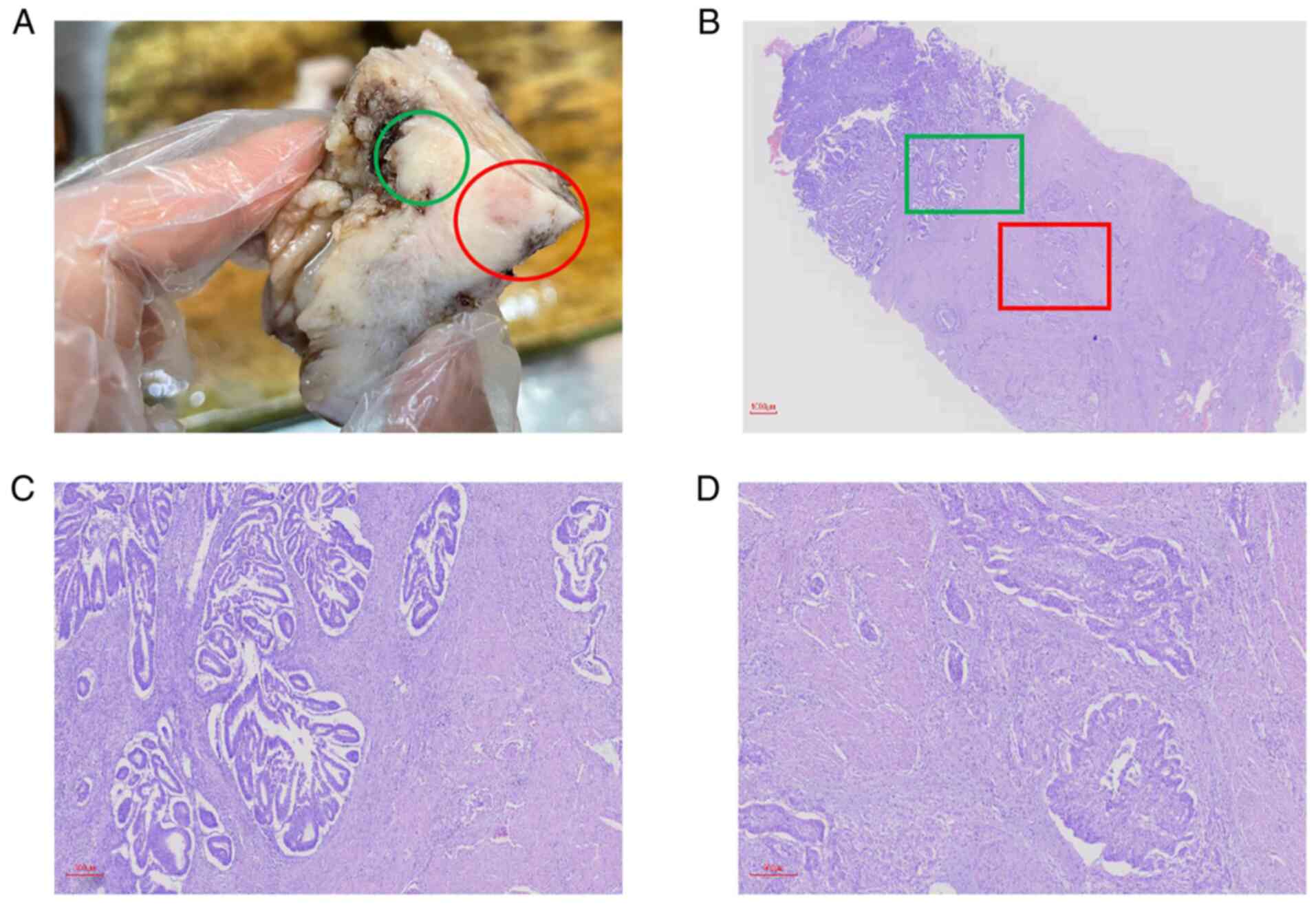
Besides, the serosa of the cervix was slightly coarse (Fig. 2A).
Microscopic characteristics on
pathologic examination
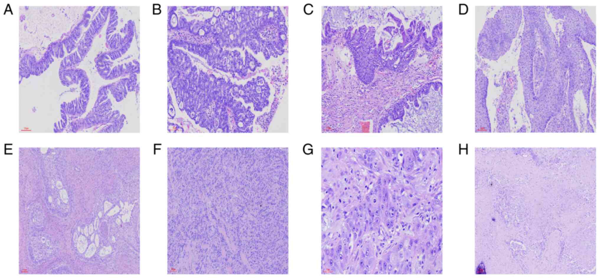
No normal ovarian tissue was found in the left and
right adnexa. In the cystic area, the cystic wall was lined with
mucinous epithelium and goblet cells were observed (magnification,
×100) (Fig. 3A). Over 10% of the
mucous epithelium of the cyst wall was stratified and part of the
epithelium was fused into a papillary or cribriform structure
(Fig. 3B). Cell atypia ranged from
mild to moderate and mitotic figures were clearly recognizable.
Fig. 3C indicates the coexistence
of mucous epithelium with the urothelium, while Fig. 3D displays a typical urothelial
tumor area. The tumor cells exhibited nest-like infiltration in the
solid area of the mass. Certain nests had a central glandular
cavity that was lined with mucous epithelium that contained
eosinophilic mucus or that exhibited necrosis (Fig. 3E). In the poorly differentiated
area, the tumor cells infiltrated into the stroma with cord-like,
trabecular and gland-like structures (Fig. 3F), and they had moderate to severe
atypia. The shape of the nuclei was round, oval, irregular or
vacuolar, with smudged chromatin and obvious nucleoli. The
cytoplasm was clear or eosinophilic and pathologic mitoses were
readily observed (magnification, ×400) (Fig. 3G). Necrosis, squamous cell
metaplasia and psammoma bodies were also observed (Fig. 3H). The mass from the uterine
endometrium exhibited well-differentiated EEC that infiltrated the
uterine myometrium to <1/2 the muscle layer (Fig. 2B), and the cervical wall was
invaded to a depth of ~2/3 the layer. A proportion of the
carcinomatous tissues featured microcystic, elongated and
fragmented (MELF) myometrial invasion (Fig. 2C). It is important to note that the
histomorphologic appearance of the mass on the cut surface of the
cervical canal wall was identical to that of the adnexal mass
(Fig. 2D) and the serosal surface
was also infiltrated by the tumor. Although the right parauterine
tissue and one lymph node from 15 resected right pelvic lymph nodes
exhibited MBT metastasis, no tumor metastasis was detected in the
resected lymph nodes from other regions.
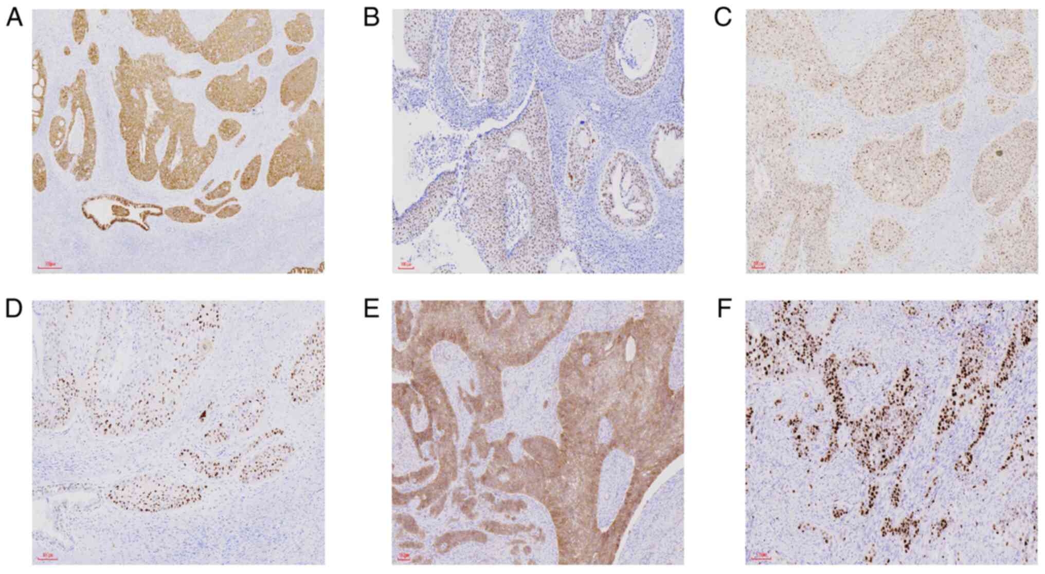
Immunohistochemical findings
The immunohistochemical findings are summarized in
Table I. Components of the MBT
from the left and right adnexa (Fig.
4) were positive for the epithelial markers cytokeratin (CK),
epithelial membrane antigen (EMA) and CK7, and for the urothelial
marker Uroplakin III (UP-III). Tissues stained positive for
steroidogenic factor 1 and weakly positive for p63, positive for
CEA in a portion of the tumor, and positive for the molecular
genetics-related proteins MDM2, C-myc, cyclin D1 and EGFR. The Ki67
hotspot index with respect to tumor cells was ~70% and tissues were
negative for estrogen receptor (ER) and progesterone receptor (PR).
Tissues were unreactive toward P16, P53 protein expression was
wild-type, and they were negative for the Müllerian duct markers
paired box 2 (Pax-2), Pax-8 and germ cell source marker spalt-like
transcription factor 4 (SALL4), as well as for the gastrointestinal
markers stabilin-2, CK20, caudal type homeobox 2, mucin 2 (MUC2)
and MUC6.
 | Table I.Immunohistochemical findings of
various carcinoma components. |
Table I.
Immunohistochemical findings of
various carcinoma components.
| Antibody | Borderline mucinous
epithelium region | Malignant Brenner
tumor region | Uterine corpus
endometrioid carcinoma |
|---|
| Pan-CK | P | P | P |
| EMA | P | P | P |
| ER | Focal P | N | P |
| PR | Focal P | N | P |
| VIM | P | P | P |
| CK7 | P | P | P |
| CK5/6 | N | P | N |
| P16 | N | N | Focal P |
| CEA | N | N | Focal P |
| P53 | Focal P | Focal P | Focal P |
| WT-1 | N | N | N |
| UP-III | N | P | N |
| SF-1 | N | P | N |
| P63 | N | Weak P | N |
| PAX2 | N | P | N |
| PAX8 | N | P | P |
| CK20 | N | N | N |
| SATB2 | N | N | N |
| CDX2 | N | N | N |
| SALL4 | N | N | N |
| MDM2 | N | P | N |
| C-myc | N | P | N |
| CyclinD1 | N | P | N |
| EGFR | N | P | N |
| Ki67, % | 20 | 70 | 50 |
Components of the borderline mucinous tumors from
the left and right adnexa were positive for EMA, CK7 and PAX8, and
all markers for MBT were negative.
Regarding UEC components, ER, PR and vimentin
antibodies reacted positively; furthermore, CK7 was positive with
CEA being locally positive. In contradistinction, P16 and Wilms'
tumor protein-1 (WT-1) markers were negative and P53 protein
exhibited wild-type expression. The mismatch repair gene-related
proteins MutL homolog 1 (MLH1), postmeiotic segregation increased 2
(PMS2), mutS homolog 2 (MSH2) and MSH6 were positive.
Pathologic diagnosis
The following observations were considered in the
diagnosis: i) Bilateral ovaries: MBT with borderline mucinous
tumor, invasion of uterine serosal surface, and cervical stroma and
infiltration of vessels; ii) UEC grade I, invasion of uterine wall
<1/2 into the muscular layer, part of the tumor infiltrated the
muscle layer in a MELF pattern, the cervical epithelium and stroma
were involved and the cervical wall was infiltrated to a depth of
<2/3 its thickness; iii) right parauterine tissue: MBT
metastasis; iv) right pelvic lymph nodes: MBT metastasis
(1/15).
The staging of the tumors was based on the American
Joint Committee on Cancer (4) and
the FIGO (5,6), as follows: i) MBT of ovary: pT3cN1M0,
stage IIIC; and ii) UEC: T2N0M0, stage II (G1).
Postoperative treatment and
follow-up
As MBT has a high degree of malignancy and
metastasizes to parauterine tissues and pelvic lymph nodes,
postoperative treatment is primarily focused on the MBT. The US
2021 National Comprehensive Cancer Network guidelines (7) recommend the use of paclitaxel +
carboplatin or paclitaxel + carboplatin + bevacizumab for
maintenance as the initial systemic treatment regimen for
epithelial ovarian cancer stages II–IV. As bevacizumab may affect
wound healing if applied less than two months after the operation,
systemic treatment with paclitaxel + carboplatin regimen was
initially adopted as an intravenous chemotherapeutic regimen.
Follow-up by telephone was implemented with the patient for four
months, the patient was treated at the hospital with chemotherapy
twice and the patient is now undergoing radiotherapy and exhibiting
disease-free survival for nearly one year.
Discussion
While synchronous primary cancers of the endometrium
and ovary are common and endometrioid cancer may manifest in both
areas (1,2), synchronous primary cancers of the UEC
and ovarian MBT are relatively unusual (1,8).
Furthermore, the coexistence of ovarian MBT and borderline mucinous
cystadenoma combined with primary UEC is even rarer and no similar
cases have been reported in the literature, to the best of our
knowledge.
Abnormal uterine bleeding (particularly
post-menopausal uterine bleeding) is etiologically associated with
endometrial cancer in 10–30% of cases (9), and the clinical manifestations of MBT
are similar to those of other malignant ovarian tumors, including
abdominal distension, abdominal pain, ovarian mass and abnormal
vaginal bleeding (10). The
present case was clinically uncovered due to abnormal uterine
bleeding and increased vaginal discharge, likely caused by
endometrial cancer with potential involvement of MBT. Although UEC
may be readily further examined by hysteroscopy and diagnosed by
fractional curettage, MBT has nonspecific features (mostly
cystic-solid masses of the ovary) and the diagnosis of MBT thereby
requires surgical excision and detailed pathologic observations
(10).
It is difficult to distinguish synchronous primary
cancers from metastatic cancers, particularly for synchronous
primary carcinomas of the same histologic subtype. In 1985,
Ulbright and Roth (11) delineated
a set of pathologic criteria to distinguish metastatic disease from
synchronous primary tumors and Scully et al (12) subsequently described more detailed
clinicopathologic features to differentiate the metastatic origin
between the ovary and the endometrium. If the pathologic findings
of the tumors in different bodily regions favor the characteristics
of independent primary cancers, a diagnosis of synchronous primary
cancers may then be made. In the present case, the diagnosis was
straightforward, since the histologic tumor types between the
ovaries and endometrium were distinct.
Identification of histologic tumor types is
principally dependent upon histomorphologic characteristics of the
tumors. Endometrioid carcinoma is the most common histotype of
endometrial carcinoma and its histologic characteristics are well
known (13). Endometrioid
carcinoma is characterized by glandular structures lined by
columnar/cuboidal cells with round/ovoidal pseudostratified nuclei
and a smooth luminal surface; nuclear atypia is most commonly of
low grade; and such histologic features were duly noted in the
present case. Furthermore, altered differentiation such as
mucinous, squamous or morular are common and are used as
confirmatory features of the endometrioid histotype (13). It is worth noting that the present
endometrioid carcinoma case exhibited the typical pattern of MELF
infiltration, which refers to the patterns of microencapsulated,
elongated and fragmented infiltration in endometrioid carcinoma;
however, its incidence is low and it is frequently observed in
certain low-grade endometrial carcinomas with deep myometrial
infiltration (14). Investigators
have ascertained that MELF infiltration is frequently accompanied
by lymph node invasion (15);
thus, if this pattern of infiltration is observed, special
attention should be paid to determine whether there is vascular
involvement and lymph node metastasis (LNM). Benign BT, borderline
BT and MBT microscopically resemble urothelium and its neoplasms
(16), and <5% of all BTs are
malignant and microscopically characterized by destructive stromal
invasion (17). In 1963, Idelson
(18) postulated diagnostic
criteria to diagnose an MBT, which stipulated the following: i) The
tumor cells conform to Brenner's morphologic characteristics; ii)
benign and borderline Brenner areas are present, preferably
exhibiting transitional changes; iii) the possibility of
pseudomucinous cystadenocarcinoma or teratoma may be excluded; and
iv) metastasis of urothelial carcinoma may also be excluded. It may
be suggested that stromal invasion by epithelial elements of MBT is
demonstrated and this is a current prerequisite according to the
most recent World Health Organization (WHO) classification
(17,19).
While UEC frequently arises from atypical
endometrial hyperplasia/endometrioid intraepithelial neoplasia [its
recognized precancerous lesion (20)], the histologic origin of ovarian BT
remains controversial. The historically proposed origins of BT have
included ovarian surface epithelium, mesonephric remnant, rete
ovarii, mucinous tumor and teratomas (3). However, more recent evidence
indicates that ovarian BT is derived from a Walthard cell nest (a
type of urothelial metaplasia) that is usually found within the
normal ovaries and fallopian tubes (21); with Walthard nests located in 50%
of patients with BT and 59% of patients with mucinous tumor
(3). The ultrastructural presence
of tumor cell cilia also indicates that BT is more likely to
originate from Walthard cell nests (16). Thus, in clinical practice, if the
mucous component is observed in the tissue slice, it may augur a
diagnosis of BT.
Immunohistochemically, low-grade UEC normally
expresses ER, PR, vimentin, CK7 and CEA; whereas p16 and WT-1 were
not expressed and P53 protein exhibited wild-type expression
(22). In addition, since MLH1,
PMS2, MSH2 and MSH6 were all positive in the present case, this
indicated that the function of mismatch repair proteins was not
lost and that the patient was not a case of Lynch
syndrome-associated endometrial carcinoma. BT features urothelial
differentiation and reflects immunophenotypes similar to those of
normal urothelium and Walthard cell nests, i.e. positivity for
UP-III, CK7, GATA3 and S100 calcium binding protein (23). Although tumor stromal cells
surrounding epithelial nests express calretinin, α-inhibin and SF1
in most benign BTs, there is a lack of expression of Müllerian
markers such as PAX8 and PAX2 and reproductive markers such as
SALL4. Of note, p63 protein that is required for urothelial
differentiation is diffusely expressed in benign and borderline BT,
but it is frequently weakly positive or negative in MBT, indicating
that p63 is involved in the malignant transformation of MBT
(19). Several studies suggested
that p16 was expressed in benign BT and absent in atypical
proliferative (borderline) BT, which may be associated with
homozygous deletion of CDKN2A in atypical proliferative BT
(24,25). In addition, malignant BT was
strongly positive for cyclinD1, RAS and EGFR (corresponding with
the molecular changes), and molecular expression rose
commensurately with the degree of BT malignancy. It has been
proposed that the combination of EGFR, RAS, cyclin D1, p16, Rb and
p53 may be employed to distinguish benign BT from
borderline/malignant BT (26).
Endometrioid carcinomas are typically associated
with microsatellite instability and mutations in phosphatase and
tensin homolog, β-catenin, K-RAS and
phosphatidylinositol-4,5-bisphosphate 3-kinase catalytic subunit α
(PIK3CA) (27). With respect to
BT, fluorescence in situ hybridization analysis identified
CDKN2A (p16 coding gene) as homozygously deleted in the epithelial
components of all borderline BT cases, but that CDKN2A remained in
all patients with benign BT (24).
The deletion of CDKN2A may thus have a critical role in the
progression from benign BT to borderline BT (24). In addition, several somatic
mutations in KRAS and PIK3CA were observed in ~30% of cases of
borderline BT (24). The
amplification of MDM2 and the cyclin D1 (CCND1) gene was confirmed
by sequencing analysis in malignant BT (25).
In the present case, ovarian MBT was complicated by
borderline mucinous cystadenoma. Studies have indicated that 25% of
mucinous tumors contain Brenner components, 16% of BTs contain
mucinous components and that Walthard nests have been detected in
50% of BTs and 59% of mucinous tumors (3). Furthermore, mucous epithelial
components are frequently localized in BT nests and transitional
cell components may be observed beneath the mucous epithelium; both
exhibit similar patterns of calcification and distribution. The BT
and mucinous tumors share similar immunohistochemical findings that
include diffuse positivity for GATA3, and a lack of Müllerian-tube
staining for PAX8 and PAX2 and the germ cell tumor marker Sall4
(28). These immunohistochemical
changes are also consistent with the molecular genetic changes.
Wang et al (29) performed
a clonal analysis of two components of 10 tissues that contained
synchronous BT and mucinous tumors by utilizing the human androgen
receptor gene and found that in 8 out of 10 cases, the pattern of
X-chromosome inactivation was identical between the two tumor
components, revealing that they were of monoclonal origin (29). Tafe et al (30) also indicated that there was a
40–75% overlap between the BT and mucinous tumors, including a
mutation in RAS and CDKN2A or an amplification of MYC, CDK4 or
CCND1. Pejovic et al (31)
demonstrated that both well-differentiated ovarian mucinous
carcinoma and its coinciding BT featured 12q 14–21 amplification.
In conclusion, it is likely that a specific relationship exists
between the two cancers in terms of morphological,
immunohistochemical and molecular changes, and it is indicated that
mucinous carcinoma and BT may be monoclonally related. Certain
authors have proposed that mucinous tumors may be formed by the
transitional epithelium of the BT consolidating into mucinous
epithelium through metaplastic modifications and the further
proliferation of mucinous epithelium (32).
The primary differential diagnosis of low-grade
endometrioid carcinoma is serous carcinoma; distinction between the
two is important, as they differ in terms of prognosis and therapy
(33). While patient features and
tumor histology are useful in making a differential diagnosis,
serous and low-grade endometrioid carcinomas may exhibit similar
growth patterns, including glandular, papillary, micropapillary and
solid forms; in addition, immunohistochemical staining (including
that for p53 and p16) may be conducive to making this distinction
(33). Loss of mismatch-repair
proteins in cancer tissue or in Lynch syndrome-associated
adenocarcinoma suggests EEC. In the 2020 classification from the
WHO (34), increased emphasis is
now put on The Cancer Genome Atlas molecular typing of endometrial
cancer so as to predict prognosis rather than only its histologic
type. When the histologic tumor type remains uncertain, application
of Gilks' binary grading system may provide more useful prognostic
information compared with FIGO grading (33). The primary differential diagnosis
with respect to MBT is transitional cell carcinoma (TCC), and while
they reflect similar morphologic features and immunohistochemical
phenotypes, the two grading systems also possess numerous
disparities. In a previous study, imaging and gross examination
suggested that TCC lacked the common calcification of MBT and that
MBT was more commonly expressed at stage I without extraovarian
spread, while ~69% of ovarian TCC was diagnosed in the advanced
stage (35). Microscopically,
although TCC was not accompanied by benign BT or borderline BT
components, it exhibited obvious malignant features. TCC usually
has diffuse strong immunoreactivity for p16, Rb and p53, while MBT
is negative. In BT, the expression of EGFR, RAS and CCND1 is
concomitantly elevated with the increase in the degree of
malignancy, but this phenomenon is lacking in TCC. There are also
numerous differences in molecular changes; for instance, a p53 gene
mutation is prominently observed in TCC (36).
Determining the appropriate treatment modality for
patients diagnosed with synchronous primary endometrial and ovarian
cancer depends upon the grade and stage of tumors in each
topographic location (37). In the
present study, surgery was first performed, followed by adjuvant
treatment in the form of chemotherapy or radiotherapy according to
tumor stage. If diagnosed as stage IA, no additional treatment was
required after surgery; if diagnosed as IIIA or II, additional
treatment was required (37). The
final diagnosis of the present case was stage IIIC of MBT in the
ovary and stage II of UEC, and therefore, radiotherapy was
performed after chemotherapy. It was previously demonstrated that
the use of a platinum-based chemotherapeutic agent plus paclitaxel
post-operatively improved patient survival (38). In the present study, it was
indicated that MDM2 amplification was associated with MBT and that
MDM2 may therefore be selected in the future as a drug target for
patients with MBT (39).
It has been reported that tumors with endometrioid
histologic features at both sites have a favorable prognosis, while
non-endometrioid tumor morphologies at both sites share a poor
prognosis (2). In one study, the
histopathologic type of the ovarian cancer component, stage of
endometrial cancer and level of CA125 at diagnosis have been
observed to have a great influence on the development of recurrence
and survival of synchronous primary carcinomas of the endometrium
and ovary (40). Therefore, it may
be posited that MBT in ovaries is a primary determining factor of
prognosis with respect to the present case. It is generally
conjectured that the prognosis of MBT is favorable, with a
five-year survival rate of MBT confined to the ovaries of 94.5%;
however, the rate involving extraovarian tissues was determined to
be 51.3% (19). It was also
reported that in patients with extrauterine metastasis, such as
that to the dura mater, lung, peritoneum, omentum, skin and bone,
the recurrence rate was 28% (19),
and the average recurrence time for MBT was 11 months (41). LNM of MBT is not common and it is
revealed in only 5.1% of patients, and when it does occur,
lymphadenectomy exerts no effect on the improvement in the
five-year survival rate of MBT (19). Although CA-125 is not specific and
its concentration is not related to the malignant degree of a
tumor, ~70% of patients with MBT have elevated CA-125 levels.
Therefore, CA-125 remains the most important tumor marker for the
post-treatment monitoring of disease recurrence (19,41).
Acknowledgements
Not applicable.
Funding
The Fund of the Women and Children's Hospital of Chongqing
Medical University provided financial support (grant no.
2021YJMS01).
Availability of data and materials
All of the data generated in the present study are
included in the figures and/or tables of this article.
Authors' contributions
YC and CZ conceived and designed the study and wrote
the original manuscript. QL and JZ analyzed data and revised the
manuscript critically for important intellectual content. All
authors read and approved the final manuscript. YC, CZ and QL
confirm the authenticity of all the raw data.
Ethics approval and consent to
participate
Not applicable.
Patient consent for publication
Written informed consent was obtained from the
patient for publication of this case report and its accompanying
images.
Competing interests
The authors declare that they have no competing
interests.
References
|
1
|
Matsuo K, Machida H, Blake EA, Holman LL,
Rimel BJ, Roman LD and Wright JD: Trends and outcomes of women with
synchronous endometrial and ovarian cancer. Oncotarget.
9:28757–28771. 2018. View Article : Google Scholar : PubMed/NCBI
|
|
2
|
Soliman PT, Slomovitz BM, Broaddus RR, Sun
CC, Oh JC, Eifel PJ, Gershenson DM and Lu KH: Synchronous primary
cancers of the endometrium and ovary: A single institution review
of 84 cases. Gynecol Oncol. 94:456–462. 2004. View Article : Google Scholar : PubMed/NCBI
|
|
3
|
Seidman JD and Khedmati F: Exploring the
histogenesis of ovarian mucinous and transitional cell (Brenner)
neoplasms and their relationship with Walthard cell nests: A study
of 120 tumors. Arch Pathol Lab Med. 132:1753–1760. 2008. View Article : Google Scholar : PubMed/NCBI
|
|
4
|
Amin MB, Edge SB, Greene FL, Byrd DR,
Brookland RK, Washington MK, Gershenwald JE, Compton CC, Hess KR,
Sullivan DC, et al: AJCC cancer staging manual. 8th edition.
Springer; New York, NY: 2017, View Article : Google Scholar
|
|
5
|
Zeppernick F and Meinhold-Heerlein I: The
new FIGO staging system for ovarian, fallopian tube, and primary
peritoneal cancer. Arch Gynecol Obstet. 290:839–842. 2014.
View Article : Google Scholar : PubMed/NCBI
|
|
6
|
Soslow RA, Tornos C, Park KJ, Malbica A,
Matias-Guiu X, Oliva E, Parkash V, Carlson J, McCluggage WG and
Gilks CB: Endometrial carcinoma diagnosis: Use of FIGO grading and
genomic subcategories in clinical practice: Recommendations of the
international society of gynecological pathologists. Int J Gynecol
Pathol. 38 (Suppl 1):S64–S74. 2019. View Article : Google Scholar : PubMed/NCBI
|
|
7
|
Armstrong DK, Alvarez RD, Bakkum-Gamez JN,
Barroilhet L, Behbakht K, Berchuck A, Chen LM, Cristea M, DeRosa M,
Eisenhauer EL, et al: Ovarian cancer, version 2.2020, NCCN clinical
practice guidelines in oncology. J Natl Compr Canc Netw.
19:191–226. 2021. View Article : Google Scholar : PubMed/NCBI
|
|
8
|
Krepinska E, Kriz JT and Laco J:
Endometroid adenocarcinoma of the uterus, borderline tumor of the
ovary and Brenner tumor of the contralateral ovary in a 63-year-old
woman. Eur J Gynaecol Oncol. 31:584–585. 2010.PubMed/NCBI
|
|
9
|
Izetbegovic S, Stojkanovic G, Ribic N and
Mehmedbasic E: Features of postmenopausal uterine haemorrhage. Med
Arch. 67:431–434. 2013. View Article : Google Scholar : PubMed/NCBI
|
|
10
|
King L, Gogoi RP, Hummel C and Smith A:
Malignant Brenner tumor: Two case reports. Case Rep Womens Health.
20:e000822018. View Article : Google Scholar : PubMed/NCBI
|
|
11
|
Ulbright T and Roth L: Metastatic and
independent cancers of the endometrium and ovary: A
clinicopathologic study of 34 cases. Hum Pathol. 16:28–34. 1985.
View Article : Google Scholar : PubMed/NCBI
|
|
12
|
Scully RE, Young RH and Clement PB: Tumors
of the ovary, maldeveloped gonads, fallopian tube, and broad
ligament. Atlas of tumor pathology. Armed Forces Institute of
Pathology; Bethesda, MD: 1998
|
|
13
|
Santoro A, Angelico G, Travaglino A,
Inzani F, Arciuolo D, Valente M, D'Alessandris N, Scaglione G,
Fiorentino V, Raffone A and Zannoni GF: New pathological and
clinical insights in endometrial cancer in view of the updated
ESGO/ESTRO/ESP guidelines. Cancers (Basel). 13:26232021. View Article : Google Scholar : PubMed/NCBI
|
|
14
|
Stewart CJ, Brennan BA, Leung YC and
Little L: MELF pattern invasion in endometrial carcinoma:
Association with low grade, myoinvasive endometrioid tumours, focal
mucinous differentiation and vascular invasion. Pathology.
41:454–459. 2009. View Article : Google Scholar : PubMed/NCBI
|
|
15
|
Pavlakis K, Messini I, Vrekoussis T,
Panoskaltsis T, Chrysanthakis D, Yiannou P and Voulgaris Z: MELF
invasion in endometrial cancer as a risk factor for lymph node
metastasis. Histopathology. 58:966–973. 2011. View Article : Google Scholar : PubMed/NCBI
|
|
16
|
Kuhn E, Ayhan A, Shih IeM, Seidman JD and
Kurman RJ: Ovarian Brenner tumour: A morphologic and
immunohistochemical analysis suggesting an origin from fallopian
tube epithelium. Eur J Cancer. 49:3839–3849. 2013. View Article : Google Scholar : PubMed/NCBI
|
|
17
|
Meinhold-Heerlein I, Fotopoulou C, Harter
P, Kurzeder C, Mustea A, Wimberger P, Hauptmann S and Sehouli J:
The new WHO classification of ovarian, fallopian tube, and primary
peritoneal cancer and its clinical implications. Arch Gynecol
Obstet. 293:695–700. 2016. View Article : Google Scholar : PubMed/NCBI
|
|
18
|
Idelson MG: Malignancy in Brenner tumors
of the ovary, with comments on histogenesis and possible estrogen
production. Obstet Gynecol Surv. 18:246–267. 1963. View Article : Google Scholar : PubMed/NCBI
|
|
19
|
Nasioudis D, Sisti G, Holcomb K, Kanninen
T and Witkin SS: Malignant Brenner tumors of the ovary; a
population-based analysis. Gynecol Oncol. 142:44–49. 2016.
View Article : Google Scholar : PubMed/NCBI
|
|
20
|
Albertini AF, Devouassoux-Shisheboran M
and Genestie C: Pathology of endometrioid carcinoma. Bull Cancer.
99:7–12. 2012. View Article : Google Scholar : PubMed/NCBI
|
|
21
|
Zheng R and Heller DS: Borderline Brenner
tumor: A review of the literature. Arch Pathol Lab Med.
143:1278–1280. 2019. View Article : Google Scholar : PubMed/NCBI
|
|
22
|
Geels YP, van der Putten LJ, van Tilborg
AA, Lurkin I, Zwarthoff EC, Pijnenborg JM, van den Berg-van Erp SH,
Snijders MP, Bulten J, Visscher DW, et al: Immunohistochemical and
genetic profiles of endometrioid endometrial carcinoma arising from
atrophic endometrium. Gynecol Oncol. 137:245–251. 2015. View Article : Google Scholar : PubMed/NCBI
|
|
23
|
Kondi-Pafiti A, Kairi-Vassilatou E,
Iavazzo Ch, Vouza E, Mavrigiannaki P, Kleanthis Ch,
Vlahodimitropoulos D and Liapis A: Clinicopathological features and
immunoprofile of 30 cases of Brenner ovarian tumors. Arch Gynecol
Obstet. 285:1699–1702. 2012. View Article : Google Scholar : PubMed/NCBI
|
|
24
|
Kuhn E, Ayhan A, Shih IeM, Seidman JD and
Kurman RJ: The pathogenesis of atypical proliferative Brenner
tumor: An immunohistochemical and molecular genetic analysis. Mod
Pathol. 27:231–237. 2014. View Article : Google Scholar : PubMed/NCBI
|
|
25
|
Wang L, Allison D and Shukla PS:
Amplification of MDM2 and loss of p16 expression: Do they have a
role in malignant transformation of ovarian Brenner tumor? Am J
Clin Pathol. 154:133–141. 2020. View Article : Google Scholar : PubMed/NCBI
|
|
26
|
Albu DF, Albu CC, Gogănău AM, Albu ŞD,
Mogoantă L, Edu A, DiŢescu D and Văduva CC: Borderline Brenner
tumors associated with ovarian cyst-case presentation. Rom J
Morphol Embryol. 57 (Suppl 2):893–898. 2016.PubMed/NCBI
|
|
27
|
Llobet D, Pallares J, Yeramian A,
Santacana M, Eritja N, Velasco A, Dolcet X and Matias-Guiu X:
Molecular pathology of endometrial carcinoma: Practical aspects
from the diagnostic and therapeutic viewpoints. J Clin Pathol.
62:777–785. 2009. View Article : Google Scholar : PubMed/NCBI
|
|
28
|
Roma AA and Masand RP: Different staining
patterns of ovarian Brenner tumor and the associated mucinous
tumor. Ann Diagn Pathol. 19:29–32. 2015. View Article : Google Scholar : PubMed/NCBI
|
|
29
|
Wang Y, Wu RC, Shwartz LE, Haley L, Lin
MT, Shih IeM and Kurman RJ: Clonality analysis of combined Brenner
and mucinous tumours of the ovary reveals their monoclonal origin.
J Pathol. 237:146–151. 2015. View Article : Google Scholar : PubMed/NCBI
|
|
30
|
Tafe LJ, Muller KE, Ananda G, Mitchell T,
Spotlow V, Patterson SE, Tsongalis GJ and Mockus SM: Molecular
genetic analysis of ovarian Brenner tumors and associated mucinous
epithelial neoplasms: High variant concordance and identification
of mutually exclusive RAS driver mutations and MYC amplification.
Am J Pathol. 186:671–677. 2016. View Article : Google Scholar : PubMed/NCBI
|
|
31
|
Pejovic T, Bürki N, Odunsi K, Fiedler P,
Achong N, Schwartz PE and Ward DC: Well-differentiated mucinous
carcinoma of the ovary and a coexisting Brenner tumor both exhibit
amplification of 12q14-21 by comparative genomic hybridization.
Gynecol Oncol. 74:134–137. 1999. View Article : Google Scholar : PubMed/NCBI
|
|
32
|
Rodríguez IM and Prat J: Mucinous tumors
of the ovary: A clinicopathologic analysis of 75 borderline tumors
(of intestinal type) and carcinomas. Am J Surg Pathol. 26:139–152.
2002. View Article : Google Scholar : PubMed/NCBI
|
|
33
|
Garg K and Soslow RA: Strategies for
distinguishing low-grade endometrioid and serous carcinomas of
endometrium. Adv Anat Pathol. 19:1–10. 2012. View Article : Google Scholar : PubMed/NCBI
|
|
34
|
Berek JS, Friedlander M and Hacker NF:
Endometrial cancer. Berek and Hacker's gynecologic oncology. Berek
JS and Hacker NF: 7th edition. Wolters Kluwer Health; Philadelphia,
PA: 2020, PubMed/NCBI
|
|
35
|
Austin RM and Norris HJ: Malignant Brenner
tumor and transitional cell carcinoma of the ovary: A comparison.
Int J Gynecol Pathol. 6:29–39. 1987. View Article : Google Scholar : PubMed/NCBI
|
|
36
|
Cuatrecasas M, Catasus L, Palacios J and
Prat J: Transitional cell tumors of the ovary: A comparative
clinicopathologic, immunohistochemical, and molecular genetic
analysis of Brenner tumors and transitional cell carcinomas. Am J
Surg Pathol. 33:556–567. 2009. View Article : Google Scholar : PubMed/NCBI
|
|
37
|
Shin W, Park SY, Kang S, Lim MC and Seo
SS: How to manage synchronous endometrial and ovarian cancer
patients? BMC Cancer. 21:4892021. View Article : Google Scholar : PubMed/NCBI
|
|
38
|
Han JH, Kim DY, Lee SW, Park JY, Kim JH,
Kim YM, Kim YT and Nam JH: Intensive systemic chemotherapy is
effective against recurrent malignant Brenner tumor of the ovary:
An analysis of 10 cases within a single center. Taiwan J Obstet
Gynecol. 54:178–182. 2015. View Article : Google Scholar : PubMed/NCBI
|
|
39
|
Shetty S, Habeeb O, Rivera C, Astbury C,
Przybycin C and Joehlin-Price AS: MDM2 amplification in malignant
Brenner tumors may play a role in progression to malignancy and aid
in separation from urothelial and other ovarian carcinomas. Hum
Pathol. 117:42–50. 2021. View Article : Google Scholar : PubMed/NCBI
|
|
40
|
Sozen H, Vatansever D, Iyibozkurt AC,
Topuz S, Ozsurmeli M, Salihoglu Y, Guzelbey B and Berkman S:
Clinicopathologic and survival analyses of synchronous primary
endometrial and epithelial ovarian cancers. J Obstet Gynaecol Res.
41:1813–1819. 2015. View Article : Google Scholar : PubMed/NCBI
|
|
41
|
Lang SM, Mills AM and Cantrell LA:
Malignant Brenner tumor of the ovary: Review and case report.
Gynecol Oncol Rep. 22:26–31. 2017. View Article : Google Scholar : PubMed/NCBI
|