Introduction
Carcinoid tumors are low-grade malignant tumors that
are most frequently located in the gastrointestinal system. The
respiratory tract is the second most frequent site and carcinoids
account for 1–2% of all lung tumors (1). Lung carcinoids are categorized into
typical carcinoids (TCs) and atypical carcinoids (ACs). This
division is dependent on the mitotic rate and necrosis. TC is
defined as <2 mitoses per 2 mm2 without necrosis. AC
is defined as ≥2 mitoses, but <10 mitoses per 2 mm2
or necrosis, or both (2).
Bronchopulmonary carcinoids are mostly found as an
incidental radiographic finding (3). They are usually observed as
well-defined pulmonary nodules, and TCs and ACs cannot be
distinguished radiographically (4).
Carcinoid tumors are macroscopically well
demarcated, tan to yellow lesions (5,6).
They consist of uniform polygonal cells with granular chromatin,
unobtrusive nucleoli and an eosinophilic cytoplasm (6). The most frequent growth patterns are
the organoid and trabecular; with an arrangement of the tumor cells
in nests or cords, respectively, with less common patterns
comprising spindle cell, pseudoglandular, papillary, rosette
formation and follicular (5,7,8).
There may also be neuroendocrine cell hyperplasia in the adjacent
airway epithelium (9,10). A recent study described the
co-existence of pulmonary carcinoid tumorlets, which are confined
neuroendocrine tumors, with chronic pulmonary inflammatory
entities, such as bronchiectasis, atelectasis and recurrent
pulmonary infections in lung tissue specimens (11).
Numerous prognostic factors for carcinoid tumors
have been described, including age, sex, tumor size, stage, type,
the type of surgical procedure performed, chemotherapy and
radiation (12–14). In addition, vascular invasion,
nuclear pleomorphism and aerogenous spread, a Ki-67 index ≥5%,
infiltrative growth and the absence of orthopedia homeobox protein
expression are considered unfavorable prognostic factors, whereas
palisading, papillary formation and pseudoglandular patterns are
favorable prognostic features (9,15,16).
However, the simultaneous pathological findings in
lung specimens of surgically resected pulmonary carcinoids and
their prognostic role have not been described to date, at least to
the best of our knowledge. The identification of prognostic factors
in lung carcinoids, which are relatively rare tumors, is
significant for a variety of reasons, such as understanding which
characteristics are predictive of outcomes, acquiring knowledge
into the biology and natural history of the disease, treatment
optimization, the inclusion of these factors in clinical trial
designs and the awareness of the possibility of recurrence or death
(17). The present study aimed to
describe the simultaneous pathological findings and determine their
association with the survival rates of patients with surgically
resected pulmonary carcinoids.
Patients and methods
Study design
From March, 2005 to March, 2019, the present study
retrospectively evaluated patients with a histological diagnosis of
a lung carcinoid who had undergone thoracic surgery. The assessment
was made with a medical history, clinical examination, chest X-ray
and computed tomography (CT) scan of the chest, brain and upper
abdomen, while a bone scintigraphy was also performed. All patients
had a pre-operative examination with a fiberoptic bronchoscope, and
in some patients, endoscopic biopsy was conducted while CT-guided
fine-needle aspiration biopsy was performed for tumors in the lung
periphery. All samples that were resected during surgery, including
the mediastinal and hilar lymph nodes, were assessed by a
pathological examination. The categorization of the tumors into TCs
and ACs was performed as stated by the World Health Organization
(WHO) (18). The estimation of
carcinoid stage was made in accordance with the 8th tumors, nodes
and metastases (ΤΝΜ) staging system for lung cancer (19). Age, sex, type of carcinoid, stage
and simultaneous pathological findings were noted, and the
association with the patient survival rates was recorded. The
present study enrolled 108 patients. AC was diagnosed in 28
patients and TC was diagnosed in 80 patients (13). Ethical approval for the present
study was obtained from the Research Ethics Committee of Athens
medical Group with file accession no. 4234. The study was in line
with the declaration of Helsinki in 1995 (as revised in Edinburgh
2000). Written informed consent was obtained from the patients for
publication of this research and accompanying images.
Histological examination
Immunohistochemistry (IHC) was performed to confirm
the diagnosis of lung carcinoids and was broadly positive for
chromogranin and synaptophysin. For histochemistry, the tissue
specimens from the patients were formalin-fixed and
paraffin-embedded, as per the standard histopathology laboratory
routine (FFPE). The fixative used was 10% formalin solution,
neutral buffered, for 24 h at room temperature. The thickness of
the sections used was 2 µm. Histochemical staining with hematoxylin
and eosin (supplied by Dako; Agilent Technologies, Inc.) was
performed using a Dako CoverStainer (Dako; Agilent Technologies,
Inc.) for ~1 h and 15 min at room temperature.
As regards immunohistochemistry, formalin-fixed
paraffin sections (2-µm-thick) were used. The staining of the
slides was performed on an AutoStainer Link 48 Dako instrument
(Dako; Agilent Technologies, Inc.). The sections were
deparaffinized and antigen retrieval was performed using Envision
Flex Target Tetrieval Solution High pH (Dako; Agilent Technologies,
Inc.) at 90°C for 20 min. The sections were then immunostained for
synaptophysin using Leica mouse anti-human synaptophysin monoclonal
antibody (Clone 27G12; Leica Biosystems, cat; NCL-SYNAP-29) at a
1:50 dilution with a 30-min incubation time at room temperature and
for chromogranin using monoclonal chromogranin A monoclonal
antibody (clone LK2H10; Thermo Fisher Scientific, Inc, cat;
MA5-13096) at a 1:400 dilution with a 25-min incubation time at
room temperature. Immunoreactions were detected using EnVision
Detection Systems (Dako; Agilent Technologies, Inc.), which
includes secondary antibody ready-to-use EnVisionTM FLEX/HRP (Code
No. K8000/K8002/K8023) solution. For the secondary antibody, the
incubation time was 20 min, at room temperature. All slides were
counterstained with Mayer's hematoxylin (Dako; Agilent
Technologies, Inc.) for 10 min at room temperature.
The categorization of the tumors into TCs and ACs
was performed by an experienced pathologist, and was based on the
evaluation of hematoxylin- and eosin-stained (histochemical)
sections. Namely, beyond the fact that all tumors had the
characteristic neuroendocrine morphology, TC had a mitotic count of
<2 mitoses/2 mm2 and lacked necrosis, whereas ACs had
2–10 mitoses/2 mm2 and/or had focal and punctuated
necrosis. The presence of simultaneous pathological findings was
noted.
Statistical analysis
The Statistical Package for Social Sciences software
(SPSS) version 13.0 (SPSS, Inc.) was used for statistical analyses.
The assessment of survival was made using Kaplan-Meier statistics,
and the comparison of the survival curves was made using the
log-rank test. Survival was estimated in units of months from
surgery. The Cox hazard-regression model was also used, including
relative risk, probability and 95% confidence intervals for
univariate analysis of the prognostic factors, while 5% was
selected as the level of statistical significance. The censoring
was random and as far as possible non-informative. In addition, the
proportionality of the hazards was tested, by examining the scaled
Schoenfeld residuals. A value of P<0.05 was considered to
indicate a statistically significant difference.
Results
Surgical resection was the only treatment that
patients received. In total, 78, 18 and 12 patients were
categorized into stages I, II and III respectively (Table I).
 | Table I.Histopathological characteristics of
the patients. |
Table I.
Histopathological characteristics of
the patients.
| Variable | TC | AC | All |
|---|
| Stage |
|
|
|
| I | 62 | 16 | 78 |
| II | 14 | 4 | 18 |
|
III | 4 | 8 | 12 |
| Recurrent
tumors | 4 | 4 | 8 |
| Stage
I | 4 | 0 | 4 |
| Stage
II | 0 | 2 | 2 |
| Stage
III | 0 | 2 | 2 |
| Deaths | 4 | 4 | 8 |
| Stage
I | 4 | 0 | 4 |
| Stage
II | 0 | 2 | 2 |
| Stage
III | 0 | 2 | 2 |
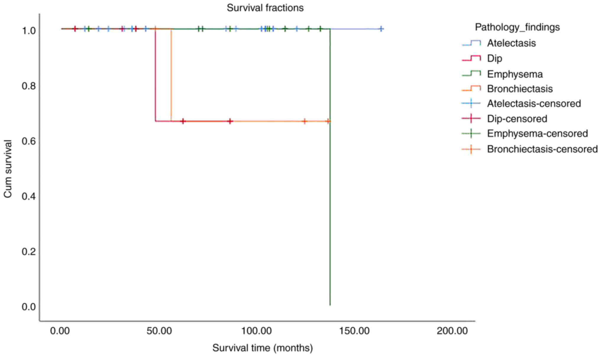
Simultaneous pathological findings were noted in the
biopsy specimens of 82 patients (56 patients with TC, 26 patients
with AC). These findings were atelectasis, emphysema,
bronchiectasis and desquamative interstitial pneumonia (DIP)
(Table II).
| Table II.Concurrent pathological findings of
pulmonary carcinoid tumors. |
Table II.
Concurrent pathological findings of
pulmonary carcinoid tumors.
| Pathological
findings | TC | AC | All |
|---|
| DIP | 4 | 7 | 11 (13.4%) |
| Emphysema | 13 | 7 | 20 (24.4%) |
| Bronchiectasis | 8 | 0 | 8 (9.8%) |
| Atelectasis | 31 | 12 | 43 (52.4%) |
The observation of the patients ended after a mean
follow-up time of 96 months. All the patients had a good attendance
at the follow-up appointments. Of the 82 patients that had
simultaneous pathological findings, 6 patients did not survive. All
the deaths were related to disease progression. In total, 4
patients were in stage II and 2 patients were in stage III, while 4
patients had AC and 2 patients had TC. There was a statistically
significant difference in survival between patients with different
simultaneous pathological findings (P=0.001). The most deaths were
observed among patients with bronchiectasis (Table III). The survival rates based on
simultaneous pathological findings are presented in Fig. 1 and Table III.
| Table III.Kaplan Meier survival analysis based
on concurrent pathological findings. |
Table III.
Kaplan Meier survival analysis based
on concurrent pathological findings.
| Characteristic | Survival time
(months) | 95% CI | No. of deaths | % | No. of
survivors | % | Log-rank test
P-value |
|---|
| Pathological
findings |
|
|
|
|
|
| 0.001 |
|
Atelectasis | 72.2 | 59.6-84.8 | 0 | 0 | 44 | 100.0 |
|
|
DIP | 46.6 | 28.7-64.5 | 2 | 18.2 | 8 | 81.8 |
|
|
Emphysema | 100.5 | 83.9-117.1 | 2 | 10 | 18 | 90.0 |
|
|
Bronchiectasis | 91.0 | 55.8-126.1 | 2 | 25 | 6 | 75.0 |
|
As regards all the patients, there was a total of
eight deaths. According to the Cox regression univariate analysis
for all the patients, the type of carcinoid was associated with
patient survival, with improved survival rates in patients with
TCs, while age, sex, stage and simultaneous pathological findings
were not associated with patient survival (Table IV). The results of the comparison
between the overall survival of patients with TCs and ACs is
presented in Table SI. The
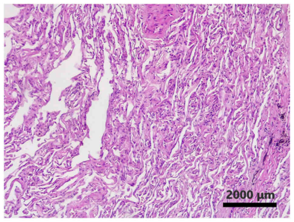
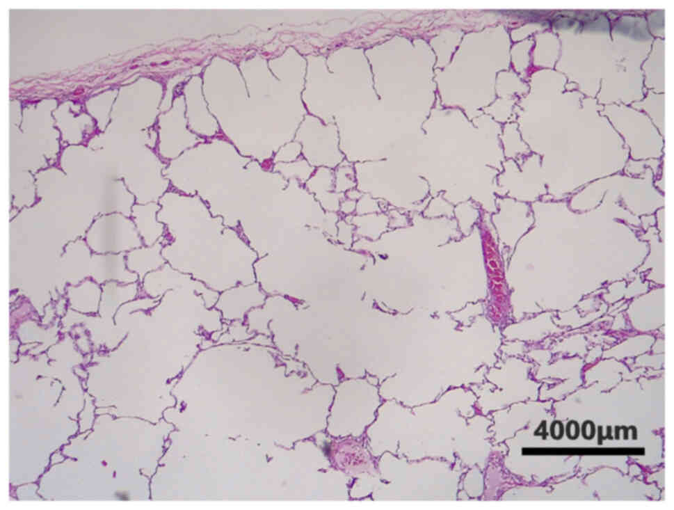
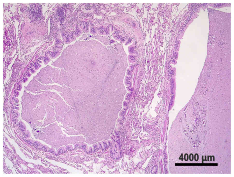
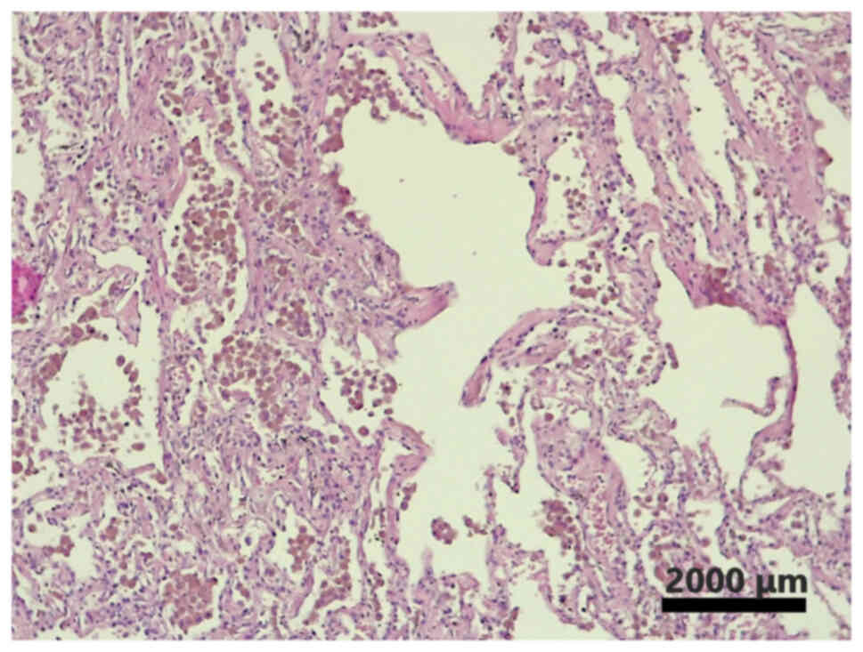
simultaneous pathological findings in the biopsy specimens of
patients with surgically resected lung carcinoids are illustrated
in Fig. 2, Fig. 3, Fig.
4, 5.
| Table IV.Univariate analysis of prognostic
factors analyzed by Cox's hazard-regression model. |
Table IV.
Univariate analysis of prognostic
factors analyzed by Cox's hazard-regression model.
| Variable | Exp(B) | P-value | 95% CI for
Exp(B) |
|---|
| Pathological
findings |
| 0.064 |
|
|
Atelectasis vs. DIP | 0.114 | 0.043 | 0.014-0.029 |
|
Atelectasis vs. emphysema | 0.000 | 0.954 | 0.000-1,
281E+188 |
|
Atelectasis vs.
bronchiectasis | 1.270 | 0.813 | 0.176-9.177 |
| Pathological
findings (presence vs. absence) | 1.120 | 0.976 | 0.269-3.873 |
| Age at surgery
(>45 vs. <45 years) | 2.767 | 0.244 | 0.499-15.35 |
| Sex (Male vs.
female) | 1.237 | 0.743 | 0.346-4.424 |
| Histological type
(typical vs. atypical) | 0.191 | 0.001 | 0.053-0.687 |
| Stage |
| 0.131 |
|
| I vs.
II | 0.491 | 0.886 | 0.087-2.787 |
| I vs.
III | 2.157 | 0.867 | 0.394-11.81 |
Discussion
According to the results of the present study,
atelectasis was related to the most favorable outcome among
patients with simultaneous pathological findings, with a survival
rate of 100%. Atelectasis is a term describing the loss of lung
volume as a result of the collapse of lung tissue. According to the
underlying pathophysiological mechanisms, atelectasis is classified
as obstructive atelectasis, which is the consequence of a blockage
of the airways, and non-obstructive atelectasis. It is also
classified according to the amount of lung involved (lobar,
segmental, subsegmental atelectasis), or the location involved
(specific lobe or segment) (20).
Non-obstructive atelectasis is caused by the loss of contact
between the visceral and parietal pleural membranes, the
compression of lung parenchyma, surfactant dysfunction, the
scarring or infiltration of lung tissue, and strong vertical
acceleration forces; it is thus divided into passive, compressive,
adhesive, cicatricial, replacement, acceleration, rounded and
plate-like atelectasis (21).
To the best of our knowledge, the present study is
the first to describe atelectasis as a prognostic factor of
survival in lung neuroendocrine tumors. Atelectasis has been
reported as a prognostic factor in other types of lung cancer. More
specifically, atelectasis has been shown to be associated with the
prolonged survival of patients with non-small cell lung carcinoma
(NSCLC) (22–25) even at advanced stages (26). In lung cancer, atelectasis
traditionally develops due to endobronchial obstruction and rarely
due to the compression of a mass or pleural effusion (25). It has been hypothesized that the
prolonged survival of patients with atelectasis may be associated
with decreased intratumoral blood flow and nutrition due to
vascular shunts in the adjacent atelectatic region, leading to the
decreased release of inflammatory cytokines from malignant cells.
Other hypotheses are the susceptibility of infection and the
alteration of immunity in atelectasis and the concentric growth of
the tumor, resulting in early presentation and diagnosis (25). These mechanisms may explain the
protective effect of atelectasis in patients with lung carcinoids.
However, according to another study, preoperative obstructive
pneumonitis and atelectasis can predict poor survival independently
in patients with NSCLC (27).
In the present study, patients with emphysema and
pulmonary carcinoids presented with a survival rate of 90%.
Pulmonary emphysema is a pathological definition. It demonstrates
the abnormal permanent enlargement of the airspaces distal to the
terminal bronchioles. This enlargement coexists with the
destruction of their walls without conspicuous fibrosis (28). In addition, emphysema has not been
studied as a prognostic factor for patients with lung carcinoids.
Emphysema can develop in patients with pulmonary carcinoids through
the check-valve effect of an endobronchial tumor and the
compensatory effect of the residual lung (29). Emphysema, as estimated with imaging
methods, has been reported as a prognostic factor in other types of
lung cancer. The severity of lung emphysema has been found to be
associated with a decreased survival rate, the development of
post-operative complications in early-stage cancer and the
recurrence of resected lung cancer in patients with NSCLC (30–32),
as well as with a poor prognosis of patients with small cell lung
carcinoma (33).
Bronchiectasis is a lung disorder that is mostly
caused by bronchial inflammation and characterized by the permanent
dilation of the airways due to bronchial wall destruction (34). According to the results of the
present study, patients with bronchiectasis had a worse outcome
compared to patients with other simultaneous pathological findings.
Bronchiectasis have, in some cases, been described as the first
finding before a diagnosis of a pulmonary carcinoid. Bronchiectasis
develop due to recurrent pneumonia in cases that a lung carcinoid
leads to bronchial obstruction. The persistence of inflammation is
a major factor in the pathogenesis of bronchiectasis (35–38).
In addition, it has been reported that the presence of
bronchiectasis is a predisposing factor for the genesis of
pulmonary neuroendocrine tumors (39–41).
To date, bronchiectasis has not been studied as a prognostic factor
in lung carcinoids or in other types of lung cancer, at least to
the best of our knowledge.
DIP is an interstitial lung disease associated with
smoking. The accumulation of macrophages in alveoli is the
underlying mechanism of its pathogenesis, leading to interstitial
inflammation and fibrosis. However, DIP has been mentioned as a
result of other exposures and disease conditions, such as
occupational exposures, medications and autoimmune diseases
(42). Moreover, it has been
reported that DIP is a pattern of pulmonary reaction that
accompanies other pulmonary lesions, such as eosinophilic
granulomas and rheumatoid nodules (42). To the best of our knowledge, the
present study is the first to report the co-existence of DIP with
lung carcinoids and the impact of DIP on the survival of patients
with lung carcinoids that underwent thoracic surgery. Patients with
DIP and pulmonary carcinoids had a survival rate of 81.8%.
Of note, there was not a statistically significant
difference in survival rates between patients with and without
simultaneous pathological findings. It has been reported that
monocyte-derived myeloid cells in lung carcinoid tissues have equal
to slightly lower expression scores of numerous gene profiles
associated with inflammation and the immune response when compared
to normal tissues, indicating that the lung carcinoid immune
microenvironment is predominated by non-inflammatory
monocyte-derived myeloid cells, without mentioning if this finding
is associated with better outcomes (43). However, it remains to be determined
whether this finding is associated with improved outcomes.
The present study has some limitations. The present
study was one of the largest on lung carcinoids in Greece. The data
rely on a large number of patients with an accurate follow-up
period. However, this was a single-center study and only a limited
number of disease-specific deaths were recorded. Therefore, a
multivariate analysis could not be performed. Another limitation of
the present study is the absence of control group which could be
patients with other types of lung cancer. Thus, further larger
multi-center, prospective studies are required to evaluate the role
of these pathological findings in the outcomes of patients with
surgically resected lung carcinoids.
In conclusion, to the best of our knowledge, the
present study is the first to examine the impact of the
simultaneous pathological findings of resected lung tissue
specimens on the survival of patients with pulmonary carcinoids.
There was a statistically significant difference in the survival
rates of patients with resected lung carcinoids with different
simultaneous pathological findings. However, further studies are
required to assess the role of these findings in the survival of
these patients.
Supplementary Material
Supporting Data
Acknowledgements
Not applicable.
Funding
Funding: No funding was received.
Availability of data and materials
The datasets used and/or analyzed during the current
study are available from the corresponding author on reasonable
request.
Authors' contributions
JD, JJ and EZ conceptualized the study. AP performed
the immunohistochemical examination and prepared the tables. VEG,
KM, CD, SC, PP, AAF and AG advised on patient treatment, and wrote
and prepared the draft of the manuscript. JD and DAS analyzed the
data and provided critical revisions. NT and PS performed the
statistical analysis. VEG and EZ confirm the authenticity of all
the data. All authors contributed to manuscript revision and have
read and approved the final version of the manuscript.
Ethics approval and consent to
participate
Ethical approval for the present study was obtained
from the Research Ethics Committee of Athens medical Group with
file accession no. 4234. The study was in line with the declaration
of Helsinki in 1995 (as revised in Edinburgh 2000). Written
informed consent was obtained from the patients for publication of
this research and accompanying images.
Patient consent for publication
Not applicable.
Competing interests
DAS is the Editor-in-Chief for the journal, but had
no personal involvement in the reviewing process, or any influence
in terms of adjudicating on the final decision, for this article.
The other authors declare that they have no competing
interests.
References
|
1
|
Yang Z, Wang Z, Duan Y and Xu S:
Clinicopathological characteristics and prognosis of resected cases
of carcinoid tumors of the lung. Thorac Cancer. 7:633–638. 2016.
View Article : Google Scholar : PubMed/NCBI
|
|
2
|
Kosmidis PA: Treatment of carcinoid of the
lung. Curr Opin Oncol. 16:146–149. 2004. View Article : Google Scholar
|
|
3
|
Fink G, Krelbaum T, Yellin A, Bendayan D,
Saute M, Glazer M and Kramer MR: Pulmonary carcinoid: Presentation,
diagnosis, and outcome in 142 cases in Israel and review of 640
cases from the literature. Chest. 119:1647–1651. 2001. View Article : Google Scholar
|
|
4
|
Fraser RS, Muller NL, Colman N and Pare
PD: Fraser and Pare's diagnosis of diseases of the chest.
Neuroendocrine neoplasms. 4th edition. Philadelphia: WB Saunders
Co; pp. 1229–1250. 1999
|
|
5
|
Colby TV, Koss M and Travis WD: Atlas of
tumor pathology. Tumors of the lower respiratory tract. Armed
Forces Institute of Pathology; Washington, DC: 1995
|
|
6
|
Soga J and Yakuwa Y: Bronchopulmonary
carcinoids: An analysis of 1,875 reported cases with special
reference to a comparison between typical carcinoids and atypical
varieties. Ann Thorac Cardiovasc Surg. 5:211–219. 1999.PubMed/NCBI
|
|
7
|
Mark EJ, Quay SC and Dickersin GR:
Papillary carcinoid tumor of the lung. Cancer. 48:316–324. 1981.
View Article : Google Scholar : PubMed/NCBI
|
|
8
|
Travis WD, Linnoila RI, Tsokos MG,
Hitchcock CL, Cutler GB Jr, Nieman L, Chrousos G, Pass H and
Doppman J: Neuroendocrine tumors of the lung with proposed criteria
for large-cell neuroendocrine carcinoma. An ultrastructural,
immunohistochemical, and flow cytometric study of 35 cases. Am J
Surg Pathol. 15:529–553. 1991. View Article : Google Scholar
|
|
9
|
Travis WD, Rush W, Flieder DB, Falk R,
Fleming MV, Gal AA and Koss MN: Survival analysis of 200 pulmonary
neuroendocrine tumors with clarification of criteria for atypical
carcinoid and its separation from typical carcinoid. Am J Surg
Pathol. 22:934–944. 1998. View Article : Google Scholar
|
|
10
|
Miller RR and Müller NL: Neuroendocrine
cell hyperplasia and obliterative bronchiolitis in patients with
peripheral carcinoid tumors. Am J Surg Pathol. 19:653–658. 1995.
View Article : Google Scholar
|
|
11
|
Wang J, Ren S, Liu Y, Guo K, Chen X, Wang
Z and Chen R: Carcinoid tumorlets co-existing with chronic
pulmonary inflammatory processes: Imaging findings and histological
appearances. Med Sci Monit. 26:e9260142020.
|
|
12
|
Huang Y, Yang X, Lu T, Li M, Zhao M, Yang
X, Ma K, Wang S, Zhan C, Liu Y and Wang Q: Assessment of the
prognostic factors in patients with pulmonary carcinoid tumor: A
population-based study. Cancer Med. 7:2434–2441. 2018. View Article : Google Scholar : PubMed/NCBI
|
|
13
|
Georgakopoulou VE, Zygouris E, Nikokiris
C, Damaskos C, Pierrakou A, Garmpis N, Garmpi A, Sklapani P,
Aravantinou A, Trakas N, et al: Predictive indicators of survival
in patients with surgically resected lung carcinoid tumors at a
Greek medical center. Cureus. 12:e103002020.PubMed/NCBI
|
|
14
|
Georgakopoulou VE, Zygouris E, Damaskos C,
Pierrakou A, Papalexis P, Garmpis N, Aravantinou-Fatorou A,
Chlapoutakis S, Diamantis E, Nikokiris C, et al: Prognostic value
of the immunohistochemistry markers CD56, TTF-1, synaptophysin,
CEA, EMA and NSE in surgically resected lung carcinoid tumors. Mol
Clin Oncol. 16:312022. View Article : Google Scholar
|
|
15
|
Papaxoinis G, Lamarca A, Quinn AM, Mansoor
W and Nonaka D: Clinical and pathologic characteristics of
pulmonary carcinoid tumors in central and peripheral locations.
Endocr Pathol. 29:259–268. 2018. View Article : Google Scholar
|
|
16
|
Beasley MB, Thunnissen FB, Brambilla E,
Hasleton P, Steele R, Hammar SP, Colby TV, Sheppard M, Shimosato Y,
Koss MN, et al: Pulmonary atypical carcinoid: Predictors of
survival in 106 cases. Hum Pathol. 31:1255–1265. 2000. View Article : Google Scholar
|
|
17
|
Halabi S and Owzar K: The importance of
identifying and validating prognostic factors in oncology. Semin
Oncol. 37:e9–e18. 2010. View Article : Google Scholar
|
|
18
|
Travis WD, Brambilla E, Nicholson AG,
Yatabe Y, Austin JHM, Beasley MB, Chirieac LR, Dacic S, Duhig E,
Flieder DB, et al: The 2015 World Health Organization
classification of lung tumors: Impact of genetic, clinical and
radiologic advances since the 2004 classification. J Thorac Oncol.
10:1243–1260. 2015. View Article : Google Scholar
|
|
19
|
Feng SH and Yang ST: The new 8th TNM
staging system of lung cancer and its potential imaging
interpretation pitfalls and limitations with CT image
demonstrations. Diagn Interv Radiol. 25:270–279. 2019. View Article : Google Scholar
|
|
20
|
Woodring JH and Reed JC: Types and
mechanisms of pulmonary atelectasis. J Thorac Imaging. 11:92–108.
1996. View Article : Google Scholar : PubMed/NCBI
|
|
21
|
Müller NL, Fraser RS, Colman NC and Paré
PD: Radiologic diagnosis of diseases of the chest. WB Saunders;
Philadelphia, PA: 2001
|
|
22
|
Tepavac A, Secen N, Sazdanic Velikic D,
Popovic G and Perin B: Atelectasis: Positive or negative prognostic
factor on outcome of patients with non-small cell lung cancer? J
BUON. 15:679–683. 2010.PubMed/NCBI
|
|
23
|
Gu K, Lee HY, Lee K, Choi JY, Woo SY, Sohn
I, Kim HK, Choi YS, Kim J, Zo JI and Shim YM: Integrated evaluation
of clinical, pathological and radiological prognostic factors in
squamous cell carcinoma of the lung. PLoS One. 14:e02232982019.
View Article : Google Scholar : PubMed/NCBI
|
|
24
|
Hasbek Z, Yucel B, Salk I, Turgut B,
Erselcan T, Babacan NA and Kacan T: Potential impact of atelectasis
and primary tumor glycolysis on F-18 FDG PET/CT on survival in lung
cancer patients. Asian Pac J Cancer Prev. 15:4085–4089. 2014.
View Article : Google Scholar : PubMed/NCBI
|
|
25
|
Bulbul Y, Eris B, Orem A, Gulsoy A, Oztuna
F, Ozlu T and Ozsu S: Pulmonary atelectasis and survival in
advanced non-small cell lung carcinoma. Ups J Med Sci. 115:176–180.
2010. View Article : Google Scholar
|
|
26
|
Dediu M, Crisan E, Radut M, Tarlea A,
Median D, Alexandru A, Vremes G and Gal C: The favorable prognostic
significance of atelectasis in patients with advanced non-small
cell lung cancer: Results of a prospective observational study.
Lung Cancer. 63:271–276. 2009. View Article : Google Scholar : PubMed/NCBI
|
|
27
|
Pang Z, Ding N, Dong W, Ni Y, Zhang T, Qu
X, Du J and Liu Q: Prognostic effects of preoperative obstructive
pneumonitis or atelectasis and comparison with tumor size in
non-small cell lung cancer. J Thorac Dis. 9:768–778. 2017.
View Article : Google Scholar : PubMed/NCBI
|
|
28
|
Standards for the diagnosis and care of
patients with chronic obstructive pulmonary disease, . American
thoracic society. Am J Respir Crit Care Med. 152:S77–S121.
1995.PubMed/NCBI
|
|
29
|
Liu S, Ko S and Chen W: Bronchial
carcinoid tumor presenting with complete lobar collapse and
unilateral lung emphysema. Clin Imaging. 24:159–161. 2000.
View Article : Google Scholar : PubMed/NCBI
|
|
30
|
Sato S, Nakamura M, Shimizu Y, Goto T,
Koike T, Ishikawa H and Tsuchida M: The impact of emphysema on
surgical outcomes of early-stage lung cancer: A retrospective
study. BMC Pulm Med. 19:732019. View Article : Google Scholar : PubMed/NCBI
|
|
31
|
Lee SJ, Yoo JW, Ju S, Cho YJ, Kim JD, Kim
SH, Jang IS, Jeong BK, Lee GW, Jeong YY, et al: Quantitative
severity of pulmonary emphysema as a prognostic factor for
recurrence in patients with surgically resected non-small cell lung
cancer. Thorac Cancer. 10:421–427. 2019. View Article : Google Scholar : PubMed/NCBI
|
|
32
|
Gao YH, Guan WJ, Liu Q, Wang HQ, Zhu YN,
Chen RC and Zhang GJ: Impact of COPD and emphysema on survival of
patients with lung cancer: A meta-analysis of observational
studies. Respirology. 21:269–279. 2016. View Article : Google Scholar : PubMed/NCBI
|
|
33
|
Lee HY, Kim EY, Kim YS, Ahn HK and Kim YK:
Prognostic significance of CT-determined emphysema in patients with
small cell lung cancer. J Thorac Dis. 10:874–881. 2018. View Article : Google Scholar : PubMed/NCBI
|
|
34
|
Georgakopoulou VE, Trakas N, Damaskos C,
Garmpis N, Karakou E, Chatzikyriakou R, Lambrou P and Tsiafaki X:
Neutrophils to lymphocyte ratio as a biomarker in bronchiectasis
exacerbation: A retrospective study. Cureus.
12:e97282020.PubMed/NCBI
|
|
35
|
Meng Z and Linian H: A typical carcinoid
tumor of the lung presenting as diffuse cystic bronchiectasis. J
Coll Physicians Surg Pak. 30:229–230. 2020. View Article : Google Scholar : PubMed/NCBI
|
|
36
|
Charokopos N, Tsiamita M, Karkoulias K,
Koumoundourou D, Aletra C, Dougenis D and Spiropoulos K: Carcinoid
tumour behind bronchiectasis. Monaldi Arch Chest Dis. 65:110–113.
2006.PubMed/NCBI
|
|
37
|
Janah H, Jabri H, Bopaka RG, Khattabi WE
and Afif H: Localised bronchiectasis revealing carcinoid tumor. Pan
Afr Med J. 24:2782016.(In French). View Article : Google Scholar : PubMed/NCBI
|
|
38
|
Ghobadi H, Farzaneh E and Darvishkhah H:
Carcinoid tumor with localized bronchiectasis. Tanaffos. 12:56–60.
2013.PubMed/NCBI
|
|
39
|
Liu B, Pu Q, Liu L and Che G: Facial
flushing due to multifocal tumorlets in the lung with
bronchiectasis. Ann Thorac Surg. 88:641–642. 2009. View Article : Google Scholar : PubMed/NCBI
|
|
40
|
Klinke F, Bosse U and Höfler H: The
tumorlet carcinoid in bronchiectasis-changed lungs. An example of a
multifocal, endocrine tumor. Pneumologie. 44 (Suppl 1):S607–S609.
1990.(In German).
|
|
41
|
Canessa PA, Santini D, Zanelli M and
Capecchi V: Pulmonary tumourlets and microcarcinoids in
bronchiectasis. Monaldi Arch Chest Dis. 52:138–139. 1997.PubMed/NCBI
|
|
42
|
Chakraborty RK, Basit H and Sharma S:
Desquamative Interstitial Pneumonia. StatPearls Treasure Island
(FL): StatPearls Publishing; August 12–2020
|
|
43
|
Bischoff P, Trinks A, Wiederspahn J,
Obermayer B, Pett JP, Jurmeister P, Elsner A, Dziodzio T, Rückert
JC, Neudecker J, et al: The single-cell transcriptional landscape
of lung carcinoid tumors. Int J Cancer. 150:2058–2071. 2022.
View Article : Google Scholar : PubMed/NCBI
|