Introduction
Gastric cancer (GC) is a major health threat, with
the highest morbidity and mortality rates among malignant tumors of
the digestive system (1). GC has a
strong heterogeneity in terms of biological characteristics,
primary tumor sites, pathogenesis, pathological types and molecular
types, which may result in varying biological behaviors and
prognoses, making it challenging to treat. a-fetoprotein
(AFP)-producing GC (AFPGC) is a special and rare type of GC with an
incidence of 1.3–15% (2). Due to
the rapid progression of AFPGC and its high rate of liver
metastasis, some patients with AFPGC are inoperable at the time of
diagnosis. Furthermore, those who undergo radical surgical
resection are more likely to experience postoperative recurrence
and metastasis, and chemotherapy is less effective for AFPGC than
for gastric adenocarcinoma (3).
The standard first-line treatment for advanced or
recurrent GC is chemotherapy combined with immunotherapy. For cases
with human epidermal growth factor receptor-2 (HER-2)
amplification, the use of trastuzumab in combination with a
molecular targeted drug is recommended. The chemotherapy regimen is
commonly a combination of two or three fluorouracil- and
platinum-based drugs (4,5). The second- and third-line treatments
mainly include single-drug therapies with a low efficacy. At
present, there is no universal standard treatment regimen. As a
result, the selection of drugs may be quite random. Although an
experimental treatment plan can be applied, it may delay the
treatment process and thus worsen the patients' quality of life.
There is currently no efficient method for the assessment of a
cancer patient's sensitivity to chemotherapeutic drugs. Therefore,
the accurate selection of medical treatment, particularly mediated
via effective drug-screening models, is urgently necessary to
improve adjuvant treatment or second-line therapy.
Patient-derived xenografts (PDXs) can be used for
the pre-evaluation of therapeutic outcomes (6). In contrast to cell line-derived tumor
models, PDX models retain the genetic characteristics of the human
tumor specimens from which they were derived (7). PDX models are generated by the
implantation of fresh human tumor tissues into immunodeficient
mice, to reduce rejection of the tumor cells by the mouse. PDX
models can effectively simulate the treatment response of the
parent tumors and provide information that may aid the selection of
therapeutic targets and protocols (8,9). To
date, the application of traditional PDX models in individualized
treatment has failed due to the low success rate and lengthy
implantation period of the models. PDX modeling is also expensive,
technically cumbersome and requires a large amount of tumor tissue
(10). Mini-PDX is a drug
sensitivity test model that maintains the oncogenicity of a
patients' tumor cells, in which cells from human tumor tissues are
injected into immunocompromised mice within special capsules to
create tumor xenografts; this is a promising model accompanied by
reduced complexity and faster turnaround compared with traditional
PDX models.
The present study aimed to evaluate the application
of the mini-PDX model combined with genetic analysis of tissue
samples to guide the individualized treatment of a patient with
metastatic AFPGC and HER-2-positive GC.
Materials and methods
Patient and ethics
A 74-year-old male patient with gastric cancer was
included in the study. The patient underwent gastroscopy at Wuxi
Hospital Affiliated to Nanjing University of Chinese Medicine
(Wuxi, China) on December 17, 2018. The study protocol was approved
by the Institutional Review Board of Wuxi Hospital of Traditional
Chinese Medicine (approval no. 201809001J01-01) and the
Institutional Animal Care and Use Committee (IACUC) of Shanghai
LIDE Biotech Co., Ltd. (approval no. LDIACUC007).
Histopathological examination
A portion of the tumor tissue was detached and fixed
by immersion in 10% neutral buffered formalin solution (pH 7.4) for
12 h at 24°C. The portion was then cut transversely, paraffin
embedded and arranged as 3-µm sections, which were stained with
hematoxylin and eosin (H&E) for 20 min at 24°C for the
microscopic assessment of tumor tissue. The tissues were examined
under a light microscope in a random order.
Immunohistochemistry
Tumor tissues were fixed with 4% paraformaldehyde
for 24 h at 24°C, and the fixed tissues were dehydrated and
embedded in paraffin. Later, 4-µm sections of the paraffin-embedded
tissues were prepared. Endogenous peroxidase activity was blocked
by incubating the tissues at 25°C with 0.6%
H2O2 in methanol for 20 min. Sections were
subsequently blocked with 10% normal horse serum (Wuhan Boster
Biological Technology, Ltd.) for 5 min at 25°C. Next, the sections
were incubated at 4°C overnight with AFP antibody (1:50; cat. no.
ab130748; Abcam). The slides were then incubated with the secondary
antibody Goat Anti-Rabbit IgG (1:2,000; cat. no. ab205718; Abcam)
for 30 min at room temperature. Subsequently, DAB chromogen (cat.
no. ab64238; Abcam) was added to the tissue and incubated for 1–10
min, depending on the desired stain intensity. The sections were
rinsed four times in PBS and were subsequently stained with
hematoxylin and hydrochloric acid alcohol for 20 min at 24°C,
followed by washing with tap water for 10 min. After dehydration
and transparency, the tissue sections were analyzed under a
bright-field Olympus BX-40 light microscope (Olympus Corporation).
The classification of nuclear AFP expression was assessed using the
following scores: Unstained, 0; <25% positive cells, 1+; 25–50%
positive cells, 2+; 50–75% positive cells, 3+; and >75% positive
cells, 4+.
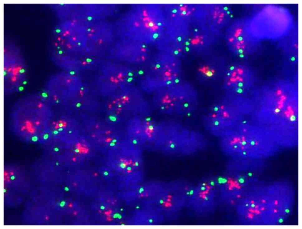
Fluorescence in situ hybridization
(FISH)
A PathVysion HER-2 DNA probe kit was purchased from
Vysis Inc. (Abbott Molecular, Inc.). A fluorescence microscope
(Bx51; Olympus Corporation) was used to view the FISH staining. The
HER-2/neu gene FISH test was performed on the tumor tissue
according to the instructions and requirements of the kit.
Mini-PDX assay
A Mini-PDX assay was carried out using the OncoVee
MiniPDX® kit (LIDE Biotech Co., Ltd.) in the joint
animal laboratory of Wuxi Hospital and Shanghai LIDE Biotech Co.,
Ltd. under the guidance of a researcher from LIDE. Briefly, the
tumor from the patient's gastroesophageal junction was harvested
and washed with Hanks' balanced salt solution (HBSS) to remove
non-tumor and necrotic tumor tissue. The tumor tissues were minced
and incubated with collagenase at 37°C for 1–2 h. After this, the
tumor cells were incubated with human anti-CD45 microbeads (cat.
no. 130-045-80, Miltenyi Biotec, Inc.) and human anti-fibroblast
microbeads (cat. no. 130-050-601; Miltenyi Biotec, Inc.) at 4°C for
30 min in the dark, at a final fixed volume of 220 µl, including 20
µl beads, 106 cells and buffer (PBS and 1% FBS).
Subsequently, tumor cell suspensions without blood cells or
fibroblasts were eluted with 1 ml buffer and collected. The
remaining cell suspension was transferred to HBSS-washed capsules
made of hollow fiber membranes with a pore size allowing the
passage of <500-kDa molecules; eventually each capsule contained
2,000 cells. A fiber system delivered media to the cells in a
manner similar to the delivery of blood through capillary networks
in vivo. A total of 8 female severe combined
immunodeficiency mice (6–8 weeks old) with average weight of ~24 g
(range, 22–28 g) were housed and monitored under
specific-pathogen-free conditions. The housing conditions comprised
a temperature of 20–26°C, humidity of 40–70% and a 12-h light/dark
cycle (11). Throughout the
experiment, all mice were able to eat and drink freely; feed and
water were autoclaved and replaced twice a week. For subcutaneous
implantation, the capsule was inserted through the subcutaneous
tissue via a special needle without any anesthesia being necessary.
One day after implantation of the tumor cell-containing capsule,
the tumor-bearing mice were randomized to the following groups:
Vehicle; docetaxel + S-1; capecitabine + oxaliplatin; and
irinotecan + S-1. S-1 is tegafur-gimeracil-oteracil, also known as
tegio. The treatment regimens were as follows: 20 mg/kg docetaxel
intraperitoneally (ip) every fourth day ×2 (Q4D*2) + 10 mg/kg S-1
ip every day for 5 days (QD*5); 400 mg/kg capecitabine orally (po)
every day for 7 days + 5 mg/kg oxaliplatin ip once weekly; and 50
mg/kg irinotecan ip Q4D*2 + 5 mg/kg S-1 po QD*5 (12). Each mouse received 6 capsules at
the same time and each regimen was tested in two mice. The final
results were calculated as the average of six repeated trials. The
relative change in body weight (RCBW) was calculated each day as
follows: RCBW=(BWi-BW0)/BW0 ×100%, where BWi is the average weight
on day i during drug administration and BW0 is the average weight
at the first administration. After 7 days, the tumor
cell-containing capsules were removed. After the experiment, the
mice were euthanized via CO2 inhalation in a euthanasia
chamber (50% chamber replacement rate/min) followed by cervical
dislocation, or by the administration of 0.1% ketamine sodium (100
mg/kg, intraperitoneal injection) (13) followed by cervical dislocation.
The viability of the cells was evaluated using a
CellTiter-Glo® Luminescent Cell Viability Assay kit
(G7571; Promega Corporation), following the manufacture's protocol.
The tumor cell proliferation rate relative to that of the control
group after the 7-day treatment was calculated using the following
equation: Relative proliferation rate=T/C ×100%, where T and C are
the cell viability values for the treatment and control group,
respectively. A lower relative proliferation rate indicated a
higher inhibitory effect of the drugs on the tumor cells.
Next-generation sequencing (NGS)
DNA was extracted from formalin-fixed,
paraffin-embedded (FFPE) tumor tissue samples using the QIAamp DNA
FFPE Tissue kit (Qiagen GmbH). In addition, 3 ml peripheral blood
from the patient was collected in an EDTA Vacutainer tube (BD
Diagnostics) and processed within 4 h. Peripheral blood lymphocytes
(PBLs) were separated by centrifugation at 1,600 × g for 10 min at
room temperature and used for the extraction of germline genomic
DNA, which served as the reference for germline variation and
single nucleotide polymorphism identification. The DNA
concentration in the PBLs and tissues was measured using a
Qubit™ Flex kit and the Qubit DNA Assay kit (both
Invitrogen; Thermo Fisher Scientific, Inc.). DNA sequencing was
carried out using 2×75-bp scanned-end reads on an Illumina HiSeq
3000 system (Illumina, Inc.) and the KAPA DNA Library Preparation
kit (cat. no. KK8234; Kapa Biosystems; Roche Diagnostics); the
loading concentration was 40.02 ng/µl DNA sequencing was applied to
a panel of 73 cancer-related genes in gastric adenocarcinoma.
Targeted gene sequencing was performed in the tumor and paired PBLs
with an average sequencing depth of 3850X and 431X, respectively.
More than 99% of the readings were mapped to areas where the tumor
and matched PBL samples were targeted. Sequencing data were
analyzed using software at default parameters. Adaptor sequences
and low-quality reads were removed. The clean reads were aligned to
the reference human genome (hg19) using Burrows-Wheeler Aligner
(version 0.7.12-r1039) (14).
Realignment and recalibration were performed using Genome Analysis
Toolkit (version 3.4-46-gbc02625) (15). Single nucleotide variants were
called using MuTect (version 1.1.4) (16).
Statistical analysis of mini-PDX
Statistical analyses were performed using SPSS
version 22.0 (IBM Corp.). One-way ANOVA followed by Tukey's post
hoc test was used for evaluating differences among groups.
P<0.05 was considered to indicate a statistically significant
difference.
Results
Clinical data
The 74-year-old male patient presented with a
bloated and uncomfortable upper abdomen and underwent gastroscopy
on December 17, 2018. Gastroscopy revealed a large ulcer from the
cardia to the fundus of the stomach. The pathology examination
confirmed adenocarcinoma. Abdominal contrast-enhanced computed
tomography (CT) revealed a mass in the lesser curvature of the
stomach. Combined with the medical history, the mass was initially
considered as a swollen lymph node. The gastric wall was thickened
in the cardiac part, gastric fundus and lesser curvature of the
stomach. Bronchiectasis with infection was detected in the right
lung. Considering that the patient had a history of bronchiectasis
for >10 years, his extremely poor pulmonary function indicated
that he was an inappropriate candidate for GC surgery. A serum test
performed in January 2019 revealed an AFP level of >1,210 ng/ml,
while the levels of carcinoembryonic antigen, carbohydrate antigen
19-9 and carbohydrate antigen 72-4 were all within the normal
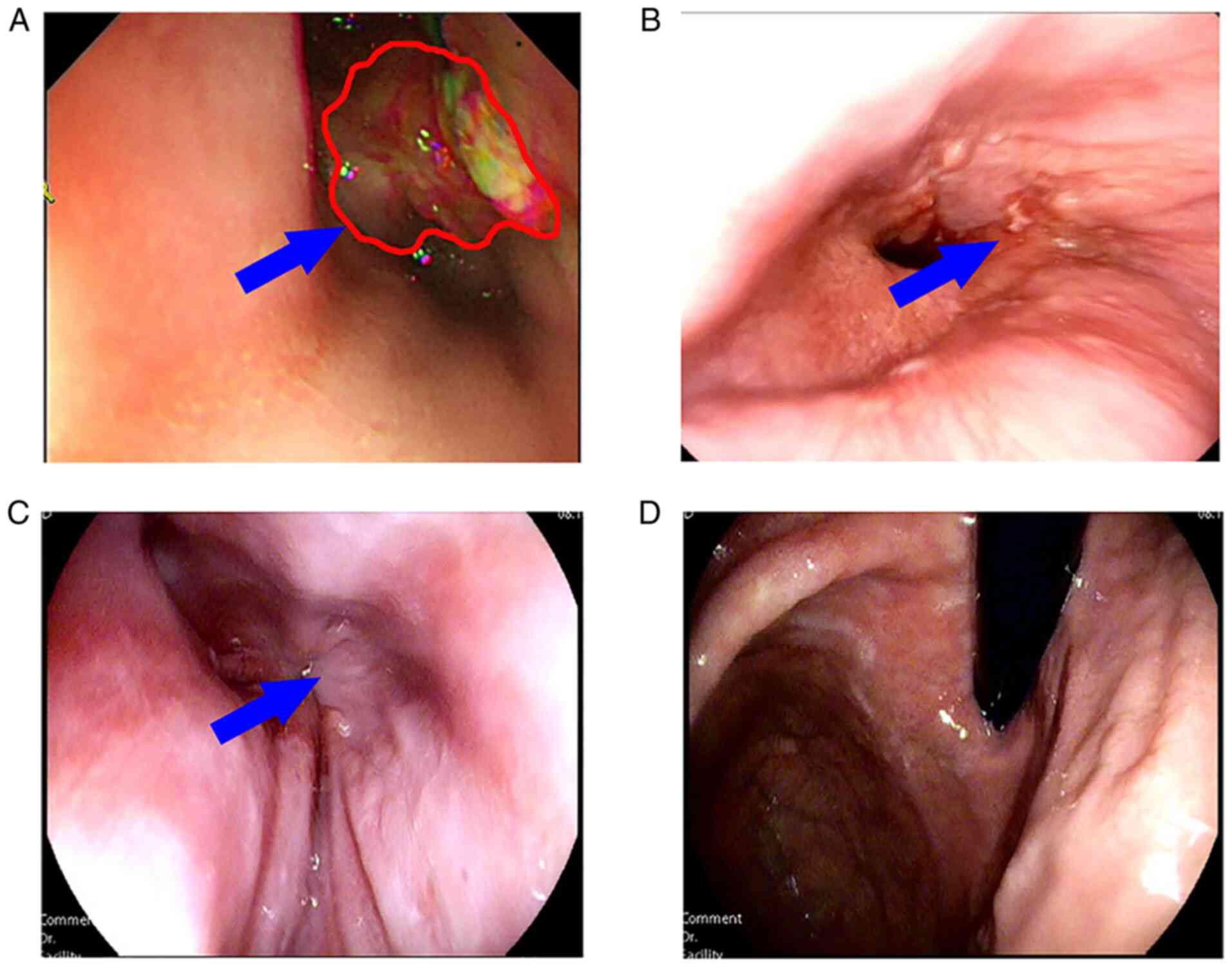
range. Gastroscopy showed the presence of carcinoma of the lower
esophagus near the cardia, which the endoscope could not pass
(Fig. 1A) due to obstruction of
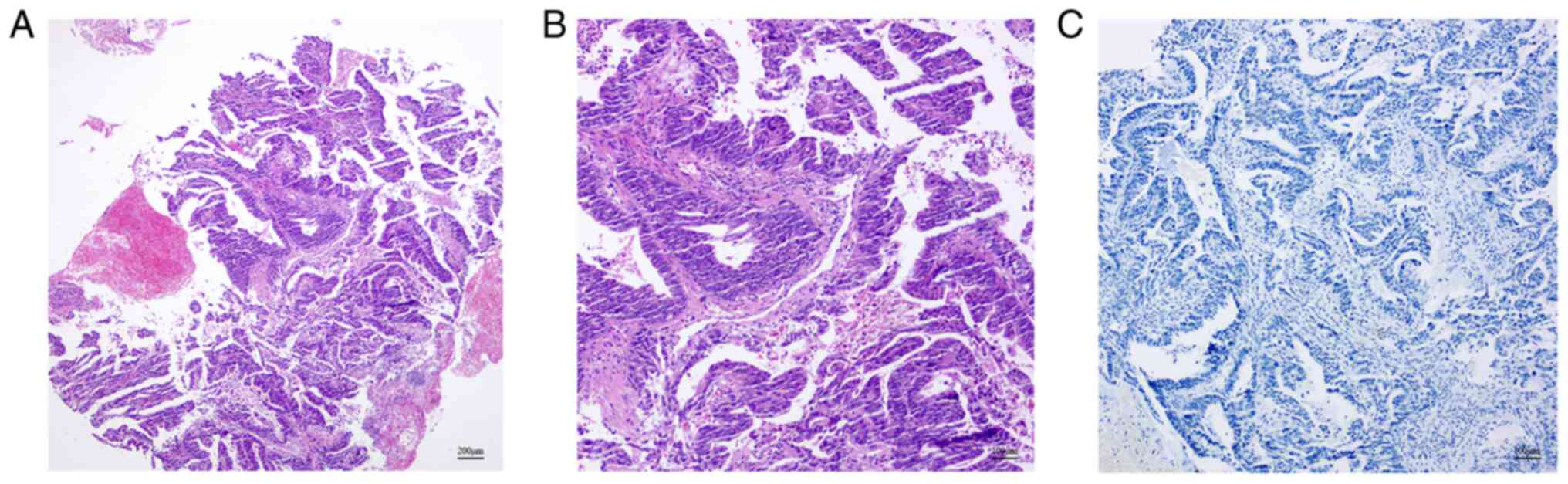
the lumen. At the same time, tumor tissue was biopsied for the
mini-PDX assay. Pathology confirmed adenocarcinoma (Fig. 2). Immunohistochemical assay showed
the following results: HER-2 (2+), epidermal growth factor receptor
(EGFR)+ (weak), Ki-67+ (~50%), P53-+ (diffuse) and AFP-. The FISH
assay indicated HER-2 amplification (Fig. 3). Contrast-enhanced CT examination
of the upper abdomen revealed swollen lymph nodes with a size of
~4.5×3.5 cm in the lesser curvature of the stomach (Fig. 4A).
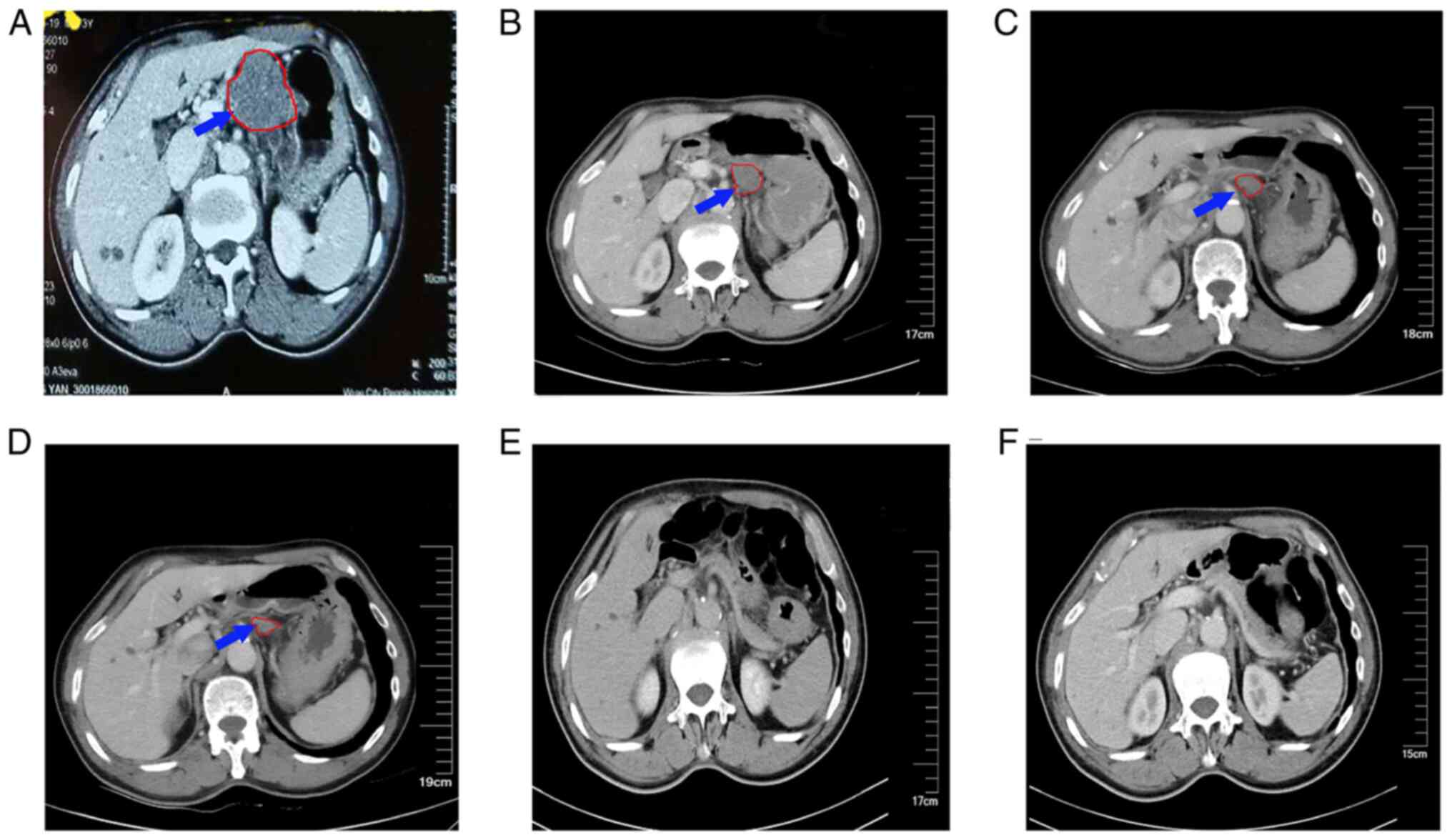 | Figure 4.Abdominal enhanced CT in which the
blue arrow and red outline indicate swollen lymph nodes and their
boundary at the lesser curvature of stomach, which gradually shrank
and then disappeared after treatment. (A) Abdominal CT scan
performed on December 17, 2018, showing lymph nodes in the lesser
curvature of the stomach ~4.5×3.5 cm in size. (B) Abdominal CT scan
carried out on March 7, 2019, showing that the lymph nodes in the
lesser curvature of the stomach had decreased to ~ 3.5×2.5 cm. (C)
Abdominal CT scan performed on May 23, 2019, showing a further
reduction in the size of the lymph nodes to ~2.5×1.5 cm. (D)
Abdominal CT carried out on October 21, 2019, indicating that the
size of the lymph nodes was ~2×1.1 cm. Abdominal contrast-enhanced
CT scans performed on (E) August 18, 2020 and (F) May 11, 2021,
showing that the enlarged lymph nodes in the lesser curvature had
disappeared. CT, computed tomography. |
Mini-PDX assay
In this assay, after 7 days of drug treatment, the
mini-PDX devices were removed from the mice and the relative
proliferation rates of the tumor cells were determined. The results
revealed that tumor cell proliferation in the three treatment
groups [irinotecan + S-1 (tumor (T)/control (C)=16%], docetaxel +
S-1 (T/C=29%) and capecitabine + oxaliplatin (T/C=40%)] was
significantly lower than that in the control group, demonstrating
the strong inhibitory effect of the three regimens on the growth of
tumor cells. The irinotecan + S-1 group had the lowest relative
tumor cell proliferation rate, which indicated that this regimen
had the strongest inhibitory effect (Fig. 5A). Furthermore, no significant
reduction in body weight of mice was observed in any group during
the experiment, which suggested that the dosage of the drug regimen
was within the safe range (Fig.
5B).
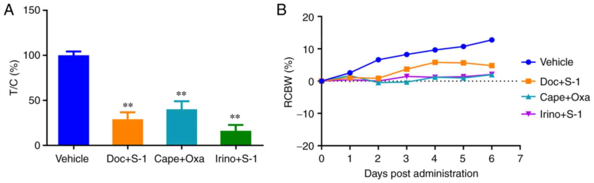 | Figure 5.Response of the mini-PDX to various
chemotherapy regimens and the change of body weight during drug
delivery. (A) The proliferation of tumor cells in mini-PDX implants
was observed in mice treated with vehicle, Doc + S-1, Cape + Oxa
and Irino + S-1. The drug treatment began 1 day after tumor cell
implantation. The tumor cell proliferation rate relative to that of
the control group was calculated after the 7-day treatment.
**P<0.001 vs. the vehicle group. (B) The change in body weight
of the mice was monitored. Throughout the experiment, no
significant reduction in the weight of mice in each group was
detected, suggesting that the dosage of the drug regimen was within
the safe range. T, tumor; C, control; Doc, docetaxel; Cape,
capecitabine; Oxa, oxaliplatin; Irino, irinotecan; S-1,
tegafur-gimeracil-oteracil (tegio); RCBW, relative change in body
weight. |
NGS
The following somatic genetic changes were detected
in the primary tumor: Tumor protein P53 [c.659A>G (p.Y220C);
52.6%], ataxia-telengiectasia mutated [ATM; c.125A>G (p.H42R);
48.1%] and MutS homolog 6 [c.3254C(8>7) (p.F1088Sfs*2); 5.4%]
mutation, and ERBB2 (HER-2) amplification. In addition, the
following genomic biomarkers associated with chemotherapeutic drugs
were detected: Capecitabine and fluoropyrimidines
[dihydropyrimidine dehydrogenase (DPYD) rs3918290 (DPYD*2A), DPYD
rs55886062 (DPYD 1679 T>G) and DPYD rs67376798 (DPYD 2846
A>T)] and irinotecan [UDP glucuronosyltransferase family member
1 A1 (UGT1A1) rs8175347 (UGT1A1*28, TA6/7) and UGT1A1 rs4148323
(UGT1A1*6, GG)], which indicates that these drugs had a low risk of
side effects.
Chemotherapy
The results of the mini-PDX assay indicated that the
irinotecan + S-1 treatment regimen had the strongest inhibitory
effect on the tumor cells, and the results of NGS indicated that
capecitabine, fluoropyrimidines and irinotecan had a low risk of
side effects. The combination of the results from both assays
suggested that irinotecan + S-1 was an appropriate regimen for
chemotherapy in this patient.
Patients with concurrent HER-2 amplification can be
treated with a combination of trastuzumab and targeted therapy.
Considering the patient's advanced age, poor pulmonary function and
lymph node metastasis in the lesser curvature of the stomach, the
patient and his family requested medical treatment and refused
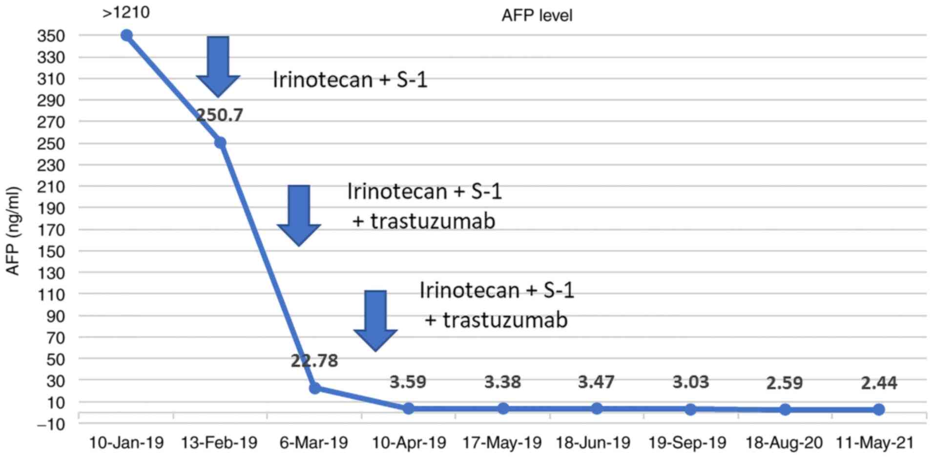
surgery. From January 22, 2019, the treatment plan comprising a
regimen of 300 mg irinotecan (day 1) plus 40 mg S-1 (twice daily,
days 2–15) plus 350 mg trastuzumab (once every 3 weeks) was
initiated. After the second course of treatment, the patient's AFP
level was reduced to 22.78 ng/ml, which was maintained within the
normal range thereafter (Fig. 6).
Following two courses of treatment, gastroscopy showed that the
size of the lesion was significantly reduced, allowing the
endoscope to pass through the stenosis. In addition, the lymph
nodes of the lesser curvature of the stomach gradually decreased in
size and then disappeared (Figs.
1B and 4B-D). After six
courses of chemotherapy, the patient continued to take four courses
of S-1 capsules and trastuzumab for a year. Afterwards, the drug
was discontinued and the patient was followed up regularly. In May
2021, gastroscopy did not show any tumor (Fig. 1C and D) and there was no sign of
recurrence or metastasis (Fig. 4E and
F). To date, the patient has been experiencing a high quality
of life.
Discussion
GC is a common malignancy. A large number of
patients with GC are diagnosed in the middle- or late-stage, and
AFPGC is associated with a poorer prognosis than other types of GC
(17). AFPGC is reported to
comprise two types, one of which is hepatoid adenocarcinoma of the
stomach (HAS), a rare aggressive tumor with hepatocellular
differentiation (18,19). The differentiation of HAS is an
important feature, particularly in hepatic lymph node metastasis
(20). However, the positive rate
of AFP in patients with HAS is 54–87%, indicating that AFP is not
present in all HAS cases (18,19).
The other type of AFPGC is the embryonic gastrointestinal type,
which constitutes 11.1-26.7% of all cases of AFPGC. The intestinal
mucosa of this type can mimic embryonic intestinal mucosa and
produce AFP (21). Regardless of
the type, serum AFP-positive GC is more malignant than other types
of GC, and more prone to hepatic lymph node metastasis.
Pathological stage, serum AFP level, age and hepatic metastasis are
independent risk factors for the prognosis of AFPGC (22). Therefore, there is an urgent
requirement for in vivo human models of AFPGC to determine
the sensitivity of drugs used for the personalized treatment of
cancer patients.
The PDX model can accurately reflect patients'
sensitivity to drugs in certain types of cancer (23). However, the limitations of the PDX
model inhibit its broad application in clinical practice: Firstly,
the PDX model has a low success rate and requires a large quantity
of tumor tissue, which can only be obtained from surgically removed
tumors; secondly, PDX modeling requires 4–8 months to determine the
efficacy of treatment for a specific cancer, and the time lag
between the transplantation of tumor tissue into mice and the
initiation of treatment limits its extensive application (7,24).
To overcome these limitations, the mini-PDX model has emerged to
assist clinicians in the selection of chemotherapeutic agents. The
mini-PDX model requires only a small number of tumor cells and
rapidly analyzes drug sensitivity in an average testing time of 7
days, enabling patients to receive individualized chemotherapy over
a clinically relevant time frame. A previous study showed a strong
consistency between mini-PDX- and PDX-based drug sensitivity
predictions for a variety of solid tumors, including lung cancer,
pancreatic cancer and stomach cancer, with an overall response
consistency of 89%, indicating that the mini-PDX-based drug
sensitivity model can be used to predict the outcome of patients
with cancer (12).
Patients with unresectable AFPGC are recommended to
be treated with chemotherapy. In a previous study on patients with
HAS receiving the same chemotherapy regimen, it was shown that the
disease-free and disease-specific survival rates of patients
receiving neoadjuvant chemotherapy were significantly higher than
those undergoing postoperative chemotherapy (25). Therefore, neoadjuvant chemotherapy
is recommended for patients with liver and/or distant metastases at
the initial diagnosis. Several chemotherapeutic regimens, including
cisplatin combined with fluorouracil and epirubicin, or irinotecan
combined with mitomycin, have been reported to effectively slowdown
the progression of AFPGC (26). In
the present study, using the mini-PDX model, it was identified that
irinotecan combined with S-1 had the strongest antitumor effect
among the regimens tested, and NGS suggested that irinotecan would
be less toxic. Satisfactory outcomes and complete remission (CR)
were achieved using the combination of irinotecan and S-1 with
trastuzumab, and no third-degree or more toxic side effects were
observed. The patient had HER-2 amplification, as confirmed by the
FISH and NGS results. A previous study showed that the positive
rate of HER-2 in patients with HAS was 42.68%, which was higher
than that in patients with other types of GC (27). However, a review of studies on
AFPGC suggested that when compared with GC with a normal AFP level,
vascular endothelial growth factor is upregulated in AFPGC while
the HER-2 expression level is not significantly different (28). In GC, the interaction between
hepatocyte growth factor and its receptor c-Met can promote mitosis
and cell migration, which promotes tumor development. A previous
study demonstrated that the positive rate of c-Met in patients with
AFPGC was higher than that in patients with AFP-negative GC.
Moreover, it revealed that the difference in c-Met expression
levels between the AFP (+) cells and AFP (−) cells in AFPGC tissues
was not statistically significant (29). In the present study, the NGS of the
tumor tissue did not suggest c-Met amplification. Instead, the
patient was revealed to have HER-2 amplification, and promising
results were obtained using trastuzumab. A previous study on the
treatment of patients with GC of different HER-2 statuses revealed
that HER-2-positive patients treated with trastuzumab had a median
overall survival of 22.3 months (30). At the time of writing, the present
case had survived for more than 32 months with CR, which has rarely
been reported. Since immunotherapy has not been validated as a
first-line treatment for patients with advanced GC, no
immunohistochemistry of programmed death-ligand 1 was performed in
the present case, and no programmed cell death protein 1 (PD-1)
inhibitor was administered. However, if the condition progresses
again, according to the KEYNOTE-811 clinical trial (31), PD-1 inhibitors may also be included
in the therapeutic scheme. In addition, the NGS results identified
the presence of ATM mutations. ATM is a tumor suppressor gene
(32). It interacts with numerous
proteins that co-localize at sites of DNA damage, where it plays an
important role in genome stability and the repair of DNA damage.
Therefore, its mutation may lead to homologous recombination repair
deficiency. However, PARP inhibitors can comprehensively kill tumor
cells with homologous recombination defects (33). Notably, patients who initially
respond to PARP inhibitors may later develop resistance.
In conclusion, the present study supports the use of
a mini-PDX model in combination with the NGS to optimize the
clinical management of metastatic gastric adenocarcinoma.
Acknowledgements
The authors would like to thank to Dr Yuange He
(Geneplus-Beijing Institute, Beijing, China) and Dr Zhenle Bi from
(Shanghai LIDE Biotech, Co., Ltd., Shanghai, China) for their
technological support.
Funding
The study was financially supported by the Precision Medicine
Special Project of Wuxi Municipal Health Commission (grant no.
J2011801).
Availability of data and materials
The datasets used and/or analyzed during the current
study are available from the corresponding author on reasonable
request.
Authors' contributions
CJ and XZ were responsible for the conception and
design of the study. CJ provided administrative support. XX, YD, SG
and TG performed clinical study and analyzed clinical data. XX and
BZ analyzed and interpreted the data. FZ performed the Mini-PDX
experiment. All authors contributed to writing the manuscript. All
authors read and approved the final version of the manuscript. CJ
and XZ confirm the authenticity of all the raw data.
Ethics approval and consent to
participate
This study was performed in accordance with the
Declaration of Helsinki. The study protocol was approved by the
Institutional Review Board of Wuxi Hospital of Traditional Chinese
Medicine (approval no. 201809001J01-01) and the Institutional
Animal Care and Use Committee (IACUC) of Shanghai LIDE Biotech Co.,
Ltd. (approval no. LDIACUC007). Written informed consent was
obtained from the patient prior to enrollment.
Patient consent for publication
The patient provided consent for publication of the
data.
Competing interests
FZ is an employee of Shanghai LIDE Biotech, Co.,
Ltd., who manufactured the miniPDX kit used for the animal
experiments. The other authors declare that they have no competing
interests.
Glossary
Abbreviations
Abbreviations:
|
PDX
|
patient-derived xenograft
|
|
NGS
|
next-generation sequencing
|
|
AFPGC
|
a-fetoprotein-producing gastric
cancer
|
|
HAS
|
hepatoid adenocarcinoma of the
stomach
|
|
S-1
|
tegafur-gimeracil-oteracil
|
References
|
1
|
International Agency for Research on
Cancer (IARC), . World Cancer Day: Breast overcancer takes lung
cancer as leading cause of worldwidecancer. IARC showcases key
research projects to address breast cancer. IARC; Lyon: 2021,
https://www.iarc.who.int/wp-content/uploads/2021/02/pr294_e.pdfFebruary
4–2021
|
|
2
|
Yang J, Wang R, Zhang W, Zhuang W, Wang M
and Tang C: Clinicopathological and prognostic characteristics of
hepatoid adenocarcinoma of the stomach. Gastroenterol Res Pract.
2014:1405872014. View Article : Google Scholar
|
|
3
|
Wang YK and Zhang XT: AFP-producing
gastric cancer and hepatoid gastric cancer. Zhonghua Zhong Liu Za
Zhi. 39:801–807. 2017.(In Chinese).
|
|
4
|
Jang Y, Peng Z, Wei B, et al: Lymph node
metastasis of early submucosal gastric cancer: A report of 290
cases. World Chin J Dig. 19:2970–2973. 2011. View Article : Google Scholar
|
|
5
|
Wang J, Tian SD and Chen XY: Advances in
the treatment of advanced gastric cancer. Chin Clin Oncol.
37:171–175. 2010.(In Chinese).
|
|
6
|
Aparicio S, Hidalgo M and Kung AL:
Examining the utility of patient-derived xenograft mouse models.
Nat Rev Cancer. 15:311–316. 2015. View
Article : Google Scholar : PubMed/NCBI
|
|
7
|
Hidalgo M, Amant F, Biankin AV, Budinská
E, Byrne AT, Caldas C, Clarke RB, de Jong S, Jonkers J, Mælandsmo
GM, et al: Patient-derived xenograft models: An emerging platform
for translational cancer research. Cancer Discov. 4:998–1013. 2014.
View Article : Google Scholar : PubMed/NCBI
|
|
8
|
Izumchenko E, Meir J, Bedi A, Wysocki PT,
Hoque MO and Sidransky D: Patient-derived xenografts as tools in
pharmaceutical development. Clin Pharmacol Ther. 99:612–621. 2016.
View Article : Google Scholar : PubMed/NCBI
|
|
9
|
Zhang L, Nomie K, Zhang H, Bell T, Pham L,
Kadri S, Segal J, Li S, Zhou S, Santos D, et al: B-cell lymphoma
patient-derived xenograft models enable drug discovery and are a
platform for personalized therapy. Clin Cancer Res. 23:4212–4223.
2017. View Article : Google Scholar : PubMed/NCBI
|
|
10
|
Byrne AT, Alférez DG, Amant F, Annibali D,
Arribas J, Biankin AV, Bruna A, Budinská E, Caldas C, Chang DK, et
al: Interrogating open issues in cancer precision medicine with
patient-derived xenografts. Nat Rev Cancer. 17:254–268. 2017.
View Article : Google Scholar : PubMed/NCBI
|
|
11
|
Xian Y, Xie Y, Song B, Ou Z, Ouyang S, Xie
Y, Yang Y, Xiong Z, Li H and Sun X: The safety and effectiveness of
genetically corrected iPSCs derived from β-thalassaemia patients in
nonmyeloablative β-thalassaemic mice. Stem Cell Res Ther.
11:2882020. View Article : Google Scholar : PubMed/NCBI
|
|
12
|
Zhang F, Wang W, Long Y, Liu H, Cheng J,
Guo L, Li R, Meng C, Yu S, Zhao Q, et al: Characterization of drug
responses of mini patient-derived xenografts in mice for predicting
cancer patient clinical therapeutic response. Cancer Commun (Lond).
38:602018. View Article : Google Scholar : PubMed/NCBI
|
|
13
|
Overmyer KA, Thonusin C, Qi NR, Burant CF
and Evans CR: Impact of anesthesia and euthanasia on metabolomics
of mammalian tissues: Studies in a C57BL/6J mouse model. PLoS One.
10:e01172322015. View Article : Google Scholar : PubMed/NCBI
|
|
14
|
Li H and Durbin R: Fast and accurate short
read alignment with Burrows-Wheeler transform. Bioinformatics.
25:1754–1760. 2009. View Article : Google Scholar : PubMed/NCBI
|
|
15
|
Aaron MK, Matthew H, Eric B, Sivachenko A,
Cibulskis K, Kernytsky A, Garimella K, Altshuler D, Gabriel S, Daly
M and DePristo MA: The genome analysis toolkit: A MapReduce
framework for analyzing next-generation DNA sequencing data. Genome
Res. 20:1297–1303. 2010. View Article : Google Scholar : PubMed/NCBI
|
|
16
|
Cibulskis K, Lawrence MS, Carter SL,
Sivachenko A, Jaffe D, Sougnez C, Gabriel S, Meyerson M, Lander ES
and Getz G: Sensitive detection of somatic point mutations in
impure and heterogeneous cancer samples. Nat Biotechnol.
31:213–219. 2013. View
Article : Google Scholar
|
|
17
|
Wang D, Li C, Xu Y, Xing Y, Qu L, Guo Y,
Zhang Y, Sun X and Suo J: Clinicopathological characteristics and
prognosis of alpha-fetoprotein positive gastric cancer in Chinese
patients. Int J Clin Exp Pathol. 8:6345–6355. 2015.
|
|
18
|
Lin CY, Yeh HC, Hsu CM, Lin WR and Chiu
CT: Clinicopathologial features of gastric hepatoid adenocarcinoma.
Biomed J. 38:65–69. 2015. View Article : Google Scholar : PubMed/NCBI
|
|
19
|
Xiao C, Wu F, Jiang H, Teng L, Song F,
Wang Q and Yang H: Hepatoid adenocarcinoma of the stomach: Nine
case reports and treatment outcomes. Oncol Lett. 10:1605–1609.
2015. View Article : Google Scholar
|
|
20
|
Liu X, Cheng Y, Sheng W, Lu H, Xu Y, Long
Z, Zhu H and Wang Y: Clinicopathologic features and prognostic
factors in alpha-fetoprotein-producing gastric cancers: Analysis of
104 cases. J Surg Oncol. 102:249–255. 2010. View Article : Google Scholar
|
|
21
|
Motoyama T, Aizawa K, Watanabe H, Fukase M
and Saito K: alpha-Fetoprotein producing gastric carcinomas: A
comparative study of three different subtypes. Acta Pathol Jpn.
43:654–661. 1993.PubMed/NCBI
|
|
22
|
Chen Y, Qu H, Jian M, Sun G and He Q: High
level of serum AFP is an independent negative prognostic factor in
gastric cancer. Int J Biol Markers. 30:e387–e393. 2015. View Article : Google Scholar : PubMed/NCBI
|
|
23
|
Garber K: From human to mouse and back:
‘Tumorgraft’ models surge in popularity. J Natl Cancer Inst.
101:6–8. 2009. View Article : Google Scholar
|
|
24
|
Hwang CI, Boj SF, Clevers H and Tuveson
DA: Preclinical models of pancreatic ductal adenocarcinoma. J
Pathol. 238:197–204. 2016. View Article : Google Scholar
|
|
25
|
Kochi M, Fujii M, Kaiga T, Takahashi T,
Morishita Y, Kobayashi M, Kasakura Y and Takayama T: FLEP
chemotherapy for alpha-fetoprotein-producing gastric cancer.
Oncology. 66:445–449. 2004. View Article : Google Scholar : PubMed/NCBI
|
|
26
|
Back SK, Han SW, Oh DY, Im SA, Kim TY and
Bang YJ: Clinicopathologic characteristics and treatment outcomes
of hepatoid adenocarcinoma of the stomach, a rare but unique
subtype of gastric cancer. BMC Gastroentero. 11:562011. View Article : Google Scholar
|
|
27
|
Giuffrè G, Ieni A, Barresi V, Caruso RA
and Tuccari G: HER2 status in unusual histological variants of
gastric adenocarcinomas. J Clin Pathol. 65:237–241. 2012.
View Article : Google Scholar
|
|
28
|
Fang Y, Wang L, Li GM, et al: Expression
of c-Met, VEGF, EGFR and Her-2 in AFP positive gastric cancer. Chin
J Cancer. 26:662–669. 2016.(In Chinese).
|
|
29
|
Amemiya H, Kono K, Mori Y, Takahashi A,
Ichihara F, Iizuka H, Sekikawa T and Matsumoto Y: High frequency of
c-Met expression in gastric cancers producing alpha-fetoprotein.
Oncology. 59:145–151. 2000. View Article : Google Scholar : PubMed/NCBI
|
|
30
|
Qin S, Ji J, Xu RH, Wang W, Tang Y, Bi F,
Li J, Wang K, Xu JM, Fan Q, et al: Treatment patterns and outcomes
in chinese patients with gastric cancer by HER2 status: A
noninterventional registry study (EVIDENCE). Oncologist.
26:e1567–e1580. 2021. View Article : Google Scholar
|
|
31
|
Chung HC, Bang YJ, S Fuchs C, Qin SK,
Satoh T, Shitara K, Tabernero J, Van Cutsem E, Alsina M, Cao ZA, et
al: First-line pembrolizumab/placebo plus trastuzumab and
chemotherapy in HER2-positive advanced gastric cancer: KEYNOTE-811.
Future Oncol. 17:491–501. 2021. View Article : Google Scholar
|
|
32
|
Russell R, Perkhofer L, Liebau S, Lin Q,
Lechel A, Feld FM, Hessmann E, Gaedcke J, Güthle M, Zenke M, et al:
Loss of ATM accelerates pancreatic cancer formation and
epithelial-mesenchymal transition. Nat Commun. 6:76772015.
View Article : Google Scholar : PubMed/NCBI
|
|
33
|
Farmer H, McCabe N, Lord CJ, Tutt AN,
Johnson DA, Richardson TB, Santarosa M, Dillon KJ, Hickson I,
Knights C, et al: Targeting the DNA repair defect in BRCA mutant
cells as a therapeutic strategy. Nature. 434:917–921. 2005.
View Article : Google Scholar : PubMed/NCBI
|