Introduction
Lung cancer is one of the most common malignant
tumor types. According to the latest statistics from the World
Health Organization in 2021, 2.2 million patients are living with
lung cancer worldwide, among which 1.8 million die; lung cancer
ranks highest in terms of death rate from cancer and the patients
are mostly male (1). Non-small
cell lung cancer (NSCLC) is the most common type of lung cancer,
accounting for 80–85% of cases. These mainly include lung
adenocarcinoma (LUAD) and lung squamous cell carcinoma (LUSC).
NSCLC mostly spreads along the trachea and alveolar wall, and as
these areas are rich in blood vessels, local infiltration and
hematogenous dissemination occur early, and it easily involves the
pleura and causes pleural effusion, and numerous patients are
diagnosed at this time; however, hematogenous and disseminated
metastases have already occurred at that stage, and patients are no
longer eligible for surgical treatment. Therefore, the 5-year
survival rate of patients with advanced NSCLC is only ~10%
(1). The treatment of advanced
lung cancer mainly comprises chemotherapy and targeted therapy.
Targeted therapy has been a hot research topic in recent years.
Genetic testing of patients with NSCLC is used to select targeted
therapeutic drugs. Current common targets include EGFR, echinoderm
microtubule-associated protein-like 4-anaplastic lymphoma kinase,
VEGF, programmed death receptor 1, HER2 and KRAS (2).
The secreted protein acidic and cysteine rich
(SPARC) gene is located on the human chromosome Sq31.3-q32 and
contains 10 exons. It is a small-molecule glycoprotein rich in
cysteine and is closely related to cellular secretion. It is
involved in various biological processes such as tumor angiogenesis
and tissue repair and remodeling (3), and is able to regulate cell adhesion
and proliferation through different signaling pathways. Members of
the SPARC family all contain a special EC domain, and the EC domain
contains the EF hand model. SPARC family members may be divided
into different taxa: SPARC, Hevin, SMOC1 and follistatin-like
protein, which are five taxa with sequence homology of different EC
structural domains, of which the SPARC gene is the more important
gene in the family (4). Studies
have indicated that SPARC is abnormally highly expressed in liver
cancer tissues and is related to the formation of microvessels in
tumor tissues (5). In gastric
cancer, SPARC regulates the epithelial-mesenchymal transition of
gastric cancer through the Slug pathway, thereby promoting gastric
cancer metastasis and leading to poor prognosis (6). Previous studies have indicated that
the expression of SPARC in gastric cancer is correlated with the
expression of E-cadherin, Slug and Vimentin, and the positive
expression of SPARC is negatively correlated with the survival rate
of patients with gastric cancer. Positive expression of E-cadherin
is positively correlated with the survival rate of patients with
gastric cancer (7). At the same
time, it has been indicated that SPARC may affect the metastasis of
lung cancer through the Wnt/β-catenin signaling pathway. The
expression of β-catenin, c-myc and cyclin-D1 in the Wnt/β-catenin
signaling pathway was increased in lung cancer tissues. After SPARC
knockout, the expression of β-catenin, c-myc and cyclin-D1
decreased. It is inferred that SPARC is able to affect the
epithelial-mesenchymal transition of NSCLC through the
Wnt/β-catenin signaling pathway and promote invasion and metastasis
of NSCLC. The high expression of SPARC in liver cancer and bladder
cancer may inhibit apoptosis of tumor cells through the PI3K/AKT
signaling pathway, while promoting the proliferation, invasion and
metastasis of tumor cells. SPARC is an upregulated gene in
CHD1L-induced hepatocellular carcinoma (8–11).
In human NSCLC cells expressing SPARC induced by TGF-β, the
expression of CDH1 did not change, while SPARC was strongly
expressed in metastatic NSCLC. It is also expressed in
triple-negative NSCLC and intraductal carcinoma in situ. The
intensity of SPARC expression is related to the stage of breast
ductal carcinoma in situ. The expression of SPARC in poorly
differentiated ductal carcinoma in situ is stronger than
that in well-differentiated cases, suggesting that SPARC may be
related to the epithelial-mesenchymal transition of NSCLC (12).
Previous studies have indicated that SPARC is highly
expressed in gastric cancer, pancreatic cancer, cervical cancer and
liver cancer, but the role of SPARC in lung cancer remains elusive.
The present study confirmed the relationship between SPARC mRNA and
protein expression levels and survival prognosis,
clinicopathological factors and immune infiltration mechanisms in
NSCLC by meta-analysis and bioinformatics analysis. It provided a
theoretical basis for the SPARC gene as a potential key factor in
the occurrence and development of NSCLC.
Materials and methods
Literature search and data
extraction
The PubMed (http://www.ncbi.nlm.nih.gov/pubmed), Web of Science
(http://webofscience.com), Wanfang database
(https://www.wanfangdata.com.cn/) and
Chinese National Knowledge Infrastructure (CNKI) database
(http://www.cnki.net/) were searched from
inception until February 2022 by using the following key words:
‘SPARC’ and ‘lung cancer’ or ‘NSCLC’ or ‘lung adenocarcinoma’ or
‘lung squamous cell carcinoma’. The present study was conducted in
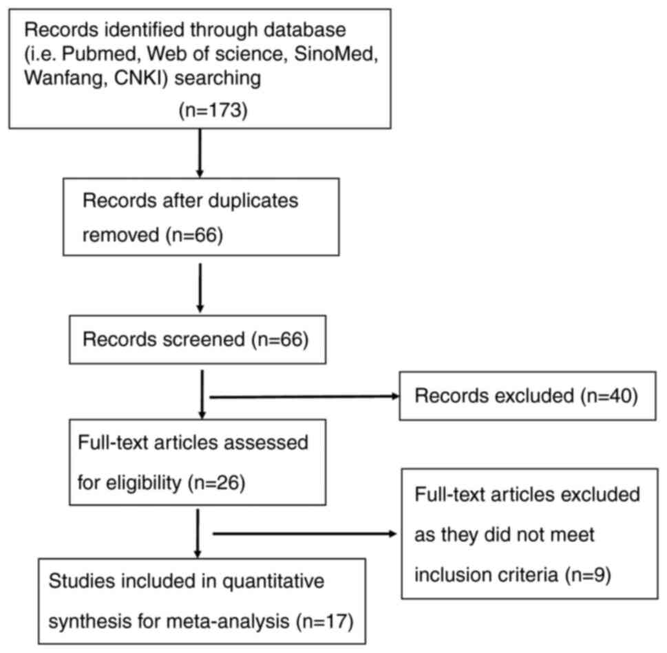
strict accordance with the PRISMA guidelines (13). A total of 173 articles were
screened after an initial search by reading abstracts and excluding
duplicate studies. After further screening (SS and YRZ), we
continued the careful screening of 26 articles for full-text
reading. Ultimately, only 17 articles were included in the study of
this paper and all case data were sourced from China. The study
inclusion criteria were as follows: i) Patients with NSCLC; ii)
immunohistochemical detection of SPARC; iii) articles containing
SPARC expression and clinicopathological parameters; iv) none of
the patients received chemotherapy or radiotherapy prior to
surgery. The exclusion criteria were as follows: i) Abstracts, case
reports, reviews and conference proceedings; ii) duplicate
publications; iii) unclear diagnoses; iv) the expression of SPARC
was investigated by western blot, reverse
transcription-quantitative PCR, cDNA microarray or transcriptome
sequencing.
Data extraction and quality
assessment
As presented in Table
I, the eligible paper information was extracted by two
reviewers (GYM and ZGZ), including the first author's name,
publication year, patient's country, antibody company, number of
cases and controls, cancer risk and follow-up results. According to
the Newcastle-Ottawa Oncomine scale (14), the quality of the studies was
independently assessed by two reviewers. The criteria to judge the
quality of the studies included sample selection, comparability and
determination of results.
 | Table I.Main characteristics of eligible
studies. |
Table I.
Main characteristics of eligible
studies.
| First author,
year | Country | Antibody
supplier | Total cases, n | Events, n | Risk of cancer | Follow-up
outcome | Quality score | (Refs.) |
|---|
| Andriani et al,
2018 | Italy | NS | 57 | 28 | Raised | NS | 8 | (17) |
| Duan et al,
2017 | China | Invitrogen; Thermo
Fisher Scientific, Inc. | 32 | 18 | Raised | Negative | 7 | (19) |
| Koumiya et al,
2016 | Japan | Abcam | 200 | 145 | Raised | NS | 7 | (20) |
| Koukourakis et al,
2003 | Greece | Abcam | 113 | 107 | Raised | NS | 9 | (28) |
| Zhang et al,
2012 | China | Fuzhou Maixin
Biotech Co., Ltd. | 89 | 63 | Raised | NS | 8 | (25) |
| Zheng et al,
2014 | China | Bostik | 71 | 44 | Raised | NS | 8 | (29) |
| Zheng et al,
2015 | China | NS | 71 | 40 | Raised | NS | 8 | (30) |
| Huang et al,
2012 | China | R&D Systems,
Inc. | 105 | 57 | Raised | NS | 8 | (26) |
| Xu et al, 2019 | China | Abcam | 90 | 54 | Raised | NS | 8 | (31) |
| Kurtul et al,
2014 | Turkey | BIOSS | 84 | NS | Raised | Negative | 8 | (22) |
| Fabrizio et al,
2020 | Italy | Cell Signaling
Technology, Inc. | 21 | NS | Raised | NS | 7 | (15) |
| Yin et al,
2016 | China | NS | NS | NS | Raised | NS | 7 | (24) |
| Gao et al,
2020 | China | NS | NS | NS | Raised | NS | 8 | (16) |
| Li et al, 2018 | USA | NS | NS | NS | NS | NS | 7 | (18) |
| Ma et al, 2006 | China | NS | NS | NS | NS | NS | 7 | (27) |
| Zheng et al,
2016 | China | NS | NS | NS | Raised | Negative | 8 | (21) |
| Grant et al,
2014 | USA | Prolytix | 19 | 7 | Raised | NS | 8 | (23) |
Meta-analysis
SPARC expression was estimated according to the
clinicopathological parameters and the odds ratio (OR) and its 95%
CI were used to express the effect size in patients with NSCLC.
First, the χ2 test was used to assess the heterogeneity
of the original study. When heterogeneity was not significant,
i.e., P≤0.1, a fixed-effects model (Mantel-Haenszel method) was
used. When P>0.1, a random-effects model (Der Simonian and Laird
method) was used. I2 statistics were used to quantify
the effect of heterogeneity at cutoffs of 25, 50 and 75%,
respectively. Heterogeneity in the results was considered
significant if P<0.1 and I2>50%. Meta-analysis was
performed based on the random-effects model. If P>0.1 and
I2<50%, it was considered that there was no
significant heterogeneity among the study results and the
fixed-effects model was used for meta-analysis. Funnel plots were
used to evaluate publication bias, and Begg's test and Egger's test
were used to evaluate whether the funnel plots were consistent.
Furthermore, to perform a sensitivity analysis on the aggregated
results, one study was deleted at a time and the impact of a single
study on the results was thereby examined. In the sensitivity
analysis, none of the studies affected the combined HR and OR,
which indicated that the results were stable (results not shown).
Meta-analysis was performed using RevMan 5.2 software and SPSS
software (version 10.0; SPSS, Inc.) with the t-test. A two-sided
P<0.05 was considered to indicate statistical significance.
Bioinformatics analysis
The prognostic value of SPARC mRNA expression in
NSCLC was analyzed using the Kaplan-Meier plotter (KM-plotter;
http://www.kmplot.com). The influence of the
expression of SPARC on overall survival, recurrence-free survival,
distant metastasis-free survival and post-progression survival in
all patients was determined. The correlation of the expression of
SPARC with clinicopathological features was also examined. To
investigate SPARC gene expression, the Oncomine database
(www.oncomine.org), an extensive database of tumor
microarray data, including microarray and gene expression data, was
used. The database may be used to analyze differences in gene
expression and to classify clinical information about tumor
patients. The expression differences of SPARC mRNA in cancer
tissues and normal tissues were compared. The gene expression and
clinicopathological data of SPARC were downloaded from The Cancer
Genome Atlas (TCGA; www.cancer.gov) database using R software
TCGA-assembler. The data were collated and SPARC mRNA expression in
NSCLC was analyzed. Furthermore, the clinicopathological data and
prognosis of tumor patients were analyzed. Cox hazard regression
models were used to perform univariate and multivariate analyses.
The model analyzed the effects of risk factors, hazard ratios and
95% CIs. The TCGA data and GTEx data from the GEPIA online analysis
website (http://gepia.cancer-pku.cn/) were
used to analyze the expression of SPARC mRNA in NSCLC tissues and
normal lung tissues. SPARC mRNA expression was analyzed according
to library online analysis and mining site exploration
subpopulations by performing TCGA number analysis on the UALCAN
online website (http://ualcan.path.uab.edu/). According to the mRNA
expression value of each gene, cancer patients were automatically
divided into a high expression group and low expression group for
comparison. P<0.05 was considered to indicate a statistically
significant difference. Based on the Human Protein mapping database
[the Human Protein Atlas (HPA); https://www.proteinatlas.org/], the SPARC mRNA and
protein levels in NSCLC and normal lung tissues were analyzed and
compared. Representative immunohistochemical images of NSCLC
tissues with varying levels of SPARC protein expression were
downloaded. Based on the website of the Timer database (http://http://timer.cistrome.org/), the relationship
between immune cells and survival prognosis in NSCLC, as well as
the relationship between lung cancer and related immune cell
infiltration and SPARC gene expression, were analyzed. The scores
were mainly calculated for 35% and a survival time of 60 months. It
was fitted automatically by software which uses the R language
package and the main algorithms are based on genetic marker method
and deconvolution. The R language package immunedeconv was used,
which integrates the six algorithms TIMER, xCell, MCP-counter,
CIBERSORT, EPIC and quanTIseq.
Statistical analysis
Revman Version 5.3 (Cochrane Collaboration) was
adopted to perform the meta-analysis. Comparison between the case
group and the control group was indicated by OR and 95% CI.
I2 statistics were used to determine heterogeneity
between study results. Publication bias was evaluated by funnel
plots and the asymmetry of the funnel plot was tested by Begg's and
Egger's tests. The Cox risk regression model was used for
univariate and multivariate analysis. The model analyzed the impact
of risk factors, risk ratios and 95% CI. P<0.05 was considered
to indicate statistical significance. All data were analyzed using
SPSS 19.0 software (IBM Corp).
Results
Literature search and publication
bias
The final 17 articles included in the present study
discussed the relationship between SPARC expression and
clinicopathologically or prognostically relevant expression markers
in NSCLC (Fig. 1) (15–31).
Clinicopathological characteristics of NSCLC included histological
grade, TNM stage, presence of lymph node metastasis and patient
gender (Table I). As presented in
Fig. 2, funnel plots were used to
examine the heterogeneity among studies. The results of Egger's
test indicated that there was no significant publication bias in
the present meta-analysis. It was indicated that SPARC expression
was able to distinguish between cancer and normal tissue, and was
related to the parameters of lymph node metastasis, TNM stage and
gender (Fig. 2A-D).
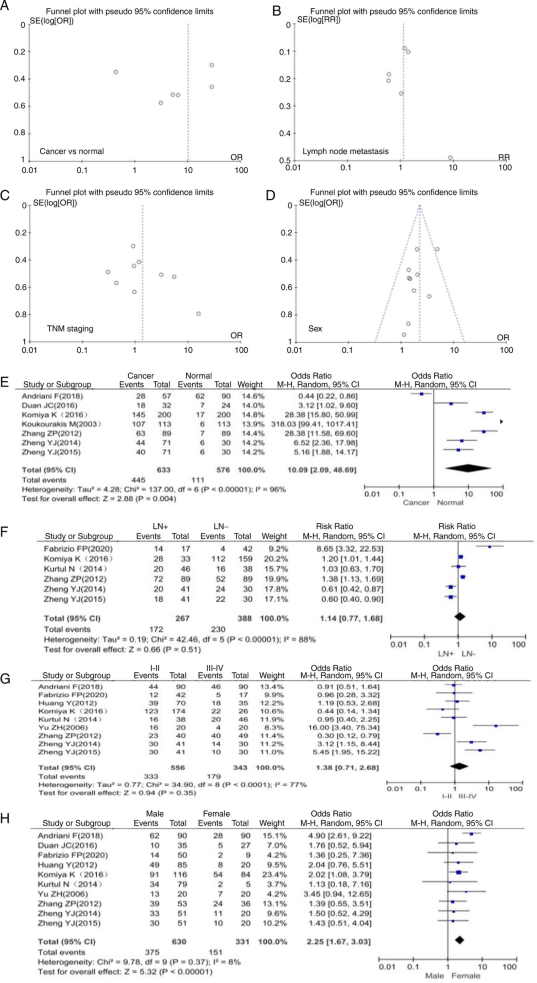 | Figure 2.Funnel plots of publication bias
detected in NSCLC expressing SPARC. Publication bias was analyzed
based on the relationship between SPARC expression and
clinicopathological features of NSCLC. This included (A) the
relationship between cancer and normal lung tissue, (B) lymph node
metastasis, (C) TNM staging and (D) gender. Forest plot of SPARC
expression and clinicopathological features of NSCLC. (E) Cancer
and normal tissue, (F) lymph node metastasis, (G) TNM staging and
(H) gender. SPARC, secreted protein acidic and cysteine rich;
NSCLC, non-small cell lung cancer; SE, standard error; OR, odds
ratio; M-H, Mantel-Haentzel; LN, lymph node involvement; df,
degrees of freedom. |
Association between SPARC expression
and clinicopathological characteristics of patients with NSCLC
according to the OR forest plot
A total of 7 articles included data on 633 patients
with NSCLC and 576 normal controls. Compared with normal tissues,
the expression of SPARC in lung cancer tissues was significantly
upregulated (OR=10.09, 95% CI 2.09-48.69, P=0.004; Fig. 2E). The SPARC gene was expressed at
higher levels in NSCLC than in normal lung tissue. There was high
heterogeneity among study results (P=0.004, I2=96%),
which may be due to potential selection bias among study subjects
and no medical history in the control group. The present
meta-analysis indicated that SPARC expression was not significantly
associated with TNM stage and lymph node metastasis in patients
with NSCLC (Fig. 2F and G). The
expression of SPARC was closely related to the gender of the
patients and males were more likely to develop NSCLC than females
(OR=2.25, 95% CI 1.67-3.03, P<0.00001; Fig. 2H).
Relationship between SPARC expression
and prognosis of NSCLC
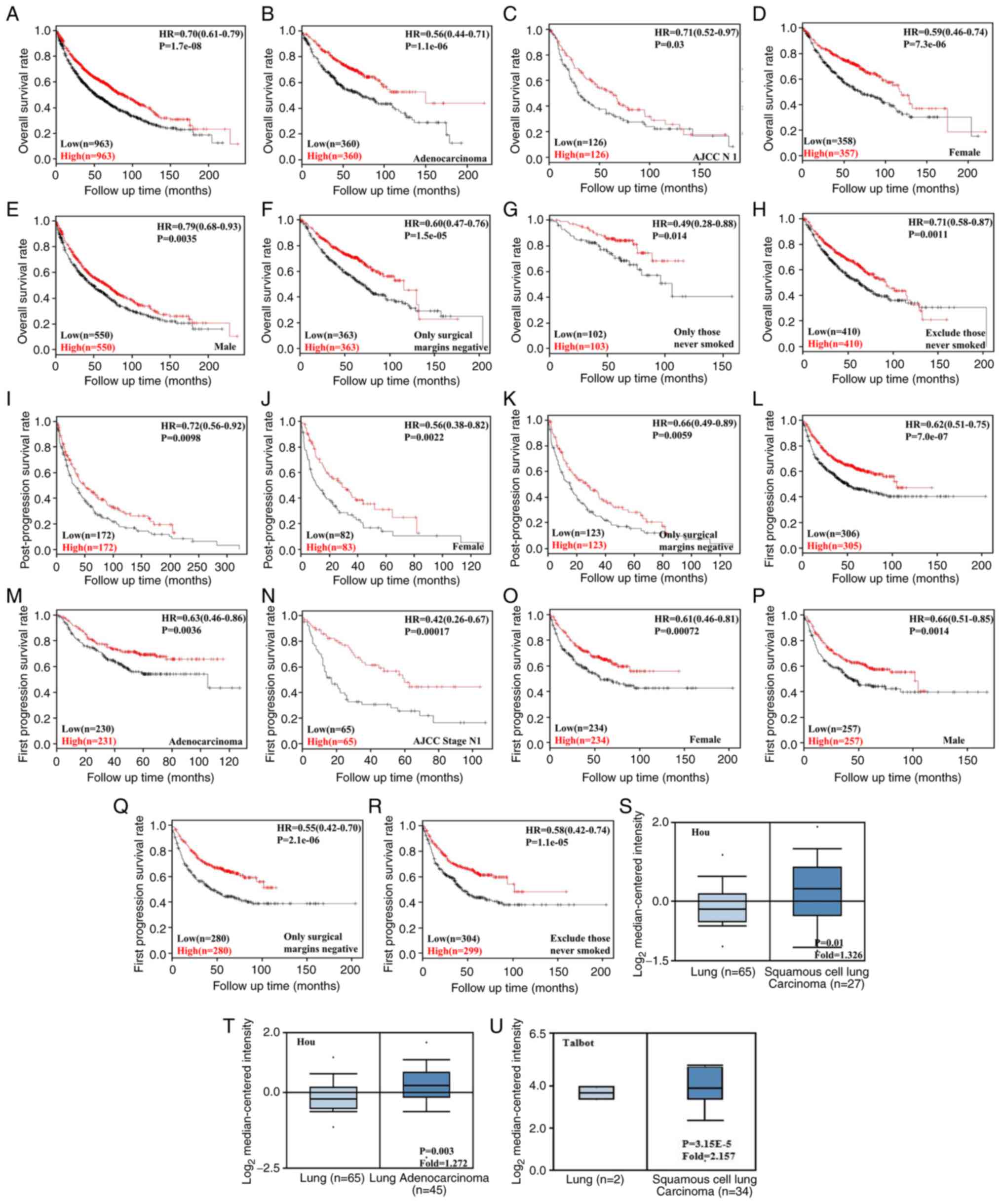
Using KM-Plotter, it was indicated that SPARC
expression levels were positively correlated with overall survival
of patients with different stages of NSCLC. In addition, there was
a positive correlation with the survival rate of patients with
different tumor types, gender and smoking history (P<0.05).
Increased SPARC expression levels were positively associated with
first progression survival and post-progression survival
(P<0.05). Patients with NSCLC and high SPARC expression levels
had a longer survival time (P<0.05). These data suggested that
the SPARC gene is closely associated with patient survival
prognosis (P<0.05, Fig.
3A).
The association between SPARC expression and the
bioinformatics signature of NSCLC in the Oncomine database was then
analyzed. The databases published by Hou and Talbot suggested that
the mRNA expression of SPARC was higher in LUSC than in normal lung
tissue; furthermore, according to the database by Hou, SPARC
expression in LUAD was also higher than that in normal lung tissue
(P<0.05, Fig. 3B).
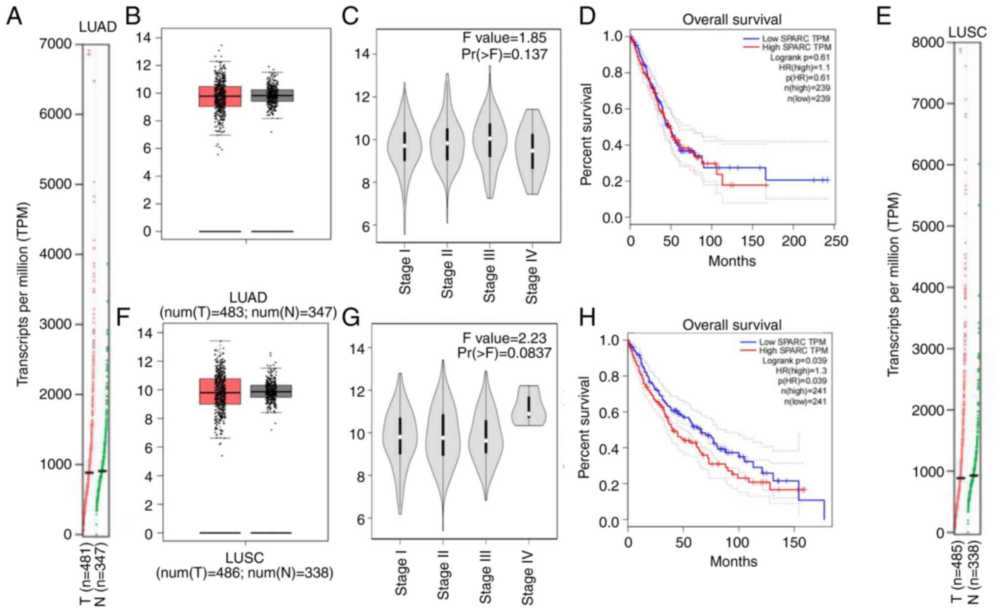
In the GEPIA database, the mRNA expression of SPARC
in LUAD tissue was higher than that in normal lung tissue (Fig. 4A and B), SPARC expression was not
significantly associated with the stage of LUAD, but when it was
analyzed in Stage I to Stage III, SPARC expression levels were
enhanced with the increase in stage; however, the level of SPARC
expression was slightly decreased again in Stage IV (Fig. 4C). Kaplan-Meier analysis with the
log-rank test suggested that among 239 patients with LUAD, the
SPARC gene was not significantly associated with OS. When SPARC
expression levels were reduced, the survival and prognostic
practices of LUSC patients tended to be prolonged (Fig. 4D). Higher SPARC expression levels
were associated with a shorter survival time of patients. The mRNA
expression levels of SPARC were not significantly different between
normal lung tissues and LUSC, with an increasing trend of
expression in LUSC (Fig. 4E and
F). This indicates that the mRNA expression levels of SPARC and
protein expression levels in LUSC are in agreement with each other,
suggesting that the analysis results are credible. SPARC expression
is upregulated in LUSC. SPARC expression was associated with the
staging of LUSC but this was not significant (P=0.0837). SPARC mRNA
expression was higher in LUSC Stage IV than in other stages
(Fig. 4G). Kaplan-Meier analysis
with the log-rank test indicated that among 241 patients with LUSC,
the expression levels of the SPARC gene had a significant influence
on OS (P=0.039; Fig. 4H), with a
higher expression level of SPARC mRNA indicating a shorter survival
time of patients.
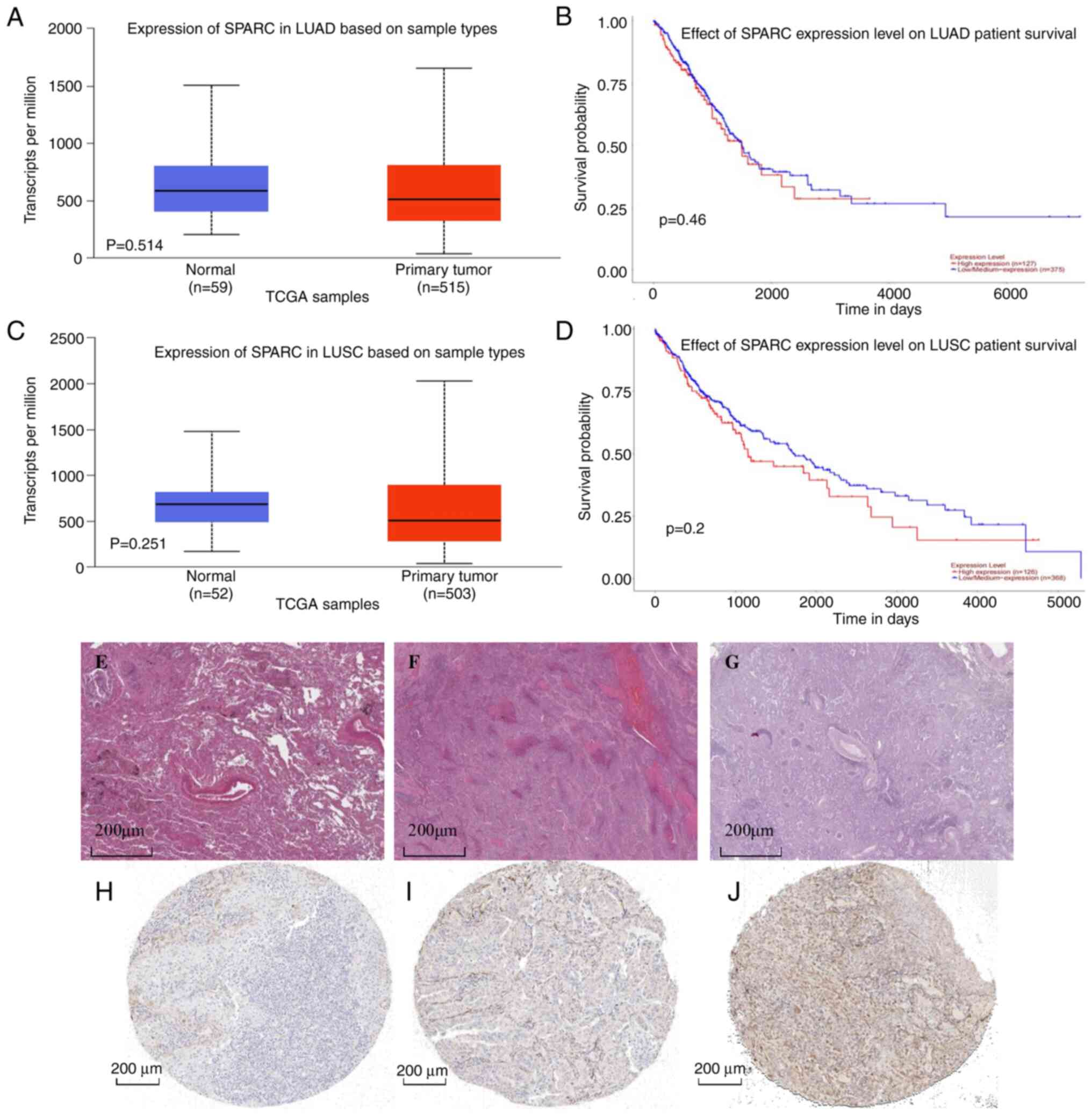
In the UALCAN database, the TCGA dataset was
analyzed, comprising 515 LUAD tissues and 59 normal lung tissues.
SPARC mRNA expression levels were upregulated in LUAD compared with
normal lung tissue (P>0.05; Fig.
5A). The level of SPARC gene expression was not significantly
associated with the prognosis of patients with LUAD (Fig. 5B). Furthermore, 503 LUSC tissues
and 52 lung tissues included in the TCGA dataset were analyzed. In
LUSC, SPARC mRNA expression levels were higher than those in normal
lung tissue, but the differential expression was not significant
(P>0.05; Fig. 5C). The SPARC
gene expression level was not significantly associated with the
prognosis of patients with LUSC (Fig.
5D), but the trends still indicated that patients with LUSC had
a better prognosis when the SPARC mRNA expression level was lower.
In the HPA database, the pathological morphology was compared among
normal lung tissue, LUAD and LUSC (Fig. 5E-G). Screening analysis suggested
that the expression level of SPARC protein in normal lung tissue
was lower than that in LUAD. In cancer tissue, positivity of
immunohistochemical staining was observed in the membrane and
cytoplasm (Fig. 5H and I); SPARC
protein was highly expressed in LUSC tissue and low in normal lung
tissue, and positivity of immunohistochemical staining was present
in the cytoplasm (Fig. 5J). This
trend of expression was closely related to the mRNA expression
level and the analytic results were consistent with this
regard.
In the Timer database, the infiltration of immune
cells closely related to lung cancer tissue was as follows: B
cells, NK, CD4+T, CD8+T, monocytes and granulocytes all had
different degrees of infiltration in NSCLC and normal lung tissue
(Fig. 6A and B). The difference in
the degree of infiltration of malignant tumor tissue was
statistically significant (P<0.05). A correlation analysis of
the proportion of liver immune cells in NSCLC patients according to
SPARC levels was then performed. In LUAD, an inverse relationship
between the proportion of B cells among immune cells and SPARC gene
expression was observed. In LUSC, the eight most important immune
cell types were positively correlated with the level of SPARC gene
expression. Among immune cells, the infiltration of various cells
was strongly correlated with the prognosis of patients with NSCLC.
A higher degree of immune-cell infiltration was linked to a longer
survival time of patients with LUAD and LUSC, with a positive
association (Fig. 6C and D), among
which B cells and dendritic cells exhibited the greatest
association (P<0.05). It was observed that the expression of
SPARC was closely related to the immune-cell infiltration mechanism
in NSCLC.
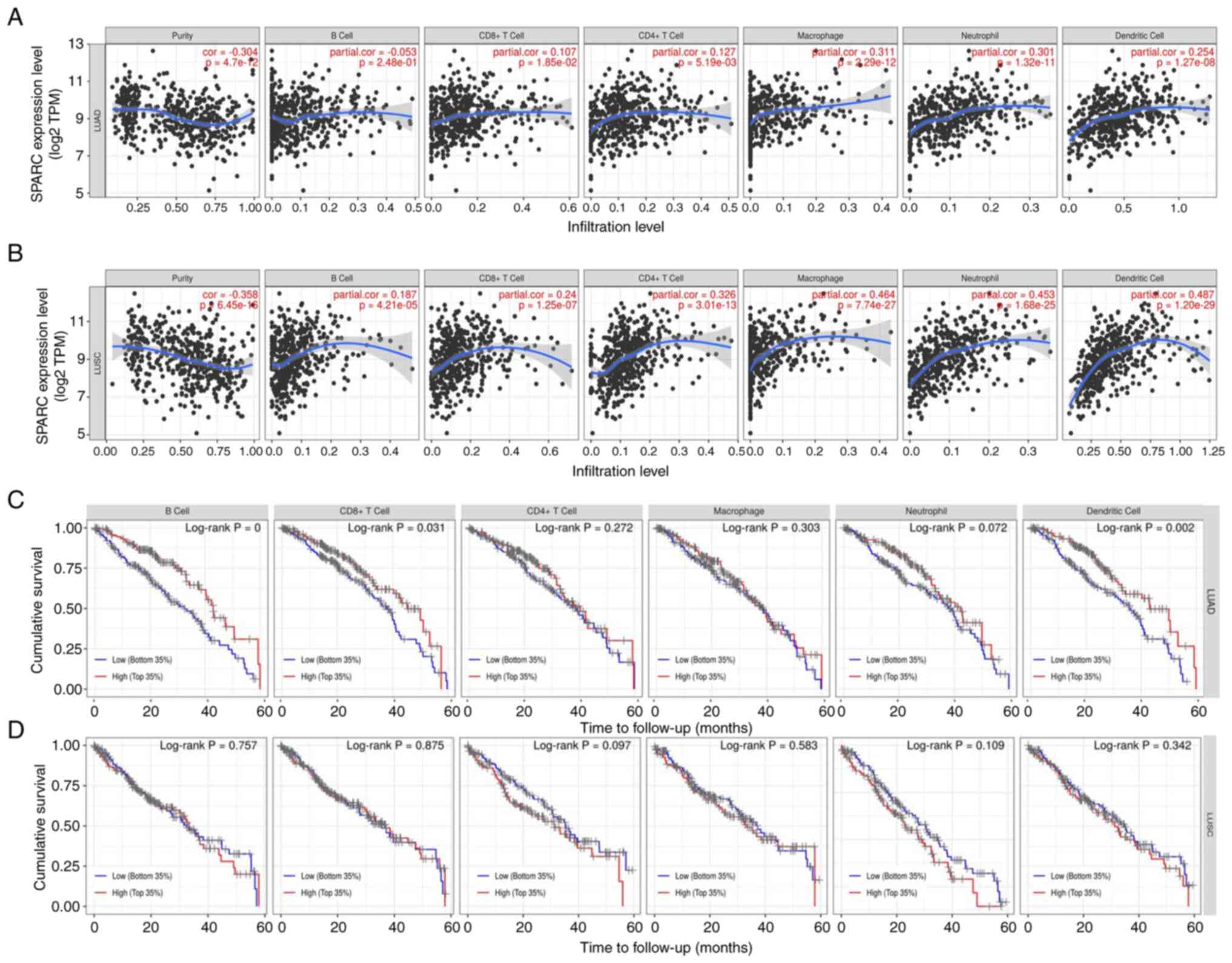 | Figure 6.(A) Correlation of SPARC expression
levels with immune cells (CD4+ T cells, macrophages, B cells, CD8+
T cells, neutrophils and dendritic cells) in LUAD. (B) Correlation
of SPARC expression levels with immune cells (CD4+ T cells,
macrophages, B cells, CD8+ T cells, neutrophils and dendritic
cells) in LUSC. The infiltration level is displayed on the X-axis
and the expression of SPARC on the Y-axis. (C) Relationship between
the degree of immune-cell infiltration and survival prognosis of
patients with LUAD. (D) Relationship between the degree of
immune-cell infiltration and survival prognosis of patients with
LUSC. The time to follow-up (months) is presented on the X-axis and
the cumulative survival on the Y-axis. SPARC, secreted protein
acidic and cysteine rich; LUAD, lung adenocarcinoma; LUSC, lung
squamous cell carcinoma; TPM, transcripts per million; cor,
correlation coefficient. |
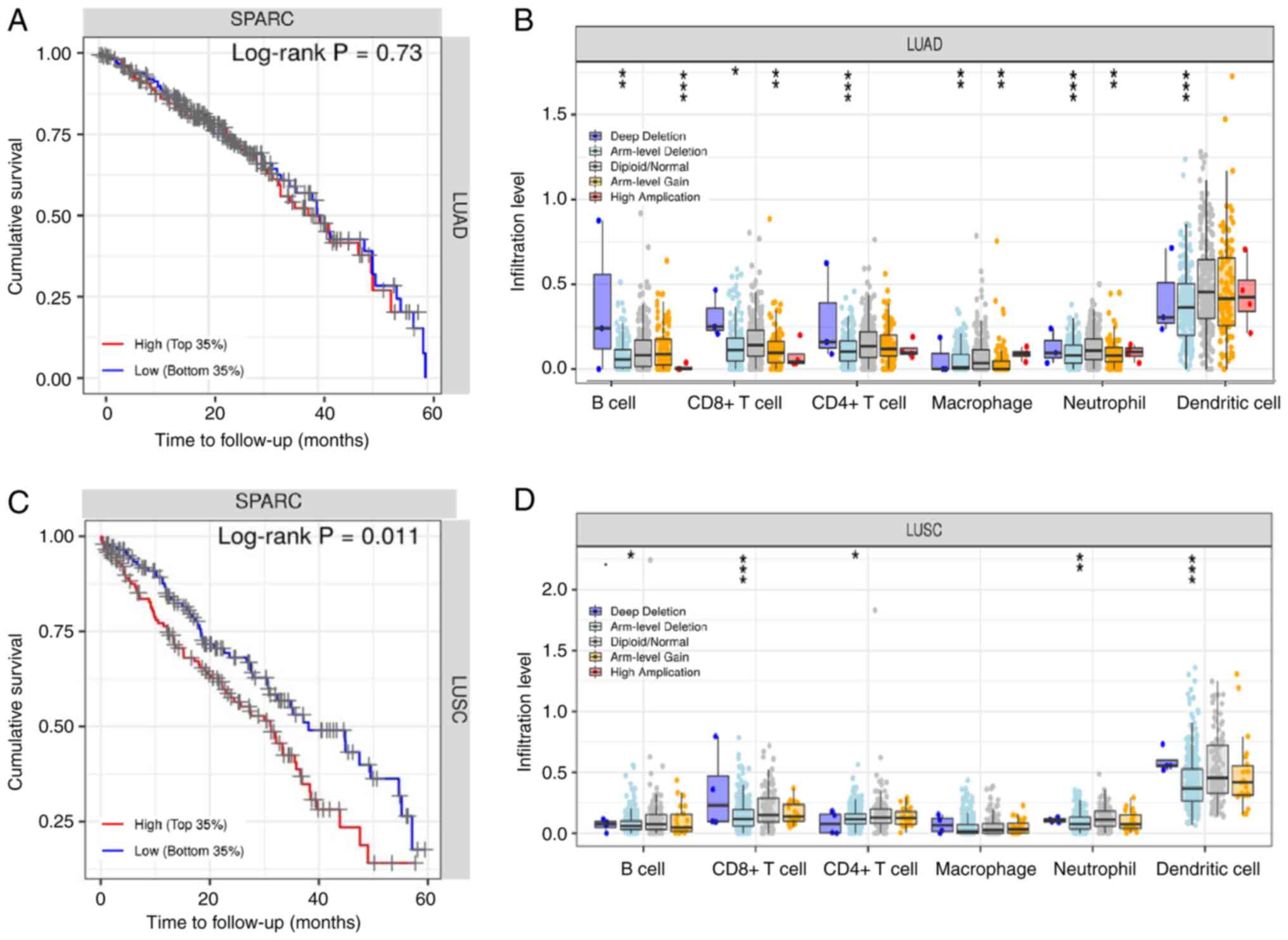
To further estimate the influence on SPARC on
survival, patients with lung cancer were automatically divided into
two groups based on SPARC protein expression levels: High
expression and low expression. The SPARC gene had a non-significant
effect on the survival rate of patients with LUAD (Fig. 7A). For LUAD copy number changes,
deep deletions of immune cells (B cells, CD8+ T cells, neutrophils
and dendritic cells) were greater than arm level deletions,
diploid/normal, arm level gain and high amplification (Fig. 7B; Table SI). In LUSC, the SPARC expression
level had a significant influence on survival and prognosis
(P<0.05; Fig. 7C). In patients
with LUSC, a higher the expression of SPARC protein was associated
with a shorter survival time and unfavorable prognosis (P<0.05).
In terms of LUSC copy number changes, deep deletions of immune
cells (B cells, CD8+ T cells, neutrophils, and dendritic cells)
were greater than arm level deletions, diploid/normal, arm level
gain and high amplification, and the copy number trends of LUAD and
LUSC were consistent (Fig. 7D;
Table SI). Cox univariate
analysis of TCGA data indicated that TNM stage, lymph node
metastasis, distant metastasis and depth of invasion were
negatively associated with patient prognosis (P<0.05; Table II). Cox multivariate analysis
suggested that SPARC expression levels and TNM stage were risk
factors affecting the prognosis of patients with NSCLC (P<0.05;
Table III).
| Table II.Univariate analysis of prognostic
risk factors in patients with lung cancer from TCGA database. |
Table II.
Univariate analysis of prognostic
risk factors in patients with lung cancer from TCGA database.
|
Characteristics | Patients, n
(%) | Hazard ratio (95%
CI) | P-value |
|---|
| Sex |
| 1.090
(0.879-1.352) | 0.433 |
|
Male | 593 (59.8) |
|
|
|
Female | 398 (40.2) |
|
|
| Age, years |
| 1.203
(0.940-1.541) | 0.142 |
|
<60 | 268 (27.0) |
|
|
|
≥60 | 724 (73.0) |
|
|
| TNM staging |
| 2.058
(1.639-2.584) | <0.001 |
|
I–II | 782 (79.9) |
|
|
|
III–IV | 197 (20.1) |
|
|
| Depth of
invasion |
| 1.486
(1.153-1.912) | 0.002 |
| - | 285 (28.7) |
|
|
| + | 708 (71.3) |
|
|
| Lymph node
metastasis |
| 1.695
(1.374-2.092) | <0.001 |
| - | 636 (65.2) |
|
|
| + | 339 (34.8) |
|
|
| Distant
metastasis |
| 2.001
(1.244-3.247) | 0.004 |
| - | 375 (92.4) |
|
|
| + | 31 (7.6) |
|
|
| Table III.Multivariate analysis of
clinicopathological variables influencing the survival of patients
with lung cancer in TCGA dataset. |
Table III.
Multivariate analysis of
clinicopathological variables influencing the survival of patients
with lung cancer in TCGA dataset.
| Clinicopathological
parameter | Hazard ratio (95%
CI) | P-value |
|---|
| SPARC
expression |
|
|
|
(+/-) | 0.697
(0.547-0.887) | 0.003 |
| Sex |
|
|
|
(female/male) | 0.966
(0.954-1.242) | 0.786 |
| Age |
|
|
| (≥60
vs. <60 years) | 1.234
(0.926-1.645) | 0.152 |
| TNM stage |
|
|
|
(III–IV/I–II) | 1.684
(1.230-2.306) | 0.001 |
| Depth of
invasion |
|
|
|
(+/-) | 1.316
(0.966-1.794) | 0.082 |
| Lymph node
metastasis |
|
|
|
(+/-) | 1.282
(0.978-1.681) | 0.072 |
| Distant
metastasis |
|
|
|
(+/-) | 1.273
(0.781-2.254) | 0.408 |
Discussion
The incidence of NSCLC is increasing year by year
worldwide and most patients are at the advanced stage when they are
diagnosed. At present, the clinical early diagnosis of lung cancer
includes lung enhanced CT, position emission computed tomography
and bronchoscopy. The improvement of diagnostic methods has
increased the early diagnosis rate of lung cancer (32). The current treatment methods for
lung cancer mainly include surgery, chemotherapy, targeted therapy,
interventional therapy, immune cell infusion therapy, Traditional
Chinese Medicine therapy and adjustment of living habits (33). At present, the five-year survival
rate of lung cancer is ~10% and the invasion and metastasis of lung
cancer are the main causes of death of patients. Finding new lung
cancer targets may help improve the rate of early diagnosis, find
new directions for treatment and improve the survival rate of
patients. Among the multiple pathways involved in
epithelial-mesenchymal transition are the Wnt/β-catenin signaling
pathway and the PI3K/AKT signaling pathway (2,34).
SPARC is a highly conserved extracellular
mesenchymal protein. Its main role is to prevent cell adhesion,
regulate cell differentiation, prevent cell spreading, inhibit cell
response to specific growth factors and regulate the production of
extracellular matrix and MMPs; SPARC may also directly bind to
VEGF, inhibit the VEGF pathway and prevent the binding of VEGF and
its receptors to each other (35).
At the same time, SPARC is able to bind to the platelet-derived
growth factor and indirectly impair angiogenesis by downregulating
MMPs and TGF-β1, thereby inhibiting tumors. invasion and metastasis
(36). SPARC has an inhibitory
role in various tumor types, including colorectal, pancreatic,
prostate and ovarian cancer, while it has a promoting role in
others such as liver cancer. In liver cancer and bladder cancer,
highly expressed SPARC is able to inhibit tumor cell apoptosis
through the PI3K/AKT signaling pathway, while promoting tumor cell
proliferation, invasion and metastasis (37). SPARC overexpression is associated
with poor prognosis in urothelial carcinoma. Alterations in the
TGF-β signaling pathway may contribute to the dysregulation of
SPARC, which in turn contributes to poor prognosis of
adenocarcinoma. As a highly conserved and multi-domain
proteoglycan, SPARC has an acidic N-terminal domain, including a
chemosensitive N-terminal region, a follistatin homology region and
a C-terminal Ca2+ binding region. SPARC is able to pass
through the folic acid-containing structure. Domain and EC domain
proteins have an effect on the activity of membrane-type MMP
(38), and serum MMP-9 levels in
patients with lung cancer with lymph node metastasis were
significantly higher than in those without lymph node metastasis.
SPARC may have a role in the treatment of NSCLC, which is mostly
treated with chemotherapy regimens such as paclitaxel, and studies
have indicated that high SPARC gene expression has an important
impact on the drugs used to treat NSCLC (39–41).
The experimental results of the present study also suggested that
the protein expression level of SPARC was higher in NSCLC than in
normal lung tissues and that patients in the high SPARC expression
group had unfavorable prognosis, which is consistent with the
trends indicated by previous studies (24,42).
At present, the commonly used immunohistochemical indicators for
the diagnosis and differential diagnosis of lung cancer mainly
include NapsinA, TTF-1, P40 and CK5/6. However, since the above
indicators cannot achieve a high level of specificity and
sensitivity at the same time, they are generally used in
combination for detection (43,44),
and it is necessary to find more suitable detection indicators. In
primary pancreatic cancer, aberrant methylation of the SPARC gene
promoter region leads to silencing of gene expression. SPARC mRNA
was indicated to be expressed in non-neoplastic pancreatic duct
epithelial cells, but not found in pancreatic cancer cell lines,
which indirectly suggests that silencing of the SPARC gene may be
one of the processes leading to the occurrence of pancreatic cancer
(15,45), which may be used for initial
screening of early pancreatic cancer. SPARC increases the
phosphorylation level of AKT in gliomas through the PI3K/AKT
pathway and significantly inhibits the activity of EGF in ovarian
cancer (46,47). In gastric cancer, SPARC expression
was negatively correlated with the degree of differentiation, lymph
node metastasis and Lauren classification. Cox analysis indicated
that lymph node metastasis was an independent prognostic factor in
patients with gastric cancer (48,49).
Furthermore, the SPARC gene was observed to be overexpressed in
oral squamous carcinoma and hepatocellular carcinoma and to have a
significant effect on the prognosis of patients with hepatocellular
carcinoma (50,51). Various studies reported that the
mRNA expression level of SPARC in cancer tissue was significantly
higher than that in normal adjacent tissue. SPARC was indicated to
be highly expressed in NSCLC tissues (52), and its expression is related to the
gender and TNM stage of patients. The Cox analysis performed in the
present study suggested that TNM stage and lymph node metastasis
were risk factors affecting the prognosis of patients. Furthermore,
PCR results indicated that SPARC mRNA was highly expressed in NSCLC
tissues. SPARC expression was increased in mouse models with an
epithelial-mesenchymal transition phenotype and SPARC expression
had an effect on lung function in both human bronchial epithelial
cells and mouse models (53,54).
The SPARC fragment binding with the present ligand protein
inhibited tumor cell adhesion, while promoting spreading and
stimulating tumor cell migration and invasion, suggesting an
important effect of SPARC expression on lung cancer development and
progression (55–57). Univariate analysis suggested that
TNM stage, lymph node metastasis, distant metastasis and smoking
history were associated with poor prognosis in patients with NSCLC.
Cox proportional hazards regression model analysis indicated that
SPARC expression and TNM stage were important factors affecting the
survival time of patients with NSCLC. The expression of SPARC at
the protein and mRNA levels indicated the same trend in NSCLC, and
the expression levels of the two had a negative effect on survival
and prognosis of patients. A linear relationship (positive or
negative correlation) is expected for the following reasons: First,
gene expression is regulated at different levels. Genes may have
carcinogenic or tumor-inhibitory effects, while the regulation of
the transcription level is only an intermediate link (58). Furthermore, post-transcriptional,
translational and post-translational regulation all contribute to
the expression of the final protein. Finally, mRNA degradation,
protein degradation and modified folding may lead to inconsistent
mRNA and protein expression levels for a given protein. In
addition, the population selected in the dataset for clinical
pathological data statistics and survival prognosis analysis may
exhibit regional or genetic differences and certain differences in
human physique and living environment are likely to be present.
Increases in the serum levels of SPARC may occur as part of
biological functions and processes such as tumors and obesity; of
note, SPARC expression levels were upregulated in the lung during
hypoxia induction in a mouse model, and the hypoxia-inducible
factor 2A signaling pathway induces SPARC expression, which in turn
has an effect on proliferation and apoptosis of tumor cells in
humans (59–61). It has also been indicated that the
SPARC signaling pathway induces albumin-bound paclitaxel, which
enhances initial chemoresistance of patients with lung cancer and
this may also be used to optimize individual gene therapy regimens,
which are important for mechanistic studies, animal models and
clinical treatment evaluation in NSCLC (62). At the same time, it was confirmed
that the expression level of SPARC and the immune cells related to
the occurrence and development of lung cancer interact with each
other and the infiltration degree of immune cells is closely
related to the prognosis of patients with NSCLC.
In summary, the available evidence suggested that
SPARC protein expression is significantly associated with NSCLC and
its different clinicopathological features, and is also positively
correlated with SPARC mRNA expression. The expression levels of
SPARC mRNA may be used to predict the corresponding protein levels,
suggesting that SPARC may have an important role in the development
of NSCLC and is closely associated with the molecular mechanisms of
immune infiltration. Thus, the present systematic evaluation
enhances the current knowledge on the pathogenesis of NSCLC and may
provide a new target for screening and gene therapy of NSCLC.
However, due to the limited number and quality of included studies,
the above findings require to be validated by further high-quality,
large-sample, rigorously designed studies. The present
meta-analysis has several limitations. First, the potential
publication bias comes from the fact that the published results
were predominantly positive. Furthermore, the patients analyzed in
the study were from Asia only. Due to the different level of
medical development in different regions, the experimental methods
to detect SPARC expression may also be different, which may affect
the results. In addition, survival data were extracted from the
survival curves, which may have affected the results. Finally, the
small sample size may have affected the statistical weight of
certain articles. SPARC protein expression was indicated to be
upregulated during the development of NSCLC. Overall, SPARC had an
inhibitory effect on lung cancer development and progression, while
being associated with favorable prognosis. Therefore, SPARC protein
expression may be used as a potential marker to predict the
prognosis of patients with NSCLC and, combined with
chemotherapeutic agents such as paclitaxel, it may be a new
clinical treatment option, which is expected to provide a new
therapeutic strategy.
In conclusion, SPARC was indicated to have a complex
role in the development of tumors. Furthermore, SPARC expression
levels were upregulated in patients with NSCLC. mRNA and protein
expression of the SPARC gene were positively associated with
immune-cell infiltration. In NSCLC, differences in SPARC expression
levels and TNM stage may be used as independent prognostic factors.
SPARC expression may be used as a marker to estimate the prognosis
of tumor patients and is closely related to the molecular
mechanisms of immune cells, which provides a new approach and idea
for gene immunotherapy.
Supplementary Material
Supporting Data
Acknowledgements
Not applicable.
Funding
Funding: No funding was received.
Availability of data and materials
All data used in this paper are from published
articles and public data platforms, including KM plotter
(kmplot.com), oncomine database (www.oncomine.org) and the TCGA database (www.cancer.gov).
Authors' contributions
GYM and ZGZ performed the meta-analysis and wrote
the manuscript. SS and YRZ obtained all the data in this
meta-analysis and made critical changes to the content of the
article. GYM and ZGZ confirmed the authenticity of all the raw
data. ZBG and WWB analyzed the data from KM-plotter and TCGA. All
authors read and approved the final manuscript.
Ethics approval and consent to
participate
Not applicable.
Patient consent for publication
Not applicable.
Competing interests
The authors declare that they have no competing
interests.
References
|
1
|
Zhao J, Cheng M, Gai J, Zhang R, Du T and
Li Q: SPOCK2 serves as a potential prognostic marker and correlates
with immune infiltration in lung adenocarcinoma. Front Genet.
11:5884992020. View Article : Google Scholar : PubMed/NCBI
|
|
2
|
Sun W, Feng J, Yi Q, Xu X, Chen Y and Tang
L: SPARC acts as a mediator of TGF-β1 in promoting
epithelial-to-mesenchymal transition in A549 and H1299 lung cancer
cells. Biofactors. 44:453–464. 2018. View Article : Google Scholar : PubMed/NCBI
|
|
3
|
Zhang S, Jin J, Tian X and Wu L:
hsa-miR-29c-3p regulates biological function of colorectal cancer
by targeting SPARC. Oncotarget. 8:104508–104524. 2017. View Article : Google Scholar : PubMed/NCBI
|
|
4
|
Chen H, Gu XH, Zhou Y, Ge Z, Wang B, Siok
WT, Wang G, Huen M, Jiang Y, Tan LH and Sun Y: A genome-wide
association study identifies genetic variants associated with
mathematics ability. Sci Rep. 7:403652017. View Article : Google Scholar : PubMed/NCBI
|
|
5
|
Zhang F, Zhang Y, Da J, Jia Z, Wu H and Gu
K: Downregulation of SPARC expression decreases cell migration and
invasion involving epithelial-mesenchymal transition through the
p-FAK/p-ERK pathway in esophageal squamous cell carcinoma. J
Cancer. 11:414–420. 2020. View Article : Google Scholar : PubMed/NCBI
|
|
6
|
Zhang S, Xiang X, Liu L, Yang H, Cen D and
Tang G: Bioinformatics analysis of hub genes and potential
therapeutic agents associated with gastric cancer. Cancer Manag
Res. 13:8929–8951. 2021. View Article : Google Scholar : PubMed/NCBI
|
|
7
|
Aghamaliyev U, Gaitantzi H, Thomas M,
Simon-Keller K, Gaiser T, Marx A, Yagublu V, Araos J, Cai C, Valous
NA, et al: Downregulation of SPARC is associated with
epithelial-mesenchymal transition and low differentiation state of
biliary tract cancer cells. Eur Surg Res. 60:1–12. 2019. View Article : Google Scholar : PubMed/NCBI
|
|
8
|
Naczki C, John B, Patel C, Lafferty A,
Ghoneum A, Afify H, White M, Davis A, Jin G, Kridel S and Said N:
SPARC Inhibits metabolic plasticity in ovarian cancer. Cancers
(Basel). 10:3852018. View Article : Google Scholar : PubMed/NCBI
|
|
9
|
Zhang Y, Wang Y, Zheng G, Liu Y, Li J,
Huang H, Xu C, Zeng Y, Zhang X, Qin J, et al: Follistatin-like 1
(FSTL1) interacts with Wnt ligands and Frizzled receptors to
enhance Wnt/β-catenin signaling in obstructed kidneys in vivo. J
Biol Chem. 298:1020102022. View Article : Google Scholar : PubMed/NCBI
|
|
10
|
Chen L, Zhou SJ, Xu Y, Liao QM, Zou YS and
Pei H: CCAR2 promotes a malignant phenotype of osteosarcoma through
Wnt/β-catenin-dependent transcriptional activation of SPARC.
Biochem Biophys Res Commun. 580:67–73. 2021. View Article : Google Scholar : PubMed/NCBI
|
|
11
|
Ye Z, Chen J, Hu X, Yang S, Xuan Z, Lu X
and Zhao Q: SPOCK1: A multi-domain proteoglycan at the crossroads
of extracellular matrix remodeling and cancer development. Am J
Cancer Res. 10:3127–3137. 2020.PubMed/NCBI
|
|
12
|
Bhat AA, Wani HA, Ishaq S, Waza AA, Malik
RA, Shabir I, Jeelani S, Kadla S, Qureshie W, Masood A and Majid S:
Promoter hypermethylation and its impact on expression of MGMT gene
in the GIT malignant patients of Kashmiri origin. Cancer Invest.
35:116–121. 2017. View Article : Google Scholar : PubMed/NCBI
|
|
13
|
Page MJ, McKenzie JE, Bossuyt PM, Boutron
I, Hoffmann TC, Mulrow CD, Shamseer L, Tetzlaff JM, Akl EA, Brennan
SE, et al: The PRISMA 2020 statement: An updated guideline for
reporting systematic reviews. PLoS Med. 18:e10035832021. View Article : Google Scholar : PubMed/NCBI
|
|
14
|
Stang A: Critical evaluation of the
Newcastle-Ottawa scale for the assessment of the quality of
nonrandomized studies in meta-analyses. Eur J Epidemiol.
25:603–605. 2010. View Article : Google Scholar : PubMed/NCBI
|
|
15
|
Fabrizio FP, Sparaneo A, Fontana A, Mazza
T, Graziano P, Pantalone A, Parente P, Centra F, Orlando N,
Trombetta D, et al: Potential prognostic role of SPARC methylation
in non-small-cell lung cancer. Cells. 9:15232020. View Article : Google Scholar : PubMed/NCBI
|
|
16
|
Gao M, Hu Q and Li C: Effects of
overexpression of cysteine-rich acidic secreted protein on the
sensitivity of albumin binding paclitaxel in non-small cell lung
cancer cells. Chin Med. 15:52020.
|
|
17
|
Andriani F, Landoni E, Mensah M,
Facchinetti F, Miceli R, Tagliabue E, Giussani M, Callari M, De
Cecco L, Colombo MP, et al: Diagnostic role of circulating
extracellular matrix-related proteins in non-small cell lung
cancer. BMC Cancer. 18:8992018. View Article : Google Scholar : PubMed/NCBI
|
|
18
|
Li X, Kabolizadeh P, Yan D, Qin A, Zhou J,
Hong Y, Guerrero T, Grills I, Stevens C and Ding X: Improve
dosimetric outcome in stage III non-small-cell lung cancer
treatment using spot-scanning proton arc (SPArc) therapy. Radiat
Oncol. 13:352018. View Article : Google Scholar : PubMed/NCBI
|
|
19
|
Duan J, Hao Y, Wan R, Yu S, Bai H, An T,
Zhao J, Wang Z, Zhuo M and Wang J: Efficacy and safety of weekly
intravenous nanoparticle albumin-bound paclitaxel for non-small
cell lung cancer patients who have failed at least two prior
systemic treatments. Thorac Cancer. 8:138–146. 2017. View Article : Google Scholar : PubMed/NCBI
|
|
20
|
Komiya K, Nakamura T, Nakashima C,
Takahashi K, Umeguchi H, Watanabe N, Sato A, Takeda Y, Kimura S and
Sueoka-Aragane N: SPARC is a possible predictive marker for
albumin-bound paclitaxel in non-small-cell lung cancer. Onco
Targets Ther. 9:6663–6668. 2016. View Article : Google Scholar : PubMed/NCBI
|
|
21
|
Zheng PF, Han FC, Guo QX, Wang J, Shan BB
and Zhang F: Progress of SPARC and albumin-bound paclitaxel in
non-small cell lung cancer. Chin J Cliicians (Electronic Edition).
10:2136–2140. 2016.(In Chinese).
|
|
22
|
Kurtul N, Eroglu C, Unal D, Tasdemir EA,
Orhan O, Zararsiz G, Baran M, Kaplan B and Kontas O: Prognostic
value of SPARC expression in unresectable NSCLC treated with
concurrent chemoradiotherapy. Asian Pac J Cancer Prev.
15:8911–8916. 2014. View Article : Google Scholar : PubMed/NCBI
|
|
23
|
Grant JL, Fishbein MC, Hong LS, Krysan K,
Minna JD, Shay JW, Walser TC and Dubinett SM: A novel molecular
pathway for Snail-dependent, SPARC-mediated invasion in non-small
cell lung cancer pathogenesis. Cancer Prev Res (Phila). 7:150–160.
2014. View Article : Google Scholar : PubMed/NCBI
|
|
24
|
Yin H, Wang Y, Chen W, Zhong S, Liu Z and
Zhao J: Drug-resistant CXCR4-positive cells have the molecular
characteristics of EMT in NSCLC. Gene. 594:23–29. 2016. View Article : Google Scholar : PubMed/NCBI
|
|
25
|
Zhang ZP, Wang Z, Liu XY, Shi M, Chen G,
Zhang B, Li Z and Song L: Correlation of KLF4 and SPARC expression
with the clinical characteristics of non-small cell lung cancer.
Zhongguo Fei Ai Za Zhi. 15:720–724. 2012.(In Chinese). PubMed/NCBI
|
|
26
|
Huang Y, Zhang J, Zhao YY, Jiang W, Xue C,
Xu F, Zhao HY, Zhang Y, Zhao LP, Hu ZH, et al: SPARC expression and
prognostic value in non-small cell lung cancer. Chin J Cancer.
31:541–548. 2012.PubMed/NCBI
|
|
27
|
Ma YG, Lou Y and Zhao HR: Study on
expression and promoter methylation of Apaf-1 gene in non-small
cell lung cancer. Chin J Cancer Prev Treat. 13:116–119. 2006.(In
Chinese).
|
|
28
|
Koukourakis MI, Giatromanolaki A, Brekken
RA, Sivridis E, Gatter KC, Harris AL and Sage EH: Enhanced
expression of SPARC/osteonectin in the tumor-associated stroma of
non-small cell lung cancer is correlated with markers of
hypoxia/acidity and with poor prognosis of patients. Cancer Res.
63:5376–5380. 2003.PubMed/NCBI
|
|
29
|
Zheng Y, Tang J, Zhang Z, Zhang Z, Zhang X
and Feng P: Expression and significance of SPARC and TGFβ1 in lung
cancer. Cancer Prev Treat Res. 41:593–597. 2014.
|
|
30
|
Zheng YJ, Zhang ZH, Zhang XL, Tang JH,
Zhang ZL and Feng P: Expression and significance of SPARC and CD105
in lung cancer tissues. Shandong Medicine. 55:64–65. 2015.(In
Chinese).
|
|
31
|
Xu J, Yang S, Gu X, Shen H, Wang L, Xu W,
Fang L, Mao Y, Xu L, Chen Y, et al: SPARC correlates with
unfavorable outcome and promotes tumor growth in lung squamous cell
carcinoma. Exp Mol Pathol. 110:1042762019.(In Chinese). View Article : Google Scholar : PubMed/NCBI
|
|
32
|
Yan J, Zhang J, Zhang X, Li X, Li L, Li Z,
Chen R, Zhang L, Wu J, Wang X, et al: SPARC is down-regulated by
DNA methylation and functions as a tumor suppressor in T-cell
lymphoma. Exp Cell Res. 364:125–132. 2018. View Article : Google Scholar : PubMed/NCBI
|
|
33
|
Wong SL and Sukkar MB: The SPARC protein:
An overview of its role in lung cancer and pulmonary fibrosis and
its potential role in chronic airways disease. Br J Pharmacol.
174:3–14. 2017. View Article : Google Scholar : PubMed/NCBI
|
|
34
|
John B, Naczki C, Patel C, Ghoneum A,
Qasem S, Salih Z and Said N: Regulation of the bi-directional
cross-talk between ovarian cancer cells and adipocytes by SPARC.
Oncogene. 38:4366–4383. 2019. View Article : Google Scholar : PubMed/NCBI
|
|
35
|
Ma J, Gao S, Xie X, Sun E, Zhang M, Zhou Q
and Lu C: SPARC inhibits breast cancer bone metastasis and may be a
clinical therapeutic target. Oncol Lett. 14:5876–5882.
2017.PubMed/NCBI
|
|
36
|
Yu XZ, Guo ZY, Di Y, Yang F, Ouyang Q, Fu
DL and Jin C: The relationship between SPARC expression in primary
tumor and metastatic lymph node of resected pancreatic cancer
patients and patients' survival. Hepatobiliary Pancreat Dis Int.
16:104–109. 2017. View Article : Google Scholar : PubMed/NCBI
|
|
37
|
Murakawa M, Aoyama T, Miyagi Y, Kobayashi
S, Ueno M, Morimoto M, Numata M, Yamamoto N, Tamagawa H, Yukawa N,
et al: The impact of SPARC expression on the survival of pancreatic
ductal adenocarcinoma patients after curative resection. J Cancer.
10:627–633. 2019. View Article : Google Scholar : PubMed/NCBI
|
|
38
|
Hung JY, Yen MC, Jian SF, Wu CY, Chang WA,
Liu KT, Hsu YL, Chong IW and Kuo PL: Secreted protein acidic and
rich in cysteine (SPARC) induces cell migration and epithelial
mesenchymal transition through WNK1/snail in non-small cell lung
cancer. Oncotarget. 8:63691–63702. 2017. View Article : Google Scholar : PubMed/NCBI
|
|
39
|
Liu G, Zhao L, Qin A, Grills I,
Deraniyagala R, Stevens C, Zhang S, Yan D, Li X and Ding X: Lung
stereotactic body radiotherapy (SBRT) using spot-scanning proton
Arc (SPArc) therapy: A feasibility study. Front Oncol.
11:6644552021. View Article : Google Scholar : PubMed/NCBI
|
|
40
|
Li C, Hou X, Yuan S, Zhang Y, Yuan W, Liu
X, Li J, Wang Y, Guan Q and Zhou Y: High expression of TREM2
promotes EMT via the PI3K/AKT pathway in gastric cancer:
Bioinformatics analysis and experimental verification. J Cancer.
12:3277–3290. 2021. View Article : Google Scholar : PubMed/NCBI
|
|
41
|
Yao L, Zhou Y, Li J, Wickens L, Conforti
F, Rattu A, Ibrahim FM, Alzetani A, Marshall BG, Fletcher SV, et
al: Bidirectional epithelial-mesenchymal crosstalk provides
self-sustaining profibrotic signals in pulmonary fibrosis. J Biol
Chem. 297:1010962021. View Article : Google Scholar : PubMed/NCBI
|
|
42
|
Bertino EM, Williams TM, Nana-Sinkam SP,
Shilo K, Chatterjee M, Mo X, Rahmani M, Phillips GS,
Villalona-Calero MA and Otterson GA: Stromal caveolin-1 is
associated with response and survival in a phase II trial of
nab-paclitaxel with carboplatin for advanced NSCLC patients. Clin
Lung Cancer. 16:466–474. 2015. View Article : Google Scholar : PubMed/NCBI
|
|
43
|
Toyota K, Murakami Y, Kondo N, Uemura K,
Nakagawa N, Takahashi S and Sueda T: Impact of secreted protein
acidic and rich in cysteine (SPARC) expression on prognosis after
surgical resection for biliary carcinoma. J Gastrointest Surg.
21:990–999. 2017. View Article : Google Scholar : PubMed/NCBI
|
|
44
|
Ma Y, Zhu J, Chen S, Ma J, Zhang X, Huang
S, Hu J, Yue T, Zhang J, Wang P, et al: Low expression of SPARC in
gastric cancer-associated fibroblasts leads to stemness
transformation and 5-fluorouracil resistance in gastric cancer.
Cancer Cell Int. 19:1372019. View Article : Google Scholar : PubMed/NCBI
|
|
45
|
Siddiqui MA, Gollavilli PN, Ramesh V,
Parma B, Schwab A, Vazakidou ME, Natesan R, Saatci O, Rapa I,
Bironzo P, et al: Thymidylate synthase drives the phenotypes of
epithelial-to-mesenchymal transition in non-small cell lung cancer.
Br J Cancer. 124:281–289. 2021. View Article : Google Scholar : PubMed/NCBI
|
|
46
|
Wang B and Zhang Z, Tang J, Tao H and
Zhang Z: Correlation between SPARC, TGFβ1, Endoglin and
angiogenesis mechanism in lung cancer. J Biol Regul Homeost Agents.
32:1525–1531. 2018.PubMed/NCBI
|
|
47
|
Wan S, Meyer AS, Weiler SME, Rupp C, Tóth
M, Sticht C, Singer S, Thomann S, Roessler S, Schorpp-Kistner M, et
al: Cytoplasmic localization of the cell polarity factor scribble
supports liver tumor formation and tumor cell invasiveness.
Hepatology. 67:1842–1856. 2018. View Article : Google Scholar : PubMed/NCBI
|
|
48
|
Dimas DT, Perlepe CD, Sergentanis TN,
Misitzis I, Kontzoglou K, Patsouris E, Kouraklis G, Psaltopoulou T
and Nonni A: The prognostic significance of Hsp70/Hsp90 expression
in breast cancer: A systematic review and meta-analysis. Anticancer
Res. 38:1551–1562. 2018.PubMed/NCBI
|
|
49
|
Alcaraz LB, Mallavialle A, David T, Derocq
D, Delolme F, Dieryckx C, Boissière-Michot F, Simony-Lafontaine J,
du Manoir S, Huesgen PF, et al: Cathepsin D exacerbates
SPARC-driven aggressiveness by limited proteolysis in
triple-negative breast cancer. bioRxiv. 2020.
|
|
50
|
Poomsawat S, Kosanwat T, Meesakul O and
Sanguansin S: Epithelial and fibroblast SPARC expression patterns
in oral leukoplakia and oral squamous cell carcinoma. Oral Surg
Oral Med Oral Pathol Oral Radiol. 134:e44–e50. 2022. View Article : Google Scholar : PubMed/NCBI
|
|
51
|
Gao ZW, Liu C, Yang L, He T, Wu XN, Zhang
HZ and Dong K: SPARC overexpression promotes liver cancer cell
proliferation and tumor growth. Front Mol Biosci. 8:7757432021.
View Article : Google Scholar : PubMed/NCBI
|
|
52
|
Sanità G, Armanetti P, Silvestri B,
Carrese B, Calì G, Pota G, Pezzella A, d'Ischia M, Luciani G,
Menichetti L and Lamberti A: Albumin-modified melanin-silica hybrid
nanoparticles target breast cancer cells via a SPARC-dependent
mechanism. Front Bioeng Biotechnol. 8:7652020. View Article : Google Scholar : PubMed/NCBI
|
|
53
|
Kehlet SN, Manon-Jensen T, Sun S, Brix S,
Leeming DJ, Karsdal MA and Willumsen N: A fragment of SPARC
reflecting increased collagen affinity shows pathological relevance
in lung cancer-implications of a new collagen chaperone function of
SPARC. Cancer Biol Ther. 19:904–912. 2018. View Article : Google Scholar : PubMed/NCBI
|
|
54
|
Xing P, Zhu Y, Shan L, Chen S, Hao X and
Li J: The role of weekly nanoparticle albumin bound paclitaxel
monotherapy as second line or later treatment for advanced NSCLC in
China. Oncotarget. 8:87442–87454. 2017. View Article : Google Scholar : PubMed/NCBI
|
|
55
|
Croci GA, Au-Yeung RKH, Reinke S, Staiger
AM, Koch K, Oschlies I, Richter J, Poeschel V, Held G, Loeffler M,
et al: SPARC-positive macrophages are the superior prognostic
factor in the microenvironment of diffuse large B-cell lymphoma and
independent of MYC rearrangement and double-/triple-hit status. Ann
Oncol. 32:1400–1409. 2021. View Article : Google Scholar : PubMed/NCBI
|
|
56
|
Zhang X, Xie J, Sun H, Wei Q and Nong G:
miR-29a-3p regulates the epithelial-mesenchymal transition via the
SPARC/ERK signaling pathway in human bronchial epithelial cells.
Int J Mol Med. 48:1712021. View Article : Google Scholar : PubMed/NCBI
|
|
57
|
Alcaraz LB, Mallavialle A, David T, Derocq
D, Delolme F, Dieryckx C, Mollevi C, Boissière-Michot F,
Simony-Lafontaine J, Du Manoir S, et al: A 9-kDa matricellular
SPARC fragment released by cathepsin D exhibits pro-tumor activity
in the triple-negative breast cancer microenvironment.
Theranostics. 11:6173–6192. 2021. View Article : Google Scholar : PubMed/NCBI
|
|
58
|
Paulsson J and Micke P: Prognostic
relevance of cancer-associated fibroblasts in human cancer. Semin
Cancer Biol. 25:61–68. 2014. View Article : Google Scholar : PubMed/NCBI
|
|
59
|
Ghanemi A, Yoshioka M and St-Amand J:
Measuring exercise-induced secreted protein acidic and rich in
cysteine expression as a molecular tool to optimize personalized
medicine. Genes (Basel). 12:18322021. View Article : Google Scholar : PubMed/NCBI
|
|
60
|
Ghanemi A, Yoshioka M and St-Amand J:
Secreted protein acidic and rich in cysteine as a molecular
physiological and pathological biomarker. Biomolecules.
11:16892021. View Article : Google Scholar : PubMed/NCBI
|
|
61
|
Veith C, Vartürk-Özcan I, Wujak M, Hadzic
S, Wu CY, Knoepp F, Kraut S, Petrovic A, Gredic M, Pak O, et al:
SPARC, a novel regulator of vascular cell function in pulmonary
hypertension. Circulation. 145:916–933. 2022. View Article : Google Scholar : PubMed/NCBI
|
|
62
|
Yuan S, Xu J, Zhou B, Zhou Y, Lang M, Cao
J, Liu Z, Yang S, Gao S and Hao J: SOX8 affects tumoral SPARC
expression by regulating EZH2 to attenuate effectiveness of
albumin-bound paclitaxel in PDAC. Int J Biol Sci. 18:911–922. 2022.
View Article : Google Scholar : PubMed/NCBI
|