Introduction
Infantile fibrosarcoma (IFS) is a malignant
fibroblastic tumor and occurs in younger children under the age of
2 years (1,2). IFS occurs most frequently in the
extremities or trunk and less frequently in the abdomen or the
retroperitoneum. Metastasis at diagnosis is uncommon (<4%)
(1,2). IFS is pathologically described as
hypercellular tumors comprising monomorphic spindle cells with
scant cytoplasm, including a hemangiopericytoma-like vascular
pattern (3). The immunochemistry
demonstrates the variable expression patterns of smooth muscle
actin (SMA), CD34, S100, and CD30 (4,5).
Most cases of IFS harbor an ETV6-NTRK3 gene fusion,
resulting in the expression of pan-TRK in the immunochemistry
(2,6).
The recent accessibility to clinical next-generation
sequencing (NGS) revealed ETV6-NTRK3 negative spindle cell
sarcomas resembling IFS morphologically. The spindle cell sarcomas
described above involve other kinase genes such as NTRK1/2, RET,
MET, and BRAF, each with various gene partners (7–11).
BRAF encodes a serine/threonine RAF kinase,
regulates the MAP kinase/ERK signaling pathway, and causes
tumorigenesis. In some solid tumors and hematological malignancies,
the activating mutations in BRAF, typically resulting in
V600E were identified and emerged as potential therapy targets
(12,13). However, the biological and clinical
characteristics of BRAF-altered spindle cell sarcomas
resembling IFS morphologically remain to be elucidated. Herein, we
report a pediatric case of spindle cell sarcoma with
KIAA1549-BRAF resembling IFS morphologically.
Case report
A 20-month-female was transferred to our hospital
for the treatment of an intrathoracic tumor. She had no remarkable
family history. At the age of 3 weeks, a subcutaneous tumor in the
right buttock was incidentally noted by a family doctor. Because
the tumor gradually increased in size, the total resection of the
tumor was performed at the age of 12 months. She was diagnosed with
IFS and followed up care without any additional therapies. However,
she relapsed as left intrathoracic tumors at the age of 20 months.
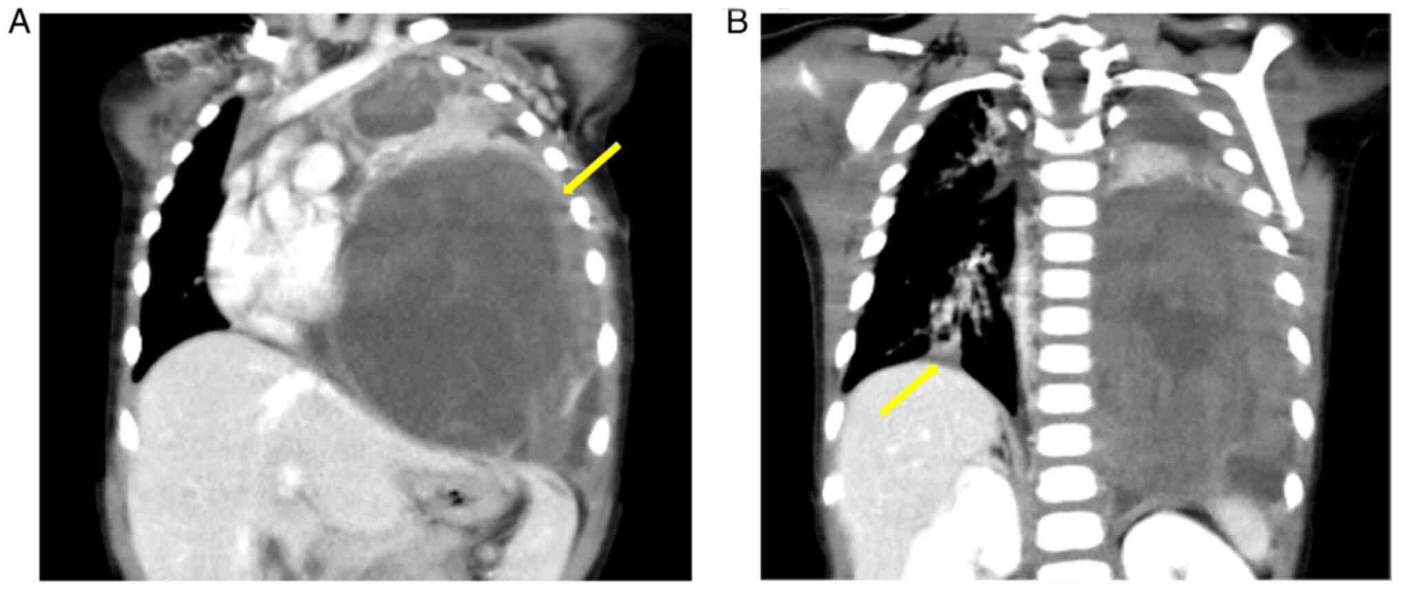
The enhanced computed tomography (CT) at transfer showed left
intrathoracic tumor of 5×4 cm with severe mediastinal shift, as
well as right intrathoracic tumor of 1.5×1.5 cm (Fig. 1A and B). Enhanced CT with 5 mm
slice thickness was performed using 640-multislice CT scanners
(Aquilion ONE, Canon Medical Systems Corportion, Otawara, Japan).
Iopamiron® (Bayer, Osak, Japan) was used as iodinated
contrast medium. No other metastatic diseases were detected. The
biopsy of the left intrathoracic tumor revealed the presence of
IFS. The VDC-IE chemotherapy containing vincristine, doxorubicin,
cyclophosphamide, ifosfamide, and etoposide, induced a significant
reduction in tumor size. She was alive with disease receiving
therapy for 8 months since the initiation of chemotherapy.
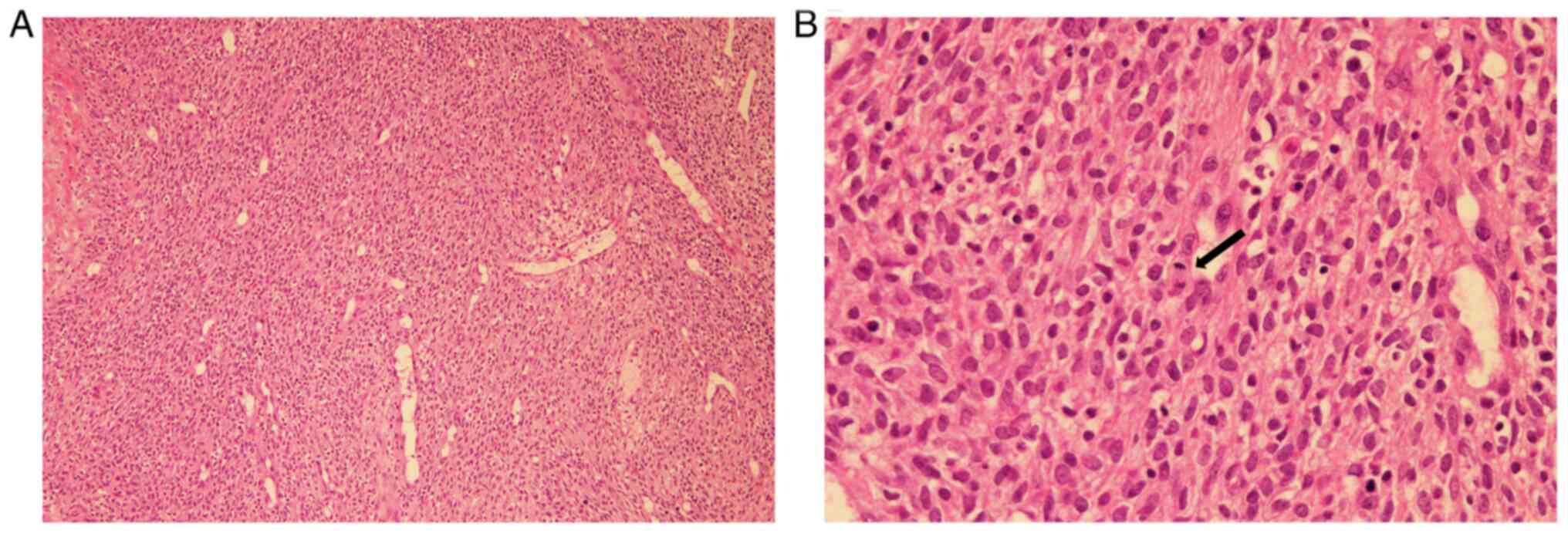
The tumor cells were composed of spindle cells,
arranged into intersecting fascicules with focal
hemagiopericytomatous pattern (Fig.
2A). The mitoses in the tumor were counted at 10 per 10
high-power fields (Fig. 2B). The
procedure of H&E pathological staining was as follows. Resected
specimens were immediately fixed with 10% formalin neutral buffer
solution for 48 h at room temperature. Fixed sections were embedded
in paraffin and 4-µm-thick tissue sections were stained with
hematoxylin and eosin solutions (Sakura, Tokyo, Japan).
Immunohistochemical staining was performed on 4-µm-thick sections
using a fully automated systems [Bench Mark GX System (Roche,
Rotkreuz, Switzerland) or Leica Bond-max (Leica Biosystems, Buffalo
Grove, IL, USA)] and the following primary antibodies; desmin
(clone D33; prediluted) (IR606; Dako, Calpinteria, CA, USA), Wilms
tumor gene 1 (WT-1) (6F-H2; dilution 1:1) (41386; NICHIREI, Osaka,
Japan), pan-TRK [VENTANA Pan-TRK (EPR17341) Assay] (790–7026;
Roche), S-100 (polyclonal; dilution 1:2,000) (Z3011; Dako), SMA
(clone 1A4; dilution 1:300) (IR611; Dako), CD34 (clone NU-4A1;
dilution 1:4) (413111; NICHIREI, Osaka Japan), CD99 (clone O13;
prediluted) (790–4452; Roche), NK2 homeobox 2(NKX2.2) (rabbit
polyclonal; dilution 1:50) (NBP1-82554; Novus Biologicals,
Littleton, CO, USA), myogenic differentiation 1 (MyoD1) (mouse
monoclonal; dilution 1:250) (ab133627, abcam, USA), and
pan-cytokeratin (clone AE1/AE3; dilution 1:100) (IR053; Dako). The
reaction of secondary antibody and following DAB
(3,3′-Diaminobenzidine) reaction were performed using ultraView
Universal DAB detection kit (109431; Roche) for Bench Mark GX
System or BOND Polymer Refine Detection (DS9800; Leica Biosystems)
for Leica Bond-max. Appropriate positive control sections were
mounted on the same slide glasses. By immunohistochemistry, the
right buttock and left intrathoracic tumor cells focally expressed
desmin (Fig. 3A). WT-1 was
detected in the cytoplasm of tumor cells, not nucleus (Fig. 3B). The both tumor cells were
negative for pan-TRK, S-100, SMA, CD34, CD99, NKX2.2, MyoD1, and
pan-cytokeratin (Fig. 3C-J).
Interphase fluorescence in situ hybridization (FISH) was
extracted from formalin-fixed paraffin-embedded tissue (FFPE). ETV6
break apart probe (Vysis LSI ETV6 Dual Color, Break Apart Probe
Kit) (VYSIS/Abbott, Abbott Park, IL) was used for the detection of
rearrangement of ETV6 (12p13). The Sure FISH CIC 5′ BA probe and
the Sure FISH CIC 3′BA probe (Agilent Technologies, Cedar Creek,
USA) were used for the detection of CIC (19q13.2) rearrangement.
FISH analysis for ETV6 and CIC revealed negative
split signals in the right buttock tumor cells. Using the left
intrathoracic tumor tissues, KIAA1549-BRAF fusion transcript
was identified by the comprehensive genomic profiling with
NGS-FoundationOne™, which examines the whole coding
sequence of 315 cancer-related genes and introns from 28 genes
often rearranged or altered in cancer. Other genomic alterations
identified using FoundationOne™ were STK11 F354L
and RET A45V. Subsequently, KIAA1549-BRAF fusion
transcript was confirmed by reverse transcription-polymerase chain
reaction (RT-PCR) (Fig. 4A). While
several types of KIAA1549-BRAF fusion transcripts were
previously reported (14), the
transcript composed of KIAA1549 exon 10 fused to BRAF
exon 9 was exclusively detected in our case (Fig. 4B). The procedure of sanger sequence
was as follows. RNA was extracted from FFPE tissue of the right
buttock tumor using The RNAstormTM kit (Cell Data Science, CA,
USA). For RT-PCR, cDNA was synthesized from 1.0 µg of total RNA
using a SuperScriptTM IV First-Strand Synthesis System
(ThermoFisher Scientific, Oslo, Norway). PCR reaction was performed
using a GoTaq® DNA polymerase (Promega, WI, USA) with
KIAA1549-forward (5′-GATTGTTGTCATCCTCTACTGG-3′) and BRAF-reverse
(5′-CCTCCATCACCACGAAATCCTT-3′) primers. PCR conditions were initial
denaturation at 95°C for 1 min, 40 cycles of 95°C for 30 sec,
annealing at 51°C for 30 sec, 72°C for 30 sec, and a final
extension at 72°C for 5 min. PCR products were electrophoresed on
2.0% agarose gel and purified with Wizard® SV Gel and
PCR Clean-up System (Promega, WI, USA). BigDye Terminator v3.1
Cycle Sequencing kit (Thermo Scientific, USA) was used for
terminator cycling sequencing reactions for Sanger sequencing of
purified PCR products on the 3730×l DNA Analyzer (Thermo Fisher
Scientific, USA).
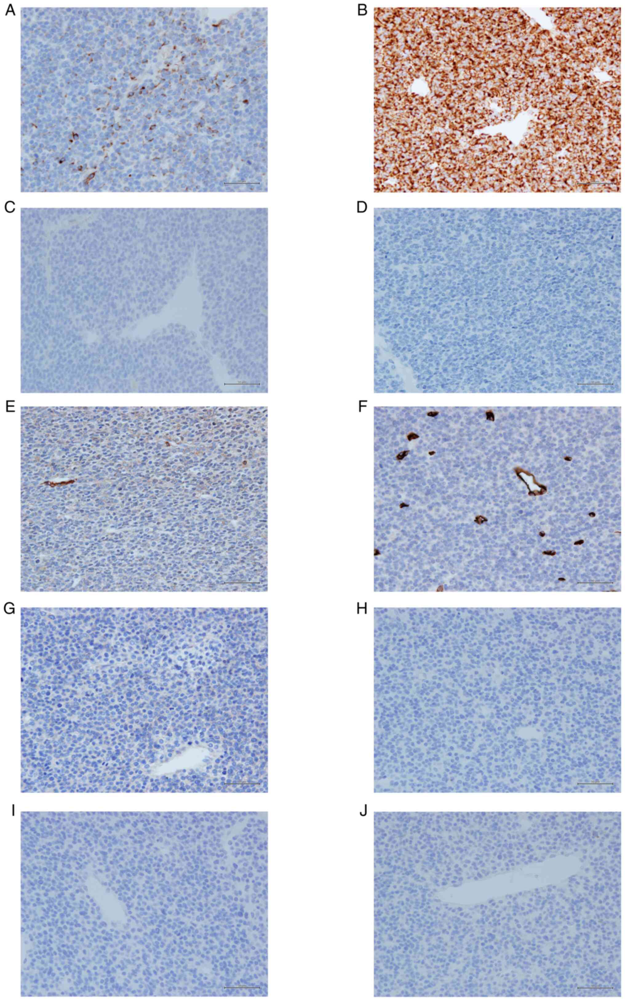 | Figure 3.IHC staining (magnification, ×400).
(A) Weak positive expression of desmin. (B) Cytoplasmic expression
of WT-1. Negative IHC staining results for (C) pan-TRK, (D) S-100,
(E) SMA, (F) CD34, (G) CD99, (H) NKX2.2, (I) MyoD1 and (J)
pan-cytokeratin. IHC, immunohistochemistry; WT-1, wilms tumor gene
1; pan-TRK, pan-tropomyosin receptor kinase; SMA, smooth muscle
actin; NKX2.2, NK2 homeobox 2; MyoD1, myogenic differentiation
1. |
Discussion
Herein, we reported a pediatric case of spindle cell
sarcoma with KIAA1549-BRAF resembling IFS morphologically.
Although the present case was initially diagnosed with IFS
morphologically, a comprehensive genomic profiling with NGS led to
a more precise diagnosis. Because sarcomas in pediatrics are rare
and heterogenous, the elucidation of genomic profiling in pediatric
sarcomas using NGS can contribute to an appropriate diagnosis and
targetable therapies.
The characteristics of BRAF-altered spindle
cell sarcomas resembling IFS morphologically are not well
understood (10,11,15–18).
To identify the clinical characteristics of BRAF-altered
spindle cell sarcomas resembling IFS morphologically, we conducted
a literature search of all reports. A literature search of all
reports was conducted. The following keywords were used in the
electronic databases PubMed with no date of publication
limitations: ‘infantile fibrosarcoma’ OR ‘spindle cell sarcoma’ OR
‘spindle cell neoplasm’ combined with ‘BRAF’. From the titles and
abstracts, we excluded non-English language studies, meeting
presentations, and commentaries. The article titles, abstracts, and
full papers were examined, and the reports not containing
BRAF-altered IFS, spindle cell sarcoma, or spindle cell neoplasm
were excluded. URL was as follows: http://pubmed.ncbi.nlm.nih.gov/?term=(((infantile%20fibrosarcoma)%20OR%20(spindle%20cell%20sarcoma)%20OR%20(spindle%20cell%20neoplasm)))%20AND%20(BRAF)&sort=date
We identified 24 cases of spindle cell sarcoma with
BRAF-rearrangement or mutation. The median age at diagnosis
was 5.5 months (range: 0–69 years). Seven of 24 cases (30%) were
diagnosed over 2 years of age and in three cases over 20 years of
age. The most common site of tumors was extremities (6 cases, 25%).
Immunohistochemical identification of expression of CD34 was
observed in 8 (47%) cases, S-100 in 4 (21%) cases, and SMA in 6
(40%) cases. The expression of pan-Trk was present in one (9.0%)
case. BRAF-rearrangements, including fusions of BRAF
kinase domain, were detected in 17 cases (68%). The fusions were as
follows: KIAA1549-BRAF, AGAP3-BRAF, CUX1-BRAF, DAAM1-BRAF,
EPB41L2-BRAF, MCC-BRAF, NPF1-BRAF, OSBP-BRAF, PDE10A-BRAF,
SEPT7-BRAF, TEX4-BRAF, FOXN3-BRAF, and TRIP11-BRAF
(10,11,15–18).
KIAA1549-BRAF fusion was detected in two cases as in our
case (10,19). BRAF point mutations were
present in four cases and BRAF-internal duplication (ID) in
three cases (10,16,17).
One case contained BRAF point mutation and
BRAF-rearrangement. The identification of ETV6-NTRK3
transcript was performed in 12 cases by FISH for ETV6 or
whole genome sequencing. In two cases, ETV6-NTRK3 fusion and
BRAF-ID coexisted (17).
One case harbored two distinct BRAF fusions
(FOXN3-BRAF and TRIP11-BRAF) (10). Three of 13 cases (23%) had
metastasis at diagnosis. All the three cases occurred in adults and
were refractory to the conventional chemotherapy. Median follow-up
period was 10 months (3i60 months) and 2 patients (8%) died of
disease. In our review of BRAF-altered spindle cell sarcomas
resembling IFS morphologically, most cases occurred in younger
children under the age of 2 years, showed nonspecific patterns of
immunohistochemistry staining, and harbored a BRAF fusion.
These results were consistent with our case.
KIAA1549-BRAF fusion is considered as a
recurrent oncogenic driver in pilocytic astrocytoma (19). KIAA1549-BRAF fusion leads to
loss of N-terminal regulatory domain of BRAF and subsequent
activation of the kinase domain, thereby resulting in constitutive
activation of BRAF (20).
Although different fusion variants were identified in
KIAA1549-BRAF, the transcript detected in our case was
composed of KIAA1549 exon 10 fused to BRAF exon 9 and
contained the intact BRAF kinase domain. Trametinib, a MEK
inhibitor has recently been demonstrated to be effective in
low-grade glioma with BRAF fusions including
KIAA1549-BRAF (21,22). Subbiah et al (15) reported the effectiveness of the
combination therapy of sorafenib, temsirolimus, and bevacizumab for
a spindle cell sarcoma with KIAA1549-BRAF, which was
refractory to the conventional chemotherapies. Because of limited
data, further studies are needed to determine the effectiveness of
BRAF-targeted therapy including MEK inhibitor for
BRAF-altered spindle cell sarcomas morphologically
resembling IFS.
In conclusion, we report a pediatric case of spindle
cell sarcoma with KIAA1549-BRAF morphologically resembling
IFS. The elucidation of genomic profiling by NGS may assist us in
making an appropriate diagnosis and selecting new therapeutic
options for ETV6-NTRK3 negative spindle cell sarcomas
morphologically resembling IFS.
Acknowledgements
Not applicable.
Funding
Funding: No funding was received.
Availability of data and materials
The dataset used and/or analyzed during the current
study are available from the corresponding author on reasonable
request.
Authors' contributions
TF, SU, MY, DH and YK participated in the conception
and design of the study. MY, SH, AK, AS, KK, TI, AU, KM, TH, MK,
and TS were involved in the analysis and interpretation of the data
for the pathological diagnosis. MY, TM, AT, NY, MK and TS were
involved in the analysis and interpretation of data for
comprehensive genomic profiling using NGS. AU, KM and TH performed
surgery. MY and MK performed the histological examination of the
tumors. TF and SU drafted the initial manuscript. TF, SU, MY, KK,
MK, DH and YK critically revised the article for important
intellectual content. TH, TS, DH and YK confirmed the authenticity
of all the raw data. All authors have read and approved the final
manuscript.
Ethics approval and consent to
participate
The present study was approved by the Institutional
Review Board of Kobe Children's Hospital (R3-62; Kobe, Japan).
Written informed consent was obtained from the patient's
parents.
Patient consent for publication
The patient's parents provided written informed
consent for the publication of any associated data.
Competing interests
The authors declare that they have no competing
interests.
References
|
1
|
Orbach D, Rey A, Cecchetto G, Oberlin O,
Casanova M, Thebaud E, Scopinaro M, Bisogno G, Carli M and Ferrari
A: Infantile fibrosarcoma: Management based on the European
experience. J Clin Oncol. 28:318–323. 2010. View Article : Google Scholar : PubMed/NCBI
|
|
2
|
Orbach D, Brennan B, De Paoli A, Gallego
S, Mudry P, Francotte N, van Noesel M, Kelsey A, Alaggio R,
Ranchère D, et al: Conservative strategy in infantile fibrosarcoma
is possible: The European paediatric soft tissue sarcoma study
group experience. Eur J Cancer. 57:1–9. 2016. View Article : Google Scholar : PubMed/NCBI
|
|
3
|
Chung EB and Enzinger FM: Infantile
fibrosarcoma. Cancer. 38:729–739. 1976. View Article : Google Scholar : PubMed/NCBI
|
|
4
|
Davis JL, Lockwood CM, Stohr B, Boecking
C, Al-Ibraheemi A, DuBois SG, Vargas SO, Black JO, Cox MC, Luquette
M, et al: Expanding the spectrum of pediatric NTRK-rearranged
mesenchymal tumors. Am J Surg Pathol. 43:435–445. 2019. View Article : Google Scholar : PubMed/NCBI
|
|
5
|
Coffin CM, Jaszcz W, O'Shea PA and Dehner
LP: So-called congenital-infantile fibrosarcoma: Does it exist and
what is it? Pediatr Pathol. 14:133–150. 1994. View Article : Google Scholar : PubMed/NCBI
|
|
6
|
Rudzinski ER, Lockwood CM, Stohr BA,
Vargas SO, Sheridan R, Black JO, Rajaram V, Laetsch TW and Davis
JL: Pan-Trk immunohistochemistry identifies NTRK rearrangements in
pediatric mesenchymal tumors. Am J Surg Pathol. 42:927–935. 2018.
View Article : Google Scholar : PubMed/NCBI
|
|
7
|
Pavlick D, Schrock AB, Malicki D, Stephens
PJ, Kuo DJ, Ahn H, Turpin B, Allen JM, Rosenzweig M, Badizadegan K,
et al: Identification of NTRK fusions in pediatric mesenchymal
tumors. Pediatr Blood Cancer. 64:e264332017. View Article : Google Scholar : PubMed/NCBI
|
|
8
|
Davis JL, Vargas SO, Rudzinski ER, López
Marti JM, Janeway K, Forrest S, Winsnes K, Pinto N, Yang SE,
VanSandt M, et al: Recurrent RET gene fusions in paediatric spindle
mesenchymal neoplasms. Histopathology. 76:1032–1041. 2020.
View Article : Google Scholar : PubMed/NCBI
|
|
9
|
Flucke U, van Noesel MM, Wijnen M, Zhang
L, Chen CL, Sung YS and Antonescu CR: TFG-MET fusion in an
infantile spindle cell sarcoma with neural features. Genes
Chromosomes Cancer. 56:663–667. 2017. View Article : Google Scholar : PubMed/NCBI
|
|
10
|
Penning AJ, Al-Ibraheemi A, Michal M,
Larsen BT, Cho SJ, Lockwood CM, Paulson VA, Liu YJ, Plank L,
Fritchie K, et al: Novel BRAF gene fusions and activating point
mutations in spindle cell sarcomas with histologic overlap with
infantile fibrosarcoma. Mod Pathol. 34:1530–1540. 2021. View Article : Google Scholar : PubMed/NCBI
|
|
11
|
Kao YC, Fletcher CDM, Alaggio R, Wexler L,
Zhang L, Sung YS, Orhan D, Chang WC, Swanson D, Dickson BC and
Antonescu CR: Recurrent BRAF gene fusions in a subset of pediatric
spindle cell sarcomas: Expanding the genetic spectrum of tumors
with overlapping features with infantile fibrosarcoma. Am J Surg
Pathol. 42:28–38. 2018. View Article : Google Scholar : PubMed/NCBI
|
|
12
|
El-Osta H, Falchook G, Tsimberidou A, Hong
D, Naing A, Kim K, Wen S, Janku F and Kurzrock R: BRAF mutations in
advanced cancers: Clinical characteristics and outcomes. PLoS One.
6:e258062011. View Article : Google Scholar : PubMed/NCBI
|
|
13
|
Davies H, Bignell GR, Cox C, Stephens P,
Edkins S, Clegg S, Teague J, Woffendin H, Garnett MJ, Bottomley W,
et al: Mutations of the BRAF gene in human cancer. Nature.
417:949–954. 2002. View Article : Google Scholar : PubMed/NCBI
|
|
14
|
Ross JS, Wang K, Chmielecki J, Gay L,
Johnson A, Chudnovsky J, Yelensky R, Lipson D, Ali SM, Elvin JA, et
al: The distribution of BRAF gene fusions in solid tumors and
response to targeted therapy. Int J Cancer. 138:881–890. 2016.
View Article : Google Scholar : PubMed/NCBI
|
|
15
|
Subbiah V, Westin SN, Wang K, Araujo D,
Wang WL, Miller VA, Ross JS, Stephens PJ, Palmer GA and Ali SM:
Targeted therapy by combined inhibition of the RAF and mTOR kinases
in malignant spindle cell neoplasm harboring the KIAA1549-BRAF
fusion protein. J Hematol Oncol. 7:82014. View Article : Google Scholar : PubMed/NCBI
|
|
16
|
Mitsis D, Opyrchal M, Zhao Y, Kane Iii JM,
Cheney R and Salerno KE: Exceptional clinical response to
BRAF-targeted therapy in a patient with metastatic sarcoma. Cureus.
7:e4392015.PubMed/NCBI
|
|
17
|
Wegert J, Vokuhl C, Collord G, Del
Castillo Velasco-Herrera M, Farndon SJ, Guzzo C, Jorgensen M,
Anderson J, Slater O, Duncan C, et al: Recurrent intragenic
rearrangements of EGFR and BRAF in soft tissue tumors of infants.
Nat Commun. 9:23782018. View Article : Google Scholar : PubMed/NCBI
|
|
18
|
Hughes CE, Correa H, Benedetti DJ, Smith
B, Sumegi J and Bridge J: Second report of PDE10A-BRAF fusion in
pediatric spindle cell sarcoma with infantile fibrosarcoma-like
morphology suggesting PDE10A-BRAF fusion is a recurrent event.
Pediatr Dev Pathol. 24:554–558. 2021. View Article : Google Scholar : PubMed/NCBI
|
|
19
|
Zhang J, Wu G, Miller CP, Tatevossian RG,
Dalton JD, Tang B, Orisme W, Punchihewa C, Parker M, Qaddoumi I, et
al: Whole-genome sequencing identifies genetic alterations in
pediatric low-grade gliomas. Nat Genet. 45:602–612. 2013.
View Article : Google Scholar : PubMed/NCBI
|
|
20
|
Jones DT, Kocialkowski S, Liu L, Pearson
DM, Bäcklund LM, Ichimura K and Collins VP: Tandem duplication
producing a novel oncogenic BRAF fusion gene defines the majority
of pilocytic astrocytomas. Cancer Res. 68:8673–8677. 2008.
View Article : Google Scholar : PubMed/NCBI
|
|
21
|
Selt F, van Tilburg CM, Bison B, Sievers
P, Harting I, Ecker J, Pajtler KW, Sahm F, Bahr A, Simon M, et al:
Response to trametinib treatment in progressive pediatric low-grade
glioma patients. J Neurooncol. 149:499–510. 2020. View Article : Google Scholar : PubMed/NCBI
|
|
22
|
Perreault S, Larouche V, Tabori U, Hawkin
C, Lippé S, Ellezam B, Décarie JC, Théoret Y, Métras MÉ, Sultan S,
et al: A phase 2 study of trametinib for patients with pediatric
glioma or plexiform neurofibroma with refractory tumor and
activation of the MAPK/ERK pathway: TRAM-01. BMC Cancer.
19:12502019. View Article : Google Scholar : PubMed/NCBI
|