Introduction
The jumonji domain-containing protein 6 (JMJD6)
plays a vital role in epigenetic regulation and demonstrates the
tyrosine kinase activity (1). The
JMJD6 has been reported in cellular proliferation and migration
(2). Abnormal over expression of
JMJD6 may contribute to the development of different types of
cancer (breast cancer, malignant melanoma, oral cancer, lung
adenocarcinoma, hepatocellular carcinoma, ovarian cancer,
colorectal cancer, glioblastoma, glioma) (3–11).
Overexpression of the JMJD6 gene promotes cell proliferation
and migration and enhances tumor growth in vivo (2). JMJD6 expression has been linked to a
poor prognosis in lung adenocarcinoma, hepatocellular carcinoma,
ovarian cancer, and colorectal cancer.
JMJD6 was reported as a regulatory gene that works
with Myc to promote tumorigenesis (12). Cancer cells in which EMT
(epithelial-mesenchymal transition) is induced acquire invasive and
metastatic potential. JMJD6 overexpression increases tumor volume,
cause EMT, and enhances invasion in breast cancer (12). JMJD6 protein was found in intestinal
glands where the intestinal epithelium is constantly regenerating,
according to Wang et al (9).
These reports suggested that JMJD6 may be involved in intestinal
cell proliferation and may be a new biomarker for colorectal cancer
development.
Generally, an immune response to aberrant tumor
antigens produces autoantibodies early in carcinogenesis and the
autoantibodies are frequently elevated in patients even at early
disease stages due to an antigen expression within the tumor
(13). Serum p53 autoantibodies
have been reported as the most common autoantibody and are used as
a standard biomarker in patients with colorectal cancer (14). Since the JMJD6 protein is
specifically exhibited in cancer cells, it may cause
autoantibodies. Nevertheless, there are no reports of the analysis
of autoantibodies against JMJD6 in solid cancer patients.
Therefore, this study aimed to analyze the anti-JMJD6 antibody in
patients with colorectal cancer and to evaluate the
clinicopathological and prognostic significance of the anti-JMJD6
antibody.
Materials and methods
Patients and sera
Overall, 167 patients with colorectal cancer who
underwent radical surgery at Toho University Omori Medical Center
were evaluated between April 2007 and May 2012. The control group
comprised 96 healthy subjects provided by a health screening
clinic, Port Square Kashiwado Clinic. The patients comprised 97
male and 70 female patients (mean age, 64.9 years; range, 33–90
years). The pathological stages were as follows; stage I (n=47),
stage II (n=56), stage III (n=49), and stage IV (n=15). The control
group comprised 51 healthy male and 45 healthy female (mean age, 58
years; range, 50–76 years).
The study was conducted following the guidelines
Ethical statement of the Declaration of Helsinki and approved by
the Ethics Committee of Faculty of Medicine, Toho University
(approval no. A18103_A17052_A16035_A16001_26095_25024_24038_22047),
Chiba University Graduate School of Medicine (approval no.
2018-320) (Japan), and Port Square Kashiwado Clinic, Kashiwado
Memorial Foundation (approval no. 2012-001). Before surgery, serum
samples were obtained and frozen at −80°C until analysis. Written,
informed consent was obtained from all subjects. The patient's
medical records were retrospectively reviewed according to the
ethics committee of Toho University Omori Medical Center (approval
nos. M21038_20197_19213 and
M21320_21039_20200_30196_19056_18002).
Purification of recombinant JMJD6 and
detection of s-JMJD6-antibody by amplified luminescent proximity
homology assay-linked immunosorbent assay (Alpha-LISA)
Serum samples were obtained before surgery and
stored frozen at −80°C until use. Glutathione S-transferase (GST)
and GST-fused JMJD6 proteins were purified as described previously
(15–17). s-JMJD6 Ab levels were assessed using
an amplified luminescent proximity homology assay-linked
immunosorbent assay (Alpha-LISA), as described previously (15–17).
Briefly, Alpha-LISA was performed in 384-well microtiter plates
(white opaque OptiPlate, PerkinElmer) containing either 2.5 µl of
1:100 diluted serum and 2.5 µl of 10 µg/ml of GST or GST-JMJD6
protein in AlphaLISA buffer (25-mM HEPES, pH 7.4, 0.1% casein, 0.5%
Triton X-100, 1 mg/ml dextran-500, and 0.05% Proclin-300). The
reaction mixture was incubated at room temperature for 6–8 h,
following which anti-human IgG-conjugated acceptor beads (2.5 µl at
40 µg/ml) and glutathione-conjugated donor beads (2.5 µl at 40
µg/ml) were added and incubated further at room temperature in the
dark for 1–21 days. Chemical emissions were read on an EnSpire
Alpha microplate reader (PerkinElmer). Specific reactions were
estimated by subtracting the emission photon counts of the GST
controls from the counts of GST-JMJD6 proteins.
Statistical analysis
The cutoff value for detecting colorectal cancer was
calculated using the receiver operating characteristic curve.
Patients with a cutoff value greater than 5,720 were categorized as
serum anti-JMJD6-Ab positive. Using 5,720 as the cutoff value,
patients with colorectal cancer were categorized into the
s-JMJD6-Abs-positive group (n=61) and s-JMJD6-Abs-negative group
(n=106), and the following analyses were performed.
Clinicopathologic factors and prognosis were
compared between the Ab-positive and Ab-negative groups using the
Mann-Whitney U test or Fisher's exact probability test.
Clinicopathological parameters associated with survival were
assessed by univariate analysis with a log-rank test based on
Kaplan-Meier survival curves. Multivariate analysis was conducted
using the Cox proportional hazards model. All statistical analyses
were performed using EZR (Saitama Medical Center, Jichi Medical
University; Saitama, Japan) (18),
a graphical user interface of R (The R Foundation for Statistical
Computing; version 2.13.0). P<0.05 was considered to indicate a
statistically significant difference.
Results
Comparison of s-JMJD6-Abs positivity
rates
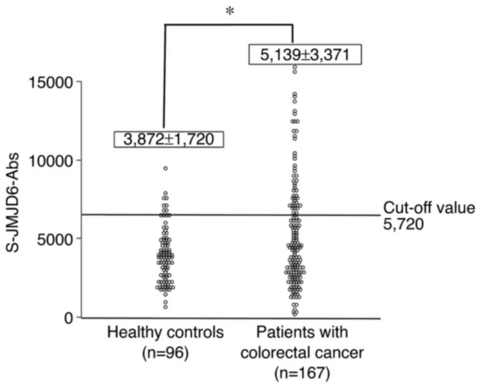
A comparison of anti-JMJD6 antibody levels in serum
from healthy controls and colorectal cancer patients is shown in
Fig. 1. The serum antibody
(s-JMJD6-Ab) levels against JMJD6 examined using Alpha-LISA. The
cutoff value for detecting colorectal cancer was calculated using
the receiver operating characteristic curve (ROC) and determined to
be 5,720 (Fig. S1). The positive
rate of s-JMJD6-Abs in colorectal cancer was significantly higher
than that of healthy controls (37 vs. 14%, P<0.05; Fig. 1). The associations of positivity of
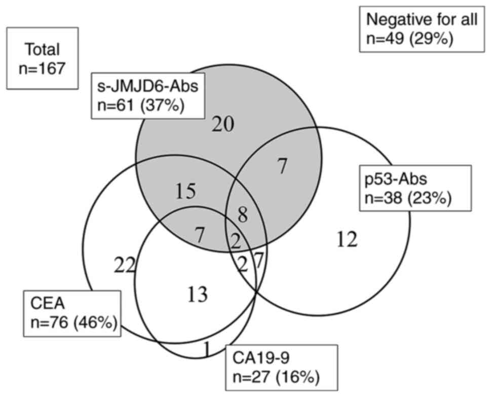
s-JMJD6-Abs, CEA, CA19-9, and p53-Abs are shown in Fig. 2. Overall, 20 patients (12%) were
solely positive for s-JMJD6-Abs. Therefore, entirely negative
patients for tumor markers were reduced from 88 (53%) by
combinatory use of CEA, CA19-9 to 49 (29%) by combinatory use of
CEA, CA19-9, p53-Abs and s-JMJD6-Abs (Fig. 2).
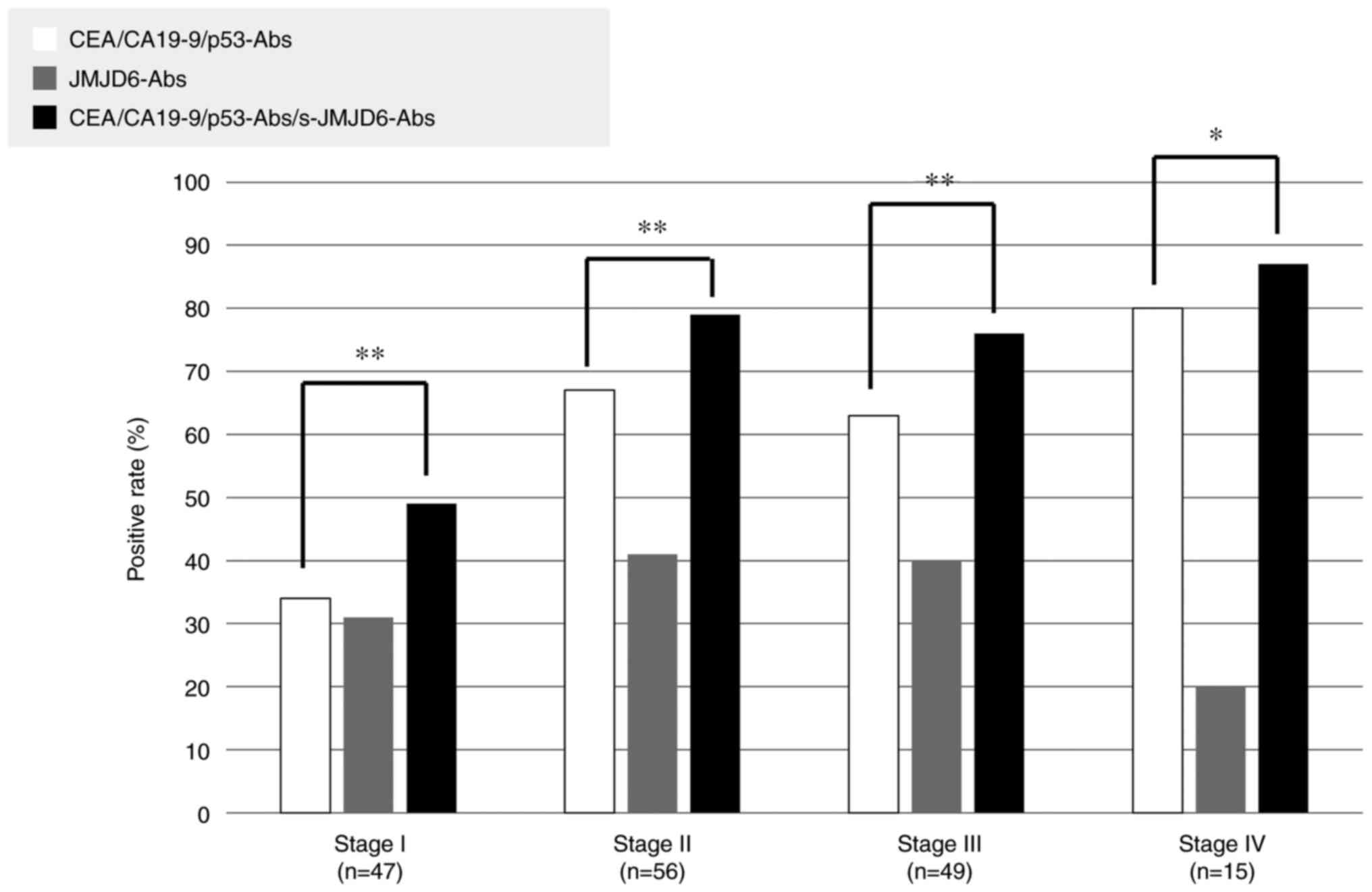
Comparisons of the positive rates according to tumor
stages are shown in Fig. 3. The
positive combinatory rates of CEA, CA19-9, p53-Abs were related to
tumor progression (stage I: 34%, stage II: 68%, stage III: 63%,
stage IV: 80%). Nevertheless, the positive rates of s-JMJD6-Abs
were not associated with tumor progression (stage I: 32%, stage II:
41%, stage III: 41%, stage IV: 17%). Combinatory use of all these
four markers significantly increased the positive rate than the
combinatory use of CEA, CA19-9, and p53-Abs (Fig. 3).
Comparison of clinicopathological
factors between s-JMJD6-Ab-positive and s-JMJD6-Ab-negative
groups
s-JMJD6-Ab positivity was significantly higher in
the elderly group (P=0.03). In contrast, no other
clinicopathological backgrounds demonstrated a significant
association with s-JMJD6-Ab positivity. Additionally, no
correlation was noticed between s-JMJD6-Abs and other tumor markers
(Table I).
 | Table I.Comparison of pretreatment JMJD6-Ab
level with clinicopathological factors. |
Table I.
Comparison of pretreatment JMJD6-Ab
level with clinicopathological factors.
| Variable | Low JMJD6-Ab group
<5,720, n=106 (%) | High JMJD6-Ab group
≧5,720, n=61 (%) | P-valuea |
|---|
| Sex |
|
| 0.15 |
|
Female | 49 (70) | 21 (30) |
|
| Male | 57 (59) | 40 (41) |
|
| Age, years |
|
| 0.03 |
|
<65 | 52 (73) | 19 (27) |
|
| ≥65 | 54 (56) | 42 (44) |
|
| Tumor depth |
|
| 0.61 |
|
pT1pT2 | 34 (67) | 17 (33) |
|
|
pT3pT4 | 72 (62) | 44 (38) |
|
| Nodal status |
|
| 1 |
|
Negative | 68 (64) | 39 (36) |
|
|
Positive | 38 (63) | 22 (37) |
|
| Stage |
|
| 0.17 |
|
I/II/III | 93 (62) | 58 (38) |
|
| IV | 13 (81) | 3 (19) |
|
| Distant
metastasis |
|
| 0.26 |
|
Negative | 94 (62) | 58 (38) |
|
|
Positive | 12 (80) | 3 (20) |
|
| Histology |
|
| 0.65 |
| Muc,
Poor | 4 (80.0) | 1 (20) |
|
| Tub | 101 (63) | 60 (37) |
|
| CEA, 5 ng/ml |
|
| 0.20 |
|
Negative | 62 (68) | 29 (32) |
|
|
Positive | 44 (58) | 32 (42) |
|
| CA19-9, 37
U/ml |
|
| 0.67 |
|
Negative | 90 (64) | 50 (36) |
|
|
Positive | 16 (59) | 11 (41) |
|
| p53-Abb |
|
| 0.34 |
|
Negative | 79 (65) | 42 (35) |
|
|
Positive | 21 (55) | 17 (45) |
|
Prognostic impact of s-JMJD6-Abs
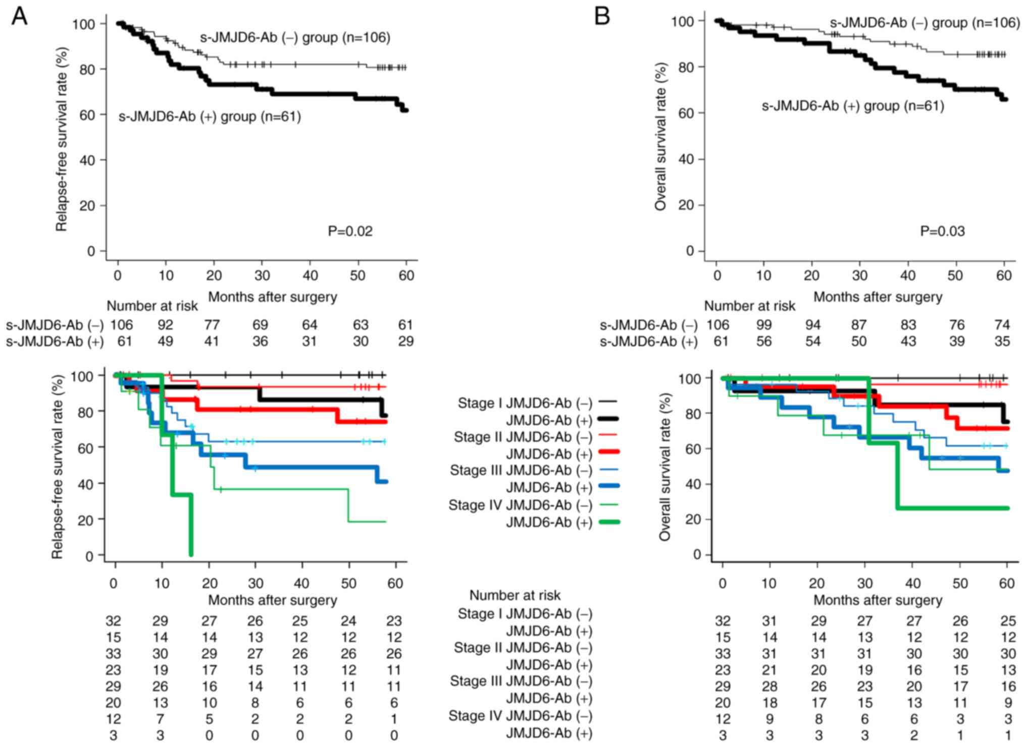
Comparisons of prognosis between the
s-JMJD6-Ab-positive group and s-JMJD6-Ab-negative groups are shown
in Fig. 4. Recurrence-free survival
was significantly worse in the high s-JMJD6-Ab group than in the
low s-JMJD6-Ab group (P=0.02; Fig.
4A). Overall survival was also significantly worse in the high
s-JMJD6-Ab group than in the low s-JMJD6-Ab group (P=0.03; Fig. 4B). When the prognosis of the
antibody-positive and antibody-negative groups was compared by
stage, the antibody-positive group had a significantly poorer
prognosis in stages I, II, and III than the antibody-negative
groups. The antibody-positive group had a worse prognosis in stage
IV, albeit this difference was not statistically significant.
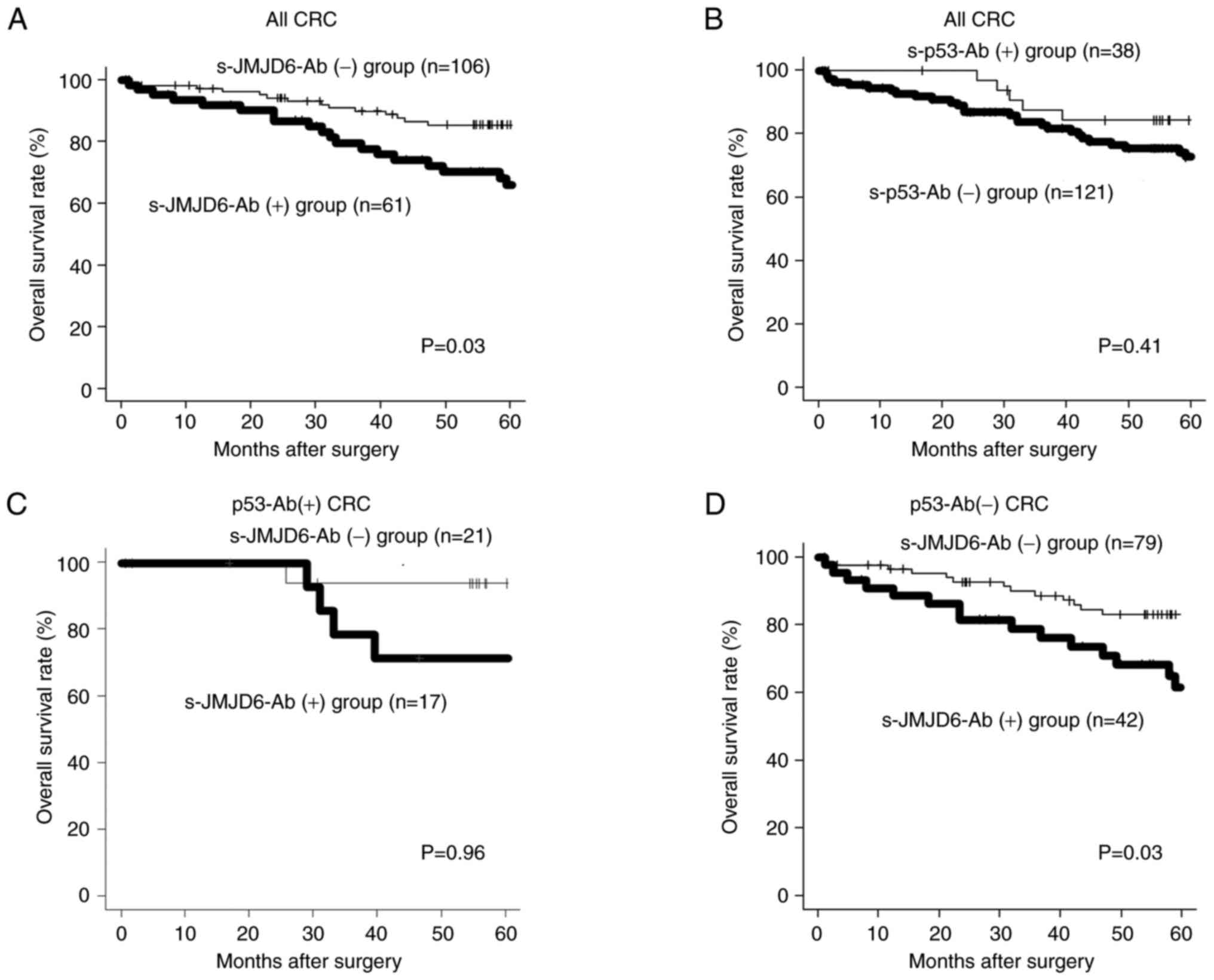
For all colorectal cancers, overall survival was
significantly worse in the high s-JMJD6-Ab group than in the low
s-JMJD6-Ab group (P=0.03; Fig. 5A).
On the other hand, there was no significant difference in overall
survival with or without p53 antibody (P=0.41; Fig. 5B). In p53 antibody-positive cases,
no prognostic difference was observed between the presence or
absence of s-JMJD6-Ab group (Fig.
5C). On the other hand, among p53-Abs-negative cases,
s-JMJD6-Ab-positive cases had a worse prognosis than
p53-Abs-negative cases (P=0.03, Fig.
5D).
Univariate and multivariate analyzes
of relapse-free survival
In the univariate analysis of recurrence-free
survival, tumor depth (P<0.01), lymph node metastasis
(P<0.01), distant metastasis (P<0.01), CA19-9 positivity
(P<0.01), and high JMJD6-Abs (P=0.02) were significant poor
prognostic factors regarding Relapse-free survival (Table II). In multivariate analysis, tumor
depth (P=0.01), lymph node metastasis (P<0.01), distant
metastasis (P<0.01), CA19-9 positivity (P=0.04), and high
JMJD6-Abs (P<0.01) were independent poor prognostic factors.
| Table II.Univariate and multivariate analysis
of clinicopathological factors to predict relapse-free
survival. |
Table II.
Univariate and multivariate analysis
of clinicopathological factors to predict relapse-free
survival.
|
|
| Multivariate
analysis |
|---|
|
|
|
|
|---|
| Variable | Univariate
P-valuea | HRb | 95% CIc |
P-valued |
|---|
| Sex |
|
|
|
|
|
Male | 0.42 |
|
|
|
|
Female |
|
|
|
|
| Age, years |
|
|
|
|
|
<65 | 0.27 |
|
|
|
|
≧65 |
|
|
|
|
| Tumor depth |
|
|
|
|
|
pT3pT4 | <0.01 | 4.59 | 1.36-15.57 | 0.01 |
|
pT1pT2 |
|
|
|
|
| Nodal status |
|
|
|
|
|
Positive | <0.01 | 3.30 | 1.67-6.52 | <0.01 |
|
Negative |
|
|
|
|
| JMJD6-Abs |
|
|
|
|
|
≥5,720 | 0.02 | 2.39 | 1.28-4.46 | <0.01 |
|
<5,720 |
|
|
|
|
| Distant
metastasis |
|
|
|
|
|
Positive | <0.01 | 3.29 | 1.52-7.14 | <0.01 |
|
Negative |
|
|
|
|
| Histology |
|
|
|
|
| Muc,
Poor | 0.36 |
|
|
|
|
Tub |
|
|
|
|
| CEA |
|
|
|
|
|
Positive | 0.32 | 0.71 | 0.36-1.39 | 0.32 |
|
Negative |
|
|
|
|
| CA19-9 |
|
|
|
|
|
Positive | <0.01 | 2.26 | 1.06-4.81 | 0.04 |
|
Negative |
|
|
|
|
| p53-Ab |
|
|
|
|
|
Positive | 0.41 |
|
|
|
|
Negative |
|
|
|
|
Univariate and multivariate analyzes
of overall survival
Similarly, in a univariate analysis of overall
survival (Table III), tumor
invasiveness (P<0.01), lymph node metastasis (P<0.01),
distant metastasis (P<0.01), CA19-9 (P<0.01) and high
JMJD6-Abs (P=0.03) were significant poor prognostic factors. In
multivariate analysis, tumor depth (P=0.03), lymph node metastasis
(P<0.01), distant metastasis (P<0.01), and high JMJD6-Abs
(P=0.01) were independent poor prognostic factors.
| Table III.Univariate and multivariate analysis
of clinicopathological factors to predict overall survival. |
Table III.
Univariate and multivariate analysis
of clinicopathological factors to predict overall survival.
|
|
| Multivariate
analysis |
|---|
|
|
|
|
|---|
| Variable | Univariate P
valuea | HRb | 95% CIc |
P-valued |
|---|
| Sex |
|
|
|
|
|
Male | 0.30 |
|
|
|
|
Female |
|
|
|
|
| Age, years |
|
|
|
|
|
<65 | 0.24 |
|
|
|
|
≥65 |
|
|
|
|
| Tumor depth |
|
|
|
|
|
pT3pT4 | <0.01 | 3.85 | 1.14-12.99 | 0.03 |
|
pT1pT2 |
|
|
|
|
| Nodal status |
|
|
|
|
|
Positive | <0.01 | 3.70 | 1.87-7.34 | <0.01 |
|
Negative |
|
|
|
|
| JMJD6-Abs |
|
|
|
|
|
≥5,720 | 0.03 | 2.14 | 1.16-3.94 | 0.01 |
|
<5,720 |
|
|
|
|
| Distant
metastasis |
|
|
|
|
|
Positive | <0.01 | 3.05 | 1.43-6.48 | 0.01 |
|
Negative |
|
|
|
|
| Histology |
|
|
|
|
| Muc,
Poor | 0.28 |
|
|
|
|
Tub |
|
|
|
|
| CEA |
|
|
|
|
|
Positive | 0.23 | 0.73 | 0.37-1.48 | 0.39 |
|
Negative |
|
|
|
|
| CA19-9 |
|
|
|
|
|
Positive | <0.01 | 2.13 | 0.98-4.63 | 0.06 |
|
Negative |
|
|
|
|
| p53-Ab |
|
|
|
|
|
Positive | 0.41 |
|
|
|
|
Negative |
|
|
|
|
Discussion
This study analyzed the anti-JMJD6 antibody in
colorectal cancer patients and evaluated the clinicopathological
and prognostic significance of the anti-JMJD6 antibody. The
positive rate of s-JMJD6-Abs in patients with colorectal cancer was
37%. s-JMJD6-Abs along with CEA/CA19-9/s-p53-Abs was 71%.
s-JMJD6-Abs showed no correlation with TNM factors. However, the
presence of s-JMJD6-Abs was an independent poor prognostic
factor.
The mechanism by which the JMJD6 protein generates
autoantibodies is thought to be the induction of autoantibodies due
to the leakage of antigens into the blood due to overexpression in
the cancer cells and subsequent cancer cell destruction. Taking p53
as an example, in cancers with p53 mutations, Mdm2, one of p53′s
target genes, is not induced, resulting in increased intracellular
p53 protein levels. Therefore, when cancer cells are destroyed,
many antigens leak into the blood, and it is speculated that p53
autoantibodies also increase. It has been reported that the JMJD6
protein is overexpressed in many cancer cells, including colon
cancer, suggesting a mechanism similar to that of the p53
autoantibody production.
Because of the high frequency of advanced disease,
the prognosis of JMJD6 immunostaining-positive patients with cancer
was poor (2). We analyzed
recurrence patterns in our current research population between
antibody-positive and antibody-negative groups and found no
significant difference in recurrence patterns (data not shown).
Interestingly, such prognostic impact of JMJD6-Abs was evident in
p53-Ab-negative cases. Because JMJD6 has been reported to suppress
the activity of the tumor suppressor gene p53 via p53 hydroxylation
(9), JMJD6 may suppress the
activity of wild-type p53 and promote carcinogenesis in
p53-Ab-negative cases. On the other hand, in the case of
p53-Ab-positive cases, since p53 is mutated and does not function,
it is thought that the presence or absence of JMJD6 has little
prognostic effect. s-JMJD6-Abs did not affect intracellular JMJD6,
suggesting that tissue destruction of cells with high levels of
JMJD6 expression increases autoantibodies. In other words, we
believe that the elevation of JMJD6-Abs is a result of its high
expression in cancer cells. Although we could not directly compare
the results with immunostaining, the antibody-positive cases
probably fared worse than the immunostaining-positive ones. It has
been reported that increased JMJD6 expression is associated with
poor prognosis in colorectal cancer (9), and our findings imply that the
s-JMJD6-Ab positivity may reflect increased JMJD6 exhibition in
cancerous cells.
Since JMJD6 works as a transcriptional and splicing
regulator for histone and non-histone proteins via arginine
demethylation or lysine hydroxylation, reducing JMJD6 enzyme
activity might be a promising new cancer therapy. For instance,
inhibitors of lysine demethylase activity are entering the clinical
trials (19). A small molecule
inhibitor that can hinder the enzymatic activity of JMJD6 has been
found (20). Such molecules
suppress JMJD6-dependent cancer cell growth, including cervical
cancer cells and hepatocellular carcinoma. Therefore, JMJD6
inhibitors may be a future option in cancer therapy. Clinical
trials of molecularly-targeted agents should also assess whether to
target immunostaining-positive tissue or antibody-positive
patients. An autoantibody monitoring-based treatment technique may
be effective in circumstances when monitoring the progress of
therapy or tissue biopsy is challenging.
The positive rates of s-JMJD6-Abs slightly increased
in stage II. However, no elevations were observed in stages III or
IV. This tendency is frequent in autoantibody markers in colorectal
cancer (21,22). The weakness of immune reactions
against tumor antigens in the liver metastases may be why antibody
titers decrease in stage IV patients. Notably, a tendency for the
positive rate to decrease in stage IV compared with stage III has
also been noticed in the other autoantibodies in the different
types of cancer (23). The decrease
in the autoantibody levels may be attributable to the immune system
breakdown or the absorption of autoantibodies by antigens leaked
due to excessive tissue destruction.
The limitations of this study are as follows. First,
we did not evaluate the association between protein expression and
the s-JMJD6-Ab reaction using the resected specimens although the
antibody reactions may reflect protein expression. Second, the
relationship between changes in antibody titer and recurrence could
not be analyzed because of the lack of postoperative antibody titer
changes and postoperative monitoring data. Third, regarding the
grade of malignancy, it is implied that it is related to resistance
to therapeutic drugs. Forth, relatively high false positive rate in
heathy controls. It was found that the s-JMJD6-Ab-positive rate
tended to be high in elderly people aged 65 and over and men.
Antibody markers are highly sensitive and have the
potential to detect early-stage cancers. Thus far, precise data on
the recurrence and/or treatment time are limited and unusable. In
conclusion, although the presence of s-JMJD6-Abs was not
substantially connected with TNM variables or stage in patients
with colorectal cancer, it was an independent poor prognostic
factor, suggesting that it is a valuable biomarker for predicting
the malignant potential of colorectal cancer.
Supplementary Material
Supporting Data
Acknowledgements
The authors would like to thank Dr. Xiao-Meng Zhang
(Department of Biochemistry and Genetics, Graduate School of
Medicine, Chiba University) for identification and purification of
the antigen.
Funding
This study was partly supported by Grant-in-Aid for Scientific
Research (grant nos. 15K10117, 16K10520, 19K09451 and 21K08695)
from the Ministry of Education, Culture, Sports, Science and
Technology of Japan.
Availability of data and materials
The datasets used and/or analyzed in the present
study are available from the corresponding author on reasonable
request.
Authors contributions
KY, HS, TH, HT and YI conceived and designed the
current study. KY, MU, KF and HT acquired patient samples. KY, MU
and KF contributed to the acquisition of the patient's
clinicopathological data. KY, MI, MU, KF, SYL, BSZ and TH analyzed
patient data. KY and HS drafted the manuscript. HS and TH confirm
the authenticity of all the raw data. All authors read and approved
the final manuscript.
Ethics approval and consent to
participate
The study was conducted following the Ethical
statement of the Declaration of Helsinki guidelines. The collection
of serum samples was approved by the Ethics Committee of Faculty of
Medicine, Toho University (approval no. A18103_A17052_A16035_A16001
_26095_25024_24038_22047), Chiba University Graduate School of
Medicine (approval no. 2018-320), and Port Square Kashiwado Clinic,
Kashiwado Memorial Foundation (approval no. 2012-001). Written
informed consent was obtained from all subjects. Retrospective
analysis of patients' medical records was approved by the ethics
committee of Toho University Omori Medical Center (approval nos.
M21038_20197_19213 and M21320_21039_20200_30196_19056_18002).
Patient consent for publication
No applicable.
Competing interests
Professor Hideaki Shimada received research grants
from the Medical & Biological Laboratories Co., Ltd. (Nagoya,
Japan). All other authors have no competing interests.
References
|
1
|
Yang J, Chen S, Yang Y, Ma X, Shao B, Yang
S, Wei Y and Wei X: Jumonji domain-containing protein 6 protein and
its role in cancer. Cell Prolif. 53:e127472020. View Article : Google Scholar : PubMed/NCBI
|
|
2
|
Poulard C, Rambaud J, Lavergne E,
Jacquemetton J, Renoir JM, Tredan O, Chabaud S, Treilleux I, Corbo
L and Le Romancer M: Role of JMJD6 in breast tumourigenesis. PLoS
One. 10:e01261812015. View Article : Google Scholar : PubMed/NCBI
|
|
3
|
Lee YF, Miller LD, Chan XB, Black MA, Pang
B, Ong CW, Salto-Tellez M, Liu ET and Desai KV: JMJD6 is a driver
of cellular proliferation and motility and a marker of poor
prognosis in breast cancer. Breast Cancer Res. 14:R852012.
View Article : Google Scholar : PubMed/NCBI
|
|
4
|
Liu X, Si W, Liu X, He L, Ren J, Yang Z,
Yang J, Li W, Liu S, Pei F, et al: JMJD6 promotes melanoma
carcinogenesis through regulation of the alternative splicing of
PAK1, a key MAPK signaling component. Mol Cancer. 16:1752017.
View Article : Google Scholar : PubMed/NCBI
|
|
5
|
Lee CR, Lee SH, Rigas NK, Kim RH, Kang MK,
Park NH and Shin KH: Elevated expression of JMJD6 is associated
with oral carcinogenesis and maintains cancer stemness properties.
Carcinogenesis. 37:119–128. 2016. View Article : Google Scholar : PubMed/NCBI
|
|
6
|
Zhang J, Ni SS, Zhao WL, Dong XC and Wang
JL: High expression of JMJD6 predicts unfavorable survival in lung
adenocarcinoma. Tumour Biol. 34:2397–2401. 2013. View Article : Google Scholar : PubMed/NCBI
|
|
7
|
Wan J, Liu H, Yang L, Ma L, Liu J and Ming
L: JMJD6 promotes hepatocellular carcinoma carcinogenesis by
targeting CDK4. Int J Cancer. 144:2489–2500. 2019. View Article : Google Scholar : PubMed/NCBI
|
|
8
|
Zheng H, Tie Y, Fang Z, Wu X, Yi T, Huang
S, Liang X, Qian Y, Wang Xi, Pi R, et al: Jumonji domain-containing
6 (JMJD6) identified as a potential therapeutic target in ovarian
cancer. Signal Transduct Target Ther. 4:242019. View Article : Google Scholar : PubMed/NCBI
|
|
9
|
Wang F, He L, Huangyang P, Liang J, Si W,
Yan R, Han X, Liu S, Gui B, Li W, et al: JMJD6 promotes colon
carcinogenesis through negative regulation of p53 by hydroxylation.
PLoS Biol. 12:e10018192014. View Article : Google Scholar : PubMed/NCBI
|
|
10
|
Miller TE, Liau BB, Wallace LC, Morton AR,
Xie Q, Dixit D, Factor DC, Kim LJY, Morrow JJ, Wu Q, et al:
Transcription elongation factors represent in vivo cancer
dependencies in glioblastoma. Nature. 547:355–359. 2017. View Article : Google Scholar : PubMed/NCBI
|
|
11
|
Zhou DX, Zhou D, Zhan SQ, Wang P, Qin K,
Gan W and Lin XF: Inhibition of JMJD6 expression reduces the
proliferation, migration and invasion of neuroglioma stem cells.
Neoplasma. 64:700–708. 2017. View Article : Google Scholar : PubMed/NCBI
|
|
12
|
Aprelikova O, Chen K, El Touny LH,
Brignatz-Guittard C, Han J, Qiu T, Yang HH, Lee MP, Zhu M and Green
JE: The epigenetic modifier JMJD6 is amplified in mammary tumors
and cooperates with c-Myc to enhance cellular transformation, tumor
progression, and metastasis. Clin Epigenetics. 8:382016. View Article : Google Scholar : PubMed/NCBI
|
|
13
|
Zaneker P, Gray ES and Ziman MR:
Autoantibody production in cancer-the humoral immune response
toword autologous antigens in cancer patients. Autoimmun Rev.
15:477–483. 2016. View Article : Google Scholar : PubMed/NCBI
|
|
14
|
Ushigome M, Shimada H, Miura Y, Yoshida K,
Kaneko T, Koda T, Nagashima Y, Suzuki T, Kagami S and Funahashi K:
Changing pattern of tumor markers in recurrent colorectal cancer
patients before surgery to recurrence: Serum p53 antibodies, CA19-9
and CEA. Int J Clin Oncol. 25:622–632. 2020. View Article : Google Scholar : PubMed/NCBI
|
|
15
|
Sumazaki M, Shimada H, Ito M, Shiratori F,
Kobayashi E, Yoshida Y, Adachi A, Matsutani T, Iwadate Y, Mine S,
et al: Serum anti-LRPAP1 is a common biomarker for digestive organ
cancers and atherosclerotic diseases. Cancer Sci. 111:4453–4464.
2020. View Article : Google Scholar : PubMed/NCBI
|
|
16
|
Hiwasa T, Wang H, Goto K, Mine S, Machida
T, Kobayashi E, Yoshida Y, Adachi A, Matsutani T, Sata M, et al:
Serum anti-DIDO1, anti-CPSF2, and anti-FOXJ2 antibodies as
predictive risk markers for acute ischemic stroke. BMC Med.
19:1312021. View Article : Google Scholar : PubMed/NCBI
|
|
17
|
Li SY, Yoshida Y, Kobayashi E, Kubota M,
Matsutani T, Mine S, Machida T, Maezawa Y, Takemoto M, Yokote K, et
al: Serum anti-AP3D1 antibodies are risk factors for acute ischemic
stroke related with atherosclerosis. Sci Rep. 11:134502021.
View Article : Google Scholar : PubMed/NCBI
|
|
18
|
Kanda Y: Investigation of the freely
available easy-to-use software ‘EZR’ for medical statistics. Bone
Marrow Transplant. 48:452–458. 2013. View Article : Google Scholar : PubMed/NCBI
|
|
19
|
Maes T, Carceller E, Salas J, Ortega A and
Buesa C: Advances in the development of histone lysine demethylase
inhibitors. Curr Opin Pharmacol. 23:52–60. 2015. View Article : Google Scholar : PubMed/NCBI
|
|
20
|
Ran T, Xiao R, Huang Q, Yuan H, Lu T and
Liu W: In silico discovery of JMJD6 inhibitors for cancer
treatment. ACS Med Chem Lett. 10:1609–1613. 2019. View Article : Google Scholar : PubMed/NCBI
|
|
21
|
Ushigome M, Shimada H, Nabeya Y, Shiratori
F, Soda H, Takiguchi N, Hoshino I, Kuwajima A, Kaneko T and
Funahashi K: Possible predictive significance of serum RalA
autoantibodies on relapse-free survival in patients with colorectal
cancer. Mol Clin Oncol. 14:182021. View Article : Google Scholar : PubMed/NCBI
|
|
22
|
Ushigome M, Nabeya Y, Soda H, Takiguchi N,
Kuwajima A, Tagawa M, Matsushita K, Koike J, Funahashi K and
Shimada H: Multi-panel assay of serum autoantibodies in colorectal
cancer. Int J Clin Oncol. 23:917–923. 2018. View Article : Google Scholar : PubMed/NCBI
|
|
23
|
Takashi S, Satoshi Y, Akihiko O, Naoya Y,
Yusuke T, Kentaro M, Yu O, Yasuaki N, Koichi Y, Takashi F, et al:
Clinical impact of preoperative serum p53 antibody titers in 1487
patients with surgically treated esophageal squamous cell
carcinoma: A multi-institutional study. Esophagus. 18:65–71. 2021.
View Article : Google Scholar : PubMed/NCBI
|