Spandidos Publications Logo
  • About
    • About Spandidos
    • Aims and Scopes
    • Abstracting and Indexing
    • Editorial Policies
    • Reprints and Permissions
    • Job Opportunities
    • Terms and Conditions
    • Contact
  • Journals
    • All Journals
    • Oncology Letters
      • Oncology Letters
      • Information for Authors
      • Editorial Policies
      • Editorial Board
      • Aims and Scope
      • Abstracting and Indexing
      • Bibliographic Information
      • Archive
    • International Journal of Oncology
      • International Journal of Oncology
      • Information for Authors
      • Editorial Policies
      • Editorial Board
      • Aims and Scope
      • Abstracting and Indexing
      • Bibliographic Information
      • Archive
    • Molecular and Clinical Oncology
      • Molecular and Clinical Oncology
      • Information for Authors
      • Editorial Policies
      • Editorial Board
      • Aims and Scope
      • Abstracting and Indexing
      • Bibliographic Information
      • Archive
    • Experimental and Therapeutic Medicine
      • Experimental and Therapeutic Medicine
      • Information for Authors
      • Editorial Policies
      • Editorial Board
      • Aims and Scope
      • Abstracting and Indexing
      • Bibliographic Information
      • Archive
    • International Journal of Molecular Medicine
      • International Journal of Molecular Medicine
      • Information for Authors
      • Editorial Policies
      • Editorial Board
      • Aims and Scope
      • Abstracting and Indexing
      • Bibliographic Information
      • Archive
    • Biomedical Reports
      • Biomedical Reports
      • Information for Authors
      • Editorial Policies
      • Editorial Board
      • Aims and Scope
      • Abstracting and Indexing
      • Bibliographic Information
      • Archive
    • Oncology Reports
      • Oncology Reports
      • Information for Authors
      • Editorial Policies
      • Editorial Board
      • Aims and Scope
      • Abstracting and Indexing
      • Bibliographic Information
      • Archive
    • Molecular Medicine Reports
      • Molecular Medicine Reports
      • Information for Authors
      • Editorial Policies
      • Editorial Board
      • Aims and Scope
      • Abstracting and Indexing
      • Bibliographic Information
      • Archive
    • World Academy of Sciences Journal
      • World Academy of Sciences Journal
      • Information for Authors
      • Editorial Policies
      • Editorial Board
      • Aims and Scope
      • Abstracting and Indexing
      • Bibliographic Information
      • Archive
    • International Journal of Functional Nutrition
      • International Journal of Functional Nutrition
      • Information for Authors
      • Editorial Policies
      • Editorial Board
      • Aims and Scope
      • Abstracting and Indexing
      • Bibliographic Information
      • Archive
    • International Journal of Epigenetics
      • International Journal of Epigenetics
      • Information for Authors
      • Editorial Policies
      • Editorial Board
      • Aims and Scope
      • Abstracting and Indexing
      • Bibliographic Information
      • Archive
    • Medicine International
      • Medicine International
      • Information for Authors
      • Editorial Policies
      • Editorial Board
      • Aims and Scope
      • Abstracting and Indexing
      • Bibliographic Information
      • Archive
  • Articles
  • Information
    • Information for Authors
    • Information for Reviewers
    • Information for Librarians
    • Information for Advertisers
    • Conferences
  • Language Editing
Spandidos Publications Logo
  • About
    • About Spandidos
    • Aims and Scopes
    • Abstracting and Indexing
    • Editorial Policies
    • Reprints and Permissions
    • Job Opportunities
    • Terms and Conditions
    • Contact
  • Journals
    • All Journals
    • Biomedical Reports
      • Information for Authors
      • Editorial Policies
      • Editorial Board
      • Aims and Scope
      • Abstracting and Indexing
      • Bibliographic Information
      • Archive
    • Experimental and Therapeutic Medicine
      • Information for Authors
      • Editorial Policies
      • Editorial Board
      • Aims and Scope
      • Abstracting and Indexing
      • Bibliographic Information
      • Archive
    • International Journal of Epigenetics
      • Information for Authors
      • Editorial Policies
      • Editorial Board
      • Aims and Scope
      • Abstracting and Indexing
      • Bibliographic Information
      • Archive
    • International Journal of Functional Nutrition
      • Information for Authors
      • Editorial Policies
      • Editorial Board
      • Aims and Scope
      • Abstracting and Indexing
      • Bibliographic Information
      • Archive
    • International Journal of Molecular Medicine
      • Information for Authors
      • Editorial Policies
      • Editorial Board
      • Aims and Scope
      • Abstracting and Indexing
      • Bibliographic Information
      • Archive
    • International Journal of Oncology
      • Information for Authors
      • Editorial Policies
      • Editorial Board
      • Aims and Scope
      • Abstracting and Indexing
      • Bibliographic Information
      • Archive
    • Medicine International
      • Information for Authors
      • Editorial Policies
      • Editorial Board
      • Aims and Scope
      • Abstracting and Indexing
      • Bibliographic Information
      • Archive
    • Molecular and Clinical Oncology
      • Information for Authors
      • Editorial Policies
      • Editorial Board
      • Aims and Scope
      • Abstracting and Indexing
      • Bibliographic Information
      • Archive
    • Molecular Medicine Reports
      • Information for Authors
      • Editorial Policies
      • Editorial Board
      • Aims and Scope
      • Abstracting and Indexing
      • Bibliographic Information
      • Archive
    • Oncology Letters
      • Information for Authors
      • Editorial Policies
      • Editorial Board
      • Aims and Scope
      • Abstracting and Indexing
      • Bibliographic Information
      • Archive
    • Oncology Reports
      • Information for Authors
      • Editorial Policies
      • Editorial Board
      • Aims and Scope
      • Abstracting and Indexing
      • Bibliographic Information
      • Archive
    • World Academy of Sciences Journal
      • Information for Authors
      • Editorial Policies
      • Editorial Board
      • Aims and Scope
      • Abstracting and Indexing
      • Bibliographic Information
      • Archive
  • Articles
  • Information
    • For Authors
    • For Reviewers
    • For Librarians
    • For Advertisers
    • Conferences
  • Language Editing
Login Register Submit
  • This site uses cookies
  • You can change your cookie settings at any time by following the instructions in our Cookie Policy. To find out more, you may read our Privacy Policy.

    I agree
Search articles by DOI, keyword, author or affiliation
Search
Advanced Search
presentation
Oncology Letters
Join Editorial Board Propose a Special Issue
Print ISSN: 1792-1074 Online ISSN: 1792-1082
Journal Cover
May-2023 Volume 25 Issue 5

Full Size Image

Sign up for eToc alerts
Recommend to Library

Journals

International Journal of Molecular Medicine

International Journal of Molecular Medicine

International Journal of Molecular Medicine is an international journal devoted to molecular mechanisms of human disease.

International Journal of Oncology

International Journal of Oncology

International Journal of Oncology is an international journal devoted to oncology research and cancer treatment.

Molecular Medicine Reports

Molecular Medicine Reports

Covers molecular medicine topics such as pharmacology, pathology, genetics, neuroscience, infectious diseases, molecular cardiology, and molecular surgery.

Oncology Reports

Oncology Reports

Oncology Reports is an international journal devoted to fundamental and applied research in Oncology.

Experimental and Therapeutic Medicine

Experimental and Therapeutic Medicine

Experimental and Therapeutic Medicine is an international journal devoted to laboratory and clinical medicine.

Oncology Letters

Oncology Letters

Oncology Letters is an international journal devoted to Experimental and Clinical Oncology.

Biomedical Reports

Biomedical Reports

Explores a wide range of biological and medical fields, including pharmacology, genetics, microbiology, neuroscience, and molecular cardiology.

Molecular and Clinical Oncology

Molecular and Clinical Oncology

International journal addressing all aspects of oncology research, from tumorigenesis and oncogenes to chemotherapy and metastasis.

World Academy of Sciences Journal

World Academy of Sciences Journal

Multidisciplinary open-access journal spanning biochemistry, genetics, neuroscience, environmental health, and synthetic biology.

International Journal of Functional Nutrition

International Journal of Functional Nutrition

Open-access journal combining biochemistry, pharmacology, immunology, and genetics to advance health through functional nutrition.

International Journal of Epigenetics

International Journal of Epigenetics

Publishes open-access research on using epigenetics to advance understanding and treatment of human disease.

Medicine International

Medicine International

An International Open Access Journal Devoted to General Medicine.

Journal Cover
May-2023 Volume 25 Issue 5

Full Size Image

Sign up for eToc alerts
Recommend to Library

  • Article
  • Citations
    • Cite This Article
    • Download Citation
    • Create Citation Alert
    • Remove Citation Alert
    • Cited By
  • Similar Articles
    • Related Articles (in Spandidos Publications)
    • Similar Articles (Google Scholar)
    • Similar Articles (PubMed)
  • Download PDF
  • Download XML
  • View XML
Case Report

Primary alveolar rhabdomyosarcoma in the stomach of an adult: A case report

  • Authors:
    • Xuejiao Zhang
    • Zhiling Yue
    • Sujun Yang
  • View Affiliations / Copyright

    Affiliations: Division of Radiological Imaging Medicine, Chengde Medical University, Chengde, Hebei 067000, P.R. China, Department of Radiology, Handan Central Hospital, Handan, Hebei 056008, P.R. China
  • Article Number: 181
    |
    Published online on: March 20, 2023
       https://doi.org/10.3892/ol.2023.13767
  • Expand metrics +
Metrics: Total Views: 0 (Spandidos Publications: | PMC Statistics: )
Metrics: Total PDF Downloads: 0 (Spandidos Publications: | PMC Statistics: )
Cited By (CrossRef): 0 citations Loading Articles...

This article is mentioned in:


Abstract

Alveolar rhabdomyosarcoma (ARMS) is more common in children and rare in adults. A small number of cases in the stomach of an adult have been documented. The present study describes a case of primary gastric ARMS in a 20‑year‑old healthy female. The patient was admitted to Handan Central Hospital (Handan, China) with intermittent abdominal pain for >1 month, which was noticeable and progressively worsened after meals, with black stools. Gastroscopy revealed a 2.5‑cm ulcer on the side of the greater curvature of the gastric body, and a pathological biopsy revealed a neuroendocrine neoplasm. An enhanced abdominal computed tomography (CT) scan indicated a thickening and an ulcerated mass measuring ~4.5x2.0 cm at the gastric body, with the tumor showing poor enhancement. Finally, a laparoscopic distal gastrectomy was performed. The postoperative pathology combined with immunohistochemical staining indicated that the patient had primary alveolar RMS of the stomach. The present case suggests that an early diagnosis of gastric adenoid RMS is extremely difficult. However, this disease must be investigated in any young person who presents with a gastric mass. The current study presents a rare case of primary ARMS occurring in the stomach; however, more research on this disease is necessary to improve the clinical diagnosis and treatment.

Introduction

Rhabdomyosarcoma (RMS) is a prevalent soft-tissue sarcoma comprising striated muscle and rhabdomyoblast cells at different stages of differentiation (1,2). RMS may occur not only in areas with striated muscle but also in areas without striated muscle (3). RMS is classified as embryonal, alveolar, pleomorphic and spindle cell/sclerotic according to the World Health Organisation 2020 classification of soft-tissue and bone tumors (1,2). Alveolar RMS (ARMS) accounts for 31% of all cases and occurs commonly in individuals aged 10–25 years, mostly in the extremities, followed by the trunk or the perineural region (4). Occurrence in the stomach is extremely rare and an early definitive diagnosis is difficult (5). The tumors are highly malignant and prone to distant lymph node metastasis (6). RMS of the stomach is extremely rare, with only four previously published cases (4–7). The reported cases have included epithelioid (4,5), pleomorphic (6) and embryonal (7) RMS of the stomach. To the best of our knowledge, the present study contains the first reported case of primary gastric ARMS. There is very little information on the outcome of chemotherapy and imaging information of gastric rhabdomyosarcoma that has been reported. The present study describes a case of primary gastric alveolar RMS that was successfully treated in Handan Central Hospital (Handan, China). Moreover, the differential diagnosis is discussed in terms of diagnostic imaging.

Case report

A healthy 20-year-old female patient visited Handan Central Hospital in March 2021 with intermittent abdominal pain for >1 month, which was noticeable after meals and gradually worsened, accompanied by melena. The physical examination revealed no specific findings. The iron level was 2.5 µmol/l (reference range, 7.8-32.2 µmol/l). Gastroscopy revealed a 2.5-cm deep ulcer on the side of the lower greater curvature of the gastric body. The ulcer was covered with white moss and the surrounding mucosa was edematous. The biopsy tissue pathology revealed a neuroendocrine 
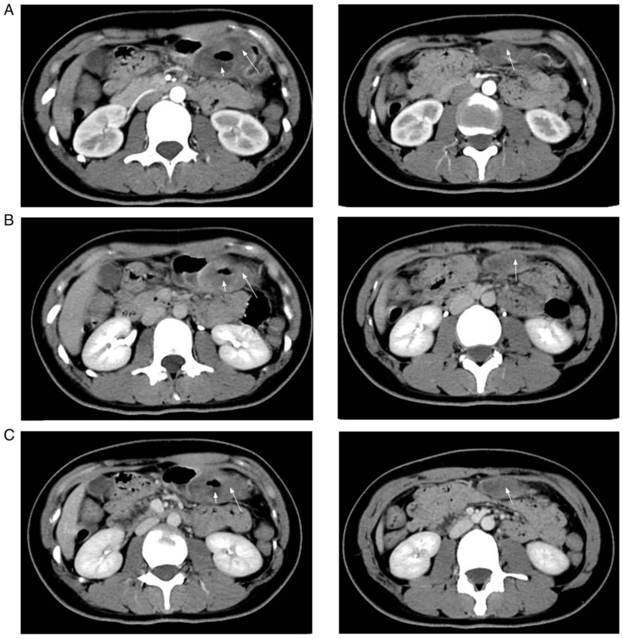
tumor. An abdominal enhancement computed tomography (CT) scan showed a thick and ulcerated distal wall of the gastric body bulging into the gastric cavity (>4.5×2.0 cm). A deep depression with poorly defined borders was also evident (Fig. 1A). In the venous (Fig. 1B) and equilibrium (Fig. 1C) phases, the ulcer was not strengthened, whereas the peripheral parenchyma was slightly strengthened. A round nodule with uneven density, slight hypointensity in the middle and well-defined borders was detected beneath the lesion (Fig. 1A). Enhancement scans revealed curved enhancement at the nodal edges. 
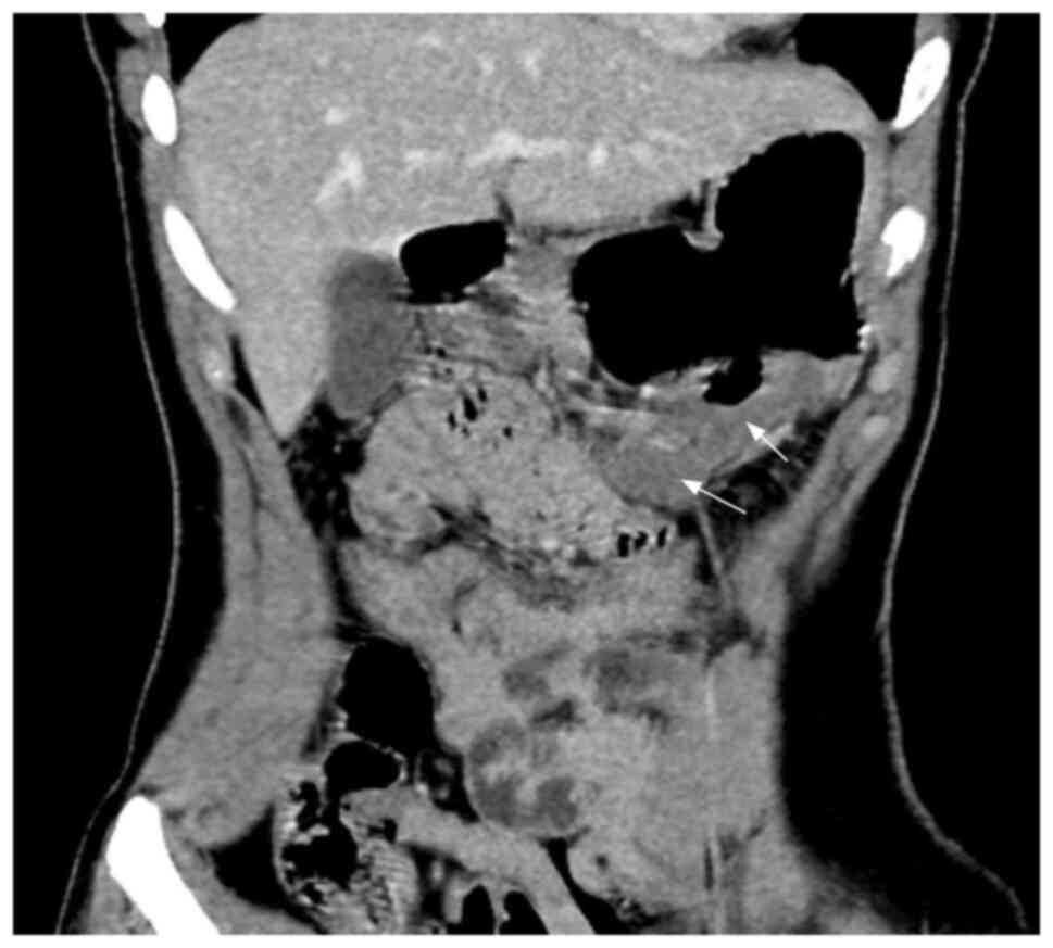
In the coronal scans, the second lesion was poorly demarcated from the gastric mucosa, and the two lesions were connected in the coronal position in the venous phase (Fig. 2). It was difficult to establish whether it was an enlarged lymph node or a tumor protruding from the gastric body. Considering the age of the patient and the present illness, the imaging diagnosis revealed lateral displacement of the greater curvature of the stomach with malignant difficulty.

Figure 1.

Abdominal enhancement computed tomography scans (axial sections). (A) Axial arterial phase image showing the thickened distal wall of the gastric body (left long arrow) and an ulcer (left short arrow), and a hypodense nodule (right arrow) below the stomach. (B) Axial delayed-phase image showing mild enhancement (long arrow) and no strengthening (left short arrow). (C) Axial venous phase image showing slight strengthening (long arrow).

Figure 2.

Coronal section computed tomography scan showing heterogeneous hypodense nodules below the lesion (arrow), indistinctly demarcated from the stomach.

A laparoscopic-assisted radical distal gastrectomy (Bi II anastomosis) was performed under general anesthesia. Intraoperatively, the tumor was located on the lateral side of the greater curvature of the gastric body (~6×5 cm) and protruded from the body. No mass was observed outside the body. The sections were 
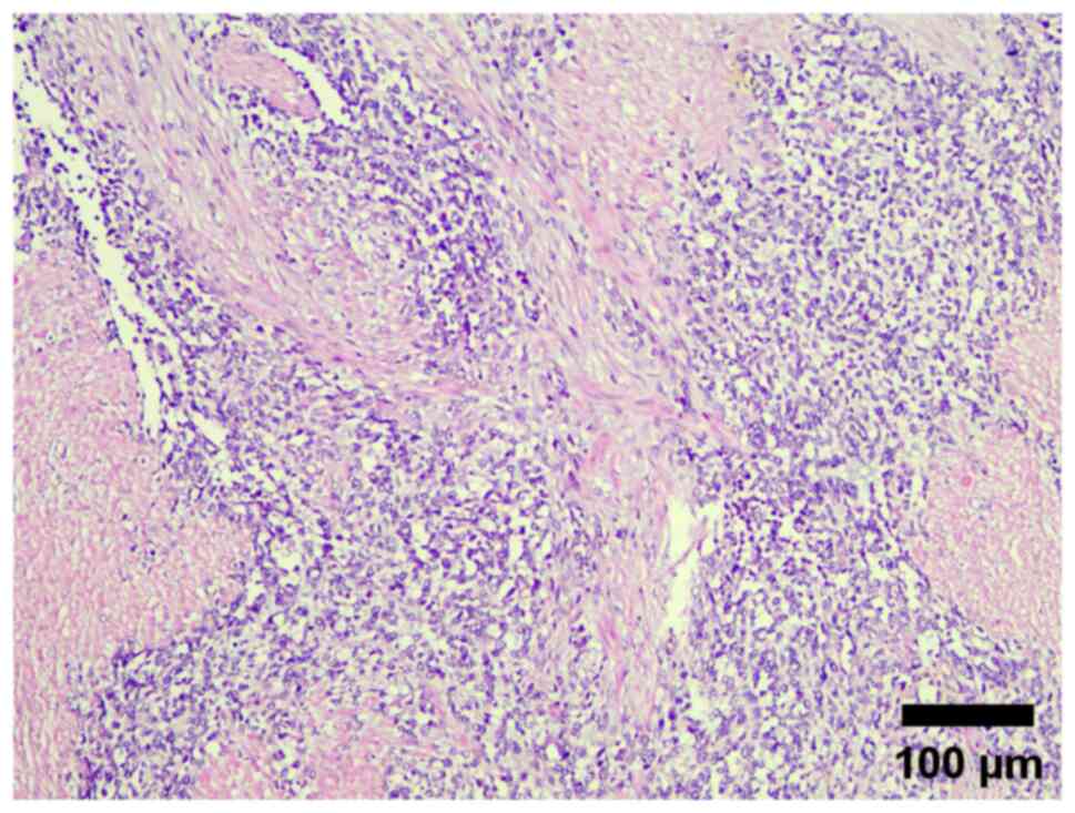
lightly counterstained with hematoxylin and eosin. Immunohistochemical staining was performed using the horseradish peroxidase complex, and reaction products were visualized by benzidine reaction. The sections were observed using a light microscope at a scale of 100 microns. The postoperative pathology showed small round tumor cells under light microscopy (Fig. 3). Immunohistochemistry showed the following results: CD56, vimentin and desmin, positive; MyoD1, nuclear weakly positive; Bcl-2 and CD99, partially weakly positive; and myogenin, myoglobin, myeloperoxidase, epithelial membrane antibody, smooth muscle actin, CK, chromogranin A, cytokeratin (CK)8/18, CK20, CD30, CD4, synaptophysin, Wilms tumor protein, anaplastic lymphoma kinase and friend leukemia virus integration, negative. The Ki67 positive index was ~70%. A definitive diagnose was difficult and so the patient was initially diagnosed with a gastric ulcer round cell malignancy. However, the specimen was sent to Renmin Hospital of Wuhan University for further testing, where fluorescence in situ hybridization (FISH) detected FOX01 gene isolation with amplification. The final diagnosis after combining immunohistochemistry and FISH testing results was gastric ARMS. The pathology of the second lesion revealed that it was a lymphatic metastasis. At 1 month post-surgery, enlarged lymph nodes were noted at the left supracostal margin and right supraclavicular region. Pathology revealed malignant metastasis. Therefore, adjuvant chemotherapy was started, and four cycles of 3 mg vindesine + 1 g cyclophosphamide and three cycles of 60 mg epirubicin were administered. Vindesine and cyclophosphamide were administered on a 4-week cycle. Epirubicin was administered on a 3-week cycle. Vindesine was administered on days 1, 8, 15 and 22 of the cycle. Cyclophosphamide was administered on days 1, 2, 3, 4 and 5 of the cycle. Epirubicin was administered on day 1 of the cycle. During this period, the leukocyte-raising therapy was performed against leukopenia due to chemotherapy-induced myeloid inhibition. The patient was eventually discharged 1 month later after the symptoms resolved and is currently doing well. The case is being followed up every 2 months.

Figure 3.

Small round tumor cells diffusely infiltrated in the intrinsic muscle layer of the gastric wall (hematoxylin and eosin staining; scale bar, 100 µm).

Discussion

RMS is a malignant mesenchymal tumor that accounts for <1% of all mesenchymal tumors in the gastrointestinal system (8). RMS of the stomach is extremely rare, with only four previously published cases (4–7). RMSs in the stomach are primary tumors, and no cases of metastasis in the stomach have been documented (3). Patients typically present with no symptoms in the early stages, followed by appetite loss and abdominal pain, or with non-specific pressure symptoms in the late stages when the tumor is large. The present case was similar to the cases documented in the literature. The signs of RMS are atypical on CT and magnetic resonance imaging (MRI), which makes the imaging diagnosis difficult and the condition easy to misdiagnose clinically. However, CT and MRI are critical in the preoperative diagnosis (the current patient did not consent to an MRI examination), as they can reveal the location of the lesion and they show the relationship between the tumor and the surrounding tissues, and the degree of invasion of the surrounding tissues. Finally, these techniques indicate the recurrence and metastasis risks after surgery (9). Therefore, it is important to raise awareness of the need for imaging for this disease.

According to the present case, the following types of gastric tumors need to be differentiated during the diagnosis of primary gastric ARMS: i) Neuroendocrine neoplasms, which often occur among the elderly, with multiple low-density small nodules (<2 cm) under the gastric mucosa, but with a density that is higher than that of muscle enhancement. The extent of uniform moderate enhancement in the arterial phase is higher than that of the gastric mucosa. Clinical manifestations include increased gastrin secretion and increased PH level. The primary pathological diagnosis of a neuroendocrine tumor, in this case, may be attributed to small round cells and immunohistochemical CD56 expression positivity. Gastrointestinal neuroendocrine tumors are more common than RMS and are easily misdiagnosed as neuroendocrine tumors. This case is that of a young patient with uneven mild and moderate enhancement on a CT enhancement scan, which can be used as a differential point. ii) Gastric stromal tumors, the most common benign tumors in the gastrointestinal tract, are often found in the upper part of the gastric fundus (7). The boundary is distinct with uniform density. Calcification is a common occurrence. In ARMS, there are few ulcers, but the boundary is unclear and the density is uneven. Calcification rarely occurs (10). Therefore, the two tumors can easily be distinguished. (iii) Gastric carcinoma often occurs among the elderly, with stiff gastric mucosa and blurred fat space around the stomach; its clinical manifestations include anemia and cachexia. ARMS often occurs in adolescents with good continuity of the gastric mucosa and no cachexia clinically. These two diseases can easily be distinguished.

In summary, although RMS is extremely rare in the adult stomach, the outcome of a young beneficiary with abdominal occupancy combined with lymph node growth should be considered a possibility, and should not be diagnosed as benign without assessment. The occurrence of distant lymph node metastasis in the present patient 1 month after surgery is consistent with the literature, which states that adult RMS is highly malignant, prone to recurrence and metastasis, and has a worse prognosis than RMS in children (11–13). Pathology is required to confirm the final diagnosis.

In conclusion, the current study presents a rare case of primary ARMS in the stomach. Efforts to increase awareness of the disease should be increased to improve the early clinical diagnosis and treatment.

Acknowledgements

The authors would like to thank Miss Shixing Zhao from the Department of Pathology, Handan Central Hospital (Handan, China) for performing the histopathological analysis.

Funding

Funding: No funding was received.

Availability of data and materials

The datasets used and/or analyzed during the current study are available from the corresponding author on reasonable request.

Authors' contributions

SY and XZ were responsible for study conception and design. XZ and ZY performed the collection and assembly of data, including obtaining the laboratory results and medical images. SY and XZ analyzed and interpreted the data. All authors wrote the manuscript. SY, ZY and XZ confirm the authenticity of all the raw data. All authors have read and approved the final manuscript.

Ethics approval and consent to participate

Not applicable.

Patient consent for publication

Written informed consent was obtained from the patient.

Competing interests

The authors declare that they have no competing interests.

Glossary

Abbreviations

Abbreviations:

CT

computed tomography

RMS

rhabdomyosarcoma

MRI

magnetic resonance imaging

References

1 

Gong QX and Fan QH: Updates of the 2020 WHO classification of the soft tissue tumors: part I. Zhonghua Bing Li Xue Za Zhi. 50:180–184. 2021.(In Chinese). PubMed/NCBI

2 

Gong QX and Fan QH: Updates of the 2020 WHO classification of soft tissue tumors: part II. Zhonghua Bing Li Xue Za Zhi. 50:314–318. 2021.(In Chinese). PubMed/NCBI

3 

Fujiie M, Yamamoto M, Taguchi K, Iwanaga A, Ohgaki K, Egashira A, Minami K, Toh Y, Oda Y and Okamura T: Gastric carcinosarcoma with rhabdomyosarcomatous differentiation: A case report and review. Surg Case Rep. 2:522016. View Article : Google Scholar : PubMed/NCBI

4 

Wang Y, Guo P, Zhang Z, Jiang RD and Li Z: Primary epithelioid rhabdomyosarcoma of the stomach: A case report and review of literature. Diagn Pathol. 14:1372019. View Article : Google Scholar : PubMed/NCBI

5 

Shah LK, Mony NJ, Mishra S and Pant B: An exceptionally rare primary epithelioid rhabdomyosarcomas of the stomach: A case report. Cureus. 14:e260462022.PubMed/NCBI

6 

Palermo M, Mastronardi LM, García RH, Solari I and Tarsitano FJ: Primary gastric rhabdomyosarcoma. Case report. Acta Gastroenterol Latinoam. 42:131–134. 2012.PubMed/NCBI

7 

Gandhi JS, Pasricha S, Gupta G, Mahanta A, Mehta A, Doudagoudar C, Goswami V and Doval DC: Synchronous embryonal rhabdomyosarcoma (NOS) of the Mid-oesophagus and stomach. J Gastrointest Cancer. 43 (Suppl 1):S217–S220. 2012. View Article : Google Scholar : PubMed/NCBI

8 

Mance M, Smuđ-Orehovec S, Vrbanović-Mijatović V and Mijatović D: Primary Alveolar rhabdomyosarcoma of the breast in a 17-year-old girl. JCO Oncol Pract. 16:93–95. 2020. View Article : Google Scholar : PubMed/NCBI

9 

Latack JT, Hutchinson RJ and Heyn RM: Imaging of rhabdomyosarcomas of the head and neck. Am J Neuroradiol. 8:353–359. 1987.PubMed/NCBI

10 

Tang W, Ren G, Cai R, He WG, Ni J and Chen J: Primary rhabdomyosarcoma in adults: Clinicopathological characteristics, CT and MRI findings. J Diagnostics Concepts Practice. 16:301–305. 2017.

11 

Heske CM and Mascarenhas L: Relapsed Rhabdomyosarcoma. J Clin Med. 10:8042021. View Article : Google Scholar : PubMed/NCBI

12 

Ferrari A, Dileo P, Casanova M, Bertulli R, Meazza C, Gandola L, Navarria P, Collini P, Gronchi A, Olmi P, et al: Rhabdomyosarcoma in adults. A retrospective analysis of 171 patients treated at a single institution. Cancer. 98:571–580. 2003. View Article : Google Scholar : PubMed/NCBI

13 

Mäkinen VN, Safwat A and Aggerholm-Pedersen N: Rhabdomyosarcoma in adults: A retrospective analysis of case records diagnosed between 1979 and 2018 in Western Denmark. Sarcoma. 2021:99488852021. View Article : Google Scholar : PubMed/NCBI

Related Articles

  • Abstract
  • View
  • Download
  • Twitter
Copy and paste a formatted citation
Spandidos Publications style
Zhang X, Yue Z and Yang S: Primary alveolar rhabdomyosarcoma in the stomach of an adult: A case report. Oncol Lett 25: 181, 2023.
APA
Zhang, X., Yue, Z., & Yang, S. (2023). Primary alveolar rhabdomyosarcoma in the stomach of an adult: A case report. Oncology Letters, 25, 181. https://doi.org/10.3892/ol.2023.13767
MLA
Zhang, X., Yue, Z., Yang, S."Primary alveolar rhabdomyosarcoma in the stomach of an adult: A case report". Oncology Letters 25.5 (2023): 181.
Chicago
Zhang, X., Yue, Z., Yang, S."Primary alveolar rhabdomyosarcoma in the stomach of an adult: A case report". Oncology Letters 25, no. 5 (2023): 181. https://doi.org/10.3892/ol.2023.13767
Copy and paste a formatted citation
x
Spandidos Publications style
Zhang X, Yue Z and Yang S: Primary alveolar rhabdomyosarcoma in the stomach of an adult: A case report. Oncol Lett 25: 181, 2023.
APA
Zhang, X., Yue, Z., & Yang, S. (2023). Primary alveolar rhabdomyosarcoma in the stomach of an adult: A case report. Oncology Letters, 25, 181. https://doi.org/10.3892/ol.2023.13767
MLA
Zhang, X., Yue, Z., Yang, S."Primary alveolar rhabdomyosarcoma in the stomach of an adult: A case report". Oncology Letters 25.5 (2023): 181.
Chicago
Zhang, X., Yue, Z., Yang, S."Primary alveolar rhabdomyosarcoma in the stomach of an adult: A case report". Oncology Letters 25, no. 5 (2023): 181. https://doi.org/10.3892/ol.2023.13767
Follow us
  • Twitter
  • LinkedIn
  • Facebook
About
  • Spandidos Publications
  • Careers
  • Cookie Policy
  • Privacy Policy
How can we help?
  • Help
  • Live Chat
  • Contact
  • Email to our Support Team