Introduction
During the past decades, the management of lung
cancer has been markedly improved with the application of targeted
therapy. Despite targeted drug therapies having resulted in notable
therapeutic benefits and prolongation of survival on the whole, the
extent of benefit of targeted therapies is not uniform. The
rearrangement during transfection (RET)
proto-oncogene encodes a transmembrane receptor tyrosine kinase,
which functions as the receptor for the growth factors of the glial
cell line-derived neurotropic factor family (1). The binding of glial cell line-derived
neurotrophic factor family ligands facilitates RET kinase
activation, which triggers the activation of signaling pathways
associated with cell proliferation, including the MAPK and PI3K
signaling pathways (1). Fusions in
RET, such as KIF5B-RET, CCDC6-RET and NCOA4-RET
consist of a type of aberration that can result in kinase
activation. RET fusions have been reported in different types of
malignancies, including 20–40% of sporadic cases of papillary
thyroid carcinoma and 1.4–2.5% of non-small cell lung cancer
(NSCLC) (2–5). Fragile sites in RET are
relatively conservative, and the chromosomal breakpoints of
RET often occur within intron 11 (6). Kinesin family member 5B
(KIF5B), described in the present case report, is the most
common RET fusion partner gene accounting for 60–80% of all
rearrangements (7,8). Similar to other types of oncogenic
RET fusions observed, KIF5B-RET proteins are likely to form
a homodimer through the coiled-coil domain of KIF5B, which causes
aberrant activation of the kinase function of RET in a manner
similar to KIF5B-ALK receptor tyrosine kinase fusions (9). The two selective RET inhibitors
approved by the China National Medical Products Administration,
pralsetinib and selpercatinib, have markedly changed the treatment
landscape for RET fusion-positive NSCLC between 2021 and
2022 (10). Unique genetic
characteristics and clinicopathological features have been observed
in patients with NSCLC harboring RET fusions (11).
Both histological and molecular subtyping have
become increasingly important as predictors of benefits of
treatment in lung cancer (12).
Further evidence has emerged which demonstrates the importance of
the molecular re-subtyping of different histology in predicting
treatment benefits for patients with lung cancer (13). There has been a marked paradigm
shift in the diagnosis and individualized clinical management of
advanced NSCLC (14). Although
tumor tissue sampling is the gold standard for molecular testing,
≤20% of samples from patients with advanced stage NSCLC may not be
adequate in quantity and quality for pathological diagnosis and
genomic profiling (15).
Circulating biomarkers such as circulating tumor DNA (ctDNA),
carcinoembryonic antigen (CEA) and cancer antigen 125 have emerged
as valuable surrogates for auxiliary diagnosis and monitoring of
treatment response in solid tumors (16). Prior studies have demonstrated that
ctDNA is superior to CEA in terms of the detection of residual
disease and early recurrence (17–19).
Both of the aforementioned biomarkers are easily acquired, exhibit
a rapid turnover and have high patient compliance, which has
increased their clinical use in monitoring treatment efficacy and
cancer progression. However, their roles still require further
elucidation.
The case of a patient with advanced NSCLC negative
for EGFR, RAS, BRAF, ALK, ROS1 and RET driver
mutations is described in the present report.
Case report
A 66-year-old male patient with no notable past
medical and family history initially presented at the Qingdao
Municipal Hospital (Qingdao, China) in April 2016 with deep vein
thrombosis in both lower extremities without pulmonary symptoms
reported. A positron emission tomography scan demonstrated a 9×16
mm fluorodeoxyglucose (FDG)-avid right lower lobe (RLL) irregular
pulmonary nodule with an FDG-avid 12×20 mm right supraclavicular
lymph node. An ultrasound-guided fine-needle aspiration with
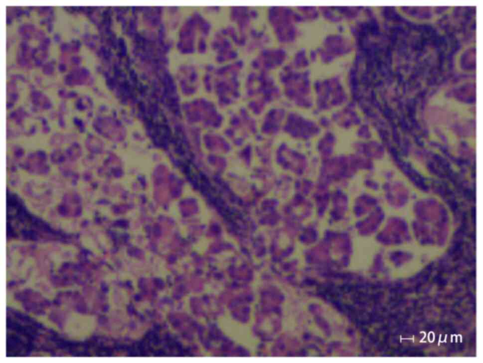
pleural and nodule biopsies confirmed unresectable metastatic
adenocarcinoma at stage IIIB (cT1N3M0; Tumor-Node-Metastasis
staging system by AJCC; Fig. 1)
(20). Analysis of tumor tissue and
peripheral blood using next-generation sequencing (NGS) revealed
the absence of driver mutations in EGFR, RAS, BRAF, ALK,
ROS1 and RET genes (Shanghai Topgen Biomedical
Technology Co., Ltd.). The KAPA HyperPlus Kit (cat. no. 7962428001;
Roche Sequencing Solutions, Inc.) that enables rapid construction
of DNA libraries was used to prepare DNA samples for sequencing.
The Agilent 2100 bioanalyzer (Agilent Technologies, Inc.) uses
capillary electrophoresis on a microchip device (LabChip 7500;
Caliper Life Sciences, Inc.), and is capable of quality check after
library dilution. Sequencing was performed on a NextSeq 550 System
(Illumina, Inc.) in 75 bp paired-end mode for short libraries using
a NextSeq 500/550 High Output Kit v2.5 and a total of 150 cycles
(cat. no. 20024907; Illumina, Inc.) with a loading concentration of
1.4 pM final library. Brain magnetic resonance imaging was negative
for intracranial disease. At the time of diagnosis of metastatic
disease, the patient was asymptomatic and the physical exam was
unremarkable with the exception of decreased breath sounds at the
right lung base.
In May 2016, four cycles (each 21 days) of induction
cisplatin (75 mg/m2) and pemetrexed (600
mg/m2) chemotherapy were performed, with endostatin (30
mg/kg) added in cycle 3 until pathological response. In June 2016,
involved-field radiotherapy alone was administered for palliative
care with a dose of 60 Gy/30 fractions delivered in 2.0 Gy/day
fractions, including the mediastinal regions and the
supraclavicular lymph drainage area, after which both lesions
continued to shrink. Subsequently maintenance with pemetrexed (600
mg/m2 on day 5) and endostatin (30 mg/kg from days 1 to
7) (21-day cycle, for 4 cycles) was performed in October 2016.
Stable disease was achieved until July 2018. The detection of a
6-mm pulmonary nodule in the right lower lobe on the follow-up CT,
along with a comprehensive evaluation of treatment efficacy,
suggested gradual disease progression. Additionally, subsequent
systemic chemotherapy with carboplatin (400 mg/m2 on day
1) and lipo-paclitaxel (240 mg/m2 on day 2) was
performed, followed by carboplatin (400 mg/m2 on day 2),
lipo-paclitaxel (240 mg/m2; on day 2) and bevacizumab
(500 mg/kg; on day 1) until stable disease in September 2018. Due
to grade III neutropenia (21)
developing during subsequent treatment, the dose and regimen were
adjusted to bevacizumab (500 mg/kg on day 0) and lipo-paclitaxel
(180 mg/m2 on day 3). In February 2019, a follow-up CT
demonstrated an enlarged 1.2 cm lesion in the RLL, which indicated
metastatic disease. Concurrent chemoradiotherapy was considered for
the patient and intensity modulated radiation therapy was
prescribed at a dose of 60 Gy/30 fractions, administered as five
fractions per week for 6 weeks. The chemotherapy schedule consisted
of bevacizumab (500 mg/kg on day 1) and lipo-paclitaxel (180
mg/m2 on day 2), and was repeated every 4 weeks for four
cycles. A follow-up CT identified right-sided pleural effusion in
September 2019. Due to gastrointestinal bleeding, potentially due
to bevacizumab intolerance, the regimens were adjusted to anlotinib
(12 mg/kg daily) alone from November 2020 to August 2021. Admission
and hospitalization occurred after 2 weeks of discomfort, including
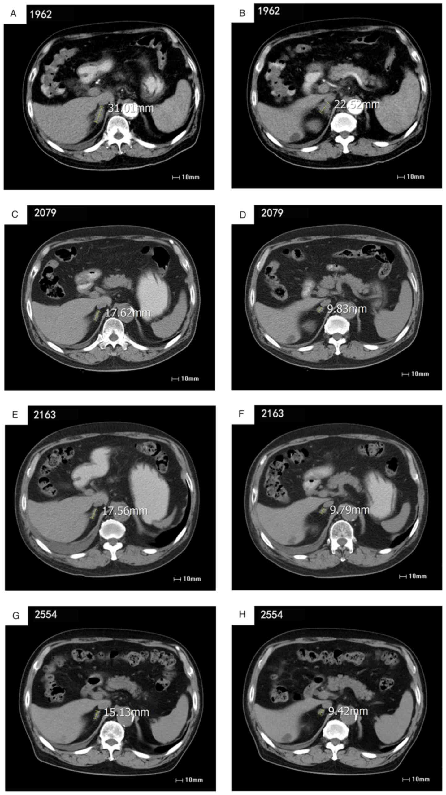
fatigue and anorexia. A follow-up CT of the metastatic abdominal
lymph node demonstrated no change from earlier images; however,
advanced adenocarcinoma at stage IV (cT1N3Mx) was confirmed
(Fig. 2A and B).
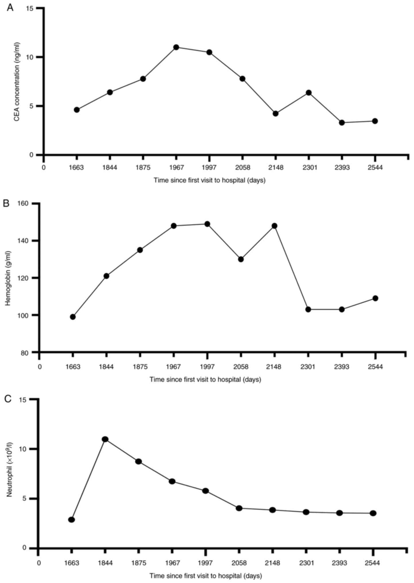
From November 2020 onwards, the effectiveness of
systemic chemotherapy was assessed through the dynamic monitoring
of serum CEA levels; this was conducted continuously for a duration
of 891 days. Between May and August 2021, the CEA levels rose from
7.0 to ~10 ng/ml, which suggested progressive disease (Fig. 3A). Peripheral blood ctDNA test using
NGS as aforementioned (Topgen Bio-Pharm Co, Ltd.) revealed a
KIF5B-RET fusion with KIF5B exon 15 fused to
RET exon 12 at a 7.4% variant allele fraction (VAF). VAF is
the percentage of sequence reads observed matching a specific DNA
variant divided by the overall coverage at that locus.
Additionally, a TP53 G244D substitution with 4.7% VAF and a
glutamate ionotropic receptor NMDA type subunit 2A (GRIN2A)
F613V substitution with 2.0% VAF were also identified (Table I).
 | Table I.Next generation sequencing results
acquired from the patient involved in this case. |
Table I.
Next generation sequencing results
acquired from the patient involved in this case.
| Days since first
visit in April 2016 | Results | Sample type |
|---|
| 20 | Negative for
EGFR, ALK and other driver genes. EGFR (−);
ALK (−) | Peripheral
blood |
| 1,969 | Detectable kinesin
family member 5B-rearranged during transfection fusion; VAF
7.4% | Peripheral
blood |
|
| TP53
c.731G>A (p.G244D); VAF 4.7% |
|
|
| GRIN2A
c.1837T>G (p.F613V); VAF 2.0% |
|
| 2,343 | GRIN2A
c.1516G>C (p.F183I); VAF 0.6% | Peripheral
blood |
In September 2021, the patient received RET
inhibitor pralsetinib orally at a reduced dose of 300 mg once a
day. An abdominal CT scan and testing of the conventional
hematological clinical indicators, such as routine blood tests,
liver function and renal function, obtained 23 days after the
initiation of therapy, showed a minor response [stable disease
according to Response Evaluation Criteria in Solid Tumors (RECIST)
v1.1; Fig. 2C and D] (17). The patient continued to take
pralsetinib orally until April 2022 for a total of 8 months of
treatment, and CEA levels continued to decrease during follow-up
tests. An abdominal CT scan of the metastatic abdominal lymph node
demonstrated no change from the last images (Fig. 2E and F). At the request of the
patient, ctDNA was monitored at periodic intervals, the RET
fusion was no longer detected by ctDNA assessment in December 2022,
and only GRIN2A F183I with 0.6% VAF was observed, and ~13 months
after the initiation of therapy, the CEA level dropped from 12
ng/ml in September 2021 to 3 ng/ml in November 2022 (Table I). The patient generally tolerated
treatment well with certain clinically relevant adverse reactions
observed, including a decreasing peripheral neutrophil count and a
slightly decreased peripheral lymphocyte count, as well as an
elevation of aspartate aminotransferase (AST) and grade II edema
(22), which required a dose
reduction to 300 mg (Fig. 3;
Table II).
| Table II.Results of blood, liver and kidney
function indicators for this patient. |
Table II.
Results of blood, liver and kidney
function indicators for this patient.
|
| Days since first
visit in April 2016 |
|
|---|
|
|
|
|
|---|
| Function
indicator | 1994 | 2079 | 2163 | 2554 | Reference
range |
|---|
| Albumin, g/l | 28a | 39a | 39a | 41 | 40-55 |
| Prealbumin,
mg/l | 281 | 413 | 436b | 376 | 200-430 |
| Aspartate
aminotransferase, U/l | 30 | 38 | 58b | 35 | 15-45 |
| Gamma glutamyl
transferase, U/l | 41 | 64b | 67b | 44 | 10-60 |
| Leucine
aminopeptidase, U/l | 55 | 80b | 73b | 51 | 30-70 |
| α-L-fucosidase,
U/l | 37 | 51b | 57b | 32 | 0-40 |
| Blood urea
nitrogen, mmol/l | 9.9b | 10.4b | 11.5b | 18.4b | 3.6–9.5 |
| Creatinine,
µmol/l | 115b | 117b | 125b | 152b | 57-111 |
| Cystatin, mg/l | 1.7b | 1.5b | 1.5b | 2.1b | 0.6–1.0 |
| Lymphocyte count,
×109/l | 0.9a | 3.0 | 2.6 | 3.1 | 1.1–3.2 |
| Lymphocyte, % | 15a | 39 | 37 | 43 | 20-50 |
| Red blood cells,
1012/l | 5.2 | 4.0a | 4.7 | 4.3a | 4.3–5.8 |
| Mean corpuscular
volume, fl | 81a | 104 | 100b | - | 82-100 |
| Mean corpuscular
hemoglobin concentration, pg | 25a | 32 | 31 | 26a | 27-34 |
| Mean corpuscular
hemoglobin concentration, g/l | 309a | 310a | 314a | 288a | 316-354 |
| Red blood cell
volume distribution width, fl | 63b | 75b | 54b | 59b | 37-54 |
At the time of submission of the present report, the
patient continued treatment with undetectable levels of
KIF5B-RET ctDNA and exhibited a decreasing serum CEA level
in the plasma for >17 months after the initiation of pralsetinib
treatment, with an ongoing minor response and stable disease
according to RECIST v1.1 on imaging, with a performance status (PS)
score of 0–1 (23–25) (Fig. 2G
and H).
The present study was performed in accordance with
the Helsinki Declaration (2013 revision) and ethical standards
approved by the Qingdao Municipal Hospital. The patient provided
written consent for the publication of the case report and
associated images.
Discussion
In the era of precision medicine and personalized
cancer treatment, the demand for tumor molecular profiling is
steadily increasing. The potential uses of liquid biopsy, such as
ctDNA testing, have been identified in the context of diagnostics,
prediction of prognosis and therapeutic efficacy, relapse
monitoring and resistance detection (26–28).
Implementing ctDNA testing for cancer diagnosis and monitoring in
clinical application fosters research in patient management,
despite the challenges and expectations it poses. The case of a
patient with advanced NSCLC negative for EGFR, RAS, BRAF, ALK,
ROS1 and RET driver mutations was described in the
present report. Periodic on-treatment CEA surveillance and ctDNA
reevaluation in the asymptomatic patient led to early
identification of further disease progression, which supported
prompt therapeutic decision-making.
Chemotherapy is considered to be the standard of
care for the first-line treatment of patients with advanced-stage,
either IIIB or IV, NSCLC with a good PS (ECOG PS<2) (23,24). A
study previously reported that the combination of pemetrexed plus
cisplatin was associated with significantly improved survival
compared with gemcitabine plus cisplatin in patients with
adenocarcinoma, with median survival time 12.6 vs. 10.9 months,
respectively and P=0.03 (29). Due
to its limited use in clinical practice, chemotherapy remains the
main treatment option for patients with RET-rearranged
advanced NSCLC. Multiple studies have shown that chemotherapy in
patients with RET fusions yields an overall response rate
(ORR) ranging from 26 to 50%, with a median progression-free
survival (PFS) time of 5 to 9.2 months for first-line treatment and
2.8 to 5.2 months for second-line treatment (30–32).
In a study of patients NSCLC and KIF5B-RET fusions compared
with patients with non-KIF5B-RET fusions, PFS was assessed
(7.8 vs. 11.2 months; P=0.847; hazard ratio, 0.902) (30). Although the survival benefit in
RET fusion-positive NSCLC is limited, chemotherapy regimens
containing pemetrexed may be relatively more effective in treating
the condition. The present case study indicated the benefit of
treatment, and revealed a >17-month PFS without detectable
KIF5B-RET ctDNA or reduced CEA levels and a >7-year overall
survival time since first-line chemotherapy with pemetrexed plus
cisplatin was initiated in April 2016. At the time of submission of
the present report, the patient continued to be on 300 mg
pralsetinib with ongoing imaging-assessed stable disease according
to RECIST v1.1 (25).
Targeted treatments based on driver genes, such as
EGFR, ALK, RET and ROS1, provide a more precise
option for the treatment of advanced NSCLC, and driver gene tests
have become an essential component of clinical practice for the
stratification of patients who are most likely to benefit from
specific tyrosine kinase inhibitors (33). The phase I/II ARROW trial of
pralsetinib in patients with advanced RET fusion-positive
NSCLC reported an ORR of 63% and a complete response of 6%. Data
from the Chinese subgroup indicated that pralsetinib was associated
with an ORR of 56% and a disease control rate of 97% in patients in
whom platinum-based therapy failed, ~50% of whom had experienced ≥3
lines of systemic treatment, and the median PFS time was 16.5
months (34,35). Clinically relevant adverse reactions
in <15% of patients who received pralsetinib in the ARROW trial
included changes in certain laboratory abnormalities, including
chemistry indices such as increased AST and decreased albumin, and
hematology indices such as decreased neutrophils and hemoglobin
(35).
A previous retrospective study reported that
cell-free ctDNA testing to detect fusion events could predict
therapy response despite assay limitations (31). It has been reported that the
detection rate of fusions in metastatic NSCLC is high at the
initial onset of disease progression, and changes in RET fusion
levels according to ctDNA testing are associated with tumor burden
and treatment process (36,37). The detection rates of high frequency
RET fusions such as KIF5B-RET and coiled-coil domain
containing 6-RET in ctDNA could be improved by NGS that has
higher sensitivity, which can benefit patients whose tissue or
cytology samples are unavailable (38,39).
There is also an increasing number of publications which have
reported the application of circulating biomarkers, such as ctDNA
and CEA, for real-time monitoring for early detection of disease
progression in advanced NSCLC (16,17).
Early reduction of circulating biomarkers after initiation of
systemic therapy has been reported to be a predictive marker of
clinical benefit in advanced tumors (27,28).
In a study of 28 patients with metastatic NSCLC who received immune
checkpoint inhibitor (anti-PD-1 or anti-PD-L1) therapy, most
patients who demonstrated a long-term benefit from immunotherapy
rapidly achieved a drop in ctDNA levels of 50–100%, and the initial
ctDNA response occurred a median of 42.5 days earlier than the
initial radiographic response (40). In a retrospective study of 40
patients with lung cancer treated with curative intent, detection
of ctDNA posttreatment preceded radiographic progression in 72% of
patients, by a median of 5.2 months (26). Early reduction in ctDNA after
initiation of systemic therapy has been reported to be a predictive
marker of clinical benefit in numerous advanced tumors (41).
For the patient in the present case study,
continuous circulating biomarker monitoring of ctDNA and CEA
resulted in the early detection of disease progression in the
absence of symptoms and molecular re-subtyping after multiple lines
of therapy, enabled a prompt adjustment in therapy, which the
patient continued to receive at the time of this report. Dynamic
monitoring of CEA has been performed since post-line treatment. The
change in CEA level combined with CT imaging is of great
significance for the management of the disease throughout the
disease course. In genetic testing, there may be heterogeneity
among samples from different disease stages, as well as
heterogeneity between primary and metastatic lesions (42). Previous studies have reported that
the mutation status of driver genes, including EGFR, ALK, BRAF,
KRAS, ROS1 and RET, may change in patients with advanced
NSCLC throughout the chemotherapy period (43,44).
RET fusion was detected in ctDNA after the patient developed
distant metastasis, which may also be the acquired RET
fusion induced by platinum-based chemotherapy. However, the
mechanism of chemotherapy-induced mutation status of driver genes
requires further study in NSCLC. Furthermore, the dosage of
pralsetinib was reduced due to an adverse reaction, but the
treatment was not interrupted based on changes in certain
laboratory parameters. In addition, targeted therapy with a RET
inhibitor such as prelsetinib resulted in a reduction of metastatic
lesion volume and a concomitant decrease in VAF value. As NGS
provides a near random sample of the DNA molecules, VAF is thus a
surrogate measure of the proportion of DNA molecules in the
original specimen carrying the variant (45). A retrospective cohort analysis of
561 patients with advanced solid cancers, including NSCLC,
pancreatic, prostate, colon and breast cancer, reported that there
was a positive association between maximal VAF levels with the
diameter or volume of all lesions, and in patients with
undetectable ctDNA, a lower ctDNA VAF was associated with improved
survival (46). Further
investigation is necessary to elucidate the relationship between
tumor mutation burden and prognosis, as well as its effect on ctDNA
levels. In addition, it is unclear whether the prognostic value of
ctDNA mutations and abundance is affected by chemotherapy or
targeted therapy, despite previous findings which suggest baseline
ctDNA levels can serve as a prognostic indicator (47). The lack of available tumor tissue
during liquid biopsy prevented NGS from detecting genetic
information in the tumor tissue, which was a limitation of the
present case report; further exploration of NGS remains necessary
for future research endeavors.
In conclusion, the case of a patient with advanced
NSCLC which was negative for EGFR, RAS, BRAF, ALK, ROS1 and
RET driver mutations at initial diagnosis, and only
RET-fusion-positive after conventional treatment is
described in the current report. ctDNA and CEA surveillance in the
patient prompted early imaging which resulted in improved
therapeutic decision-making. The current report provides further
evidence of the potential benefit of serial tests or different
combinations of tests during treatment as a useful and effective
method to evaluate treatment efficacy and detect relapse (15,16).
Further research is required to define the optimal integration of
the scheme for management throughout the disease course in patients
with advanced KIF5B-RET fusion-positive NSCLC, and to
demonstrate the subsequent clinical decision-making that is
informed by the scheme presented in the current report.
Acknowledgements
Not applicable.
Funding
Funding: No funding was received.
Availability of data and materials
The raw data of NGS can be accessed using accession
number PRJNA985255 in SRA of NCBI (https://www.ncbi.nlm.nih.gov/sra).
Authors' contributions
YB and CX drafted the manuscript. YB, XZ and HL
collected data and served as scientific advisors. CX and HL
performed data analysis and the literature review. YB, HL and CX
conducted the final critical review of the paper. YB, XZ and HL
confirm the authenticity of all the raw data. All authors read and
approved the final version of the manuscript.
Ethics approval and consent to
participate
The present study involving a human patient was
reviewed and approved by the Qingdao Municipal Hospital (approval
no. 20220616; Qingdao, China). Written informed consent was
obtained from the patient, and all procedures were conducted in
accordance with the Declaration of Helsinki. Shanghai Topgen
Biomedical Technology Co., Ltd. had a College of American
Pathologists certificate (approval no. 8671637-01) for assessing
human samples using NGS genetic testing.
Patient consent for publication
The patient provided written consent for
publication.
Competing interests
The authors declare that they have no competing
interests.
Glossary
Abbreviations
Abbreviations:
|
NSCLC
|
non-small cell lung cancer
|
|
RET
|
rearranged during transfection
|
|
CEA
|
carcinoembryonic antigen
|
|
RLL
|
right lower lobe
|
|
NGS
|
next-generation sequencing
|
|
ctDNA
|
circulating tumor DNA
|
|
PFS
|
progression-free survival
|
|
ORR
|
overall response rate
|
|
VAF
|
variant allele fraction
|
References
|
1
|
Houvras YL: Completing the Arc: Targeted
inhibition of RET in medullary thyroid cancer. J Clin Oncol.
30:200–202. 2012. View Article : Google Scholar : PubMed/NCBI
|
|
2
|
Santoro M, Melillo RM and Fusco A: RET/PTC
activation in papillary thyroid carcinoma: European journal of
endocrinology prize lecture. Eur J Endocrinol. 155:645–653. 2006.
View Article : Google Scholar : PubMed/NCBI
|
|
3
|
Kato S, Subbiah V, Marchlik E, Elkin SK,
Carter JL and Kurzrock R: RET aberrations in diverse cancers:
Next-Generation sequencing of 4,871 patients. Clin Cancer Res.
23:1988–1997. 2017. View Article : Google Scholar : PubMed/NCBI
|
|
4
|
Li W, Guo L, Liu Y, Dong L, Yang L, Chen
L, Liu K, Shao Y and Ying J: Potential unreliability of uncommon
ALK, ROS1, and RET genomic breakpoints in predicting the efficacy
of targeted therapy in NSCLC. J Thorac Oncol. 16:404–418. 2021.
View Article : Google Scholar : PubMed/NCBI
|
|
5
|
Zhang K, Chen H, Wang Y, Yang L, Zhou C,
Yin W, Wang G, Mao X, Xiang J, Li B, et al: Clinical
characteristics and molecular patterns of RET-Rearranged lung
cancer in Chinese patients. Oncol Res. 27:575–582. 2019. View Article : Google Scholar : PubMed/NCBI
|
|
6
|
Gandhi M, Dillon LW, Pramanik S, Nikiforov
YE and Wang YH: DNA breaks at fragile sites generate oncogenic
RET/PTC rearrangements in human thyroid cells. Oncogene.
29:2272–2280. 2010. View Article : Google Scholar : PubMed/NCBI
|
|
7
|
Li AY, McCusker MG, Russo A, Scilla KA,
Gittens A, Arensmeyer K, Mehra R, Adamo V and Rolfo C: RET fusions
in solid tumors. Cancer Treat Rev. 81:1019112019. View Article : Google Scholar : PubMed/NCBI
|
|
8
|
Takeuchi K, Soda M, Togashi Y, Suzuki R,
Sakata S, Hatano S, Asaka R, Hamanaka W, Ninomiya H, Uehara H, et
al: RET, ROS1 and ALK fusions in lung cancer. Nat Med. 18:378–381.
2012. View
Article : Google Scholar : PubMed/NCBI
|
|
9
|
Kohno T, Ichikawa H, Totoki Y, Yasuda K,
Hiramoto M, Nammo T, Sakamoto H, Tsuta K, Furuta K, Shimada Y, et
al: KIF5B-RET fusions in lung adenocarcinoma. Nat Med. 18:375–377.
2012. View
Article : Google Scholar : PubMed/NCBI
|
|
10
|
Chen P, Liu Y, Wen Y and Zhou C: Non-small
cell lung cancer in China. Cancer Commun (Lond). 42:937–970. 2022.
View Article : Google Scholar : PubMed/NCBI
|
|
11
|
Qiu Z, Ye B, Wang K, Zhou P, Zhao S, Li W
and Tian P: Unique genetic characteristics and clinical prognosis
of female patients with lung cancer harboring RET fusion gene. Sci
Rep. 10:103872020. View Article : Google Scholar : PubMed/NCBI
|
|
12
|
Pikor LA, Ramnarine VR, Lam S and Lam WL:
Genetic alterations defining NSCLC subtypes and their therapeutic
implications. Lung Cancer. 82:179–189. 2013. View Article : Google Scholar : PubMed/NCBI
|
|
13
|
Shim HS, Choi YL, Kim L, Chang S, Kim WS,
Roh MS, Kim TJ, Ha SY, Chung JH, Jang SJ, et al: Molecular testing
of lung cancers. J Pathol Transl Med. 51:242–254. 2017. View Article : Google Scholar : PubMed/NCBI
|
|
14
|
Chan BA and Hughes BG: Targeted therapy
for non-small cell lung cancer: Current standards and the promise
of the future. Transl Lung Cancer Res. 4:36–54. 2015.PubMed/NCBI
|
|
15
|
Lim C, Tsao MS, Le LW, Shepherd FA, Feld
R, Burkes RL, Liu G, Kamel-Reid S, Hwang D, Tanguay J, et al:
Biomarker testing and time to treatment decision in patients with
advanced non small-cell lung cancer. Ann Oncol. 26:1415–1421. 2015.
View Article : Google Scholar : PubMed/NCBI
|
|
16
|
Janse van Rensburg HJ, Spiliopoulou P and
Siu LL: Circulating biomarkers for therapeutic monitoring of
anti-cancer agents. Oncologist. 27:352–362. 2022. View Article : Google Scholar : PubMed/NCBI
|
|
17
|
Duffy MJ and Crown J: Circulating Tumor
DNA as a biomarker for monitoring patients with solid cancers:
Comparison with standard protein biomarkers. Clin Chem.
68:1381–1390. 2022. View Article : Google Scholar : PubMed/NCBI
|
|
18
|
Kotani D, Oki E, Nakamura Y, Yukami H,
Mishima S, Bando H, Shirasu H, Yamazaki K, Watanabe J, Kotaka M, et
al: Molecular residual disease and efficacy of adjuvant
chemotherapy in patients with colorectal cancer. Nat Med.
29:127–134. 2023. View Article : Google Scholar : PubMed/NCBI
|
|
19
|
Zhao H, Chen KZ, Hui BG, Zhang K, Yang F
and Wang J: Role of circulating tumor DNA in the management of
early-stage lung cancer. Thorac Cancer. 9:509–515. 2018. View Article : Google Scholar : PubMed/NCBI
|
|
20
|
Lim W, Ridge CA, Nicholson AG and
Mirsadraee S: The 8th lung cancer TNM classification and clinical
staging system: Review of the changes and clinical implications.
Quant Imaging Med Surg. 8:709–718. 2018. View Article : Google Scholar : PubMed/NCBI
|
|
21
|
Kaba H, Fukuda H, Yamamoto S and Ohashi Y:
Reliability at the National Cancer Institute-Common Toxicity
Criteria version 2.0. Gan To Kagaku Ryoho. 31:1187–1192. 2004.(In
Japanese). PubMed/NCBI
|
|
22
|
Brodovicz KG, McNaughton K, Uemura N,
Meininger G, Girman CJ and Yale SH: Reliability and feasibility of
methods to quantitatively assess peripheral edema. Clin Med Res.
7:21–31. 2009. View Article : Google Scholar : PubMed/NCBI
|
|
23
|
Azam F, Latif MF, Farooq A, Tirmazy SH,
AlShahrani S, Bashir S and Bukhari N: Performance status assessment
by using ECOG (Eastern Cooperative Oncology Group) score for cancer
patients by oncology healthcare professionals. Case Rep Oncol.
12:728–736. 2019. View Article : Google Scholar : PubMed/NCBI
|
|
24
|
Kelly CM and Shahrokni A: Moving beyond
Karnofsky and ECOG performance status assessments with new
technologies. J Oncol. 2016:61865432016. View Article : Google Scholar : PubMed/NCBI
|
|
25
|
Eisenhauer EA, Therasse P, Bogaerts J,
Schwartz LH, Sargent D, Ford R, Dancey J, Arbuck S, Gwyther S,
Mooney M, et al: New response evaluation criteria in solid tumours:
Revised RECIST guideline (version 1.1). Eur J Cancer. 45:228–247.
2009. View Article : Google Scholar : PubMed/NCBI
|
|
26
|
Chaudhuri AA, Chabon JJ, Lovejoy AF,
Newman AM, Stehr H, Azad TD, Khodadoust MS, Esfahani MS, Liu CL,
Zhou L, et al: Early detection of molecular residual disease in
localized lung cancer by circulating tumor DNA profiling. Cancer
Discov. 7:1394–1403. 2017. View Article : Google Scholar : PubMed/NCBI
|
|
27
|
Arrieta O, Villarreal-Garza C,
Martínez-Barrera L, Morales M, Dorantes-Gallareta Y, Peña-Curiel O,
Contreras-Reyes S, Macedo-Pérez EO and Alatorre-Alexander J:
Usefulness of serum carcinoembryonic antigen (CEA) in evaluating
response to chemotherapy in patients with advanced non small-cell
lung cancer: A prospective cohort study. BMC Cancer. 13:2542013.
View Article : Google Scholar : PubMed/NCBI
|
|
28
|
Kuo YS, Zheng MY, Huang MF, Miao CC, Yang
LH, Huang TW and Chou YT: Association of divergent carcinoembryonic
antigen patterns and lung cancer progression. Sci Rep. 10:20662020.
View Article : Google Scholar : PubMed/NCBI
|
|
29
|
Scagliotti GV, Parikh P, von Pawel J,
Biesma B, Vansteenkiste J, Manegold C, Serwatowski P, Gatzemeier U,
Digumarti R, Zukin M, et al: Phase III study comparing cisplatin
plus gemcitabine with cisplatin plus pemetrexed in
chemotherapy-naive patients with advanced-stage non-small-cell lung
cancer. J Clin Oncol. 26:3543–3551. 2008. View Article : Google Scholar : PubMed/NCBI
|
|
30
|
Shen T, Pu X, Wang L, Yu Z, Li J, Zhang Y,
Liang X, Chen H, Xu C, Song Z and Wang W: Association between RET
fusions and efficacy of pemetrexed-based chemotherapy for patients
with advanced NSCLC in China: A multicenter retrospective study.
Clin Lung Cancer. 21:e349–e354. 2020. View Article : Google Scholar : PubMed/NCBI
|
|
31
|
Gautschi O, Milia J, Filleron T, Wolf J,
Carbone DP, Owen D, Camidge R, Narayanan V, Doebele RC, Besse B, et
al: Targeting RET in patients with RET-Rearranged lung cancers:
Results from the global, multicenter RET registry. J Clin Oncol.
35:1403–1410. 2017. View Article : Google Scholar : PubMed/NCBI
|
|
32
|
Drilon A, Bergagnini I, Delasos L, Sabari
J, Woo KM, Plodkowski A, Wang L, Hellmann MD, Joubert P, Sima CS,
et al: Clinical outcomes with pemetrexed-based systemic therapies
in RET-rearranged lung cancers. Ann Oncol. 27:1286–1291. 2016.
View Article : Google Scholar : PubMed/NCBI
|
|
33
|
König D, Savic Prince S and Rothschild SI:
Targeted therapy in advanced and metastatic non-small cell lung
cancer. An update on treatment of the most important actionable
oncogenic driver alterations. Cancers (Basel). 13:8042021.
View Article : Google Scholar : PubMed/NCBI
|
|
34
|
Zhou Q, Wu Y, Chang J, Wang H, Fan Y, Wang
K, Wu G, Nian W, Sun Y, Sun M, et al: Efficacy and safety of
pralsetinib in Chinese patients with advanced RET fusion+ non-small
cell lung cancer after platinum-based chemotherapy. J Thorac Oncol.
16:216–227. S1002021. View Article : Google Scholar
|
|
35
|
Subbiah V, Cassier PA, Siena S, Garralda
E, Paz-Ares L, Garrido P, Nadal E, Vuky J, Lopes G, Kalemkerian GP,
et al: Pan-cancer efficacy of pralsetinib in patients with RET
fusion-positive solid tumors from the phase 1/2 ARROW trial. Nat
Med. 28:1640–1645. 2022. View Article : Google Scholar : PubMed/NCBI
|
|
36
|
Supplee JG, Milan MSD, Lim LP, Potts KT,
Sholl LM, Oxnard GR and Paweletz CP: Sensitivity of next-generation
sequencing assays detecting oncogenic fusions in plasma cell-free
DNA. Lung Cancer. 134:96–99. 2019. View Article : Google Scholar : PubMed/NCBI
|
|
37
|
Rolfo C, Mack PC, Scagliotti GV, Baas P,
Barlesi F, Bivona TG, Herbst RS, Mok TS, Peled N, Pirker R, et al:
Liquid biopsy for advanced non-small cell lung cancer (NSCLC): A
statement paper from the IASLC. J Thorac Oncol. 13:1248–1268. 2018.
View Article : Google Scholar : PubMed/NCBI
|
|
38
|
Tsuta K, Kohno T, Yoshida A, Shimada Y,
Asamura H, Furuta K and Kushima R: RET-rearranged non-small-cell
lung carcinoma: A clinicopathological and molecular analysis. Br J
Cancer. 110:1571–1578. 2014. View Article : Google Scholar : PubMed/NCBI
|
|
39
|
Lin C, Wang S, Xie W, Chang J and Gan Y:
The RET fusion gene and its correlation with demographic and
clinicopathological features of non-small cell lung cancer: A
meta-analysis. Cancer Biol Ther. 16:1019–1028. 2015. View Article : Google Scholar : PubMed/NCBI
|
|
40
|
Goldberg SB, Narayan A, Kole AJ, Decker
RH, Teysir J, Carriero NJ, Lee A, Nemati R, Nath SK, Mane SM, et
al: Early assessment of lung cancer immunotherapy response via
circulating tumor DNA. Clin Cancer Res. 24:1872–1880. 2018.
View Article : Google Scholar : PubMed/NCBI
|
|
41
|
Sanz-Garcia E, Zhao E, Bratman SV and Siu
LL: Monitoring and adapting cancer treatment using circulating
tumor DNA kinetics: Current research, opportunities, and
challenges. Sci Adv. 8:eabi86182022. View Article : Google Scholar : PubMed/NCBI
|
|
42
|
Caswell DR and Swanton C: The role of
tumour heterogeneity and clonal cooperativity in metastasis, immune
evasion and clinical outcome. BMC Med. 15:1332017. View Article : Google Scholar : PubMed/NCBI
|
|
43
|
Shi X, Huang F, Chen A, Wu Z, Huang Q,
Liang Y, Zhou Q, Mo H, Li X and Zhang J: The impact of chemotherapy
on EGFR mutation status in non-small-cell lung cancer: A
meta-analysis. Open J Gen. 7:117–129. 2017. View Article : Google Scholar
|
|
44
|
Pich O, Muiños F, Lolkema MP, Steeghs N,
Gonzalez-Perez A and Lopez-Bigas N: The mutational footprints of
cancer therapies. Nat Genet. 51:1732–1740. 2019. View Article : Google Scholar : PubMed/NCBI
|
|
45
|
Strom SP: Current practices and guidelines
for clinical next-generation sequencing oncology testing. Cancer
Biol Med. 13:3–11. 2016. View Article : Google Scholar : PubMed/NCBI
|
|
46
|
Hsiehchen D, Espinoza M, Gerber DE and Beg
MS: Clinical and biological determinants of circulating tumor DNA
detection and prognostication using a next-generation sequencing
panel assay. Cancer Biol Ther. 22:455–464. 2021. View Article : Google Scholar : PubMed/NCBI
|
|
47
|
Parikh AR, Mojtahed A, Schneider JL,
Kanter K, Van Seventer EE, Fetter IJ, Thabet A, Fish MG, Teshome B,
Fosbenner K, et al: Serial ctDNA monitoring to predict response to
systemic therapy in metastatic gastrointestinal cancers. Clin
Cancer Res. 26:1877–1885. 2020. View Article : Google Scholar : PubMed/NCBI
|