Introduction
Fibroadenoma is the most common benign tumor
diagnosed in the breast, with a peak incidence in the female in
their 20s and 30s worldwide (1,2). In
principle, an asymptomatic fibroadenoma can be managed
conservatively with regular breast imaging follow-up; however, in
clinical practice, it may not be an optimal choice if the mass is
enlarged or other atypical symptoms are present (3). Carcinoma arising in a fibroadenoma of
the breast can be diagnosed by incidental biopsy or excision
although it is present in only 0.1–0.3% of all cases (4).
To the best of our knowledge, only sporadic case
reports and case series with a small sample size describing breast
carcinoma occurring within a fibroadenoma have been published
(3,5). Breast carcinoma arising within a
fibroadenoma includes lobular carcinoma in situ (LCIS),
ductal carcinoma in situ (DCIS) and, rarely, invasive
carcinoma (6). Non-invasive breast
carcinomas are more commonly found than invasive breast carcinomas
(7). An epithelial malignancy
within a fibroadenoma often mimics the features of a fibroadenoma
clinically and radiologically; therefore, early detection remains
clinically challenging. Although radiological examinations such as
ultrasonography and mammography are valuable in diagnosing
fibroadenoma, malignancy within a fibroadenoma may be
indistinguishable from benign fibroadenoma on imaging (5). Moreover, few reports have documented
the transition from fibroadenoma to breast cancer and little is
known about its clinicopathological features and prognosis
(5,8). To the best of our knowledge, no
evidence-based guidelines for diagnosis and treatment of breast
carcinoma arising within a fibroadenoma exist due to its low
incidence rate. The present study reviews the literature and
discusses the clinicopathological features and treatments of
patients with breast carcinoma arising from a fibroadenoma
diagnosed at The Third Hospital of Nanchang City (Nanchang, China)
between January 2019 and December 2021.
Materials and methods
Patients
Between January 2019 and December 2021, 6,558 of
breast carcinoma were admitted to The Third Hospital of Nanchang
City (Nanchang, China). Of them, 6,550 cases were female. All cases
diagnosed as breast carcinoma in situ or invasive carcinoma
arising within fibroadenoma were included. All histopathological
slides were reviewed by two pathologists independently to confirm
the diagnosis. Data such as age, sex, menopausal status, primary
complaint and treatment were also collected.
Pathological examination
All available hematoxylin and eosin
(H&E)-stained breast carcinoma arising within a fibroadenoma
sections were reviewed by two independent pathologists. Tumor
morphology was assessed using recently described criteria (9), including nuclear grade, mitotic rate,
presence and extent of associated carcinoma in situ and
invasive carcinoma. Immunohistochemistry was performed on
paraffin-embedded 4 µm tissue slides as described previously and
fixed in 10% formalin for 6 h at room temperature (10). First incubate the slides at 65°C for
2 h, and then deparaffinize twice for 5 min. Antigen retrieval
solution (10 mmol/l tris; 1 mmol/LEDTA; pH9.0) was 100°C for 5 min
and blocked with 2% sheep serum (Biyangtian Biotechnology Research
Institute) at room temperature. The slides were then incubated with
primary antibodies overnight at 4°C and with horseradish
peroxidase-labeled secondary antibodies and DAB for 1 h at 37°C.
Tumor immunoreactivity was assessed independently by two
pathologists. In addition, human epidermal growth factor receptor 2
(HER2) was detected by immunohistochemistry (IHC) and fluorescence
in situ hybridization (FISH) according to the latest HER2
testing recommendations from the American Society of Clinical
Oncology and the College of American Pathologists. Status is
determined. Breast Cancer 2018 (11). FISH analysis was performed as
previously reported (12).
Follow-up
All patients are followed up regularly every 3–6
months at the Breast Disease Prevention and Treatment Center of
Nanchang Third Hospital Routine physical and radiological
examinations were performed to monitor recurrence and metastasis.
The last date of follow-up was September 30, 2022.
Results
Clinical and pathological
findings
Between January 2019 and December 2021, 16 patients
were diagnosed with carcinoma arising in a fibroadenoma at The
Third Hospital of Nanchang City. The age at diagnosis ranged
between 19 and 58 years (median, 45 years). All patients were
female and most were premenopausal (87.5%). The most common initial
clinical presentation was an asymptomatic palpable mass (87.5%),
followed by nipple discharge (6.3%). Notably, two patients (12.5%)
presented because of abnormal imaging changes of fibroadenoma
during follow-up (Table I).
 | Table I.Clinicopathological characteristics of
breast carcinoma arising in a fibroadenoma. |
Table I.
Clinicopathological characteristics of
breast carcinoma arising in a fibroadenoma.
| Characteristic | Value |
|---|
| Age at diagnosis,
years (%) |
|
|
<35 | 5 (31.3) |
|
35-50 | 10 (62.5) |
|
>50 | 1 (6.3) |
| Median age (range),
years | 45.0 (19.0–58.0) |
| Sex (%) |
|
| Male | 0 (0.0) |
|
Female | 16 (100.0) |
| Clinical
presentation (%) |
|
|
Mass | 14 (87.5) |
| Nipple
discharge | 1 (6.3) |
| Laterality (%) |
|
|
Left | 8 (50.0) |
|
Right | 8 (50.0) |
|
Bilateral | 0 (0.0) |
| Fibroadenoma
diameter, cm (%) |
|
| ≤2 | 6 (37.5) |
|
2-3 | 8 (50.0) |
|
>3 | 2 (12.5) |
| Mean fibroadenoma
diameter (range), cm | 2.2 (1.0–3.7) |
| Malignant component
subtype (%) |
|
|
DCIS | 10 (62.5) |
|
LCIS | 2 (12.5) |
|
IDC | 3 (18.8) |
|
ILC | 1 (6.3) |
| T stage (%) |
|
|
Tis | 12 (75.0) |
| T1 | 4 (25.0) |
| T2 | 0 (0.0) |
| T3 | 0 (0.0) |
| T4 | 0 (0.0) |
| Axillary lymph node
involvement (%) |
|
|
Yes | 0 (0.0) |
| No | 16 (100.0) |
Consistent with a previous study (13), a small malignant tumor arising in a
relatively large fibroadenoma was observed in 14 cases (87.5%). The
mean tumor size (diameter of fibroadenoma containing the malignant
component) ranged between 1.0 and 3.7 cm (mean, 2.2 cm). However,
the malignant component in 12 of cases was non-invasive (75%).
Thus, the majority of cases in the present study were tumor in
situ (Tis) according to the latest TNM staging system (75%;
Table I) (14).
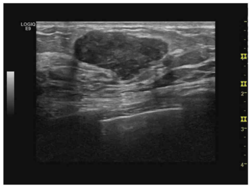
Fibroadenoma harboring a carcinoma mimics benign
fibroepithelial tumor radiologically. Ultrasonography revealed a
circumscribed, homogenous, isoechoic or hypoechoic mass (Fig. 1). A total of 5 cases (31.3%) were
categorized as breast imaging-reporting and data system (15) (BI-RADS) 4 on ultrasonography in the
present study which indicated suspicious malignancy. Mammography
findings were similar to those of benign tumors and revealed a
lobulated well-defined mass. However, several indicators of
malignant transformation, including indistinct borders and
microcalcifications, were observed in three cases (18.8%) (16).
Diagnosis, treatment and survival
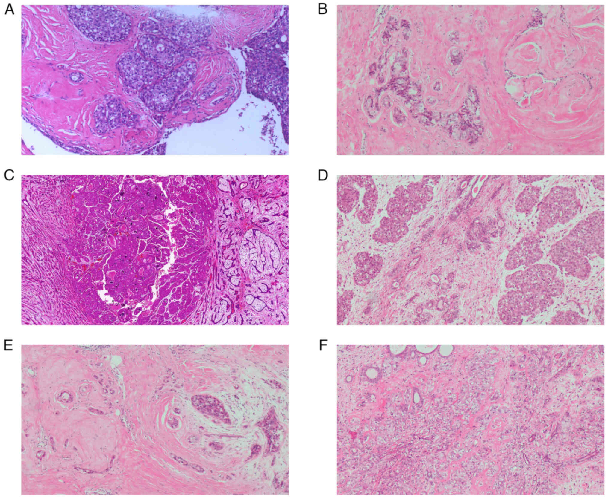
In the present study, all cases received core needle
or vacuum-assisted biopsy or excision to make a diagnosis.
Pathologically, carcinoma in situ was more common than
invasive carcinoma, with 12 non-invasive cases (75%) and four
invasive cases (25%; Fig. 2A and
B). Furthermore, DCIS was more common than LCIS (Table II; Fig.
2C and D). Invasive ductal and lobular carcinoma were diagnosed
in three (81.3%) and one case (93.8%), respectively (Fig. 2E and F). When breast carcinoma
arising within a fibroadenoma is suspected, immunohistochemical
analysis (such as CK5/6 and α-SMA) is performed to confirm the
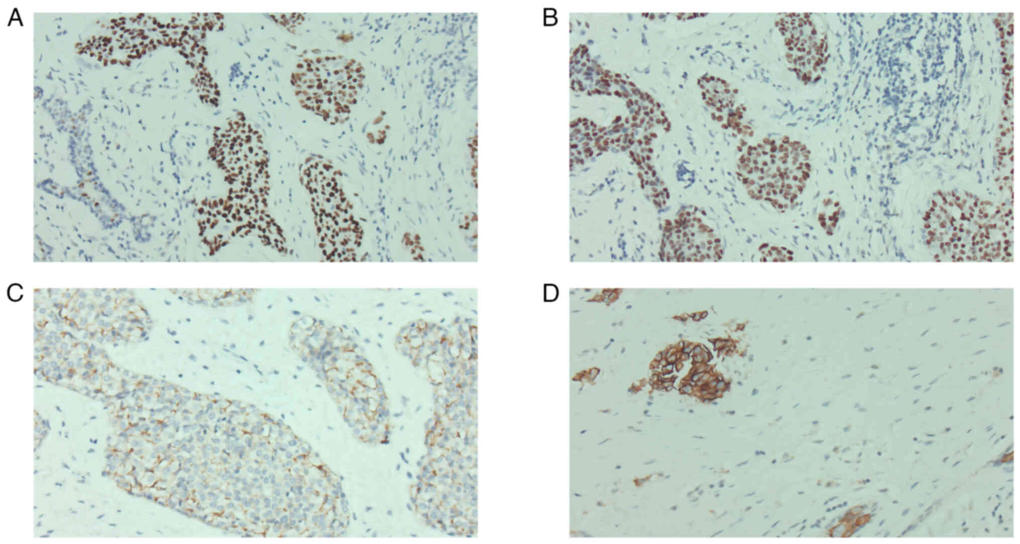
diagnosis (Fig. S1). In
immunohistochemical analyses, the majority of cases were hormone
receptor (HR)-positive (15 cases, 93.8%) and HER2-negative (14
cases, 87.5%; Fig. 3A-C). Regarding
molecular subtypes, luminal A and B, HER2-positive and
triple-negative subtypes accounted for 10 (62.5%), 5 (31.3%), 1
(6.3%) and 0 (0.0%) cases, respectively.
| Table II.Immunohistochemical analysis. |
Table II.
Immunohistochemical analysis.
| Immunohistochemical
marker | n (%) |
|---|
| ER status |
|
|
Positive | 15 (93.8) |
|
Negative | 1 (6.3) |
| PR status |
|
|
Positive | 14 (87.5) |
|
Negative | 2 (12.5) |
| HER2 status |
|
| - | 3 (18.8) |
| 1+ | 3 (18.8) |
| 2+ | 8 (50.0) |
| 3+ | 2 (12.5) |
| Ki67, % |
|
|
≤14 | 12 (75.0) |
|
>14 | 4 (25.0) |
| Molecular
subtype |
|
| Luminal
A | 10 (62.5) |
| Luminal
B | 5 (31.3) |
| HER2
overexpression | 0 (0.0) |
|
TNBC | 1 (6.3) |
Once the tumor was confirmed to be malignant,
breast-conserving surgery (BCS) as well as sentinel lymph node
biopsy (SLNB) were advised. A total of seven patients (43.6%)
received BCS and nine patients (56.3%) received mastectomy
(Table III). SLNB was performed
in all cases and no axillary lymph node involvement was observed.
Of seven patients who received BCS, six underwent radiation
therapy; one 19-year-old female patient diagnosed with DCIS arising
within a fibroadenoma rejected radiotherapy because of personal
reasons. Furthermore, chemotherapy was administered in two patients
diagnosed with invasive carcinoma. Anti-HER2-targeted therapy was
administered concurrently with chemotherapy in one of these
patients, since the immunohistochemical staining revealed
HER2-positive invasive carcinoma (Fig.
3D).
| Table III.Treatment of breast carcinoma arising
in a fibroadenoma. |
Table III.
Treatment of breast carcinoma arising
in a fibroadenoma.
| Treatment | n (%) |
|---|
| Surgery |
|
| BCS or
lumpectomy | 7 (43.8) |
|
Mastectomy | 9 (56.3) |
| Surgery of axillary
lymph nodes |
|
|
SLNB | 16 (100) |
|
ALND | 0 (0.0) |
| Chemotherapy |
|
|
Yes | 2 (12.5) |
| No | 14 (87.5) |
| Radiotherapy |
|
|
Yes | 6 (37.5) |
| No | 10 (62.5) |
| Endocrine
therapy |
|
|
Yes | 15 (93.8) |
| No | 1 (6.3) |
| HER2-targeted
therapy |
|
|
Yes | 1 (6.3) |
| No | 15 (93.8) |
Follow-up data were available for all patients. In
the follow-up period, no signs of recurrence or relapse were
observed and all patients were in good health at the last
follow-up.
Discussion
Carcinoma arising from a fibroadenoma is a poorly
understood disease of the breast, with only a few reported cases
(17,18). Unlike benign fibroadenoma, malignant
transformation within a fibroadenoma is most reported in patients
in their 40s (6,13), although it can occur at any age.
Consistent with these reports, the age of patients at diagnosis in
the present study ranged between 19 and 58 years, with a median age
of 45 years. Notably, five of the 16 patients (31.3%) were <35
years old, with the proportion of young patients with breast cancer
markedly higher than that in the whole breast cancer population
(19). This indicated that
malignant transformation within a fibroadenoma may be stimulated by
high estrogen levels in young women. Notably, the youngest patient
in the present study was a 19-year-old female patient who was
diagnosed with low-grade DCIS arising with a fibroadenoma. To the
best of our knowledge, this is the youngest case reported in the
literature so far. Therefore, it is important for clinicians to be
cognizant of potential malignant transformation within a
fibroadenoma even if the patient is young.
Breast cancer arising in fibroadenomas is difficult
to diagnose because its oncogenic components may be masked by
fibroadenoma components and has a low incidence rate.. In the
present study, the most common clinical presentation was a
well-defined, movable, asymptomatic mass (87.5%), which was similar
to a benign breast tumor. Furthermore, observations in the
radiological examinations, including ultrasonography and
mammography, are usually non-specific (20). Only five patients (31.3%) in the
present study were categorized as BI-RADS 4 on ultrasonography.
Additionally, malignant transformation indicators in mammography,
such as microcalcifications and distortion of surrounding tissue,
were only observed in three cases (18.8%) (21). As the carcinoma component may be
hidden by other components in fibroadenoma, and thus, may not be
detected by imaging examinations (16), it is difficult to differentiate
benign fibroadenomas from their counterparts with malignant
transformation within them. Therefore, tumor biopsy or excision
should potentially be suggested to make a diagnosis in this
situation. Notably, a recent study evaluated the role of dynamic
contrast-enhanced MRI in diagnosing cancerous lesions developing
within fibroadenomas (21) and
revealed that relatively low diffusion of the apparent diffusion
coefficient value suggested a malignant transformation within a
fibroadenoma. Novel potential imaging methods with high accuracy
and efficiency are clinically warranted.
Here, non-invasive carcinoma was the predominant
type of malignancy within a fibroadenoma (75%). Theoretically, the
occurrence of a breast carcinoma within a fibroadenoma has two
potential etiologies: The breast carcinoma may arise from
epithelial components of the fibroadenoma or tumors may coexist
separately (22). Previous studies
have produced inconsistent results: Several studies found that LCIS
is more common compared with DCIS since fibroadenoma is of lobular
origin (23,24), while other reports demonstrated that
LCIS and DCIS present with approximately equal frequency within a
fibroadenoma because they both originate from the terminal
duct-lobular units of the breast (25,26).
However, in the present study, DCIS was more frequent than LCIS.
These inconsistent results indicate that the exact cell origin of
the carcinoma within a fibroadenoma remains unclear.
Since few case reports have been published to date
(27,28), the immunohistochemical
characteristics as well as molecular subtype distribution of breast
carcinoma arising within a fibroadenoma remain to be determined. A
retrospective study reported that most cases are HR-positive
(25). However, the study was
reported in the ‘pre-adjuvant anti-HER2 therapy era’ and HR status
was determined few cases, which introduced possible elements of
bias. In the present immunohistochemical examination, the majority
of cases were positive for HR (93.8%) and negative for HER2
(87.5%). The luminal A and B, HER2-positive and triple-negative
molecular subtypes accounted for 10 (62.5%), five (31.3%), 0 (0.0%)
and 0 (0.0%) and one (6.3%) cases, respectively. To the best of our
knowledge, this is the first case series reporting molecular
subtype distribution in detail. Since DCIS cases have diverse
clinical and prognostic features for different molecular subtypes,
accurate diagnosis, including determination of molecular subtype,
is required for prompt treatment and prognosis.
Following diagnosis of carcinoma, BCS was performed
in seven patients and mastectomy was performed in nine patients
based on the personal preference of the patient. Although the
majority of cases were non-invasive on core needle biopsy, SLNB was
performed in all cases in case of tumor upstaging on final
pathology. A previous study (29)
reported the rate of upstaging to invasive breast cancer in
patients with DCIS identified on core needle biopsy; 36% of
patients with DCIS were ultimately diagnosed with invasive
carcinoma (29). Additionally, in
the present study, no axillary node involvement was observed. The
present results indicated that SLNB may be unnecessary in carcinoma
arising within a fibroadenoma, particularly in non-invasive
cases.
Although differences between breast carcinoma
arising from fibroadenoma and breast cancer remain unknown, a tumor
bed boost after postoperative whole breast irradiation in patients
with DCIS following conservative surgery can be recommended, as
evidenced by the BIG 3–07/TROG 07.01 trial (30). Regarding radiotherapy following BCS,
six out of seven patients completed the treatment as planned; one
19-year-old female with DCIS refused post-operative radiotherapy
for personal reasons. Adjuvant chemotherapy was administered to one
HER2-positive invasive breast cancer case. Upregulation of HER2 in
DCIS is associated with adverse clinicopathological parameters,
including higher grade, comedo necrosis, as well as worse clinical
outcome (31). To the best of our
knowledge, however, the therapeutic role of anti-HER2-targeted
therapy in HER2-positive DCIS has not been established. Therefore,
another patient diagnosed with HER2-positive DCIS arising within a
fibroadenoma was treated with post-operative radiotherapy only.
At the end of follow-up, all 16 patients included in
the present study were alive and no progression was observed. A
limitation of the present study is the follow-up period was
relatively short; further follow-up and prognosis should be
assessed in future. A second limitation of the study is the genetic
analysis of breast carcinoma arising within a fibroadenoma is not
routinely performed in daily clinical practice, therefore genetic
information of these cases was not collected. Previous studies have
demonstrated that carcinoma arising within a fibroadenoma is
associated with a favorable prognosis with adequate local therapy
(3,32), regardless of whether it is
biologically in situ or not. However, clinicians should be
alert to potentially enclosed cancerous lesions in a fibroadenoma
since they can radically alter both prognosis and treatment
outcome.
In conclusion, carcinoma arising within a
fibroadenoma is a rare malignancy of the breast. In clinical
practice, it is often encountered incidentally during pathological
examination of a benign breast mass. Non-invasive carcinoma was the
predominant type of malignancy within a fibroadenoma, although the
cell origin of the carcinoma remains to be elucidated. Notably, the
present report described the youngest case to date, although
malignant changes within fibroadenomas are typically identified in
older patients. Aggressive molecular phenotypes, including
HER2-positive or triple-negative subtypes, were identified in the
present case series. Surgical excision with or without
postoperative radiotherapy should be recommended once the
malignancy is diagnosed. Additionally, systemic treatment, such as
chemotherapy and endocrine therapy, were also suggested, especially
in the presence of invasive carcinoma with unfavorable tumor
biology or axillary lymph node metastases.
Supplementary Material
Supporting Data
Acknowledgements
The authors would like to thank Professor Jianhong
Tu (Department of Pathology, The Third Hospital of Nanchang City,
Nanchang, China) for technical assistance in preparing histology
tissue sections.
Funding
The present study was supported by the National Natural Science
Foundation of China (grant nos. 81860467, 82060482 and 81860546)
and Key Science and Technology Support Project of Nanchang (grant
no. 2019-258-13).
Availability of data and materials
The datasets used and/or analyzed during the current
study are available from the corresponding author on reasonable
request.
Authors' contributions
LX, WQ, SL and YC conceived and designed the study.
LX and QM wrote the manuscript. LL collected data. QM, YG and LL
analyzed the data. WQ performed the pathological examination and
provided experimental technical support. SL and YC wrote and
revised the manuscript. LX and YC confirm the authenticity of all
the raw data. All authors have read and approved the final
manuscript.
Ethics approval and consent to
participate
The present study was approved by the Institutional
Review Boards of The Third Hospital of Nanchang City (Nanchang,
China; approval no. K-ky2023005). Written informed consent was
obtained from all patients according to The Declaration of
Helsinki.
Patient consent for publication
Not applicable.
Competing interests
The authors declare that they have no competing
interests.
References
|
1
|
Basara Akin I and Balci P: Fibroadenomas:
A multidisciplinary review of the variants. Clin Imaging.
71:83–100. 2021. View Article : Google Scholar : PubMed/NCBI
|
|
2
|
Lerwill MF, Lee AHS and Tan PH:
Fibroepithelial tumours of the breast-a review. Virchows Arch.
480:45–63. 2022. View Article : Google Scholar : PubMed/NCBI
|
|
3
|
Feliciano YZ, Freire R, Net J and Yepes M:
Ductal and lobular carcinoma in situ arising within an enlarging
biopsy proven fibroadenoma. BMJ Case Rep. 14:e2370172021.
View Article : Google Scholar : PubMed/NCBI
|
|
4
|
Brock CM, Harper C and Tyler T:
Fibroadenoma containing lobular carcinoma in situ, an unusual
finding in a normally benign mass. J Surg Case Rep.
2020:rjaa0592020. View Article : Google Scholar : PubMed/NCBI
|
|
5
|
Shojaku H, Hori R, Yoshida T, Matsui K,
Shimada K, Takayanagi N and Noguchi K: Low-grade ductal carcinoma
in situ (DCIS) arising in a fibroadenoma of the breast during 5
years follow-up: A case report. Medicine (Baltimore).
100:e240232021. View Article : Google Scholar : PubMed/NCBI
|
|
6
|
Krishnamurthy K, Alghamdi S, Gyapong S,
Kaplan S and Poppiti RJ: A clinicopathological study of
fibroadenomas with epithelial proliferation including lobular
carcinoma in-situ, atypical ductal hyperplasia, DCIS and invasive
carcinoma. Breast Dis. 38:97–101. 2019. View Article : Google Scholar : PubMed/NCBI
|
|
7
|
Fukuda M, Nagao K, Nishimura R, Matsuda M,
Baba K, Ueno Y, Morinaga H, Omachi H and Hamada T: Carcinoma
arising in fibroadenoma of the breast-a case report and review of
the literature. Jpn J Surg. 19:593–596. 1989. View Article : Google Scholar : PubMed/NCBI
|
|
8
|
Wu YT, Wu HK, Chen ST, Chen CJ, Chen DR
and Lai HW: Fibroadenoma progress to ductal carcinoma in situ,
infiltrating ductal carcinoma and lymph node metastasis? Report an
unusual case. J Surg Case Rep. 2017:rjx0642017. View Article : Google Scholar : PubMed/NCBI
|
|
9
|
Md Nasir ND, Koh VCY, Cree IA, Ruiz BII,
Del Águila J, Armon S, Fox SB, Lakhani SR and Tan PH: Phyllodes
tumour evidence gaps mapped from the 5th edition of the WHO
classification of tumours of the breast. Histopathology.
82:704–712. 2023. View Article : Google Scholar : PubMed/NCBI
|
|
10
|
Yamamoto Y, Hayashi Y, Sakaki H and
Murakami I: Downregulation of fascin induces collective cell
migration in triple-negative breast cancer. Oncol Rep. 50:1502023.
View Article : Google Scholar : PubMed/NCBI
|
|
11
|
Wolff AC, Hammond MEH, Allison KH, Harvey
BE, Mangu PB, Bartlett JMS, Bilous M, Ellis IO, Fitzgibbons P,
Hanna W, et al: Human epidermal growth factor receptor 2 testing in
breast cancer: American society of clinical oncology/college of
American pathologists clinical practice guideline focused update. J
Clin Oncol. 36:2105–2122. 2018. View Article : Google Scholar : PubMed/NCBI
|
|
12
|
Jacobs TW, Gown AM, Yaziji H, Barnes MJ
and Schnitt SJ: Comparison of fluorescence in situ hybridization
and immunohistochemistry for the evaluation of HER-2/neu in Breast
Cancer. J Clin Oncol. 17:1974–1982. 1999. View Article : Google Scholar : PubMed/NCBI
|
|
13
|
Wu YT, Chen ST, Chen CJ, Kuo YL, Tseng LM,
Chen DR, Kuo SJ and Lai HW: Breast cancer arising within
fibroadenoma: Collective analysis of case reports in the literature
and hints on treatment policy. World J Surg Oncol. 12:3352014.
View Article : Google Scholar : PubMed/NCBI
|
|
14
|
Giuliano AE, Connolly JL, Edge SB,
Mittendorf EA, Rugo HS, Solin LJ, Weaver DL, Winchester DJ and
Hortobagyi GN: Breast Cancer-Major changes in the American Joint
Committee on Cancer eighth edition cancer staging manual. CA Cancer
J Clin. 67:290–303. 2017. View Article : Google Scholar : PubMed/NCBI
|
|
15
|
Spak DA, Plaxco JS, Santiago L, Dryden MJ
and Dogan BE: BI-RADS ® fifth edition: A summary of
changes. Diagn Interv Imaging. 98:179–190. 2017. View Article : Google Scholar : PubMed/NCBI
|
|
16
|
Shiino S, Yoshida M, Tokura M, Watase C,
Murata T, Jimbo K, Takayama S, Suto A, Satomi K, Miyagi Maeshima A,
et al: Locally advanced triple negative breast cancer arising from
fibroadenoma with complete response to neoadjuvant chemotherapy: A
case report. Int J Surg Case Rep. 68:234–238. 2020. View Article : Google Scholar : PubMed/NCBI
|
|
17
|
Razakanaivo M, Rakotoarivo T,
Andrianandrasana NO and Rafaramino F: Breast Carcinoma Arising in
Fibroadenoma in a 15-Year-Old Girl; Diagnosis and Treatment
Challenge. J Cancer Ther. 13:615–620. 2022. View Article : Google Scholar
|
|
18
|
Ni XH, An R, Shi QW and Wang CL: Low-Grade
ductal carcinoma in situ within a fibroadenoma of the breast: A
rare case report and review of the literature. Onco Targets Ther.
16:399–406. 2023. View Article : Google Scholar : PubMed/NCBI
|
|
19
|
Paluch-Shimon S, Cardoso F, Partridge AH,
Abulkhair O, Azim HA, Bianchi-Micheli G, Cardoso MJ, Curigliano G,
Gelmon KA, Gentilini O, et al: ESO-ESMO fifth International
consensus guidelines for breast cancer in young women (BCY5). Ann
Oncol. 33:1097–1118. 2022. View Article : Google Scholar : PubMed/NCBI
|
|
20
|
Ren Y, Li P and Yang Y: Imaging findings
of ductal carcinoma in situ arising within fibroadenoma. Breast J.
26:1037–1038. 2020. View Article : Google Scholar : PubMed/NCBI
|
|
21
|
Tagliati C, Lanni G, Cerimele F, Di
Martino A, Calamita V, Lucidi Pressanti G, Mingliang Y, Giuseppetti
GM, Argalia G and Giovagnoni A: Low diffusion level within a
fibroadenoma as the sole sign of ductal carcinoma in situ: A case
report. Breast Dis. 40:283–286. 2021. View Article : Google Scholar : PubMed/NCBI
|
|
22
|
Saimura M, Koga K, Anan K, Mitsuyama S and
Tamiya S: Diagnosis, characteristics, and treatment of breast
carcinomas within benign fibroepithelial tumors. Breast Cancer.
25:470–478. 2018. View Article : Google Scholar : PubMed/NCBI
|
|
23
|
Hayes BD and Quinn CM: Microinvasive
lobular carcinoma arising in a fibroadenoma. Int J Surg Pathol.
21:419–421. 2013. View Article : Google Scholar : PubMed/NCBI
|
|
24
|
Seow DYB, Tay TKY and Tan PH:
Fibroepithelial lesions of the breast: A review of recurring
diagnostic issues. Semin Diagn Pathol. 39:333–343. 2022. View Article : Google Scholar : PubMed/NCBI
|
|
25
|
Diaz NM, Palmer JO and McDivitt RW:
Carcinoma arising within fibroadenomas of the breast. A
clinicopathologic study of 105 patients. Am J Clin Pathol.
95:614–622. 1991. View Article : Google Scholar : PubMed/NCBI
|
|
26
|
Hammood ZD, Mohammed SH, Abdulla BA, Omar
SS, Naqar S, Salih AM and Kakamad FH: Ductal carcinoma in situ
arising from fibroadenoma; a rare case with review of literature.
Ann Med Surg (Lond). 75:1034492022.PubMed/NCBI
|
|
27
|
Ma XL, Kang L, Li BJ, He CN and Zhao HF:
Invasive ductal carcinoma displayed ‘basal-like’ feature arising
within a breast fibroadenoma. Breast J. 22:695–696. 2016.
View Article : Google Scholar : PubMed/NCBI
|
|
28
|
Fujimoto A, Matsuura K, Kawasaki T,
Ichinose Y, Nukui A, Hiratsuka M, Asano A, Shimada H, Osaki A and
Saeki T: Early HER2-positive breast cancer arising from a
fibroadenoma: A case report. Oxf Med Case Reports.
2021:omab0832021. View Article : Google Scholar : PubMed/NCBI
|
|
29
|
Miller-Ocuin JL, Howard-McNatt M, Levine
EA and Chiba A: Is sentinel lymph node biopsy necessary for ductal
carcinoma in situ patients undergoing mastectomy? Am Surg.
86:955–957. 2020. View Article : Google Scholar : PubMed/NCBI
|
|
30
|
Chua BH, Link EK, Kunkler IH, Whelan TJ,
Westenberg AH, Gruber G, Bryant G, Ahern V, Purohit K, Graham PH,
et al: Radiation doses and fractionation schedules in non-low-risk
ductal carcinoma in situ in the breast (BIG 3–07/TROG 07.01): A
randomised, factorial, multicentre, open-label, phase 3 study.
Lancet. 400:431–440. 2022. View Article : Google Scholar : PubMed/NCBI
|
|
31
|
Yang L, Shen M, Qiu Y, Tang T and Bu H:
Molecular subtyping reveals uniqueness of prognosis in breast
ductal carcinoma in situ patients with lumpectomy. Breast. 64:1–6.
2022. View Article : Google Scholar : PubMed/NCBI
|
|
32
|
Wu J, Sun KW, Mo QP, Yang ZR, Chen Y and
Zhong MC: Preoperational diagnosis and management of breast ductal
carcinoma in situ arising within fibroadenoma: Two case reports.
World J Clin Cases. 10:3496–3504. 2022. View Article : Google Scholar : PubMed/NCBI
|