Introduction
Oral cancer is the eighth most common cancer
worldwide with high morbidity and low survival rates, the majority
of which is oral squamous cell carcinoma (OSCC) (1). Despite recent advances in diagnostic
accuracy, treatment modalities, and combinations of various
chemotherapeutic agents that have improved quality of life,
mortality from this disease is high because of local recurrence and
distant metastasis. Such treatment failures are due to a small
population of cancer stem cells (CSCs) responsible for
tumorigenesis and resistance to conventional therapies such as
pharmacotherapy and radiotherapy (2,3).
Therefore, attempts have been made to identify CSCs
using various cell surface markers, such as CD44, its variant form
CD44v3, and CD24, in various solid tumor types including oral and
breast cancers (4–9). However, most stem cell markers
currently used to identify CSCs do not have sufficient specificity,
and a single marker is not sufficient as direct therapeutic targets
(8,10,11).
These markers are routinely used in combination for the isolation
of pure CSCs. In OSCC, CD44 or CD44v3 combined with CD24 is used
for CSC isolation. CD44v3 is an alternatively spliced variant of
CD44, a multifunctional transmembrane glycoprotein expressed in
many cancer types (12,13). CD44v3 is regulated by U2 small
nuclear RNA auxiliary factor 1 (U2AF1), an essential spliceosome
component, which enhances cancer cell proliferation and promotes
stemness of CSCs (14). On the
other hand, CD24 is a glycosylphosphatidylinositol anchor (15) that binds to the extracellular matrix
and cell membrane (16). CD24
expression in cancer is reduced by Twist, one of the well-known
Epithelial-mesenchymal transition (EMT) factors (17), and it is speculated that CD24
depletion is a prerequisite for EMT induction (8). We previously reported that
CD44v3high/CD24low cells in OSCC have CSC
characteristics, namely a self-renewal capacity and drug
resistance, and that patients with more CSCs have a less favorable
prognosis (7). On the other hand,
dysfunction of the immune system, which is crucial for
carcinogenesis and evolution of various solid tumor types (18–21),
may also be involved in treatment failure of OSCC. In particular,
immune checkpoint molecules, such as programmed cell death ligand 1
(PD-L1) and programmed cell death 1 (PD-1), play an essential role
in immune evasion of tumors (22).
They are also associated with the degree of tumor malignancy
(19–21). When tumor cells overexpress PD-L1,
this molecule binds to the PD-1 receptor of T cells around the
tumor, activating the PD-L1/PD-1 checkpoint pathway to attenuate
immune responses (23). PD-L1/PD-1
co-expression is an independent poor prognostic for OSCC (24,25).
Recent studies have shown that PD-L1/PD-1 checkpoint inhibition is
effective for treating head and neck squamous cell carcinomas
(HNSCCs), including OSCC, and is beginning to be applied clinically
(26,27). The only clinical immune checkpoint
inhibitor used for OSCC targets the PD-L1/PD-1 axis, the key
molecule in OSCC.
Recently, in HNSCC, there have been a few reports of
a potential association between PD-L1 and CSCs. CD44-positive CSCs
increase PD-L1 expression and promote T cell-mediated
immunosuppression in HNSCC (28),
and tumors expressing PD-L1 mediate immunosuppression, EMT, and CSC
phenotype (29,30). In OSCC, PD-L1 knockdown has also
been shown to suppress the induction of Akt phosphorylation and
Stat3 phosphorylation (31),
suggesting that PD-L1 may contribute to the maintenance of CSC
stemness through Akt phosphorylation. However, the relationship
between CSCs and the PD-L1/PD-1 axis in OSCC is still unclear, with
only a few reports discussing the association. Therefore, we
performed a clinicopathological study to clarify the relationship
between cell surface markers CD44v3 and CD24 and immune checkpoint
molecules PD-L1 and PD-1.
Materials and methods
Study design and patient cohort
This study was a retrospective analysis of 168
untreated OSCC patients (mean age: 69.307 years; age range: 28–95
years) who visited the Kurume University Hospital Dental and Oral
Medical Center from January 2013 to December 2015. Eligible
patients had biopsy- or resection-proven OSCC, no prior treatment
for oral cancer, and a sufficient sample volume to prepare a tissue
microarray (TMA) for immunohistochemical analysis. The design and
methods of this study complied with the ethical guidelines of the
Declaration of Helsinki and the guidelines for research involving
human subjects of the Ethical Review Committee of the Clinical
Research Center of Kurume University Hospital. Institutional review
and approval were obtained prior to commencing the study (approval
number: 22217). Written informed consent was obtained before
patient participation in the study, and clinical specimens were
obtained from each patient in accordance with the approved
guidelines.
Postoperative recurrence was defined as lymph node
(LN) metastasis and/or local recurrence. In this study, 41 of 168
patients had postoperative recurrence. Of these patients, 16 were
LN metastases, 9 were local recurrences, and 16 were LN metastases
and local recurrences. Sixteen of 168 patients died during the
observation period, and were insufficient to assess patient
prognosis by overall survival. As the primary treatment, surgery
was performed in 158 cases, of which 58 underwent cervical neck
dissection. Postoperative therapy, including radiotherapy and/or
chemotherapy, was conducted for 40 of the 168 patients.
Thirty-eight of the 41 patients with postoperative recurrence
underwent cervical neck dissection and/or reoperation. The other
three patients received systemic chemotherapy because of distant
metastases. The cases and clinicopathological features were shown
in Table I.
 | Table I.Associations of the CSC
immunophenotype with various clinicopathological characteristics
and PD-L1/PD-1 expression. |
Table I.
Associations of the CSC
immunophenotype with various clinicopathological characteristics
and PD-L1/PD-1 expression.
|
|
| CSC
immunophenotype |
|
|---|
|
|
|
|
|
|---|
| Characteristic | Cases (%) | CSC | non-CSC | P-value |
|---|
| Age, years |
|
|
| NS |
|
>60 | 40 (23.8) | 14 | 26 |
|
|
≤60 | 128 (76.2) | 32 | 96 |
|
| Sex |
|
|
| NS |
|
Male | 87 (51.8) | 25 | 62 |
|
|
Female | 81 (48.2) | 21 | 60 |
|
| Location |
|
|
| NS |
|
Tongue | 76 (45.2) | 23 | 53 |
|
|
Gingiva | 63 (37.5) | 12 | 51 |
|
| Floor
of mouth | 11 (6.5) | 4 | 7 |
|
| Buccal
mucosa | 13 (7.7) | 6 | 7 |
|
|
Palate | 3 (1.8) | 1 | 2 |
|
|
Lip | 2 (1.2) | 0 | 2 |
|
| Histological
gradea |
|
|
| NS |
| Grade
1 | 141 (83.9) | 42 | 99 |
|
| Grade
2/3 | 27 (16.1) | 4 | 23 |
|
| Lymphatic vessel
invasion |
|
|
| 0.063 |
|
Yes | 48 (28.6) | 18 | 30 |
|
| No | 120 (71.4) | 28 | 92 |
|
| pT
classification |
|
|
| NS |
|
T1/T2 | 122 (72.6) | 38 | 84 |
|
|
T3/T4 | 46 (27.4) | 8 | 38 |
|
| Nodal
statusb |
|
|
| 0.02 |
|
N(+) | 64 (38.1) | 24 | 40 |
|
|
N(−) | 104 (61.9) | 22 | 82 |
|
| Stage |
|
|
| NS |
|
I/II | 101 (60.1) | 30 | 71 |
|
|
III/IV | 67 (39.9) | 16 | 51 |
|
| Mode of invasion
(YK classification)c |
|
| NS |
|
| Grade
1/2 | 66 (39.3) | 16 | 50 |
|
| Grade
3 | 58 (34.5) | 15 | 43 |
|
| Grade
4C/4D | 44 (26.2) | 15 | 29 |
|
| Post-operative
local recurrence |
|
|
| 0.043 |
|
Yes | 25 (14.9) | 11 | 14 |
|
| No | 143 (85.1) | 35 | 108 |
|
| Post-operative
lymph node metastasis |
|
|
| 0.006 |
|
Yes | 32 (19.0) | 15 | 17 |
|
| No | 136 (81.0) | 31 | 105 |
|
| Post-operative
recurrenced |
|
|
| 0.002 |
|
Yes | 41 (24.4) | 19 | 22 |
|
| No | 127 (75.6) | 27 | 100 |
|
| PD-L1/PD-1 |
|
|
| 0.002 |
| Group
Ae | 45 (26.8) | 8 | 37 |
|
| Group
Be | 76 (45.2) | 16 | 60 |
|
| Group
Ce | 47 (28.0) | 22 | 25 |
|
TMA preparation
A TMA was used for multiple histological analyses of
tissue specimens. TMA blocks were prepared using an arraying device
(Azumaya, Tokyo, Japan). Depending on the amount of tissue
collected, two or three cores of 5 mm in diameter were punched from
the invasive front of the tumor portion of the Formalin-fixed,
paraffin-embedded (FFPE) tissue block of each patient and placed
onto the recipient TMA block. Each TMA block contained 11
cores.
Histopathological analysis
Biopsy or resected specimens were used in study.
Specimens were prepared as 4 µm-thick sections from TMA FFPE blocks
and stained with hematoxylin-eosin to confirm the diagnosis and
histological grade by experienced oral pathologists (KT and YA.).
The histopathological tumor-node-metastasis (TNM) stage was defined
by the Union for International Cancer Control TNM Classification of
Malignant Tumors, 8th edition. The mode of tumor invasion was
defined by Yamamoto-Kohama (YK) criteria as follows: grade 1,
well-defined borderline; grade 2, cords and a less marked
borderline; grade 3, a group of cells and no distinct borderline;
grade 4C, diffuse invasion of the cord-like type; grade 4D, diffuse
invasion of the widespread type (32). Patient and tumor characteristics
were shown in Table I.
Immunohistochemistry (IHC)
FFPE TMA block samples were cut at 4 µm thicknesses,
examined on a coated glass slide, and labeled with antibodies using
the Bond-III autostainer (Leica Microsystems, Newcastle, UK).
Primary antibodies were against CD44v3 (mouse anti-human monoclonal
antibody, cat. no. BMS144, 1:200 dilution, clone VFF-327 variant 3,
Bender MedSystems, Vienna, Austria), CD24 (mouse anti-human
monoclonal antibody, cat. No. NB100-64861, 1:100 dilution, clone
SN3, Novus Biologics, Briarwood, USA), PD-L1 (rabbit anti-human
monoclonal antibody, 1:50 dilution, clone 28–8, cat. no. ab205921,
Abcam, Cambridge, MA, USA), and PD-1 (mouse anti-human monoclonal
antibody, 1:100 dilution, clone NAT105, cat. no. ab52587, Abcam,
Cambridge, MA, USA). Briefly, antigen retrieval was performed by
heat treatment in epitope retrieval solution 1 (pH 6) for 30 min,
followed by attenuating non-specific protein binding by incubation
for 30 min with 10% goat serum. Anti-CD44v3 and CD24 antibodies
were incubated for 30 min. Anti-PD-L1 and -PD-1 antibodies were
treated by heat in epitope retrieval solution 2 (pH 9) for 30 min
and then incubated for 30 min. The automated system used a Refine
polymer detection system (Leica Microsystems, Newcastle, UK) with
horseradish peroxidase polymer as the secondary antibody and 3,3′
diaminobenzidine as the chromogen. Negative controls included PBS
instead of primary antibody. Expression analysis was performed to
measure the positive expression area in all cases using Win ROOF
software (version 7.4.5, Mitani Corporation, Osaka, Japan). Images
of positive cells were selected for clarity in five high-power
fields of the invasive front from each IHC specimen using a CCD
digital camera (DXM1200, Nikon, Tokyo, Japan) with light microscope
(BX43, Olympus, Tokyo, Japan). The digitized data of the positive
expression area (µm2) were measured and averaged. The
labeling index for each case was calculated as follows: for CD44v3,
CD24, and PD-L1, the denominator was all the area of all cancer
cells in the region of interest (the invasive front in the TMA
core), and the numerator was all the area of cancer cells stained
by DAB for each antibody in the same region of interest. For PD-1,
the denominator is all the area of inflammatory cells in the region
of interest around the tumor (the invasive front in the TMA core).
The numerator is all the area of inflammatory cells stained by DAB
(CD24-positive cells) in the same region of interest. The
percentage of positive cells, or labeling index, for each antibody,
was calculated as a percentage by dividing the numerator by the
denominator mentioned above. Finally, the median value was
calculated by univariate analysis (n=168) for the labeling index
for each antibody. The calculation of the labeling index was based
on the previous report (24).
mRNA extraction from TMA sample
For the extraction of RNA, each TMA block from 168
OSCC cases was used. The TMA blocks were sectioned to a thickness
of 10 µm using a Leica RM2245 microtome (Leica Microsystems K.K.,
Tokyo, Japan) with an RNase-free water-treated blade. For the
preparation of RNA from TMA blocks, the RNeasy FFPE kit (QIAGEN
GmbH, Hilden, Germany) was used according to manufacturer's
recommendations. Depending on each TMA core, 1 to 4 sections of 10
µm thickness were used per preparation of RNA. For RNA preparation
from RNAlater stabilized samples, RNeasy plus kits were used. RNA
isolation was carried out in an RNase-free environment. RNA yields
were determined by Nanodrop ND 1000 (Thermo Fisher Scientific,
Waltham, MA, USA).
Complementary DNA (cDNA) preparation
followed by reverse transcription quantitative polymerase chain
reaction (RT-qPCR) for gene expression assay
cDNA was synthesized using the Reverse Transcription
System (Promega, Madison, WI, USA) according to the manufacturer's
instructions. RT-qPCR was performed to examine the expression of
U2AF1, CD24, PD-L1, and PD-1 with ABI PRISM 7500 (Applied
Biosystems, Foster City, CA, USA). Gene expression assays and
primer and probe mixes were used for U2AF1, CD24, PD-L1, and PD-1,
and β-actin [assay IDs (Hs00739599_m1, Hs04405694_m1,
Hs00204257_m1, Hs01550088_m1, and Hs99999903_m1, respectively;
Thermo Fisher Scientific, Waltham, MA, USA)], and thermal cycler
conditions were as follows: initial incubation at 95°C for 10 min,
then 40 cycles alternating in turn with 95°C for 10 sec, 60°C for
20 sec, and 72°C for 15 sec, and then maintained at 72°C for 10
min. Comparative gene expression analysis was performed using the
2(−ΔΔCq) method (33)
with normalization to the level of internal control gene,
β-actin.
Statistical analysis
Statistical analyses were performed using JMP
software version 16 (SAS Institute Inc., Cary, NC, USA). The
Pearson χ2 test and Fisher's exact test were used to
assess the significance of between-group differences in
clinicopathological characteristics. Kendall rank correlation
coefficient was used to analyze the correlation between CD44v3 and
PD-L1 expression in IHC, PD-1 and CD24 expression in IHC, and PD-L1
and U2AF1 expression at mRNA level. Mann-Whitney U test was used to
analyze the correlation between the immunophenotype of CD44v3 and
U2AF1 at mRNA level, immunophenotype and genotype of CD24,
immunophenotype and genotype of PD-L1, and immunophenotype and
genotype of PD-1. Univariate and multivariate analyses using a Cox
proportional hazards regression model were applied to examine the
effect of clinicopathological characteristics, CSC markers (CD44v3
and CD24), and PD-L1/PD-1 co-expression on postoperative
recurrence. Survival curves were generated by the Kaplan-Meier
method, and the log-rank test was used to calculate P-values.
P<0.05 was considered statistically significant.
Results
Immunohistochemical evaluation of CSC
markers and PD-L1/PD-1
Immunohistochemical analysis was performed to
examine the expression of CD44v3, CD24, PD-L1, and PD-1 in 168 OSCC
patients. Various expression levels of CD44v3 (Fig. 1B, G, L and Q), CD24 (Fig. 1C, H, M and R), and PD-L1 (Fig. 1D, I, N and S) were observed in tumor
cell membranes at the invasive front among the cases. Various
levels of PD-1 expression were also analyzed in inflammatory cells
around the invasive front of tumors (Fig. 1E, J, O and T). The median labeling
indices of CD44v3, CD24, PD-1, and PD-L1 were 29.1, 6.1, 1.6, and
6.1%, respectively. Next, all OSCC patients were classified into
high and low expression groups for each molecule using the median
value as the cutoff. All OSCC patients were then classified into
two subgroups by the CSC immunophenotype. Specifically,
CD44v3highCD24low was designated as the CSC
group, and CD44v3low/CD24low and
CD44v3high/CD24high as the non-CSC group.
Additionally, all OSCC patients were classified into three
subgroups by the mode of PD-L1/PD-1 co-expression as follows: group
A, PD-L1low/PD-1low; group B,
PD-Lhigh1/PD-1low or
PD-L1low/PD-1high; group C,
PD-L1high/PD-1high. Based on these
classifications, four different cases with different expressions of
each cell surface marker were shown in Fig. 1, and the clinical characteristics of
each were as follows: Fig. 1A-E
(Case 1, 61 age, male, tongue), Fig.
1F-J (Case 2, 91 age, female, lower gingiva), Fig. 1K-O (Case 3, 39 age, female, tongue),
and Fig. 1P-T (Case 4, 54 age,
male, tongue), respectively. Then, the immunophenotypes of each
case are as follows: Case 1;
CD44v3low/CD24low (non-CSC pattern) and
PD-L1low/PD-1low (group A), Case 2;
CD44v3high/CD24high (non-CSC pattern) and
PD-L1high/PD-1low (group B), Case 3;
CD44v3high/CD24low (CSC pattern) and
PD-L1low/PD-1high (group B). Case 4;
CD44v3high/CD24low (CSC pattern) and
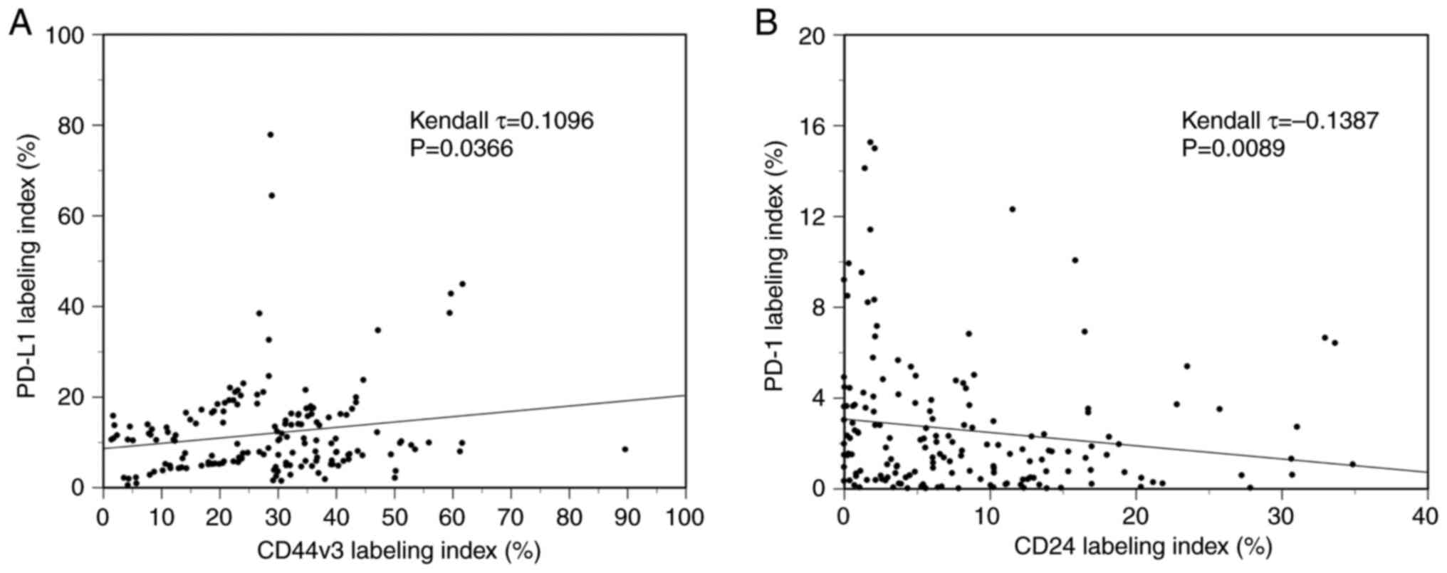
PD-L1high/PD-1high (group C). The
CD44v3-positive rate was significantly positively correlated to the
PD-L1 labeling index (τ=0.1096, P=0.0366, Kendall rank correlation
coefficient) (Fig. 2A). On the
other hand, the CD24-positive rate was significantly negatively
correlated to the PD-1 labeling index (τ=−0.1387, P=0.0089, Kendall
rank correlation coefficient) (Fig.
2B). However, as the τ-values were <0.3/-0.3, these
correlations are considered to be very weak.
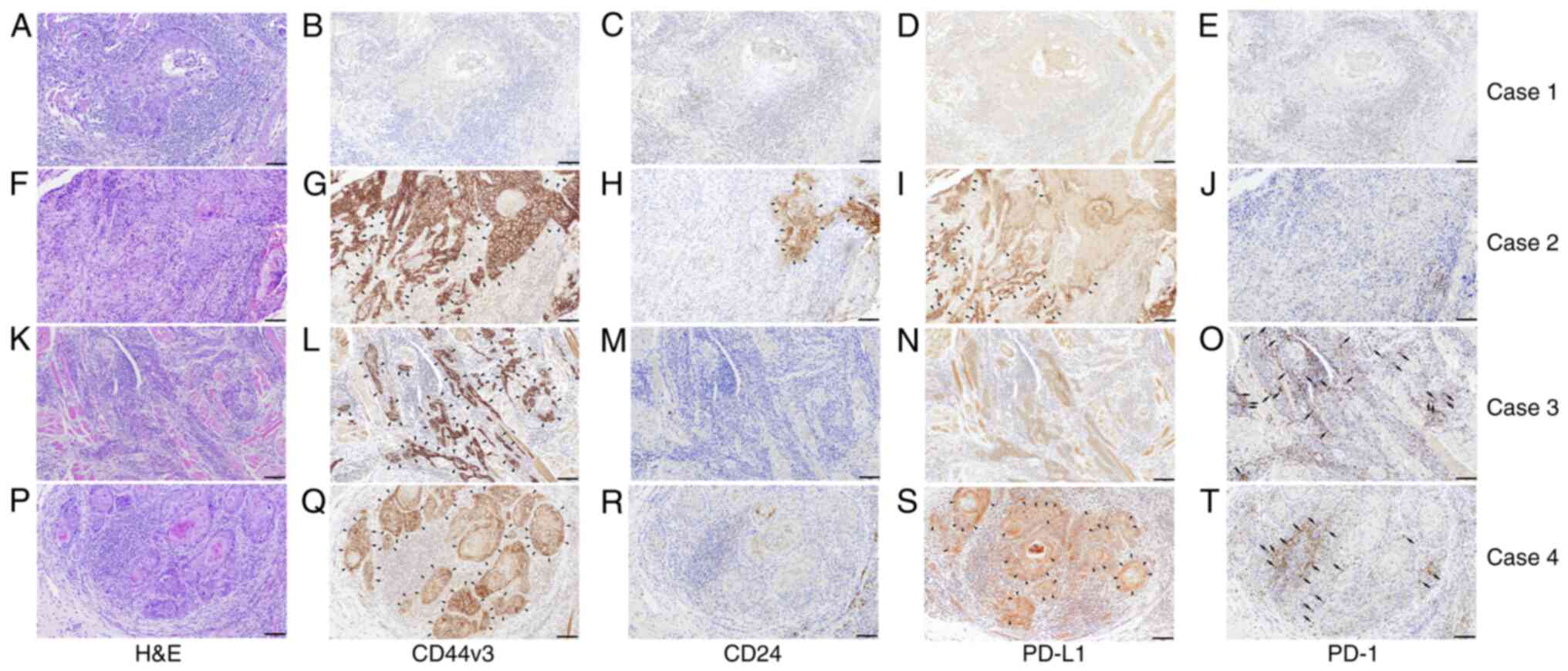 | Figure 1.Representative micrographs of H&E
staining and immunohistochemical classification in accordance with
CD44v3, CD24, PD-L1, and PD-1 expression patterns. Four different
cases with different expressions of each cell surface marker are
shown. (A-E) Photomicrographs of the same case (Case 1, 61 age,
male, tongue). (A) H&E staining. (B) CD44v3 and (C) CD24
immunoreactivity. (D) PD-L1 and (E) PD-1 immunoreactivity. This
case is classified as CD44v3low/CD24low
(non-CSC pattern) and PD-L1low/PD-1low (group
A). (F-J) Photomicrographs of the same case (Case 2, 91 age,
female, lower gingiva). (F) H&E staining. (G) CD44v3 and (H)
CD24 immunoreactivity. Both CD44v3 and CD24 are expressed on the
cancer cell membrane. (I) PD-L1 and (J) PD-1 immunoreactivity.
PD-L1 is expressed on the cancer cell membrane. This case is
classified as CD44v3high/CD24high (non-CSC
pattern) and PD-L1high/PD-1low (group B).
(K-O) Photomicrographs of the same case (Case 3, 39 age, female,
tongue). (K) H&E staining. (L) CD44v3 and (M) CD24
immunoreactivity. CD44v3 is expressed on the cancer cell membrane.
(N) PD-L1 and (O) PD-1 immunoreactivity. PD-1 expression is
observed in lymphocytes around cancer nests. This case is
classified as CD44v3high/CD24low (CSC
pattern) and PD-L1low/PD-1high (group B).
(P-T) Photomicrographs of the same case (Case 4, 54 age, male,
tongue). (P) H&E staining. (Q) CD44v3 and (R) CD24
immunoreactivity. CD44v3 is expressed on the cancer cell membrane.
(S) PD-L1 and (T) PD-1 immunoreactivity, respectively. PD-L1 is
expressed on the cancer cell membrane. PD-1 expression is observed
in lymphocytes around cancer nests. This case is classified as
CD44v3high/CD24low (CSC pattern) and
PD-L1high/PD-1high (group C). Arrowheads
indicate regions where CD44v3, CD24, or PD-L1 expression is
highlighted. Arrows indicate regions where PD-1 expression is
highlighted. Scale bars: 100 µm. H&E, hematoxylin and eosin;
PD-1, programmed cell death 1; PD-L1, programmed cell death ligand
1. |
Analysis of CSC markers-related gene
and PD-L1/PD-1 gene expression by RT-qPCR
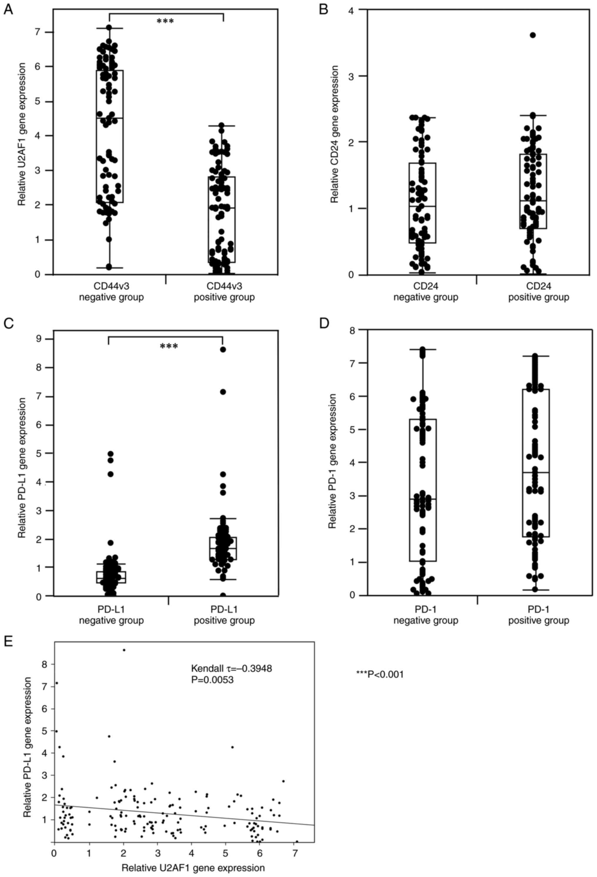
RT-qPCR was performed to examine the expression of
CSC-related molecules (U2AF1 and CD24) and PD-L1/PD-1 at mRNA level
in 168 OSCC patients. For CD24 and PD-1, no significant correlation
was found between the immunophenotype and the genotype. On the
other hand, a significant negative correlation was found between
the expression of CD44v3 in IHC and U2AF1 at mRNA level
(P<0.001, Mann-Whitney U test) (Fig.
3A). Moreover, a significant positive correlation was found
between the immunophenotype and genotype of PD-L1 (P<0.001,
Mann-Whitney U test) (Fig. 3B). In
addition, U2AF1 expression was significantly negatively correlated
to the PD-L1 expression at mRNA level (τ=−0.3948, P=0.0053, Kendall
rank correlation coefficient) (Fig.
3E). On the other hand, for PD-1 and CD24, no significant
correlation was observed between the immunophenotype in IHC and the
genotype at the mRNA level, respectively (Fig. 3C and D).
Associations of the CSC
immunophenotype with various clinicopathological factors and
PD-L1/PD-1 expression
As shown in Table I,
the associations of CSC surface markers with clinicopathological
factors were investigated in all 168 OSCC patients. The nodal
status (prevalence rates of cervical LN metastasis), postoperative
local recurrence, and postoperative LN metastasis were
significantly higher in the CSC group than in the non-CSC group
(Table I). Moreover, PD-L1/PD-1
co-expression was significantly associated with the CSC
immunophenotyped (Table I).
Conversely, other clinical factors, including age, gender,
location, histological grade, lymphatic vessel invasion, pT
classification, stage, and mode of tumor invasion, did not differ
significantly between CSC and non-CSC groups.
As shown in Table
II, the associations between postoperative recurrence-free
survival (RFS) and individual clinicopathological factors,
including CSC markers and PD-L1/PD-1 co-expression, were assessed
by univariate and multivariate analyses. In univariate analysis of
postoperative RFS, nodal status [hazard ratio (HR) 2.12, 95%
confidence interval (CI) 1.14–3.92], mode of invasion (HR 3.01, 95%
CI 1.38–6.55), CSC immunophenotype (HR 3.90, 95% CI 2.05–7.43),
PD-L1/PD-1 expression (HR 4.43, 95% CI 1.78–11.06), and CSC
immunophenotype combined with PD-L1/PD-1 co-expression (HR 9.61,
95% CI 4.58–20.16) were significant predictors of postoperative
recurrence. Further multivariate analysis showed that the CSC
immunophenotype (HR 2.89, 95% CI 1.41–5.95) and PD-L1/PD-1
co-expression (HR 3.01, 95% CI 1.38–6.55) were independent poor
prognostic factors for postoperative recurrence. No significant
association was found regarding age, gender, location, histological
grade, or pT classification in univariate and multivariate
analyses.
| Table II.Univariate and multivariate analyses
of postoperative recurrence-free survival of individual
characteristics. |
Table II.
Univariate and multivariate analyses
of postoperative recurrence-free survival of individual
characteristics.
|
| Univariate
analysis | Multivariate
analysis |
|---|
|
|
|
|
|---|
| Characteristic | HR (95% CI) | P-value | HR (95% CI) | P-value |
|---|
| Age, years |
|
|
|
|
|
>60 | Reference |
| Reference |
|
|
≤60 | 2.52
(0.99–6.43) | 0.053 | 2.16
(0.19–4.23) | 0.364 |
| Sex |
|
|
|
|
|
Male | Reference |
| Reference |
|
|
Female | 1.01
(0.54–1.85) | 0.980 | 1.45
(0.72–2.91) | 0.301 |
| Location |
|
|
|
|
|
Tongue | Reference |
| Reference |
|
|
Gingiva | 1.45
(0.72–2.91) | 0.300 | 1.05
(0.42–2.64) | 0.913 |
| Floor
of mouth | 1.53
(0.44–5.34) | 0.500 | 1.09
(0.28–4.16) | 0.904 |
| Buccal
mucosa | 1.18
(0.34–4.12) | 0.790 | 0.49
(0.13–1.91) | 0.308 |
|
Palate | 3.29
(0.75–14.55) | 0.200 | 3.87
(0.82–18.24) | 0.095 |
|
Lip | 3.74
(0.48–28.79) | 0.120 | 4.82
(1.24–32.79) | 0.180 |
| Histological
gradea |
|
|
|
|
| Grade
1 | Reference |
| Reference |
|
| Grade
2/3 | 1.55
(1.75–3.16) | 0.230 | 3.23
(1.44–7.26) | 0.410 |
| pT
classification |
|
|
|
|
|
T1/T2 | Reference |
| Reference |
|
|
T3/T4 | 1.58
(0.84–2.98) | 0.160 | 1.98
(0.77–5.09) | 0.156 |
| Nodal
statusb |
|
|
|
|
|
N(−) | Reference |
| Reference |
|
|
N(+) | 2.12
(1.14–3.92) | 0.016 | 1.49
(0.73–3.02) | 0.273 |
| Mode of invasion
(YK classification)c |
|
|
|
|
| Grade
1/2 | Reference |
| Reference |
|
| Grade
3 | 1.22
(0.57–2.60) | 0.610 | 1.13
(0.53–2.42) | 0.753 |
| Grade
4C/4D | 3.01
(1.38–6.55) | 0.006 | 1.39
(1.02–5.61) | 0.342 |
| Immunophenotype of
CSC |
|
|
|
|
|
Non-CSC | Reference |
| Reference |
|
|
CSC | 3.90
(2.05–7.43) | <0.0001 | 2.89
(1.41–5.95) | 0.039 |
| PD-L1/PD-1 |
|
|
|
|
| Group
Ad | Reference |
| Reference |
|
| Group
Bd | 1.80
(0.69–4.62) | 0.220 | 1.59
(0.61–4.17) | 0.434 |
| Group
Cd | 4.43
(1.78–11.06) | 0.001 | 3.54
(1.37–9.12) | 0.028 |
| Immunophenotype of
CSC combined with PD-L1/PD-1 |
|
|
|
|
| CSC and
Group Ad, or
non-CSC | Reference |
|
|
|
| CSC and
Group Bd | 2.62
(0.98–6.97) | 0.054 |
|
|
| CSC and
Group Cd | 9.61
(4.58–20.16) | <0.0001 |
|
|
Prognostic effect of CSC markers and
PD-L1/PD-1 co-expression in OSCC patients
To estimate the relationship between
immunoreactivity for CSC markers and PD-L1/PD-1 and the prognosis
of OSCC patients, postoperative RFS rates were calculated using the
Kaplan-Meier method. There was a significant difference in the
postoperative RFS rate between the CSC group (34.1%) and the
non-CSC group (77.0%) (log-rank test, P<0.01) (Fig. 4A). Furthermore, all OSCC patients
were classified into three subgroups by the mode of PD-L1/PD-1
expression as follows: group A,
PD-L1low/PD-1low; group B,
PD-L1high/PD-1low or
PD-L1low/PD-1high; group C,
PD-L1high/PD-1high, and there was a
significant difference in the postoperative RFS rate between group
C (45.7%) and group B (74.9%) (log-rank test, P<0.05), and
between group C and group A (83.9%) (log-rank test, P<0.01)
(Fig. 4B). Moreover, group C with a
combined CSC immunophenotype had a significantly poorer prognosis
than group B with the combined CSC immunophenotype and group A with
the combined CSC immunophenotype or non-CSC group. RFS rates in
group C with the combined CSC immunophenotype, group B with the
combined CSC immunophenotype, and group A with the combined CSC or
non-CSC immunophenotype were 0, 50.6, and 78.1%, respectively
(Fig. 4C).
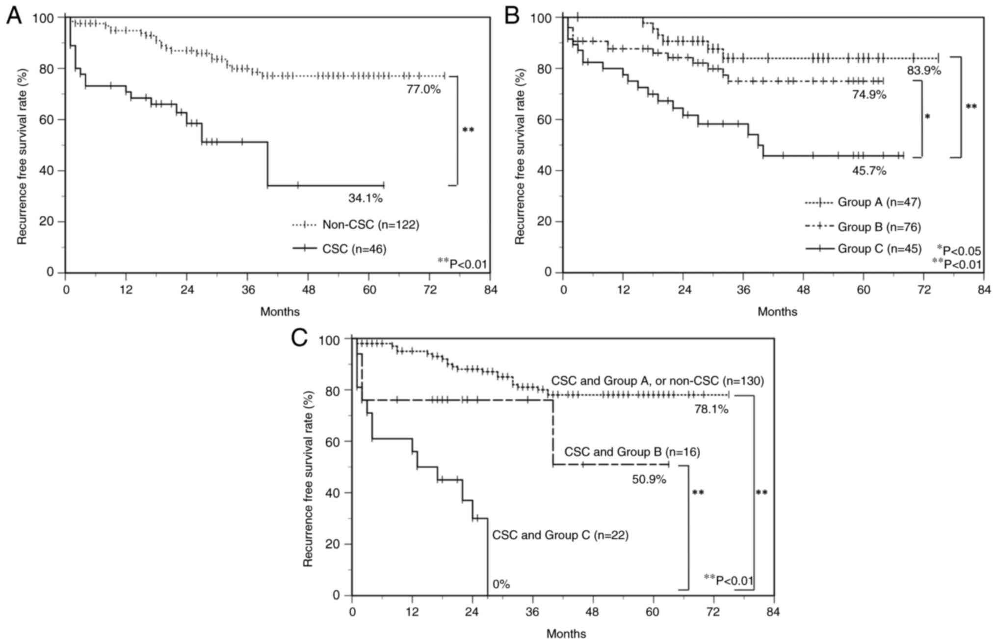 | Figure 4.Postoperative RFS curves of OSCC
patients. Cumulative survival curves of OSCC patients classified by
the expression patterns of (A) CSC markers, (B) PD-L1/PD-1, and (C)
combined CSC markers and PD-L1/PD-1. (A) CSC cases have poor
prognoses, and there is a significant difference between CSC and
non-CSC cases. (B) The time to postoperative RFS is significantly
shorter in group C. (C) The time to postoperative RFS is
significantly shorter in combined CSC and group C. Based on the
PD-L1/PD-1 immunophenotypes, group A, B, and C are characterized as
follows: Group A, PD-L1low/PD-1low; group B,
PD-L1high/PD-1low or
PD-L1low/PD-1high; group C,
PD-L1high/PD-1high. The Kaplan-Meier method
was used to construct survival curves, and the log-rank test was
used to calculate P-value. *P<0.05 and **P<0.01. CSC, cancer
stem cell; OSCC, oral squamous cell carcinoma; PD-1, programmed
cell death 1; PD-L1, programmed cell death ligand 1; RFS,
recurrence-free survival. |
Discussion
There have been only a few reports on the
relationship between immune checkpoint molecules PD-L1/PD-1 and
CSCs in HNSCCs including OSCC (28–30).
To our knowledge, this is the first study to address the prognostic
effect of CD44v3 and CD24, cell surface markers of CSCs, in terms
of PD-L1 and PD-1 co-expression in OSCC. In this study, 168 OSCC
specimens were analyzed and grouped according to the expression
results of the CSC markers, i.e., CD44v3 and CD24, and the immune
checkpoint molecules, i.e., PD-1 and its ligand PD-L1 in IHC.
We believe that CSCs in OSCC have a critical
unfavorable prognostic effect in terms of PD-L1/PD-1 co-expression
for several reasons. First, we identified the CSC immunophenotype
CD44v3high/CD24low in OSCC as a critical
unfavorable prognostic factor. Although our results may revealed
that the phenotype of CSCs was not correlate with histological
morphology, such as histological differentiation or infiltration
pattern (YK classification), it is even more critical to identify
CSCs using cell surface markers such as CD44v3 and CD24. We found
that the patient population with the CSC immunophenotype was
significantly correlated to unfavorable clinical outcomes
(postoperative LN metastasis and/or local recurrence) compared with
the patient population with the non-CSC immunophenotype
(CD44v3high/CD24high and
CD44v3low/CD24high or low), which was similar
to our previous study (7).
The second reason for the CSC prognostic effect in
terms of PD-L1/PD-1 co-expression was that univariate analysis
revealed that the PD-L1/PD-1 co-expression group was significantly
associated with unfavorable clinical outcomes compared with groups
expressing either PD-L1, PD-1, or neither. Furthermore,
multivariate analysis revealed that PD-L1/PD-1 co-expression was an
independent poor prognostic factor. These findings were consistent
with previous studies (24,25). Cancer cells stimulate PD-1 receptors
on T cell membranes via PD-L1, and these signals act as so-called T
cell brakes and inhibit cancer cell attack by T cells (19–21,23).
These findings suggest that PD-L1 expression in the tumor
parenchyma and PD-1 expression in inflammatory cells in the stroma
play an essential role in immune evasion of OSCC.
The third reason, which was most important for the
critical unfavorable prognostic effect of CSCs in terms of
PD-L1/PD-1 co-expression, was that the CSC immunophenotype and
PD-L1/PD-1 co-expression were independent unfavorable prognostic
factors for OSCC, they influenced each other, and CSC marker and
PD-L1/PD-1 co-expression had a more substantial prognostic effect.
Furthermore, our results revealed that PD-L1/PD-1 co-expression was
significantly associated with the CSC immunophenotype, suggesting
that PD-L1 and PD-1 play an essential role in maintaining CSC
stemness in OSCC. Indeed, previous reports have shown that PD-L1
plays a crucial role in CSC expansion in addition to its role as an
immune checkpoint (34,35). Moreover, in breast cancer, where
CD44high/CD24low has been reported to be a
CSC immunophenotype, the PD-L1/PD-1 axis activates Phosphoinositide
3-kinase/Akt and Extracellular signal-regulated kinase 1/2
signaling pathways to promote cancer stemness and drug resistance
(36–38). To investigate the relationship
between the Akt pathway and PD-L1/PD-1 axis in CSCs in OSCC, we
focused on U2FA1, which has been reported to be a splicing factor
of CD44v3 and a down regulator of the Akt pathway upstream of this
pathway (14). Furthermore, U2AF1
has been reported to suppress PD-L1 expression (39). In the present study, the
CD44v3-positive group showed significantly lower expression of
U2AF1 at mRNA level than that of the CD44v3-negative group.
Furthermore, a negatively significant correlation was observed
between U2AF1 and PD-L1 expression at mRNA level. These results
suggest that the downregulation of U2AF1, which acts in a
suppressive manner to activate the Akt pathway, contributes to the
promotion of CD44v3 splicing, the maintenance of CSC stemness, and
the increase in PD-L1 expression. However, the role of U2AF1 in
CSCs of OSCC remains unclear, with only one report indicating that
U2AF1 is involved in carcinogenesis from oral precancerous lesions
(40). Also, another report on the
relationship between U2AF1 and PD-L1 found no correlation between
their expression (41). On the
other hand, Geum et al (31)
support our study. They reported that PD-L1 knockdown suppressed
the induction of Akt phosphorylation. In other words, PD-L1
activates Akt and may be involved in maintaining OSCC stemness.
However, the involvement of U2AF1 in PD-L1 to Akt is still unknown
with only a few reports. Thus, further validation is needed to
elucidate the role of U2AF1 in CSCs in OSCC. To confirm the further
relationship between CSCs and PD-L1/PD-1, we examined the
expression correlation between CD44v3 and PD-L1, and CD24 and PD-1.
Regarding the relationship between CD44v3 and PD-L1, our results
revealed a significant positive correlation between CD44v3 and
PD-L1. Although these correlations are considered to be very weak
due to the low τ-value of <0.3, our results may suggest that
CSCs in OSCC express PD-L1 on the tumor cell membrane surface and
play a role in immune evasion. Previous studies support our
findings by reporting that CD44v3high exosomes have
higher levels of immunosuppressive proteins such as PD-L1 than
CD44v3low exosomes, correlating to a higher stage and LN
metastasis in OSCC patients (42).
Regarding the relationship between CD24 and PD-1, a
significant negative correlation was found between CD24 in the
tumor parenchyma and PD-1 expression in surrounding inflammatory
cells. Although these correlations are considered to be very weak
due to the low τ-value of <-0.3, our results may suggest that
downregulation of CD24 plays a role in evading host immune
responses via high PD-1 expression on the stroma of CSCs. A
previous report supports the results of our study. Kim et al
(43) reported that CD24 bound to T
cells via intracytoplasmic high-mobility group box 1, an
inflammation-associated protein, and promoted activation of CD8+
effector T cells (43). In other
words, CD24low cancer cells reduce CD8+ T cell activity
and attenuated host immune responses compared with
CD24high cancer cells. Another study also supports our
findings. Xiao et al (44)
reported that high expression of PD-1 correlated to decreased CD24
expression and cytokine production in B cells infiltrating
hepatocellular carcinoma (44).
Furthermore, in another previous report of breast cancer stem
cells, in which CD24low was considered to be an
immunophenotype of CSCs, increased expression of PD-L1 was observed
in CD24low cancer cells (36). Although this study does not directly
address the negative correlation between CD24 and PD-1, it can be
inferred that PD-1 expression is elevated along with PD-L1 in
CD24low breast cancer (36). On the other hand, Mirhashemi et
al (45) reported contradictory
results to our study. They performed RT-qPCR analysis of clinical
specimens from 15 oral epithelial dysplasia and 45 cases of OSCC.
They reported that as histologic grade increased from oral
epithelial dysplasia to low-grade OSCC and from low-grade OSCC to
high-grade OSCC, the expression of both CD44 and CD24 increased and
correlated more intensely with the expression of these two cell
surface markers. However, they also mentioned that there were
conflicting reports, including our previous report (7), on the CD24 phenotype of CSCs in OSCC,
and further verification was needed to assert that
CD44high and CD24high were the phenotypes of
CSCs in OSCC. On the contrary, Ghuwalewala et al (9) support our results. They report that
MiR-146a, which directly targets IRAK1, traf6, and numb genes in
OSCC and confers tumorigenicity, contributes to the enrichment of
CSCs in OSCC through increased expression of stem cell markers,
including CD44 and decreased CD24 levels. In addition, a previous
study also supports our results. It reveals an increase in
CD44highCD24low cells in IHC from low in
normal tissues to relatively high in OSCC (8). Thus, more data are needed before
definitive conclusions can be drawn because of the conflicting
results of previous studies on the interaction of CD24 with PD-1
and the possible tumor specificity of CD24 functions. At least
concerning CSCs in OSCC, attenuated expression of CD24 appears to
play a role in evasion of CSCs from host immunity by enhancing PD-1
expression in inflammatory cells in the tumor stroma.
Although there is no doubt that CSCs are a
significant unfavorable prognostic factor in terms of PD-L1/PD-1
co-expression, the following five points need to be considered to
ensure the validity of this study: 1. the reasons for focusing on
the invasive front of the tumor as a region of interest. 2. the
limitation of the ability to profile mRNA expression extracted from
FFPE, 3. the validity of the cutoff values used in the IHC analysis
of the four antibodies (CD44v3, CD24, PD-L1, PD-1) used in this
study, 4. the heterogeneity of expression of each antibody within
the tumor tissue, and 5. the impact of core selection in TMA. 1.
The reason for focusing on the invasive front of the tumor was that
these areas are suitable for observing cross-talk between the
parenchyma and stroma via the PD-L1/PD-1 axis, as described in a
previous report (24) 2. Regarding
the limitation of the ability to profile mRNA expression extracted
from FFPE, the expression of CD44v3 in IHC was significantly
negatively correlated with the expression of the molecules U2AF1,
and the expression of PD-L1 in IHC was significantly positively
correlated with the expression of PD-L1 at mRNA level. On the other
hand, for CD24 and PD-1, no significant difference was found
between the immunophenotype and the genotype. This discrepancy
between the immunophenotype and genotype may be due to the effect
of chemical modification to mRNA during FFPE specimen preparation
and the degradation of nucleic acids during mRNA extraction process
(46). 3. Regarding the cutoff
values, the validity of the labeling indices (median values)
obtained in this study was compared with the cutoff values
described in previous literatures on IHC studies in OSCC for each
molecule. No reports were found mentioning a cutoff value for
CD44v3 in IHC, but as far as the CD44 standard isoform is
concerned, a range of 10–50% was reported, and no uniform value
existed (47,48). As far as CD24 is concerned, only one
reference mentioned a cutoff value in IHC, which was reported as
37.4% (47). As far as PD-L1 is
concerned, the range of cutoff values in IHC was reported to be
1–20% (24,25,49).
As far as PD-1 is concerned, only one reference mentions a cutoff
value in IHC, which was reported as 30% (24). The previous reports listed above
regarding cutoff values in IHC for CD44 and PD-L1 include review
articles (25,48,49),
indicating no uniform cutoff value in the IHC study. Oliveira et
al (50) mentioned that the
cutoff values differ among studies of the same antibody because of
the different antibodies used and staining conditions in IHC.
Therefore, as is done in many IHC clinical studies, cutoff value
was established for each antibody, using the median value in this
study. In particular, we mention the criteria of PD-L1 positivity
in this study, concerning the immunohistochemical analysis of
PD-L1. For PD-L1 expression analysis, the antibody clone used in
this study (clone 28-8) was the same clone used in the companion
diagnosis of nivolumab for HNSCC treatment. In clinical practice, a
cutoff value of 1% is considered positive. However, this clinical
cutoff value is lower than the cutoff value of 6.1% adopted in this
study. The difference in cutoff values between clinical practice
and the present study may be attributed to multiple factors,
including the different staining conditions and analysis methods
used in the companion diagnosis. Another factor contributing to the
discrepancy is that only the area of the invasive portion was
selected as the stained area for PD-L1 in this study, rather than
the entire tumor. Indeed, PD-L1 expression is spatiotemporally
heterogeneous, and there is heterogeneity in PD-L1 expression rates
within tumors and at different treatment points (20,51,52).
Thus, PD-L1 expression levels are not well understood by single
treatment point or single time sampling. 4. Regarding the
heterogeneity of expression of each antibody in tumor tissue and 5.
the impact of core selection in TMA, it has been reported that the
expression of CD44(v), CD24, PD-L1, and PD-1 are all heterogeneous
in OSCC in the same tissue (48,53–56).
Boxberg et al (54) reported
that this heterogeneity in the expression of each antibody had the
risk of missing heterogeneous tumor regions when using TMA for
immunohistochemical analysis while noting that increasing the
number of cores collected per case may allow for a close match
between tissue cores and staining results on the entire slide
(56). In this study, by sampling
at least two to three cores per case, we reduced the possibility of
missing heterogeneous expression regions and, as much as possible,
ensured the validity of each molecule's expression in IHC from TMA.
In addition, we add one point of the limitations of this study. In
this study, for CD44v3, we could not perform direct RT-qPCR due to
the lack of commercially available primer.
In summary, this study revealed that CSCs in OSCC
evade host immune mechanisms and maintain CSC stemness via
PD-L1/PD-1 expression, resulting in unfavorable clinical outcomes,
suggesting that CSCs are a potential therapeutic target for immune
checkpoint inhibitors.
Acknowledgements
The authors would like to thank Ms. Kumiko Tsubone
(Kurume University School of Medicine, Dental and Oral Medical
Center) for their excellent assistance in the experiments.
Funding
This study was supported by a Grant-in-Aid for Scientific
Research (grant no. 20K18714) from the Ministry of Education,
Culture, Sports, Science and Technology of Japan.
Availability of data and materials
The datasets used and/or analyzed during the current
study are available from the corresponding author on reasonable
request.
Authors' contribution
KT and YN conceived and designed the study. KT, AK,
MN and NS performed the experiments and acquired data, and assessed
the authenticity of all the raw data to ensure its legitimacy. KT,
YA, KM, JK and HN analyzed the data. KT, YN and MN drafted the
manuscript. KT and JK confirm the authenticity of all the raw data.
All authors read and approved the final manuscript.
Ethics approval and consent to
participate
The design and methods of this study complied with
the ethical guidelines of the Declaration of Helsinki and the
guidelines for research involving human subjects of the Ethical
Review Committee of the Clinical Research Center of Kurume
University Hospital. Institutional review and approval were
obtained prior to commencing the study (approval no. 22217).
Written informed consent was obtained before patient participation
in the study, and clinical specimens were obtained from each
patient in accordance with the approved guidelines.
Patient consent for publication
Not applicable.
Competing interests
The authors declare that they have no competing
interests.
Glossary
Abbreviations
Abbreviations:
|
CSC
|
cancer stem cell
|
|
OSCC
|
oral squamous cell carcinoma
|
|
FFPE
|
formalin-fixed, paraffin-embedded
|
|
IHC
|
immunohistochemistry
|
|
RT-qPCR
|
reverse transcription quantitative
polymerase chain reaction
|
|
U2AF1
|
U2 small nuclear RNA auxiliary factor
1
|
|
HNSCC
|
head and neck squamous cell
carcinoma
|
|
LN
|
lymph node
|
|
YK
|
Yamamoto-Kohama
|
|
EMT
|
epithelial-mesenchymal transition
|
|
TMA
|
tissue microarray
|
References
|
1
|
Petersen PE: Oral cancer prevention and
control-the approach of the world health organization. Oral Oncol.
45:454–460. 2009. View Article : Google Scholar : PubMed/NCBI
|
|
2
|
Prince ME, Sivanandan R, Kaczorowski A,
Wolf GT, Kaplan MJ, Dalerba P, Weissman IL, Clarke MF and Ailles
LE: Identification of a subpopulation of cells with cancer stem
cell properties in head and neck squamous cell carcinoma. Proc Natl
Acad Sci. 104:973–978. 2007. View Article : Google Scholar : PubMed/NCBI
|
|
3
|
Kidwai F, Costea DE, Hutchison I and
Mackenzie I: The effects of CD44 down-regulation on stem cell
properties of head and neck cancer cell lines. J Oral Pathol Med.
42:682–690. 2013. View Article : Google Scholar : PubMed/NCBI
|
|
4
|
Kalish ED, Iida N, Moffat FL and
Bourguignon LY: A new CD44v3-containing isoform is involved in
tumor cell growth and migration during human breast carcinoma
progression. Front Biosci. 4:A1–A8. 1999. View Article : Google Scholar : PubMed/NCBI
|
|
5
|
Hurt EM, Kawasaki BT, Klarmann GJ, Thomas
SB and Farrar WL: CD44+CD24- prostate cells are early cancer
progenitor/stem cells that provide a model for patients with poor
prognosis. Br J Cancer. 98:756–765. 2008. View Article : Google Scholar : PubMed/NCBI
|
|
6
|
Ma F, Li H, Wang H, Shi X, Fan Y, Ding X,
Lin C, Zhan Q, Qian H and Xu B: Enriched CD44+/CD24- population
drives the aggressive phenotypes presented in triple-negative
breast cancer (TNBC). Cancer Lett. 353:153–159. 2014. View Article : Google Scholar : PubMed/NCBI
|
|
7
|
Todoroki K, Ogasawara S, Akiba J, Nakayama
M, Naito Y, Seki N, Kusukawa J and Yano H: CD44v3+/CD24- cells
possess cancer stem cell-like properties in human oral squamous
cell carcinoma. Int J Oncol. 48:99–109. 2015. View Article : Google Scholar : PubMed/NCBI
|
|
8
|
Ghuwalewala S, Ghatak D, Das P, Dey S,
Sarkar S, Alam N, Panda CK and Roychoudhury S: CD44highCD24low
molecular signature determines the cancer stem cell and EMT
phenotype in oral squamous cell carcinoma. Stem Cell Res.
16:405–417. 2016. View Article : Google Scholar : PubMed/NCBI
|
|
9
|
Ghuwalewala S, Ghatak D, Das S, Roy S, Das
P, Butti R, Gorain M, Nath S, Kundu GC and Roychoudhury S:
MiRNA-146a/AKT/β-catenin activation regulates cancer stem cell
phenotype in oral squamous cell carcinoma by targeting CD24. Front
Oncol. 11:6516922021. View Article : Google Scholar : PubMed/NCBI
|
|
10
|
Clay MR, Tabor M, Owen JH, Carey TE,
Bradford CR, Wolf GT, Wicha MS and Prince ME: Single-marker
identification of head and neck squamous cell carcinoma cancer stem
cells with aldehyde dehydrogenase. Head Neck. 32:1195–1201. 2010.
View Article : Google Scholar : PubMed/NCBI
|
|
11
|
Baillie R, Tan ST and Itinteang T: Cancer
stem cells in oral cavity squamous cell carcinoma: A review. Front
Oncol. 7:1122017. View Article : Google Scholar : PubMed/NCBI
|
|
12
|
Wang SJ, Wong G, de Heer AM, Xia W and
Bourguignon LYW: CD44 variant isoforms in head and neck squamous
cell carcinoma progression. Laryngoscope. 119:1518–1530. 2009.
View Article : Google Scholar : PubMed/NCBI
|
|
13
|
Bourguignon LYW, Wong G, Earle C and Chen
L: Hyaluronan-CD44v3 interaction with Oct4-Sox2-nanog promotes
miR-302 expression leading to self-renewal, clonal formation, and
cisplatin resistance in cancer stem cells from head and neck
squamous cell carcinoma. J Biol Chem. 287:32800–32824. 2012.
View Article : Google Scholar : PubMed/NCBI
|
|
14
|
Zhu H, Zhou W, Wan Y, Lu J, Ge K and Jia
C: CD44V3, an alternatively spliced form of CD44, promotes
pancreatic cancer progression. Int J Mol Sci. 23:120612022.
View Article : Google Scholar : PubMed/NCBI
|
|
15
|
Pirruccello SJ and LeBien TW: The human B
cell-associated antigen CD24 is a single chain sialoglycoprotein. J
Immunol. 136:3779–3784. 1986. View Article : Google Scholar : PubMed/NCBI
|
|
16
|
Fischer GF, Majdic O, Gadd S and Knapp W:
Signal transduction in lymphocytic and myeloid cells via CD24, a
new member of phosphoinositol-anchored membrane molecules. J
Immunol. 144:638–641. 1990. View Article : Google Scholar : PubMed/NCBI
|
|
17
|
Vesuna F, Lisok A, Kimble B and Raman V:
Twist modulates breast cancer stem cells by transcriptional
regulation of CD24 expression. Neoplasia. 11:1318–1328. 2009.
View Article : Google Scholar : PubMed/NCBI
|
|
18
|
Dunn GP, Bruce AT, Ikeda H, Old LJ and
Schreiber RD: Cancer immunoediting: From immunosurveillance to
tumor escape. Nat Immunol. 3:991–998. 2002. View Article : Google Scholar : PubMed/NCBI
|
|
19
|
Economopoulou P, Agelaki S, Perisanidis C,
Giotakis EI and Psyrri A: The promise of immunotherapy in head and
neck squamous cell carcinoma. Ann Oncol. 27:1675–1685. 2016.
View Article : Google Scholar : PubMed/NCBI
|
|
20
|
Zhang S, Bai X and Shan F: The progress
and confusion of anti-PD1/PD-L1 immunotherapy for patients with
advanced non-small cell lung cancer. Int Immunopharmacol.
80:1062472020. View Article : Google Scholar : PubMed/NCBI
|
|
21
|
Liu J, Chen Z, Li Y, Zhao W, Wu J and
Zhang Z: PD-1/PD-L1 checkpoint inhibitors in tumor immunotherapy.
Front Pharmacol. 12:7317982021. View Article : Google Scholar : PubMed/NCBI
|
|
22
|
Blank C, Gajewski TF and Mackensen A:
Interaction of PD-L1 on tumor cells with PD-1 on tumor-specific T
cells as a mechanism of immune evasion: Implications for tumor
immunotherapy. Cancer Immunol Immunother. 54:307–314. 2005.
View Article : Google Scholar : PubMed/NCBI
|
|
23
|
Ferris RL: Immunology and immunotherapy of
head and neck cancer. J Clin Oncol. 33:3293–3304. 2015. View Article : Google Scholar : PubMed/NCBI
|
|
24
|
Maruse Y, Kawano S, Jinno T, Matsubara R,
Goto Y, Kaneko N, Sakamoto T, Hashiguchi Y, Moriyama M, Toyoshima
T, et al: Significant association of increased PD-L1 and PD-1
expression with nodal metastasis and a poor prognosis in oral
squamous cell carcinoma. Int J Oral Max Surg. 47:836–845. 2018.
View Article : Google Scholar : PubMed/NCBI
|
|
25
|
Lenouvel D, González-Moles MÁ, Ruiz-Ávila
I, Gonzalez-Ruiz L, Gonzalez-Ruiz I and Ramos-García P: Prognostic
and clinicopathological significance of PD-L1 overexpression in
oral squamous cell carcinoma: A systematic review and comprehensive
meta-analysis. Oral Oncol. 106:1047222020. View Article : Google Scholar : PubMed/NCBI
|
|
26
|
Ferris RL, Blumenschein G Jr, Fayette J,
Guigay J, Colevas AD, Licitra L, Harrington K, Kasper S, Vokes EE,
Even C, et al: Nivolumab for recurrent squamous-cell carcinoma of
the head and neck. New Engl J Med. 375:1856–1867. 2016. View Article : Google Scholar : PubMed/NCBI
|
|
27
|
Burtness B, Harrington KJ, Greil R,
Soulières D, Tahara M, de Castro G Jr, Psyrri A, Basté N, Neupane
P, Bratland Å, et al: Pembrolizumab alone or with chemotherapy
versus cetuximab with chemotherapy for recurrent or metastatic
squamous cell carcinoma of the head and neck (KEYNOTE-048): A
randomised, open-label, phase 3 study. Lancet. 394:1915–1928. 2019.
View Article : Google Scholar : PubMed/NCBI
|
|
28
|
Lee Y, Shin JH, Longmire M, Wang H, Kohrt
HE, Chang HY and Sunwoo JB: CD44+ cells in head and neck squamous
cell carcinoma suppress t-cell-mediated immunity by selective
constitutive and inducible expression of PD-L1. Clin Cancer Res.
22:3571–3581. 2016. View Article : Google Scholar : PubMed/NCBI
|
|
29
|
Chen L, Yang QC, Li YC, Yang LL, Liu JF,
Li H, Xiao Y, Bu LL, Zhang WF and Sun ZJ: Targeting CMTM6
suppresses stem cell-like properties and enhances antitumor
immunity in head and neck squamous cell carcinoma. Cancer Immunol
Res. 8:179–191. 2020. View Article : Google Scholar : PubMed/NCBI
|
|
30
|
Wang C, Li Y, Jia L, Kim JK, Li J, Deng P,
Zhang W, Krebsbach PH and Wang CY: CD276 expression enables
squamous cell carcinoma stem cells to evade immune surveillance.
Cell Stem Cell. 28:1597–1613.e7. 2021. View Article : Google Scholar : PubMed/NCBI
|
|
31
|
Geum DH, Hwang DS, Lee CH, Cho SD, Jang
MA, Ryu MH and Kim UK: PD-L1 expression correlated with
clinicopathological factors and akt/stat3 pathway in oral SCC. Life
(Basel). 12:2382022.PubMed/NCBI
|
|
32
|
Yamamoto E, Kohama G, Sunakawa H, Iwai M
and Hiratsuka H: Mode of invasion, bleomycin sensitivity, and
clinical course in squamous cell carcinoma of the oral cavity.
Cancer. 51:2175–2180. 1983. View Article : Google Scholar : PubMed/NCBI
|
|
33
|
Livak KJ and Schmittgen TD: Analysis of
relative gene expression data using real-time quantitative PCR and
the 2(−Delta Delta C(T)) method. Methods. 25:402–408. 2001.
View Article : Google Scholar : PubMed/NCBI
|
|
34
|
Almozyan S, Colak D, Mansour F, Alaiya A,
Al-Harazi O, Qattan A, Al-Mohanna F, Al-Alwan M and Ghebeh H: PD-L1
promotes OCT4 and Nanog expression in breast cancer stem cells by
sustaining PI3K/AKT pathway activation. Int J Cancer.
141:1402–1412. 2017. View Article : Google Scholar : PubMed/NCBI
|
|
35
|
Wei F, Zhang T, Deng SC, Wei JC, Yang P,
Wang Q, Chen ZP, Li WL, Chen HC, Hu H and Cao J: PD-L1 promotes
colorectal cancer stem cell expansion by activating HMGA1-dependent
signaling pathways. Cancer Lett. 450:1–13. 2019. View Article : Google Scholar : PubMed/NCBI
|
|
36
|
Wu Y, Dutta P, Clayton S, McCloud A and
Vadgama JV: Elevated baseline serum PD-L1 level may predict poor
outcomes from breast cancer in African-American and hispanic women.
J Clin Med. 11:2832022. View Article : Google Scholar : PubMed/NCBI
|
|
37
|
Gao L, Guo Q, Li X, Yang X, Ni H, Wang T,
Zhao Q, Liu H, Xing Y, Xi T and Zheng L: MiR-873/PD-L1 axis
regulates the stemness of breast cancer cells. EBioMedicine.
41:395–407. 2019. View Article : Google Scholar : PubMed/NCBI
|
|
38
|
Hsu JM, Xia W, Hsu YH, Chan LC, Yu WH, Cha
JH, Chen CT, Liao HW, Kuo CW, Khoo KH, et al: STT3-dependent PD-L1
accumulation on cancer stem cells promotes immune evasion. Nat
Commun. 9:19082018. View Article : Google Scholar : PubMed/NCBI
|
|
39
|
Judd J, Karim NA, Khan H, Naqash AR, Baca
Y, Xiu J, VanderWalde AM, Mamdani H, Raez LE, Nagasaka M, et al:
Characterization of KRAS mutation subtypes in non-small cell lung
cancer. Mol Cancer Ther. 20:2577–2584. 2021. View Article : Google Scholar : PubMed/NCBI
|
|
40
|
Yin F, Wang J, Zhao K, Xin C, Shi Y, Zeng
X, Xu H, Li J and Chen Q: The significance of PA28γ and U2AF1 in
oral mucosal carcinogenesis. Oral Dis. 26:53–61. 2020. View Article : Google Scholar : PubMed/NCBI
|
|
41
|
Goldschmid H, Kluck K, Ball M, Kirchner M,
Allgäuer M, Winter H, Herth F, Heußel CP, Pullamsetti SS, Savai R,
et al: Spatial profiling of the microenvironment reveals low
intratumoral heterogeneity and STK11-associated immune evasion in
therapy-naïve lung adenocarcinomas. Lung Cancer. 180:1072122023.
View Article : Google Scholar : PubMed/NCBI
|
|
42
|
Theodoraki MN, Matsumoto A, Beccard I,
Hoffmann TK and Whiteside TL: CD44v3 protein-carrying tumor-derived
exosomes in HNSCC patients' plasma as potential noninvasive
biomarkers of disease activity. Oncoimmunology. 9:17477322020.
View Article : Google Scholar : PubMed/NCBI
|
|
43
|
Kim TS, Gorski SA, Hahn S, Murphy KM and
Braciale TJ: Distinct dendritic cell subsets dictate the fate
decision between effector and memory CD8+ T cell differentiation by
a CD24-dependent mechanism. Immunity. 40:400–413. 2014. View Article : Google Scholar : PubMed/NCBI
|
|
44
|
Xiao X, Lao XM, Chen MM, Liu RX, Wei Y,
Ouyang FZ, Chen DP, Zhao XY, Zhao Q, Li XF, et al: PD-1hi
identifies a novel regulatory B-cell population in human hepatoma
that promotes disease progression. Cancer Discov. 6:546–559. 2016.
View Article : Google Scholar : PubMed/NCBI
|
|
45
|
Mirhashemi M, Ghazi N, Saghravanian N,
Taghipour A and Mohajertehran F: Evaluation of CD24 and CD44 as
cancer stem cell markers in squamous cell carcinoma and epithelial
dysplasia of the oral cavity by q- RT-PCR. Dent Res J (Isfahan).
17:208–212. 2020. View Article : Google Scholar : PubMed/NCBI
|
|
46
|
Hall JS, Taylor J, Valentine HR, Irlam JJ,
Eustace A, Hoskin PJ, Miller CJ and West CM: Enhanced stability of
microRNA expression facilitates classification of FFPE tumour
samples exhibiting near total mRNA degradation. Br J Cancer.
107:684–694. 2012. View Article : Google Scholar : PubMed/NCBI
|
|
47
|
de Moraes FP, Lourenço SV, Ianez RCF, de
Sousa EA, Silva MM, Damascena AS, Kowalski LP, Soares FA and
Coutinho-Camillo CM: Expression of stem cell markers in oral cavity
and oropharynx squamous cell carcinoma. Oral Surg Oral Med Oral
Pathol Oral Radiol. 123:113–122. 2017. View Article : Google Scholar : PubMed/NCBI
|
|
48
|
Mirhashemi M, Sadeghi M, Ghazi N,
Saghravanian N, Dehghani M and Aminian A: Prognostic value of CD44
expression in oral squamous cell carcinoma: A meta-analysis. Ann
Diagn Pathol. 67:1522132023. View Article : Google Scholar : PubMed/NCBI
|
|
49
|
Nocini R, Vianini M, Girolami I, Calabrese
L, Scarpa A, Martini M, Morbini P, Marletta S, Brunelli M, Molteni
G, et al: PD-L1 in oral squamous cell carcinoma: A key biomarker
from the laboratory to the bedside. Clin Exp Dent Res. 8:690–698.
2022. View Article : Google Scholar : PubMed/NCBI
|
|
50
|
Oliveira LR, Oliveira-Costa JP, Araujo IM,
Soave DF, Zanetti JS, Soares FA, Zucoloto S and Ribeiro-Silva A:
Cancer stem cell immunophenotypes in oral squamous cell carcinoma.
J Oral Pathol Med. 40:135–142. 2011. View Article : Google Scholar : PubMed/NCBI
|
|
51
|
McLaughlin J, Han G, Schalper KA,
Carvajal-Hausdorf D, Pelekanou V, Rehman J, Velcheti V, Herbst R,
LoRusso P and Rimm DL: Quantitative assessment of the heterogeneity
of PD-L1 expression in non-small-cell lung cancer. JAMA Oncol.
2:1–9. 2015.
|
|
52
|
Liu Y, Dong Z, Jiang T, Hou L, Wu F, Gao
G, He Y, Zhao J, Li X, Zhao C, et al: Heterogeneity of PD-L1
expression among the different histological components and
metastatic lymph nodes in patients with resected lung adenosquamous
carcinoma. Clin Lung Cancer. 19:e421–e430. 2018. View Article : Google Scholar : PubMed/NCBI
|
|
53
|
Athanassiou-Papaefthymiou M, Shkeir O, Kim
D, Divi V, Matossian M, Owen JH, Czerwinski MJ, Papagerakis P,
McHugh J, Bradford CR, et al: Evaluation of CD44 variant expression
in oral, head and neck squamous cell carcinomas using a triple
approach and its clinical significance. Int J Immunopathol
Pharmacol. 27:337–349. 2014. View Article : Google Scholar : PubMed/NCBI
|
|
54
|
Boxberg M, Götz C, Haidari S, Dorfner C,
Jesinghaus M, Drecoll E, Boskov M, Wolff KD, Weichert W, Haller B
and Kolk A: Immunohistochemical expression of CD44 in oral squamous
cell carcinoma in relation to histomorphological parameters and
clinicopathological factors. Histopathology. 73:559–572. 2018.
View Article : Google Scholar : PubMed/NCBI
|
|
55
|
Dave K, Ali A and Magalhaes M: Increased
expression of PD-1 and PD-L1 in oral lesions progressing to oral
squamous cell carcinoma: A pilot study. Sci Rep. 10:97052020.
View Article : Google Scholar : PubMed/NCBI
|
|
56
|
Straub M, Drecoll E, Pfarr N, Weichert W,
Langer R, Hapfelmeier A, Götz C, Wolff KD, Kolk A and Specht K:
CD274/PD-L1 gene amplification and PD-L1 protein expression are
common events in squamous cell carcinoma of the oral cavity.
Oncotarget. 7:12024–12034. 2016. View Article : Google Scholar : PubMed/NCBI
|