Introduction
Rectal cancer is a malignant tumor type that ranks
second in incidence among all digestive tract cancers. At present,
surgical treatment of rectal cancer is the most important and
effective treatment approach (1–3).
Postoperative recurrence and metastasis are the main reasons for
treatment failure (4). Lymph node
metastasis is the main metastatic pathway of rectal cancer.
Japanese scholars found that nearly 40% of rectal cancer patients
had upward lymph node metastasis (upward metastasis along the
mesorectal lymphatic vessels) and 10–25% of patients with rectal
cancer had lateral lymph node metastasis (mainly along the
obturator, internal iliac and external iliac artery) (5,6).
Radical resection of rectal cancer following the ‘smash principle’
is routinely performed (7,8). However, whether lateral lymph node
dissection (LLND) should be performed has always been the focus of
debate (9). European and American
scholars routinely do not carry out LLND. It is noteworthy that the
presence of lateral lymph node metastasis indicates the breach of
the rectum's proper fascia barrier. This occurrence is recognized
as one of the signs of advanced rectal cancer and serves as a local
indication of the tumor's spread throughout the body (10). Expanding the scope of the operation
cannot control local recurrence and improve the 5-year survival
rate. On the contrary, it may cause further complications, increase
the risk of operation and reduce the quality of life after the
operation (9). It is emphasized
that attention should be paid to the protection of postoperative
function during radical operation and routine preoperative
radiotherapy should be performed for advanced rectal cancer with
possible lateral metastasis (7). By
contrast, in the 1970s, Japanese surgeons began to carry out
extended radical resection of rectal cancer mainly by LLND
(11). Of note, LLND may
significantly improve the survival rate and reduce the recurrence
rate, particularly for rectal cancer below the peritoneal reflux
(12). In the present study,
relevant studies were systematically and quantitatively analyzed in
order to evaluate the value of LLND in the treatment of rectal
cancer and to provide a reliable reference for further
research.
Materials and methods
Eligibility criteria
The study's inclusion criteria were delineated as
follows: i) All types of studies, irrespective of their
randomization status; ii) comparative studies that assessed the
efficacy of total mesorectal excision (TME) accompanied by LLND vs.
TME alone in patients who underwent surgical intervention for
rectal cancer; iii) adult patients who underwent curative surgery
for rectal cancer via laparoscopic, laparoscopic-assisted, or open
anterior resection or abdominoperineal resection; iv) the
intervention of interest was defined as TME with LLND; v) the
control of interest was defined as TME alone; vi) LLND encompassed
the dissection of middle and inferior rectal, internal iliac,
common iliac and obturator lymph nodes (13).
Primary and secondary outcomes
Primary and secondary endpoints were as follows: The
primary outcome measures encompassed overall survival (OS),
disease-free survival (DFS) and local recurrence. Secondary outcome
measures included postoperative complications, sexual dysfunction,
urinary dysfunction and operation time. Survival results were
presented in two formats. First, time-to-event outcomes
(time-to-event OS and time-to-event DFS) were employed to address
uncertainties stemming from varying follow-up durations across the
encompassed studies. Second, crude outcomes included OS at the
maximum follow-up, 5-year OS, DFS at the maximum follow-up and
5-year DFS. These were used to convey the proportion of patients
who survived by the conclusion of specific follow-up periods. In
terms of recurrence as an outcome, distinct assessments were
conducted for local recurrence, distant recurrence and total
recurrence.
Search methods
A total of two investigators (BZ and NN) conducted a
comprehensive search across multiple databases, including the
Cochrane Central Register of Controlled Trials [CENTRAL (https://www.cochranelibrary.com/central/about-central)],
Excerpta Medica database [EMBASE (https://www.embase.com/)], Medical Literature Analysis
and Retrieval System Online [MEDLINE, (https://www.nlm.nih.gov/medline/medline_overview.html)],
Cumulative Index to Nursing and Allied Health Literature [CINAHL
(https://www.ebsco.com/products/research-databases/cinahl-database)],
ClinicalTrials.gov, International Clinical Trials Registry Platform
[ICTRP (https://www.who.int/clinical-trials-registry-platform)]
and International Standard Randomised Controlled Trial Number
Registry [ISRCTN (https://www.isrctn.com/)]. The final literature search
was performed on November 13, 2019. In addition, to identify
further eligible studies, the references cited within the full text
of relevant articles were scrutinized. Of note, only studies
published in English and Chinese were retrieved and assessed.
Study selection and data
extraction
Following the execution of the search strategy in
the aforementioned databases, a thorough examination of the titles
and abstracts of the located articles was performed. Subsequently,
the full texts of these identified studies were acquired and
subjected to a rigorous selection process to ensure they met the
eligibility criteria. To facilitate this process, a data collection
proforma, designed in adherence to Cochrane's guidelines
(https://training.cochrane.org/handbook), was developed
and assessed using randomly selected studies. The data collection,
as depicted in the tables and figures, encompassed various aspects
of the eligible studies, including bibliometric parameters (e.g.,
the first author's name, publication year, journal name, follow-up
duration and study design), baseline patient characteristics (such
as rectal cancer stage, tumor location, neoadjuvant
chemoradiotherapy, adjuvant chemotherapy, age and gender) and
outcome measures. The entire procedure of study selection and data
extraction was carried out by a pair of reviewers (BZ and NN). In
cases of any discrepancies arising during the selection of included
studies or the data extraction process, these matters were
deliberated upon and resolved through discussion between the two
reviewers. If necessary, a third reviewer (YY) was consulted for
resolution. Importantly, this meta-analysis adhered to the
Preferred Reporting Items for Systematic Reviews and Meta-Analyses
guidelines (http://www.prisma-statement.org/).
Risk of bias assessment
The methodological rigor of randomized controlled
trials (RCTs) underwent evaluation utilizing the Cochrane tool,
which assesses a study's quality by scrutinizing aspects such as
selection, performance, detection, attrition, reporting and other
potential sources of bias. Similarly, the methodological quality of
nonrandomized comparative studies was appraised using the Risk of
Bias In Non-randomized Studies of Interventions assessment tool
(https://sites.google.com/site/riskofbiastool/welcome/home?authuser=0).
This evaluation scrutinizes the study's quality in relation to
potential biases stemming from confounding factors, participant
selection, intervention classification, deviations from intended
interventions, missing data, outcome measurement and selection of
reported results. This comprehensive assessment procedure was
conducted collaboratively by two reviewers (BZ and NN). Any
disparities that emerged during the selection of included studies
or the data extraction process were deliberated upon and resolved
through discussion between these two reviewers. If necessary, a
third reviewer (YY) was engaged to contribute to the resolution of
such matters.
Data analysis
In terms of summary measures, the odds ratio (OR)
was computed for dichotomous outcomes and the mean difference was
calculated for continuous outcomes. The unit of analysis was the
individual patient, and the analyses were conducted based on
intent-to-treat information. Data analysis was undertaken using the
Review Manager software (RevMan, version 5.3; The Nordic Cochrane
Center). Random-effects modeling was employed for the analyses.
Heterogeneity was quantified and reported as I2, as
determined by the Cochrane Q test. I2 values were
interpreted as follows: 0–50% indicating low-level heterogeneity,
50–75% indicating moderate-level heterogeneity and 75–100%
indicating high-level heterogeneity. For outcomes reported by a
minimum of 10 studies, it was attempted to create funnel plots and
assess publication bias by examining the symmetry of these funnel
plots.
Uncertainties associated with varying
follow-up periods
In the studies that were included, time-to-event
outcomes were examined. Initially, the natural logarithm of hazard
ratios (HRs) was calculated. Subsequently, the natural logarithm of
the upper and lower confidence limits provided for HRs was
determined to derive standard errors from confidence intervals
(CIs). Finally, the generic inverse variance method was applied to
construct meta-analytical models for the computation of HRs on the
natural logarithm scale.
Results
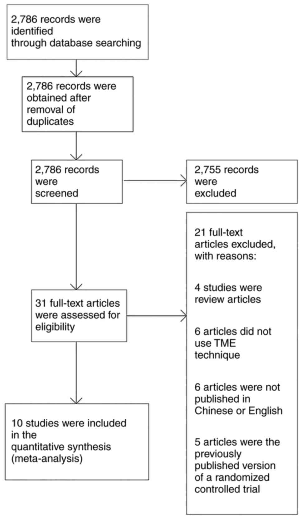
Study selection
A total of 2,786 articles were identified after
applying the search strategy in the aforementioned databases. Among
the studies that were identified through search of electronic
databases, 2,755 articles were not relevant to the topic of this
study and were excluded. The remaining 31 studies were relevant to
the topic of this study. After assessing their full texts, 21
articles were excluded (4 studies were review articles, 6 articles
did not use the TME technique, 6 articles were not published in
Chinese or English, and 5 articles were a previously published
version of an RCT). Finally, 10 studies were selected and involved
in the meta-analysis (Fig. 1)
(14–24). Tables
I and II show the baseline
characteristics of the included population. In total, 2,272
patients, including 1,101 cases in the LLND group and 1,171 cases
in the non-LLND group, were involved in the pooled analysis. In
most of the studies, the intervention involved performing TME along
with LLND for rectal cancer (15–19,21–24).
However, there was one study (21),
in which TME alone was performed as the intervention.
 | Table I.Baseline characteristics of the
included population. |
Table I.
Baseline characteristics of the
included population.
| First author,
year | Study design | Follow-up,
years | Stage | Intervention | Comparison | (Refs.) |
|---|
| Motoki, 2018 | Randomized
controlled trial | 5 | II or III | LLND+TME | TME | (15) |
| Zeng, 2019 | Retrospective
cohort | 5 | II or III | LLND+TME | TME | (16) |
| Aisu, 2018 | Retrospective
cohort | 5 | II or III | LLND+TME | TME | (17) |
| Zhang, 2020 | Retrospective
cohort | 3 | II or III | LLND+TME | TME | (18) |
| Fujita, 2012 | Retrospective
cohort | 3 | II or III | LLND+TME | TME | (19) |
| Ozawa, 2016 | Retrospective
cohort | 5 | II or III | TME | TME | (20) |
| Ogura, 2017 | Retrospective
cohort | 5 | II or III | LLND+TME | TME | (21) |
| Akiyoshi, 2014 | Retrospective
cohort | 5 | II or III | LLND+TME | TME | (22) |
| Lin, 2020 | Retrospective
cohort | 5 | II or III | LLND+TME | TME | (23) |
| Peacock, 2020 | Retrospective
cohort | 5 | II or III | LLND+TME | TME | (24) |
| Table II.Baseline characteristics of the
included population. |
Table II.
Baseline characteristics of the
included population.
| First author | Location of rectal
tumor | Age, years'
(experimental vs. control group) | Male sex
(experimental vs. control group) | (Refs.) |
|---|
| Motoki | Low | 67±14 vs.
63±12 | 167/231 vs.
112/156 | (15) |
| Zeng | Low | NR | 214/156 vs.
320/467 | (16) |
| Aisu | Low | 58±12 vs.
61±15 | 56/98 vs.
67/78 | (17) |
| Zhang | Low | 54±13 vs.
62±15 | 126/234 vs.
110/145 | (18) |
| Fujita | Low | 57±15 vs.
66±16 | 56/145 vs.
124/167 | (19) |
| Ozawa | Low | 63±12 | 145/123 vs.
112/134 | (20) |
| Ogura | Low | 58±13 vs.
61±11 | 78/99 vs.
89/102 | (21) |
| Akiyoshi | Low | 51±10 vs.
62±17 | NR | (22) |
| Lin | Low | 70±17 | 75/78 vs.
108/112 | (23) |
| Peacock | Low | 56±12 vs.
60±13 | 66/68 vs.
114/121 | (24) |
Risk of bias in the included
studies
The risk of bias and quality of the evidence of the
ROLARR trial (published as conference proceedings) as in a fully
published study: Sufficient methodological details were available
from the published protocol (25)
and authors (contacted) confirmed that no deviations from the
protocol had occurred in the conduct of the study. Of the included
trials, none had a high risk of bias on all items, while 2
(15,17) were scored as low in 6 out of 7
domains. Three trials (21,23,24)
were of unclear or low quality, with a high or unclear risk in at
least 1 of 7 domains. Fig. 2
highlights the outcomes of methodological quality assessment based
on the Cochrane tool and the Risk of Bias in Nonrandomized Studies
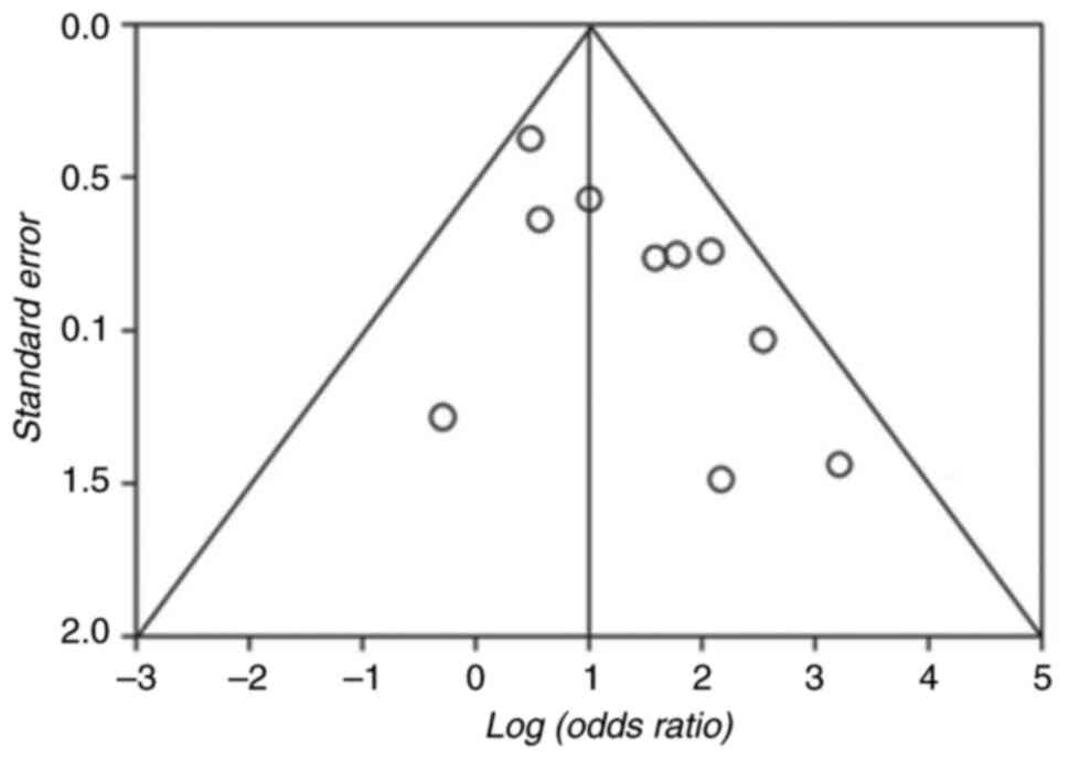
of Interventions assessment tool. Of note, as illustrated in
Fig. 3, the absence of studies that
include inconclusive or negative research findings may contribute
to publication bias in the current study.
Outcome synthesis for comparison
between the LLND+TME and TME groups
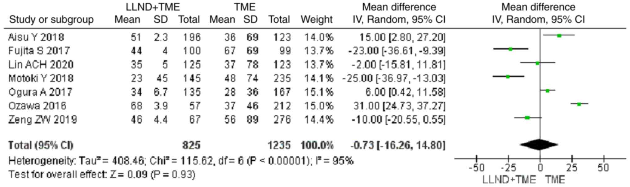
OS
Analysis of time-to-event outcome from 7 studies
revealed that cases in the LLND+TME group had longer OS than those
in the TME group, but the difference was not significant [OR=−0.73,
95% CI=(−16.26, 14.80), P>0.05]. The reported heterogeneity was
judged to be remarkable (Chi2=115.62, I2=95%)
(Fig. 4).
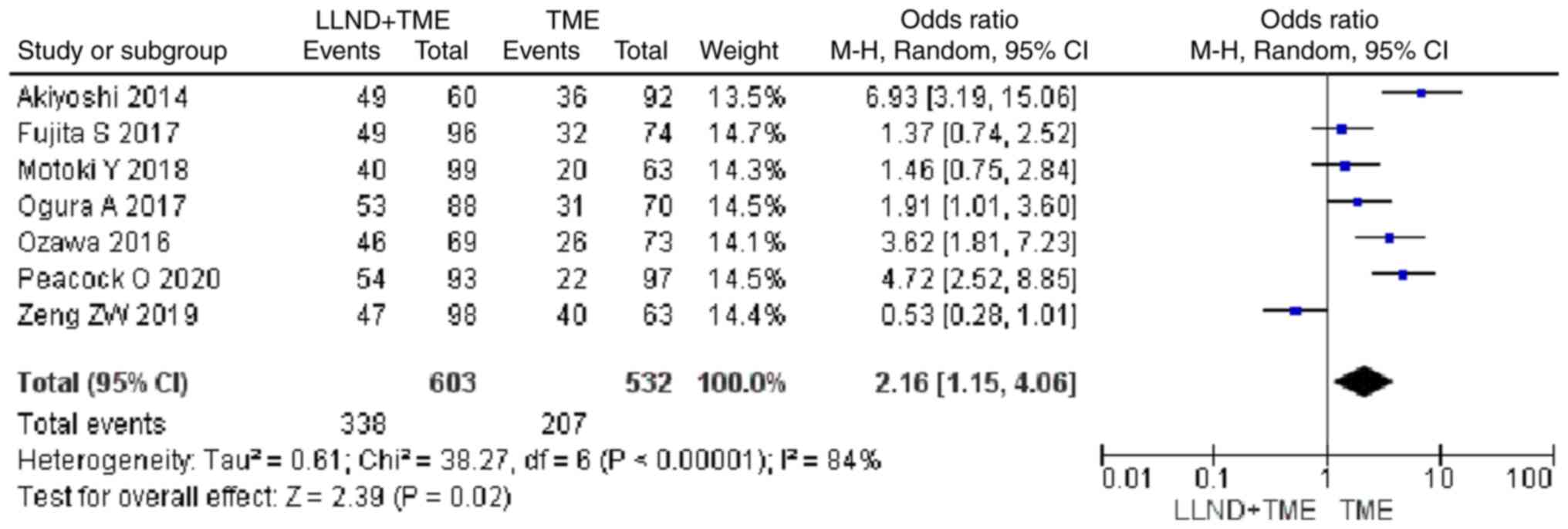
5-year survival rate
Analysis of 1,135 patients from 7 studies showed
that cases in the LLND+TME group had a higher 5-year survival rate
than those in the TME group [OR=2.16, 95% CI=(1.15, 4.06),
P<0.05]. The reported heterogeneity was judged to be high
(Chi2=38.27, I2=84%) (Fig. 5).
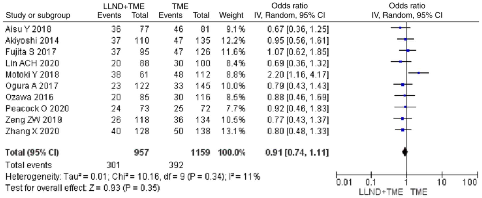
Local recurrence
Analysis of 2,116 patients from 10 studies showed
that patients in the either LLND+TME group or TME group were
similar in terms of local recurrence rate [OR=0.91, 95% CI=(0.74,
1.11), P>0.05]. The reported heterogeneity was judged to be low
(Chi2=10.16, I2=11%) (Fig. 6).
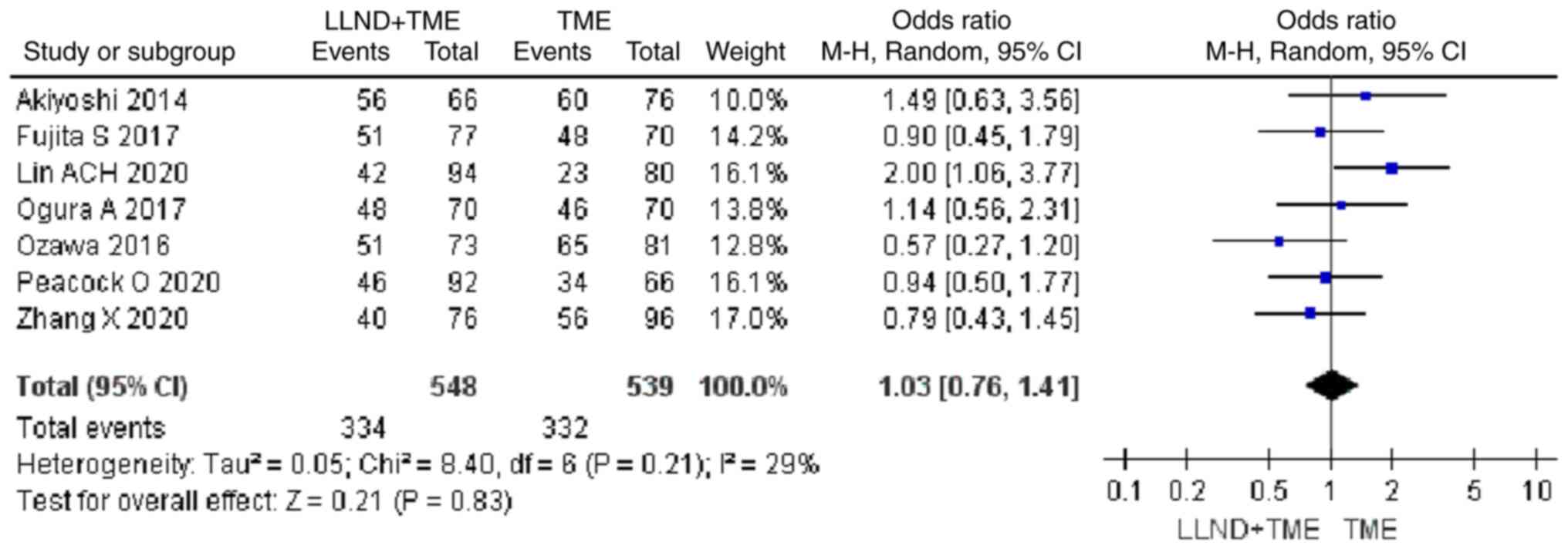
5-year DFS rate
Analysis of 1,931 patients from 7 studies showed
that patients in the either LLND+TME group or TME group were
similar in terms of 5-year DFS rate [OR=1.03, 95% CI=(0.76, 1.41),
P>0.05]. The reported heterogeneity was judged to be low
(Chi2=8.4, I2=29%) (Fig. 7).
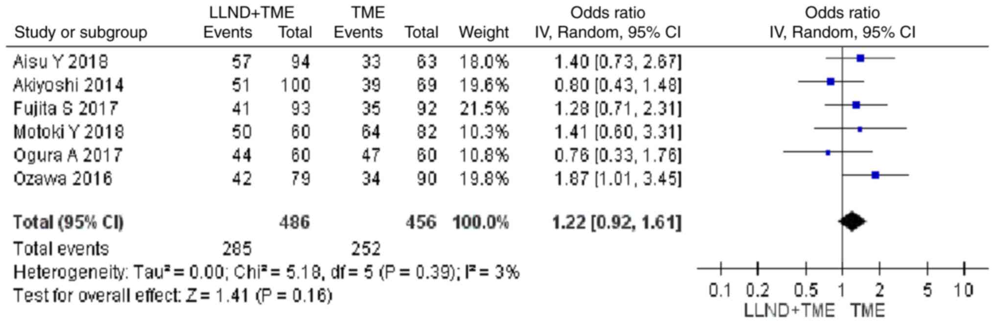
DFS at the maximum follow-up
Analysis of 942 patients from 6 studies indicated
that patients in the either LLND+TME group or TME group were
similar in terms of DFS at the maximum follow-up [OR=1.22, 95%
CI=(0.92, 1.61), P>0.05]. The reported heterogeneity was judged
to be low (Chi2=5.18, I2=3%) (Fig. 8).
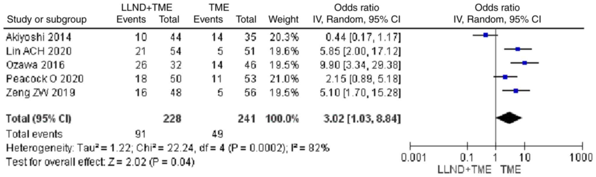
Urinary dysfunction
Analysis of 469 patients from 5 studies showed that
patients in the LLND+TME group had a higher risk of urinary
dysfunction compared with those in the TME group [OR=3.02, 95%
CI=(1.03, 8.84), P<0.05]. The reported heterogeneity was judged
to be high (Chi2=22.24, I2=82%) (Fig. 9).
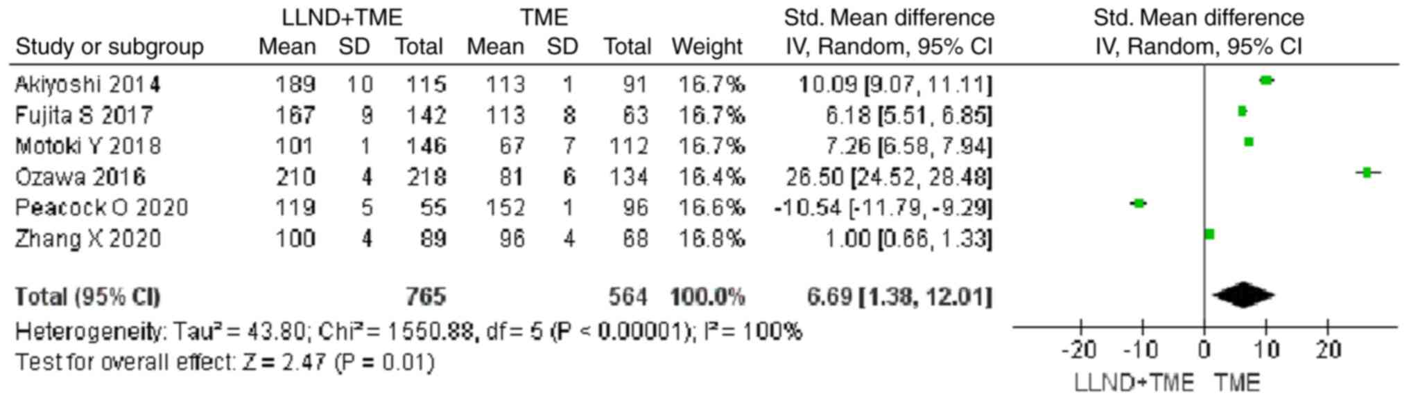
Operation time
Analysis of 1,329 patients from 6 studies revealed
that patients in the LLND+TME group had a longer operation time
compared with those in the TME group [OR=6.69, 95% CI=(1.38,
12.01), P<0.05]. The reported heterogeneity was judged to be
remarkable (Chi2=1,550.88, I2=100%) (Fig. 10).
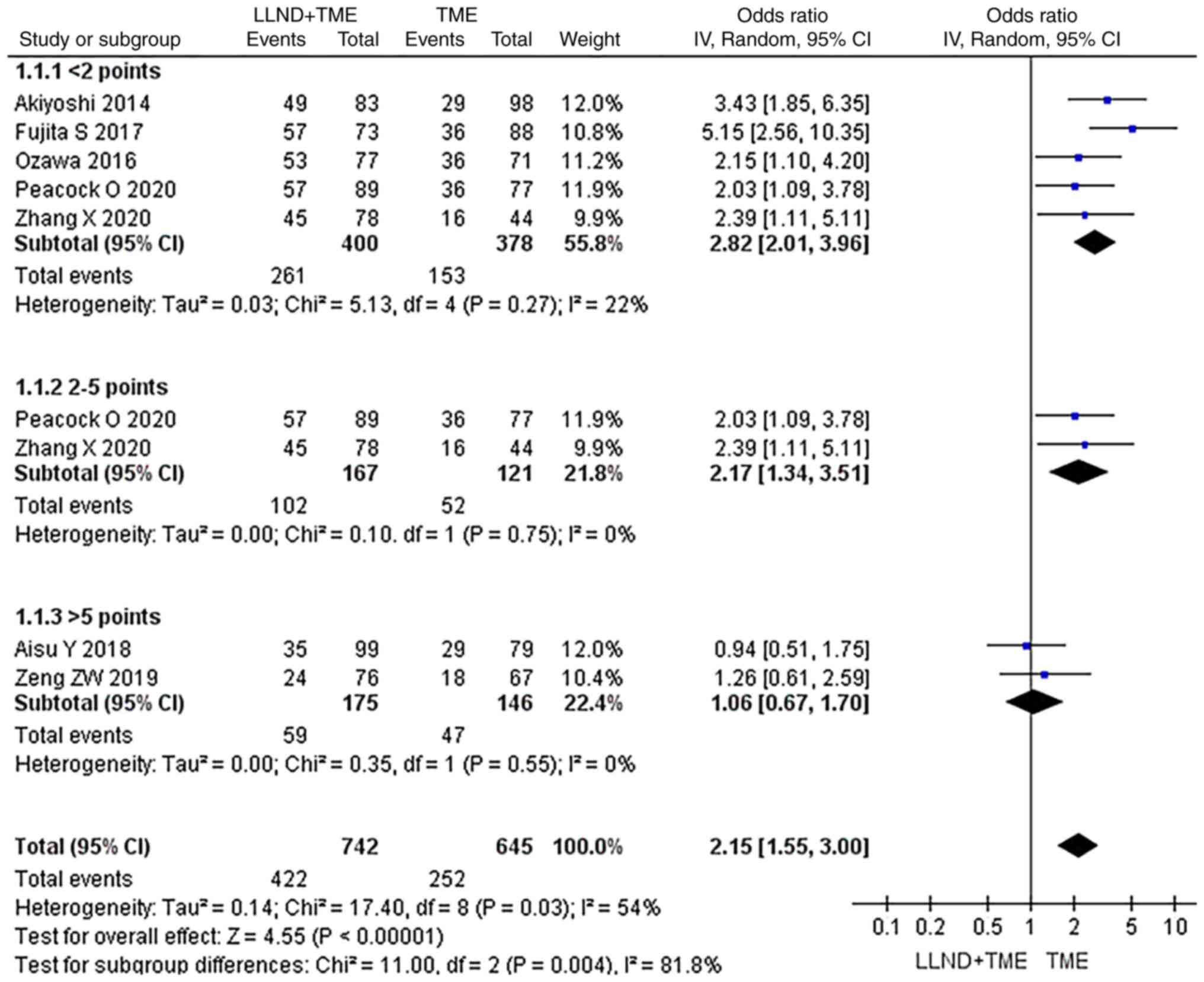
Subgroup analysis of OS data from
different research quality sources
In the subgroup analysis, the OS data from different
research sources were used and a subgroup analysis was performed
according to the influential factors of OS. They were divided into
subgroups with scores of <2, 2–5 and >5 points. In each
subgroup, patients' OS was analyzed and it was found that in all
subgroups, OS in the LLND+TME group was not significantly longer
than that in the TME group (P>0.05), indicating high reliability
of the analysis (Fig. 11).
Discussion
The results of the present meta-analysis indicated
that LLND did not increase OS, but increased the 5-year survival
rate compared to the non-LLND group. The results of the sensitivity
analysis were consistent, which suggested that the reliability of
these results was reasonable. However, Lin et al (23) also conducted a meta-analysis and
found that LLND could not improve the 5-year survival rate of
patients with rectal cancer. The different results may be explained
by the fact that the included subjects by Lin et al
(23) were mainly from Japan, which
were not representative, while the present study included eligible
patients across the world, including China. The surgical criteria
for rectal cancer in Japan were different from those in other
regions and countries. Of note, the surgical criteria for rectal
cancer in various countries can differ in several aspects. In the
US, the standard approach to surgical criteria for rectal cancer
primarily includes a combination of neoadjuvant therapies, such as
chemotherapy and radiation, followed by surgical intervention. The
objective is to shrink the tumor and increase the likelihood of
complete resection. Depending on factors, such as tumor size,
location and patient health, surgeons may perform surgical
procedures [e.g., low anterior resection (LAR) or abdominoperineal
resection (APR)]. The decision on whether to preserve the anal
sphincter or create a permanent colostomy depends on the extent of
the tumor and the patient's overall health (26). European countries, such as the UK,
Germany and France, generally follow similar principles to the US.
They emphasize neoadjuvant therapies to downsize the tumor before
surgery. Minimally invasive techniques (e.g., laparoscopic or
robotic-assisted surgeries) are frequently utilized to preserve
sphincter function whenever possible. The decision between
sphincter-preserving surgery and abdominoperineal resection depends
on the tumor's location, size and patient preference (27). In South Korea, there is a strong
emphasis on preserving anal sphincter function. Neoadjuvant
chemoradiotherapy is a common approach, followed by
sphincter-preserving surgeries, such as LAR. Surgeons often utilize
techniques that prioritize maintaining bowel continuity while
ensuring complete tumor removal (28). In Brazil, surgical criteria for
rectal cancer focus on individualized treatment plans. Neoadjuvant
therapies are employed and sphincter-preserving surgeries are
preferred whenever possible. The decision between LAR and APR
depends on the tumor's proximity to the anal sphincter and the
patient's overall health (29). In
Japan, the emphasis is often on sphincter-preserving surgeries to
maintain bowel function and quality of life. They tend to
prioritize preoperative chemoradiotherapy, followed by minimally
invasive techniques (30). Second,
the heterogeneity of the meta-analysis by Lin et al
(23) was high. Thus, the findings
of the present study are more convincing and representative of the
general world population, and the results of Lin et al
(23) are more representative of
the Japanese population. Besides, the compared results of the
perioperative situation in the present study showed that operation
time in the LLND group was longer and this may be associated with a
higher incidence of perioperative complications compared with the
non-LLND group.
Georgiou et al (31) compared conventional surgery and
expanded lymphadenectomy for rectal cancer in a meta-analysis in
2009, and in addition, various studies have summarized the effects
of generalized lymph node dissection on the prognosis of colorectal
cancer (32–34), including survival, DFS, operation
time, and urinary tract function. Certain studies included in the
meta-analysis by Georgiou et al (31) were excluded from the present study,
as they did not use extended lymphadenectomy. Surprisingly,
although the research by Georgiou et al (31) was conducted more than a decade ago,
the level of evidence remains unchanged due to the lack of
comparative data from RCTs. The data of non-RCTs may be complicated
by certain bias factors, as it was assumed that patients who
received LLND may have more severe disease than those who received
TME alone (35,36). Yang et al (37) conducted a meta-analysis of the role
of LLND in patients with rectal cancer after surgery. They found
that LLND increased the risk of urinary dysfunction and yielded a
longer operation time, which was consistent with the present
findings. However, their results suggested that LLND did not
contribute to longer 3 and 5-year cumulative OS. By contrast, the
present study indicated that the LLND group had longer OS and a
higher 5-year survival rate. This may be due to the inclusion of
different studies and populations. The present study included the
largest sample size and heterogeneity was controlled. There was no
language restriction related to Chinese. Furthermore, in the
present study, a subgroup analysis of different article quality
sources was also performed and the same conclusion was drawn. It
appears that the present findings were more promising. However, the
present results remain to be further verified by more
well-designed, large-scale studies.
The treatment of rectal cancer is different between
the West and the East (38,39). In Western countries with low-density
lipoprotein deficiency, it is common to use neoadjuvant
chemotherapy and radiation therapy before TME. Although the use of
neoadjuvant radiotherapy and chemotherapy has decreased the local
recurrence rate of low rectal cancer after surgery, metastasis to
lymph nodes in the lateral pelvis is still a major problem. In
Western countries, the standard approach for treating rectal cancer
is typically chemoradiotherapy followed by TME instead of LLND
(40,41). However, in Eastern countries,
particularly Japan, LLND is considered the preferred surgical
procedure for locally advanced lower rectal cancer (31). Numerous analyses conducted by
different authors have shown that LLND enhances OS rates and
reduces local recurrence, as evidenced by historical control
studies (42–44). The present study found that LLND has
a certain beneficial effect on the survival of patients. The
present subgroup analysis also confirmed that similar findings may
be obtained from data sources of articles with different quality.
The present subgroup analysis is currently the first one in which
the impact factors of articles and journals are considered, which
may decrease publication bias to a large extent. The current study
also confirmed that LLND is associated with a longer operation time
and the prognosis of urinary function may be adversely affected,
consistent with the results of Yang et al (37) and Georgiou et al (31). It is thought that urinary
dysfunction may be improved by minimally invasive procedures. This
suggests that LLND is recommended for colorectal cancer.
The present study also has certain limitations that
were summarized as follows: i) Due to the limitation of the number
and level of existing clinical trials, only two studies included in
the present analysis were prospective RCTs, and the remaining eight
were non-RCTs, which may produce selection bias, implementation
bias and measurement bias; ii) these studies were conducted in
different clinical centers and the surgery was performed by
different surgeons, which may produce bias; iii) the lack of
studies containing inconclusive or negative research results may be
a cause of publication bias in the present study. Consequently, the
present findings remain to be further verified by additional
studies.
In conclusion, LLND provided a specific advantage in
terms of increasing 5-year survival rate, while LLND was associated
with prolonged operation time and increased incidence of urinary
dysfunction.
Acknowledgements
Not applicable.
Funding
This study was funded by the Youth Fund of Peking University
International Hospital Research Grant (YN2020QN08).
Availability of data and materials
The datasets used and/or analyzed during the current
study are available from the corresponding author on reasonable
request.
Authors' contributions
Conception and design: BZ and YZ. Administrative
support: YZ; Provision of study materials or patients: BZ and YZ.
Collection and assembly of data: BZ, NN and YY. Data analysis and
interpretation and manuscript writing: BZ, NN, YY and YZ. BZ and YZ
confirm the authenticity of all the raw data (the pooled data). All
authors read and approved the final manuscript.
Ethics approval and consent to
participate
Not applicable.
Patient consent for publication
Not applicable.
Competing interests
The authors declare that they have no competing
interests.
References
|
1
|
Silva-Fisher JM, Dang HX, White NM, Strand
MS, Krasnick BA, Rozycki EB, Jeffers GGL, Grossman JG, Highkin MK,
Tang C, et al: Long non-coding RNA RAMS11 promotes metastatic
colorectal cancer progression. Nat Commun. 11:21562020. View Article : Google Scholar : PubMed/NCBI
|
|
2
|
Jones WF, Ahnen DJ and Schroy PC III:
Improving on-time colorectal cancer screening through lead time
messaging. Cancer. 126:247–252. 2020. View Article : Google Scholar : PubMed/NCBI
|
|
3
|
Carvalho MR, Reis RL and Oliveira JM:
Dendrimer nanoparticles for colorectal cancer applications. J Mater
Chem B. 8:1128–38. 2020. View Article : Google Scholar : PubMed/NCBI
|
|
4
|
Azzam N, AlRuthia Y, Alharbi O, Aljebreen
A, Almadi M, Alarfaj M, Alsaleh K, Almasoud A, Alsharidah M,
Alseneidi S, et al: Predictors of survival among colorectal cancer
patients in a low incidence area. Cancer Manag Res. 12:451–459.
2020. View Article : Google Scholar : PubMed/NCBI
|
|
5
|
Nahm SH, Blinman P, Butler S, Tan SYC and
Vardy J: Factors associated with fear of cancer recurrence in
breast and colorectal cancer survivors: A cross-sectional study of
cancer survivors. Asia Pac J Clin Oncol. 17:222–229. 2021.
View Article : Google Scholar : PubMed/NCBI
|
|
6
|
He E, Lew JB, Egger S, Banks E, Ward RL,
Beral V and Canfell K: Factors associated with participation in
colorectal cancer screening in Australia: Results from the 45 and
up study cohort. Prev Med. 106:185–193. 2018. View Article : Google Scholar : PubMed/NCBI
|
|
7
|
Shen MH, Chen LP, Ho TF, Shih YY, Huang
CS, Chie WC and Huang CC: Validation of the Taiwan Chinese version
of the EORTC QLQ-CR29 to assess quality of life in colorectal
cancer patients. BMC Cancer. 18:3532018. View Article : Google Scholar : PubMed/NCBI
|
|
8
|
Zeng K, Chen X, Xu MU, Liu X, Hu X, Xu T,
Sun H, Pan Y, He B and Wang S: CircHIPK3 promotes colorectal cancer
growth and metastasis by sponging miR-7. Cell Death Dis. 9:4172018.
View Article : Google Scholar : PubMed/NCBI
|
|
9
|
Crespo A, García-Suárez O, Fernández-Vega
I, Solis-Hernandez MP, García B, Castañón S and Quirós LM: Heparan
sulfate proteoglycans undergo differential expression alterations
in left sided colorectal cancer, depending on their metastatic
character. Bmc Cancer. 18:6872018. View Article : Google Scholar : PubMed/NCBI
|
|
10
|
Zhang J, Li XY, Hu P and Ding YS: lncRNA
NORAD contributes to colorectal cancer progression by inhibition of
miR-202-5p. Oncol Res. 26:1411–1418. 2018. View Article : Google Scholar : PubMed/NCBI
|
|
11
|
Liu Y, Chen X, Cheng R, Yang F, Yu M, Wang
C, Cui S, Hong Y, Liang H, Liu M, et al: The Jun/miR-22/HuR
regulatory axis contributes to tumourigenesis in colorectal cancer.
Mol Cancer. 17:112018. View Article : Google Scholar : PubMed/NCBI
|
|
12
|
Gao X, Wang C, Yu Y, Singh D, Yang L and
Zhou Z: Lateral lymph node dissection reduces local recurrence of
locally advanced lower rectal cancer in the absence of preoperative
neoadjuvant chemoradiotherapy: A systematic review and
meta-analysis. World J Surg Oncol. 18:3042020. View Article : Google Scholar : PubMed/NCBI
|
|
13
|
NHSN Patient Safety Component Manual, U.S,
. Centers for disease control and prevention, national healthcare
safety network. New York; USA: 2023
|
|
14
|
Hajibandeh S, Hajibandeh S, Matthews J,
Palmer L and Maw A: Meta-analysis of survival and functional
outcomes after total mesorectal excision with or without lateral
pelvic lymph node dissection in rectal cancer surgery. Surgery.
168:486–496. 2020. View Article : Google Scholar : PubMed/NCBI
|
|
15
|
Motoki Y, Sugimoto K, Sakisaka H, Nakano
K, Kan K, Nakaguchi K and Doi S: A case of laparoscopic surgery for
advanced rectal cancer with lateral lymph node metastasis resected
after neoadjuvant chemotherapy. Gan To Kagaku Ryoho. 45:2357–2359.
2018.(In Japanese). PubMed/NCBI
|
|
16
|
Zeng ZW, Zhang XW, Chen JJ, Huang L, Luo
SL and Kang L: Transanal lateral lymph node dissection surgery for
5 cases of mid-low rectal cancer. Zhonghua Wei Chang Wai Ke Za Zhi.
22:781–785. 2019.(In Chinese). PubMed/NCBI
|
|
17
|
Aisu Y, Kato S, Kadokawa Y, Yasukawa D,
Kimura Y, Takamatsu Y, Kitano T and Hori T: Feasibility of extended
dissection of lateral pelvic lymph nodes during laparoscopic total
mesorectal excision in patients with locally advanced lower rectal
cancer: A single-center pilot study after neoadjuvant chemotherapy.
Med Sci Monit. 24:3966–3977. 2018. View Article : Google Scholar : PubMed/NCBI
|
|
18
|
Zhang X, Zhang Y, Wei M, Wang M, Yang X,
Deng X and Wang Z: Letter to the editor regarding ‘Does adding
lateral pelvic lymph node dissection to neoadjuvant chemotherapy
improve outcomes in low rectal cancer?’. Int J Colorectal Dis.
35:2139–2140. 2020. View Article : Google Scholar : PubMed/NCBI
|
|
19
|
Fujita S, Mizusawa J, Kanemitsu Y, Saito
N, Kinugasa Y, Kanemitsu Y, Ohue M, Fujii S, Shiozawa M, Yamaguchi
T, et al: Postoperative morbidity and mortality after mesorectal
excision with and without lateral lymph node dissection for
clinical stage II or stage III lower rectal cancer (JCOG0212):
Results from a multicentre, randomised controlled, non-inferiority
trial. Lancet Oncol. 13:616–621. 2012. View Article : Google Scholar : PubMed/NCBI
|
|
20
|
Ozawa H, Kotake K, Hosaka M, Hirata A and
Sugihara K: Impact of lateral pelvic lymph node dissection on the
survival of patients with T3 and T4 low rectal cancer. World J
Surg. 40:1492–1499. 2016. View Article : Google Scholar : PubMed/NCBI
|
|
21
|
Ogura A, Akiyoshi T, Nagasaki T, Konishi
T, Fujimoto Y, Nagayama S, Fukunaga Y, Ueno M and Kuroyanagi H:
Feasibility of laparoscopic total mesorectal excision with extended
lateral pelvic lymph node dissection for advanced lower rectal
cancer after preoperative chemoradiotherapy. World J Surg.
41:868–875. 2017. View Article : Google Scholar : PubMed/NCBI
|
|
22
|
Akiyoshi T, Ueno M, Matsueda K, Konishi T,
Fujimoto Y, Nagayama S, Fukunaga Y, Unno T, Kano A, Kuroyanagi H,
et al: Selective lateral pelvic lymph node dissection in patients
with advanced low rectal cancer treated with preoperative
chemoradiotherapy based on pretreatment imaging. Ann Surg Oncol.
21:189–196. 2014. View Article : Google Scholar : PubMed/NCBI
|
|
23
|
Lin ACH, Hakim A, Kellish AS, Singh P,
Wozniak M, Kwiatt M, Gaughan J and Hong YK: Inguinal lymph node
dissection does not improve overall survival in anal cancer nodal
disease. J Surg Res. 255:13–22. 2020. View Article : Google Scholar : PubMed/NCBI
|
|
24
|
Peacock O and George JC: The landmark
series: Management of lateral lymph nodes in locally advanced
rectal cancer. Ann Surg Oncol. 27:2723–2731. 2020. View Article : Google Scholar : PubMed/NCBI
|
|
25
|
Jayne D, Pigazzi A, Marshall H, Croft J,
Corrigan N, Copeland J, Quirke P, West N, Edlin R, Hulme C and
Brown J: Robotic-assisted surgery compared with laparoscopic
resection surgery for rectal cancer: The ROLARR RCT. Southampton
(UK): NIHR Journals Library; 2019, View
Article : Google Scholar
|
|
26
|
Xynos E, Tekkis P, Gouvas N, Vini L,
Chrysou E, Tzardi M, Vassiliou V, Boukovinas I, Agalianos C,
Androulakis N, et al: Clinical practice guidelines for the surgical
treatment of rectal cancer: A consensus statement of the hellenic
society of medical oncologists (HeSMO). Ann Gastroenterol.
29:103–126. 2016.PubMed/NCBI
|
|
27
|
Babaei M, Balavarca Y, Jansen L, Gondos A,
Lemmens V, Sjövall A, Brge Johannesen T, Moreau M, Gabriel L,
Gonçalves AF, et al: Minimally invasive colorectal cancer surgery
in Europe: Implementation and outcomes. Medicine (Baltimore).
95:e38122016. View Article : Google Scholar : PubMed/NCBI
|
|
28
|
Jeong SY: Surgical management of
colorectal cancer. J Korean Med Assoc. 53:569–581. 2010. View Article : Google Scholar
|
|
29
|
Valadão M, Cesar D, Véo CAR, Araújo RO, do
Espirito Santo GF, Oliveira de Souza R, Aguiar S Jr, Ribeiro R, de
Castro Ribeiro HS, de Souza Fernandes PH and Oliveira AF: Brazilian
society of surgical oncology: Guidelines for the surgical treatment
of mid-low rectal cancer. J Surg Oncol. 125:194–216. 2022.
View Article : Google Scholar : PubMed/NCBI
|
|
30
|
Watanabe T, Muro K, Ajioka Y, Hashiguchi
Y, Ito Y, Saito Y, Hamaguchi T, Ishida H, Ishiguro M, Ishihara S,
et al: Japanese society for cancer of the colon and rectum (JSCCR)
guidelines 2016 for the treatment of colorectal cancer. Int J Clin
Oncol. 23:1–34. 2018. View Article : Google Scholar : PubMed/NCBI
|
|
31
|
Georgiou P, Tan E, Gouvas N, Antoniou A,
Brown G, Nicholls RJ and Tekkis P: Extended lymphadenectomy versus
conventional surgery for rectal cancer: A meta-analysis. Lancet
Oncol. 10:1053–1062. 2009. View Article : Google Scholar : PubMed/NCBI
|
|
32
|
Yokota M, Morikawa A, Nagahisa Y, Okabe M,
Kitagawa H and Kawamoto K: Combined use of curved scissors and the
soft coagulation system in robot-assisted lateral lymph node
dissection for rectal cancer-a video vignette. Colorectal Dis.
22:2359–2360. 2020. View Article : Google Scholar : PubMed/NCBI
|
|
33
|
Wong KY and Tan AM: Short term outcomes of
minimally invasive selective lateral pelvic lymph node dissection
for low rectal cancer. World J Gastrointest Surg. 12:178–189. 2020.
View Article : Google Scholar : PubMed/NCBI
|
|
34
|
Sabra H, Alimoradi M, El-Helou E, Azaki R,
Khairallah M and Kfoury T: Perforated sigmoid colon cancer
presenting as an incarcerated inguinal hernia: A case report. Int J
Surg Case Rep. 72:108–111. 2020. View Article : Google Scholar : PubMed/NCBI
|
|
35
|
Hojo D, Murono K, Nozawa H, Kawai K, Hata
K, Tanaka T and Ishihara S: Utility of a three-dimensional printed
pelvic model for lateral pelvic lymph node dissection. Int J
Colorectal Dis. 35:905–910. 2020. View Article : Google Scholar : PubMed/NCBI
|
|
36
|
Tsukamoto S, Fujita S and Kanemitsu Y:
Author response to: Beyond T, N and M: Can lateral lymph node
dissection treat tumour deposits in advanced low rectal carcinoma?
Br J Surg. 107:e2912020. View Article : Google Scholar : PubMed/NCBI
|
|
37
|
Yang X, Yang S, Hu T, Gu C, Wei M, Deng X,
Wang Z and Zhou Z: What is the role of lateral lymph node
dissection in rectal cancer patients with clinically suspected
lateral lymph node metastasis after preoperative chemoradiotherapy?
A meta-analysis and systematic review. Cancer Med. 9:4477–4489.
2020. View Article : Google Scholar : PubMed/NCBI
|
|
38
|
Young R, Rajkomar A, Smart P and Warrier
S: Robotic-assisted complete mesocolic excision, central vascular
ligation and para-aortic lymph node dissection in multifocal
carcinoid: A case report and technical description. Int J Surg Case
Rep. 67:262–266. 2020. View Article : Google Scholar : PubMed/NCBI
|
|
39
|
Asari M, Aoyama T, Koumori K, Uchiyama M,
Maezawa Y, Sawazaki S, Numata M, Tamagawa H, Sato T, Osaragi T, et
al: The efficacy of D3 lymph node dissection in elderly patients
with colorectal cancer. Gan To Kagaku Ryoho. 47:259–261. 2020.(In
Japanese). PubMed/NCBI
|
|
40
|
van Gijn W, Marijnen CA, Nagtegaal ID,
Kranenbarg EM, Putter H, Wiggers T, Rutten HJ, Påhlman L, Glimelius
B and van de Velde CJ; Dutch Colorectal Cancer Group, :
Preoperative radiotherapy combined with total mesorectal excision
for resectable rectal cancer: 12-Year follow-up of the multicentre,
randomised controlled TME trial. Lancet Oncol. 12:575–582. 2011.
View Article : Google Scholar : PubMed/NCBI
|
|
41
|
Sauer R, Becker H, Hohenberger W, Rödel C,
Wittekind C, Fietkau R, Martus P, Tschmelitsch J, Hager E, Hess CF,
et al: Preoperative versus postoperative chemoradiotherapy for
rectal cancer. N Engl J Med. 351:1731–1740. 2004. View Article : Google Scholar : PubMed/NCBI
|
|
42
|
Takahashi T, Ueno M, Azekura K and Ohta H:
Lateral node dissection and total mesorectal excision for rectal
cancer. Dis Colon Rectum. 43 (10 Suppl):S59–S68. 2000. View Article : Google Scholar : PubMed/NCBI
|
|
43
|
Moriya Y, Sugihara K, Akasu T and Fujita
S: Importance of extended lymphadenectomy with lateral node
dissection for advanced lower rectal cancer. World J Surg.
21:728–732. 1997. View Article : Google Scholar : PubMed/NCBI
|
|
44
|
Ueno H, Mochizuki H, Hashiguchi Y,
Ishiguro M, Miyoshi M, Kajiwara Y, Sato T, Shimazaki H and Hase K:
Potential prognostic benefit of lateral pelvic node dissection for
rectal cancer located below the peritoneal reflection. Ann Surg.
245:80–87. 2007. View Article : Google Scholar : PubMed/NCBI
|