Introduction
Breast cancer (BC) is the most commonly diagnosed
cancer worldwide, with an estimated 2.3 million new cases (11.7%)
and has ranked the fifth leading cause (6.9%) of cancer death
(1). Axillary lymph node (ALN)
status is a key prognostic factor for patients with early-stage
invasive BC and influences the clinical therapeutic schedule. The
guidelines of the American Society of Clinical Oncology and the
Z0011 trial have demonstrated that patients with early-stage BC
with <2 sentinel lymph node (SLN) metastases can be spared ALN
dissection (ALND) (2,3). SLN biopsy is still the current
standard approach for the assessment of ALN status. However, it has
been criticized for its low efficiency and invasive complications,
including upper arm numbness, lymphedema, nerve damage and hematoma
(4,5). Therefore, developing an accurate and
non-invasive method to predict ALN status before surgery remains a
challenge.
Ultrasound (US) has potential clinical benefits for
performing breast examinations due to low cost, convenience and
lack of radiation. Axillary US examination is a simple and
convenient diagnostic method to detect ALNM but its value is
limited due to its low sensitivity (26.4–69.5%), especially for
patients with a minor ALN metastatic burden (6). US-guided fine needle aspiration is an
invasive procedure and its high accuracy is influenced by the
inadequate sample collection (7).
‘Radiomics’ has attracted increased attention in
(8–10). Several studies (8–10) have
predicted the likelihood of ALNM based on US radiomics. However,
certain radiomics analyses are limited by single-modal grayscale US
images, and the values of the area under the receiver operator
characteristic (ROC) curve (AUC) were found to be relatively low
(0.71–0.73) (8,9). A recent study reported construction of
a shear wave elastography (SWE) US-based radiomics nomogram that
yielded a moderate predictive ability, with a C-index of 0.817
(10). However, the most
significant SWE radiomics features only reflected the intratumoral
heterogeneity of the tumor. This does not embody the value of SWE
sufficiently as SWE is known for its ability to show both intrinsic
and peritumoral characteristics of the tumor via qualitative and
quantitative stiffness parameters (11–13).
Huang et al (14) designed a
multimodal US (MMUS)-based auto-weighting framework for breast
cancer classification. However, the MMUS analysis only involved
B-mode, Doppler, strain elastography (SE) and SWE US and there was
lack of valuable contrast-enhanced (CEUS) data and may be not
entirely comprehensive. In addition, MMUS analysis was only used to
classify BC, not to predict ALNM, and therefore was insufficient to
guide clinical strategy. Other researchers have proposed deep
learning radiomics models and reported favorable predictive
efficacy (15,16). However, the ‘black box’ pattern and
specialized algorithms of deep learning are hard to reproduce
(14–16). Moreover, radiomics features, such as
texture or wavelet, are both obscure and complex for clinicians to
understand. Finally, the extraction procedures are not easy to
apply in clinical practice (14,16).
The aim of the present study was to establish and assess a
comprehensive, concise and easy-to-use predictive tool using the
combined conventional US, SE, SWE and CEUS features for
preoperative ALNM risk estimation in early-stage IBC.
Materials and methods
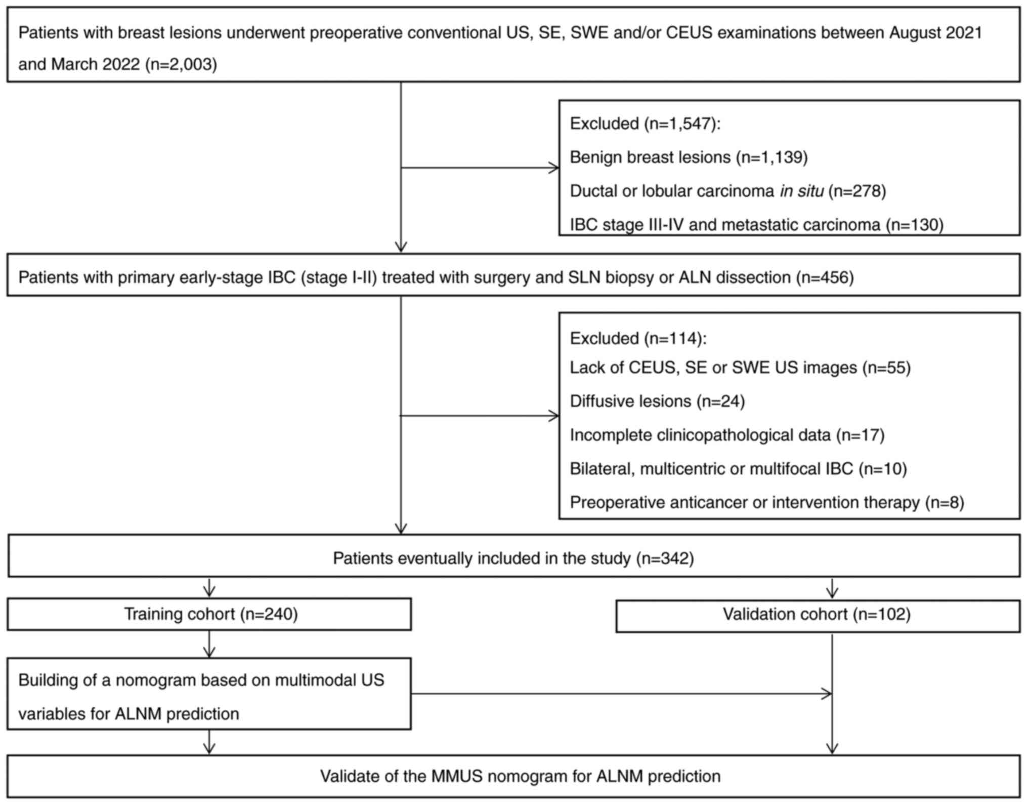
Study design and patients
The present study was a retrospective sub-analysis
of data acquired from a prospective study (17). It was conducted in accordance with
the Declaration of Helsinki and approved by the Ethics Committee of
Ruijin Hospital, School of Medicine, Shanghai Jiao Tong University
(Shanghai, China). Written informed consent was obtained from
patients. From August 2021 to March 2022, 2,003 patients with
breast lesions in the Comprehensive Breast Health Center at Ruijin
Hospital underwent preoperative conventional US, SE, SWE and/or
CEUS examination. Conventional US + elastography was the first-line
breast US examination protocol for all patients. For patients with
a suspected breast tumor, CEUS examination was performed for more
accurate differential diagnosis, to assess the extent of the tumor
or to select necessary tumor regions for biopsy. A total of 456
patients was enrolled. The inclusion criteria were as follows: i)
Diagnosis of primary early-stage IBC (stage I–II); ii) performance
of breast surgery and SLN biopsy or ALND and iii) presence of
preoperative multimodal US images of breast tumors and conventional
US images of ALN. The exclusion criteria were as follows: i) Lack
of CEUS or SE or SWE US images; ii) preoperative anticancer
(neoadjuvant therapy or chemotherapy) or intervention therapy
(biopsy or ablation) prior to US examination; iii) diagnosis of
bilateral, multicentric or multifocal IBC; iv) presence of
diffusive lesions and v) insufficient US image quality or
incomplete clinicopathological data.
A total of 342 patients (all female; mean age,
51.15±11.62 years; range, 32–88 years) passed the quality control
for final analysis and were randomly divided (7:3) into a training
(n=240) and validation cohort (n=102), according to the Transparent
Reporting of a multivariable prediction model for Individual
Prognosis or Diagnosis reporting guideline (18). Fig.
1 shows the recruitment of patients and the study design.
Clinical and pathological data
Data were obtained from the medical records. The
clinical data included patient age, symptoms, family history of BC,
history of hormone therapy and clinical tumor stage. Pathological
data included histological type, tumor grade, estrogen receptor
(ER), progesterone receptor (PR), human epidermal growth factor
receptor 2 (HER2) status, tumor proliferation rate (Ki67 levels)
and ALN status (positive or negative). All pathological
macrometastases, micrometastases or isolated tumor cells of ALNs
were defined as node-positive. A cut-off value for Ki67+
was established at 20% (19).
Molecular subtypes were classified as luminal A or B,
HER2+ and triple-negative BC, according to the
expression of ER, PR and HER2 (20).
US image acquisition and US-depicted
ALN status
All preoperative breast and axillary US examinations
were performed by two experienced radiologists (JJY and YZ) with
>15 combined years of experience in breast US and 8 combined
years of experience in performing SE, SWE and CEUS of breast
lesions, using a 3–11 MHz linear probe (Resona 8, Shenzhen Mindray
Bio-Medical Electronics Co., Ltd.). The conventional US variables,
such as tumor shape, margin, orientation, echogenicity, posterior
acoustic pattern, calcification and vascularity status, were
assessed according to the Breast Imaging Reporting and Data System
(BI-RADS) (21). The maximum size
of the breast tumor, measured by US, and the US BI-RADS category
were also assessed. The lymph node was depicted as positive if it
had ≥1 of the following suspicious US characteristics: Circular
shape, cortical thickening, calcification or cystic change, no
fatty hilum or no hilar blood flow (22,23).
SE features were classified as soft or hard as described previously
(24). The stiff rim sign in SWE
was reported as absent or present according to Zhou et al
(25). The maximum
(Emax), mean (Emean) and minimum
(Emin) elastic modulus and elastic modulus standard
deviation (ESD), calculated automatically by the US
system, were recorded. CEUS examinations were performed according
to the American Institute of Ultrasound in Medicine practice
guidelines for performing breast CEUS (26). Contrast agent SonoVue®
(Bracco S.p.A.) was reconstituted by addition of 5 ml sterile
normal saline and 25 mg lyophilized powder. Following hand
agitation, 2.4 ml contrast agent was injected through a 21-G
catheter via a peripheral vein in a bolus fashion, followed by a
flush of 5 ml saline solution. The entire CEUS process for each
patient was recorded immediately following injection of the
contrast agent and lasted for 180 sec. US images and video clips
were stored on the hard disk for subsequent analysis. The
qualitative features assessed included degree of enhancement
(hypo/isoenhancement or hyperenhancement), perfusion defect (absent
or present) and radial vessel at the tumor margin (absent or
present). Quantitative parameters, including arrival time (AT; the
time point when the microbubble arrived at the lesion), time to
peak (TTP; time point when the contrast intensity reached its
peak), peak intensity (PI; maximum intensity of the time-intensity
curve), ascending slope (AS; slope from the beginning of focal
perfusion to the peak point on the curve), descending slope (DS;
curve descent slope) and area under the time-intensity curve
(AUTIC; total volume of blood in the region of interest) were also
recorded.
Observer variability for evaluation of
the US features
In a subset of 60 randomly selected breast lesions
and 30 ALNs, interobserver variability in conventional US, SE, SWE
and CEUS of breast lesions and conventional US of ALNs were
assessed separately by two radiologists (JJY and YZ). At 1 month
after the first evaluation, one observer (JJY) reviewed all images
of the same 60 lesions and 30 ALNs for the calculation of
intra-observer variability.
Statistical analysis
Statistical analyses were performed using SPSS
(version 25.0; IBM Corp.) and R software (version 4.1.3; The R
Foundation). Continuous variables were compared using independent
t-test. Categorical variables were compared using the χ2
or Fisher's exact test. The Cohen κ statistic was used to assess
interobserver agreement. All clinicopathological factors and
conventional US features potentially associated with ALNM
(P<0.05 in univariate analysis) were used to construct a
clinicopathological model and a conventional US model. A nomogram
was formulated based on multimodal US variables by the results of
multivariate logistic analysis. Calibration was assessed using the
calibration curve with 1,000 bootstrap samples to decrease overfit
bias. The predictive performances were compared with AUC.
Additionally, net reclassification improvement (NRI) was applied to
evaluate incremental value, and decision curve analysis (DCA) to
investigate the clinical usefulness of the nomogram. The ‘rms’
package was used for nomogram and calibration curve construction,
the ‘rmda’ package for DCA and the ‘nricens’ package was used for
NRI calculation. All packages were performed from R software
(version 4.1.3; The R Foundation). P<0.05 was considered to
indicate a statistically significant difference.
Results
Clinicopathological
characteristics
A total of 342 patients were finally included in the
present study and were randomly divided into the training (n=240)
and validation (n=102) cohorts (Fig.
1). There were no significant differences in ALN positivity
between cohorts [training cohort, 35.4% (85/240); validation
cohort, 37.3% (38/102)]. The clinicopathological and US variables
were not significantly different between cohorts, demonstrating no
selection bias in the random allocation process.
Predictive factors associated with
ALNM
Table I presents the
distribution of clinicopathological characteristics in relation to
ALN status. The results revealed that older age, clinical T2 stage
compared with T1 stage, and high histological grade compared with
low or intermediate grade were significantly more likely to be
associated with ALNM in both cohorts. The other clinicopathological
factors, such as symptoms, family history of BC, history of hormone
therapy, ER, PR and HER2 status, Ki67 levels and molecular subtypes
showed no significant differences between the ALN-positive and
-negative groups. Univariate analysis of conventional US features
revealed that tumors with a larger maximum size, heterogeneous
echogenicity in comparison with homogeneous echogenicity, tumor
with calcification in comparison with no calcification, high
vascularity in comparison with absent or low vascularity and
BI-RADS category 5 in comparison with BI-RADS category 3–4C were
risk factors significantly associated with ALNM. However, tumor
shape, margin, orientation and posterior acoustic pattern showed no
significant predictive value for ALNM. In the training cohort,
US-depicted suspicious ALN positive were 65 patients, the
pathological results of ALNM were 85 patients, thus the diagnostic
sensitivity was 76.47% (65/85). US-depicted suspicious ALN negative
were 102 patients, the pathological results of ALN negative were
155 patients, thus the diagnostic specificity was 65.80% (102/155).
In the validation cohort, US-depicted suspicious ALN positive were
30 patients, the pathological results of ALNM were 38 patients, the
diagnostic sensitivity was 78.95% (30/38). US-depicted suspicious
ALN negative were 41 patients, the pathological results of ALN
negative were 64 patients, the diagnostic specificity was 64.06%
(41/64) (Table II).
 | Table I.Univariate analysis of clinical and
pathological characteristics associated with axillary lymph node
status. |
Table I.
Univariate analysis of clinical and
pathological characteristics associated with axillary lymph node
status.
| A, Training cohort
(n=240) |
|---|
|
|---|
|
| Axillary lymph node
status |
|
|---|
|
|
|
|
|---|
| Characteristic | Negative
(n=155) | Positive
(n=85) | P-value |
|---|
| Age, years (%) | 47.05±10.23 | 54.10±11.34 | <0.001 |
|
<40 | 24 (15.48) | 16 (18.82) |
|
|
40-60 | 99 (63.87) | 30 (35.30) |
|
|
>60 | 32 (20.65) | 39 (45.88) |
|
| Symptoms (%) |
|
| 0.338 |
|
Palpable mass | 136 (87.74) | 78 (91.76) |
|
|
Other | 19 (12.26) | 7 (8.24) |
|
| Family history of
breast cancer (%) |
|
| 0.507 |
| No | 125 (80.65) | 65 (76.47) |
|
|
Yes | 30 (19.35) | 20 (23.53) |
|
| History of hormone
therapy (%) |
|
| 0.390 |
| No | 124 (80.00) | 72 (84.71) |
|
|
Yes | 31 (20.00) | 13 (15.29) |
|
| Clinical T stage
(%) |
|
| 0.043 |
| T1 | 94 (60.65) | 40 (47.06) |
|
| T2 | 61 (39.35) | 45 (52.94) |
|
| Histological type
(%) |
|
| 0.874 |
|
Ductal | 123 (79.35) | 69 (81.18) |
|
| Lobular
or mixed | 27 (17.42) | 13 (15.29) |
|
|
Other | 5 (3.23) | 3 (3.53) |
|
| Histological grade
(%) |
|
| 0.002 |
|
Low | 11 (7.09) | 5 (5.88) |
|
|
Intermediate | 85 (54.84) | 26 (30.59) |
|
|
High | 59 (38.07) | 54 (63.53) |
|
| ER status (%) |
|
| 0.868 |
|
Negative | 27 (17.42) | 14 (16.47) |
|
|
Positive | 128 (82.58) | 71 (83.53) |
|
| PR status (%) |
|
| 0.990 |
|
Negative | 40 (25.81) | 22 (25.88) |
|
|
Positive | 115 (74.19) | 63 (74.12) |
|
| HER2 status
(%) |
|
| 0.885 |
|
Negative | 123 (79.35) | 57 (67.06) |
|
|
Positive | 32 (20.65) | 28 (32.94) |
|
| Ki67 levels
(%) |
|
| 0.746 |
|
≤20% | 45 (29.03) | 23 (27.06) |
|
|
>20% | 110 (70.97) | 62 (72.94) |
|
| Molecular subtype
(%) |
|
| 0.883 |
| Luminal
A | 28 (18.06) | 16 (18.82) |
|
| Luminal
B | 95 (61.29) | 51 (60.00) |
|
|
HER2-positive | 23 (14.84) | 11 (12.94) |
|
| Triple
negative | 9 (5.81) | 7 (8.24) |
|
|
| B, Validation
cohort (n=102) |
|
|
| Axillary lymph
node status |
|
|
|
|
|
|
Characteristic | Negative
(n=64) | Positive
(n=38) | P-value |
|
| Age, years (%) | 48.21±11.51 | 54.37±12.00 | <0.001 |
|
<40 | 10 (15.63) | 7 (18.42) | 0.027 |
|
40-60 | 37 (57.81) | 12 (31.58) |
|
|
>60 | 17 (26.56) | 19 (50.00) |
|
| Symptoms (%) |
|
| 0.765 |
|
Palpable mass | 56 (87.50) | 34 (89.47) |
|
|
Other | 8 (12.50) | 4 (10.53) |
|
| Family history of
breast cancer (%) |
|
| 0.922 |
| No | 50 (78.13) | 30 (78.95) |
|
|
Yes | 14 (21.87) | 8 (21.05) |
|
| History of hormone
therapy (%) |
|
| 0.804 |
| No | 51 (79.69) | 29 (76.32) |
|
|
Yes | 13 (20.31) | 9 (23.68) |
|
| Clinical T stage
(%) |
|
| 0.036 |
| T1 | 40 (62.50) | 16 (42.11) |
|
| T2 | 24 (37.50) | 22 (57.89) |
|
| Histological type
(%) |
|
| 0.971 |
|
Ductal | 51 (79.69) | 31 (81.58) |
|
| Lobular
or mixed | 11 (17.19) | 6 (15.79) |
|
|
Other | 2 (3.12) | 1 (2.63) |
|
| Histological grade
(%) |
|
| 0.026 |
|
Low | 5 (7.81) | 2 (5.26) |
|
|
Intermediate | 35 (54.69) | 12 (31.58) |
|
|
High | 24 (37.50) | 24 (63.16) |
|
| ER status (%) |
|
| 0.967 |
|
Negative | 12 (18.75) | 7 (18.42) |
|
|
Positive | 52 (81.25) | 31 (81.58) |
|
| PR status (%) |
|
| 0.747 |
|
Negative | 17 (26.56) | 8 (21.05) |
|
|
Positive | 47 (73.43) | 30 (78.95) |
|
| HER2 status
(%) |
|
| 0.937 |
|
Negative | 45 (70.31) | 27 (71.05) |
|
|
Positive | 19 (29.69) | 11 (28.95) |
|
| Ki67 levels
(%) |
|
| 0.929 |
|
≤20% | 18 (28.13) | 11 (28.95) |
|
|
>20% | 46 (71.87) | 27 (71.05) |
|
| Molecular subtype
(%) |
|
| 0.953 |
| Luminal
A | 12 (18.75) | 6 (15.79) |
|
| Luminal
B | 38 (59.38) | 23 (60.53) |
|
|
HER2-positive | 9 (14.06) | 5 (13.16) |
|
| Triple
negative | 5 (7.81) | 4 (10.52) |
|
| Table II.Univariate analysis of conventional
ultrasound features associated with axillary lymph node status. |
Table II.
Univariate analysis of conventional
ultrasound features associated with axillary lymph node status.
| A, Training cohort
(n=240) |
|---|
|
|---|
|
| Axillary lymph node
status |
|
|---|
|
|
|
|
|---|
| Feature | Negative
(n=155) | Positive
(n=85) | P-value |
|---|
| Maximum size,
cm | 2.21±1.20 | 2.93±1.82 | <0.001 |
|
<2 | 38 (24.52) | 4 (4.71) | <0.001 |
|
2-3 | 94 (60.64) | 25 (29.41) |
|
|
3-5 | 23 (14.84) | 56 (65.88) |
|
| Shape (%) |
|
| 0.366 |
|
Oval/round | 39 (25.16) | 17 (20.00) |
|
|
Irregular | 116 (74.84) | 68 (80.00) |
|
| Margin (%) |
|
| 0.538 |
|
Circumscribed | 20 (12.90) | 8 (9.41) |
|
|
Indistinct | 58 (37.42) | 33 (38.82) |
|
|
Angulation | 45 (29.03) | 24 (28.24) |
|
|
Microlobulation | 9 (5.81) | 6 (7.06) |
|
|
Spiculation | 23 (14.84) | 14 (16.47) |
|
| Orientation
(%) |
|
| 0.691 |
|
Parallel | 136 (87.74) | 77 (90.59) |
|
| Not
parallel | 19 (12.26) | 8 (9.41) |
|
|
Echogenicity |
|
| <0.001 |
|
Homogeneous | 110 (70.97) | 19 (22.35) |
|
|
Heterogeneous | 45 (29.03) | 66 (77.65) |
|
| Posterior acoustic
(%) |
|
| 0.270 |
| No
change | 39 (25.16) | 17 (20.00) |
|
|
Enhancement | 23 (14.84) | 21 (24.70) |
|
| Mixed
change | 33 (21.29) | 15 (17.65) |
|
|
Shadow | 60 (38.71) | 32 (37.65) |
|
| Calcification
(%) |
|
| <0.001 |
|
Absent | 117 (75.48) | 35 (41.18) |
|
|
Present | 38 (24.52) | 50 (58.82) |
|
| Vascularity
(%) |
|
| 0.002 |
|
Absent/low | 60 (38.71) | 16 (18.82) |
|
|
High | 95 (61.29) | 69 (81.18) |
|
| BI-RADS category
(%) |
|
| <0.001 |
| 3 | 3 (1.94) | 1 (1.18) |
|
| 4A | 24 (15.48) | 6 (7.06) |
|
| 4B | 40 (25.81) | 13 (15.29) |
|
| 4C | 59 (38.06) | 23 (27.06) |
|
| 5 | 29 (18.71) | 42 (49.41) |
|
| US-depicted
suspicious |
|
| <0.001 |
| ALN (%) |
|
|
|
|
Negative | 102 (65.81) | 20 (23.53) |
|
|
Positive | 53 (34.19) | 65 (76.47) |
|
|
| B, Validation
cohort (n=102) |
|
|
| Axillary lymph
node status |
|
|
|
|
|
| Feature | Negative
(n=64) | Positive
(n=38) | P-value |
|
| Maximum size, cm
(%) | 2.14±1.32 | 3.20±1.90 | <0.001 |
|
<2 | 14 (21.88) | 2 (5.26) | <0.001 |
|
2-3 | 40 (62.50) | 10 (26.32) |
|
|
4-5 | 10 (15.62) | 26 (68.42) |
|
| Shape (%) |
|
| 0.677 |
|
Oval/round | 14 (21.88) | 7 (18.42) |
|
|
Irregular | 50 (78.12) | 31 (81.58) |
|
| Margin (%) |
|
| 0.645 |
|
Circumscribed | 6 (9.38) | 4 (10.53) |
|
|
Indistinct | 24 (37.50) | 13 (34.21) |
|
|
Angulation | 17 (26.56) | 10 (26.32) |
|
|
Microlobulation | 4 (6.25) | 3 (7.89) |
|
|
Spiculation | 13 (20.31) | 8 (21.05) |
|
| Orientation
(%) |
|
| 0.734 |
|
Parallel | 54 (84.4) | 33 (86.84) |
|
| Not
parallel | 10 (15.6) | 5 (13.16) |
|
| Echogenicity
(%) |
|
| <0.001 |
|
Homogeneous | 45 (70.31) | 8 (21.05) |
|
|
Heterogeneous | 19 (29.69) | 30 (78.95) |
|
| Posterior acoustic
(%) |
|
| 0.489 |
| No
change | 15 (23.44) | 7 (18.42) |
|
|
Enhancement | 9 (14.06) | 10 (26.32) |
|
| Mixed
change | 14 (21.88) | 7 (18.42) |
|
|
Shadow | 26 (40.62) | 14 (36.84) |
|
| Calcification
(%) |
|
| 0.002 |
|
Absent | 45 (70.31) | 15 (39.47) |
|
|
Present | 19 (29.69) | 23 (60.53) |
|
| Vascularity
(%) |
|
| 0.019 |
|
Absent/low | 23 (35.94) | 6 (15.79) |
|
|
High | 41 (64.06) | 32 (84.21) |
|
| BI-RADS category
(%) |
|
| <0.001 |
| 3 | 2 (3.13) | 1 (2.63) |
|
| 4A | 12 (18.75) | 2 (5.26) |
|
| 4B | 15 (23.44) | 6 (15.79) |
|
| 4C | 25 (39.06) | 10 (26.32) |
|
| 5 | 10 (15.62) | 19 (50.00) |
|
| US-depicted
suspicious |
|
| <0.001 |
| ALN (%) |
|
|
|
|
Negative | 41 (64.06) | 8 (21.05) |
|
|
Positive | 23 (35.94) | 30 (78.95) |
|
Additionally, the presence of stiff rim sign,
perfusion defect and radial vessel in comparison with absent were
also risk factors significantly associated with ALNM. However, the
other MMUS variables, including SE features, Emax,
Emean, Emin, ESD, AT, TTP, PI, AS,
DS and AUTIC, demonstrated no significant association with ALNM
(Table III).
| Table III.Univariate analysis of SE, SWE and
contrast-enhanced ultrasound features associated with axillary
lymph node status. |
Table III.
Univariate analysis of SE, SWE and
contrast-enhanced ultrasound features associated with axillary
lymph node status.
| A, Training cohort
(n=240) |
|---|
|
|---|
|
| Axillary lymph node
status |
|
|---|
|
|
|
|
|---|
| Feature | Negative
(n=155) | Positive
(n=85) | P-value |
|---|
| SE (%) |
|
| 0.817 |
|
Soft | 22 (14.19) | 13 (15.29) |
|
|
Hard | 133 (85.81) | 72 (84.71) |
|
| Stiff rim sign
(%) |
|
| <0.001 |
|
Absent | 105 (67.74) | 25 (29.41) |
|
|
Present | 50 (32.26) | 60 (70.59) |
|
| SWE, kPa |
|
| 0.764 |
|
Emax | 135.15±31.08 | 133.19±29.35 |
|
|
Emin | 3.72±1.85 | 3.61±1.71 |
|
|
Emean | 24.55±8.91 | 22.23±8.06 |
|
|
ESD | 18.26±7.69 | 17.54±7.15 |
|
| Enhancement degree
(%) |
|
| 0.182 |
|
Hypo/isoenhancement | 39 (25.16) | 15 (17.65) |
|
|
Hyperenhancement | 116 (74.84) | 70 (82.35) |
|
| Perfusion defect
(%) |
|
| <0.001 |
|
Absent | 116 (74.84) | 25 (29.41) |
|
|
Present | 39 (25.16) | 60 (70.59) |
|
| Radial vessel
(%) |
|
| <0.001 |
|
Absent | 109 (70.32) | 19 (22.35) |
|
|
Present | 46 (29.68) | 66 (77.65) |
|
| AT | 0.55±0.28 | 0.51±0.23 | 0.756 |
| TTP | 11.93±3.87 | 13.12±4.24 | 0.675 |
| PI | 28.12±10.34 | 31.09±10.86 | 0.469 |
| AS | 2.38±1.79 | 2.25±1.61 | 0.780 |
| DS | −0.47±0.21 | −0.43±0.32 | 0.412 |
| AUTIC |
2,819.56±887.12 |
2,967.41±923.04 | 0.312 |
|
| B, Validation
cohort (n=102) |
|
|
| Axillary lymph
node status |
|
|
|
|
|
| Feature | Negative
(n=64) | Positive
(n=38) | P-value |
|
| SE (%) |
|
| 0.734 |
|
Soft | 10 (15.63) | 5 (13.16) |
|
|
Hard | 54 (84.37) | 33 (86.84) |
|
| Stiff rim sign
(%) |
|
| <0.001 |
|
Absent | 44 (68.75) | 11 (28.95) |
|
|
Present | 20 (31.25) | 27 (71.05) |
|
| SWE, kPa |
|
| 0.908 |
|
Emax | 136.24±2.11 | 134.33±30.48 |
|
|
Emin | 3.81±1.79 | 3.76±1.68 |
|
|
Emean | 26.89±9.57 | 25.72±7.87 |
|
|
ESD | 20.14±8.13 | 18.78±7.91 |
|
| Enhancement degree
(%) |
|
| 0.359 |
|
Hypo/isoenhancement | 13 (20.31) | 5 (13.16) |
|
|
Hyperenhancement | 51 (79.69) | 33 (86.84) |
|
| Perfusion defect
(%) |
|
| <0.001 |
|
Absent | 45 (70.31) | 10 (26.32) |
|
|
Present | 19 (29.69) | 28 (73.68) |
|
| Radial vessel
(%) |
|
| <0.001 |
|
Absent | 47 (73.44) | 9 (23.68) |
|
|
Present | 17 (26.56) | 29 (76.32) |
|
| AT | 0.54±0.25 | 0.52±0.20 | 0.821 |
| TTP | 11.14±3.96 | 12.88±4.01 | 0.761 |
| PI | 29.89±11.02 | 31.37±11.12 | 0.508 |
| AS | 2.40±1.87 | 2.31±1.70 | 0.843 |
| DS | −0.46±0.19 | −0.45±0.28 | 0.640 |
| AUTIC |
2,776.51±907.23 |
2,879.56±974.15 | 0.551 |
The variables significantly associated with ALNM in
univariate analysis were further evaluated by multivariate logistic
analysis. The maximum tumor size [odds ratio (OR), 4.312; 95% CI,
2.933–7.364], heterogeneous echogenicity (OR, 2.473; 95% CI,
1.065–6.518), stiff rim sign (OR, 6.140; 95% CI, 3.202–15.893),
perfusion defect (OR, 3.632; 95% CI, 0.772–8.644), radial vessel
(OR, 7.577; 95% CI, 3.750–20.605) and US BI-RADS category 5 (OR,
8.178; 95% CI, 3.930–22.307) were independent risk factors
significantly associated with ALNM (Table IV).
| Table IV.Multivariate logistic regression
analysis of risk factors for predicting axillary lymph node
metastasis in the training cohort. |
Table IV.
Multivariate logistic regression
analysis of risk factors for predicting axillary lymph node
metastasis in the training cohort.
| Factor | B | OR | 95% CI | P-value |
|---|
| Age, years | 0.016 | 1.029 | 0.849–1.074 | 0.302 |
| Clinical T stage,
T2 vs. T1 | −0.521 | 0.819 | 0.286–1.765 | 0.513 |
| Histological grade,
high vs. intermediate or low | 1.187 | 2.279 | 0.991–5.438 | 0.271 |
| Maximum size,
cm | 1.904 | 4.312 | 2.933–7.364 | <0.001 |
| Echogenicity,
heterogeneous vs. homogeneous | 1.245 | 2.473 | 1.065–6.518 | 0.039 |
| Calcification,
present vs. absent | −0.300 | 0.741 | 0.475–2.965 | 0.672 |
| Vascularity, high
vs. low or absent | −0.151 | 0.860 | 0.234–2.159 | 0.821 |
| Stiff rim sign,
present vs. absent | 1.267 | 6.140 | 3.202–15.893 | <0.001 |
| Perfusion defect,
present vs. absent | 1.090 | 3.632 | 0.772–8.644 | 0.002 |
| Radial vessel,
present vs. absent | 2.269 | 7.557 | 3.750–20.605 | <0.001 |
| US BI-RADS
categories, 5 vs. 3–4C | 1.432 | 8.178 | 3.930–22.307 | <0.001 |
The interobserver agreement was substantial for
breast tumor features of conventional US, SE, SWE and CEUS, with κ
values of 0.840, 0.796, 0.827 and 0.801, respectively. For ALN
status, κ value was 0.865. The intra-observer agreement was
favorable, with κ values of 0.890 and 0.915 for breast tumor US
features and ALN status, respectively.
Nomogram development and
validation
MMUS nomogram was constructed based on the six
independent risk factors determined by multivariate logistic
analysis. Probability of ALNM was obtained by summing corresponding
points from these predictive variables (Fig. 2A). The calibration curve with 1,000
bootstrap samples demonstrated a high level of consistency between
MMUS nomogram predictions and actual ALNM probabilities in both the
training and validation cohorts (Fig.
2B and C). Moreover, MMUS nomogram showed a favorable
prediction efficacy, with AUCs of 0.927 and 0.922 in the training
and validation cohorts, respectively.
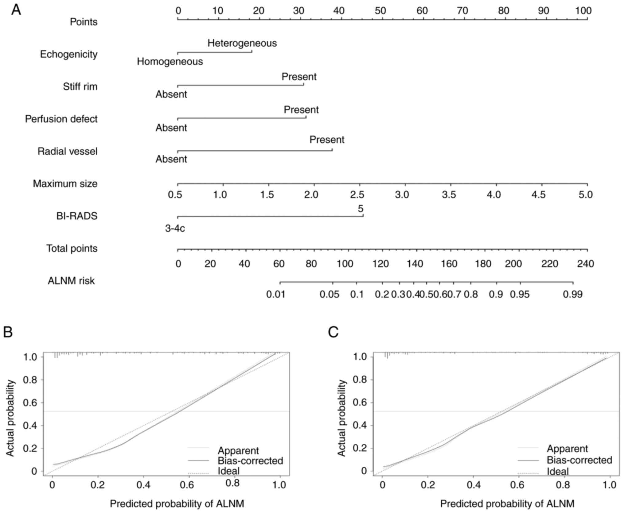 | Figure 2.MMUS nomogram and calibration curves.
(A) MMUS nomogram was developed with echogenicity, stiff rim sign,
perfect defect, radial vessel, maximum size and BI-RADS category
for the prediction of ALNM risk in the training cohort. To use the
nomogram, the different values of each variable are corresponded to
a point at the top of the graph and the total points to the bottom
line is the probability of ALNM. The calibration curves of the MMUS
nomogram in (B) training and (C) validation cohorts demonstrated
agreement between actual ALNM rate (ideal) and predictive ALNM rate
estimated by MMUS nomogram (solid line). MMUS, multimodal
ultrasound; BI-RADS, Breast Imaging and Reporting Data System;
ALNM, axillary lymph node metastasis, Predicted Pr[y=positive],
Predicted probability of axillary lymph node positive. |
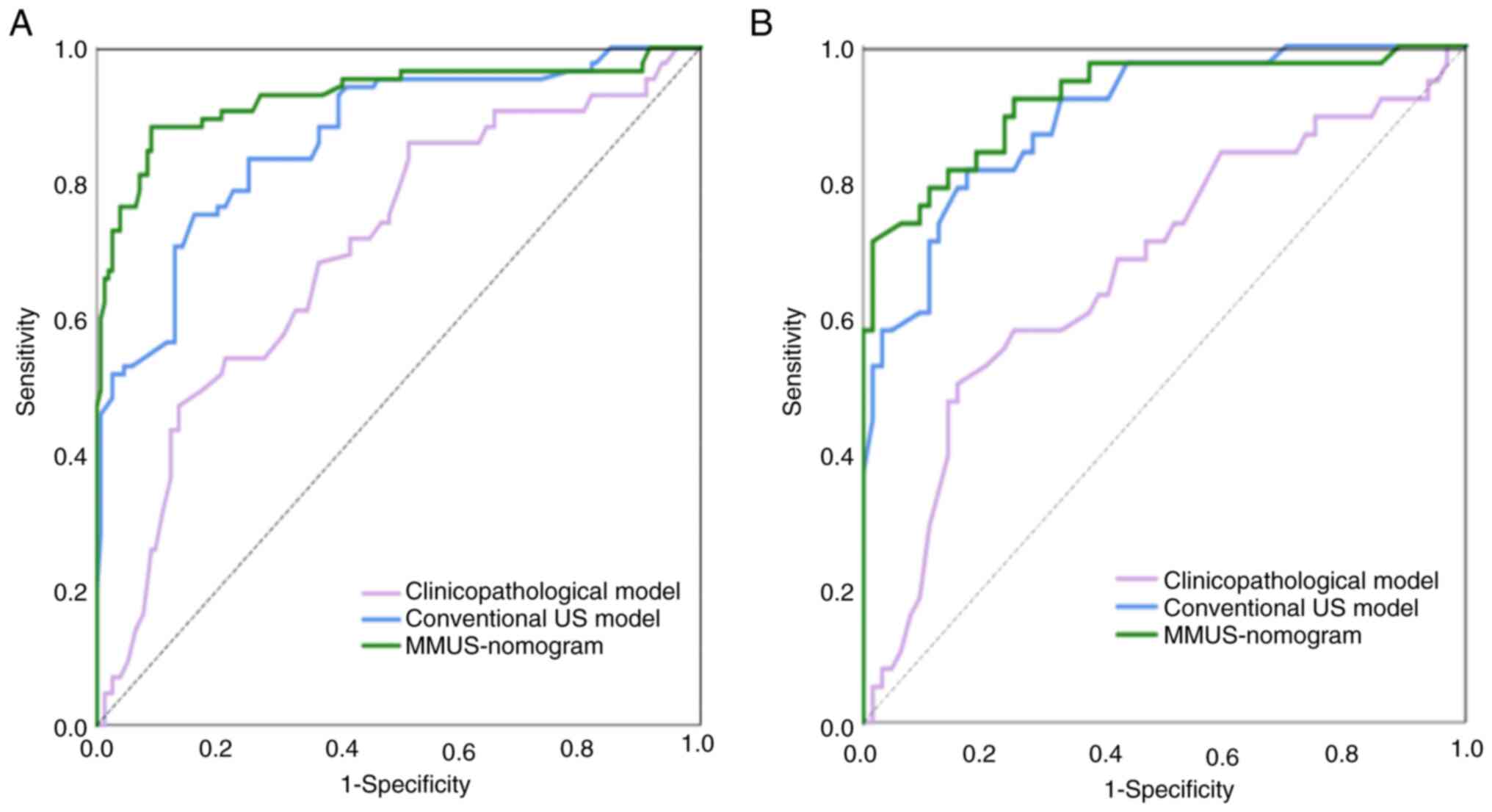
Discrimination and clinical usefulness
of MMUS nomogram
A clinicopathological model and conventional US
model were constructed to compare the predictive effect of ALNM.
The clinicopathological model was built with age, clinical tumor
(T) stage and histological grade, while the conventional US model
was built with maximum tumor size, echogenicity, calcification,
vascularity and US BI-RADS category. ROCs of the two models and the
MMUS nomogram in both cohorts are shown in Fig. 3. MMUS nomogram demonstrated an
improved predictive performance (AUC, 0.927; 95% CI, 0.891, 0.973)
compared with that of the clinicopathological model (AUC, 0.681;
95% CI, 0.594, 0.740), US-depicted ALN status (AUC, 0.710; 95% CI,
0.643, 0.780) and conventional US model (AUC, 0.867; 95% CI, 0.837,
0.904) in the training cohort. In the validation cohort, AUCs were
0.922 (95% CI, 0.880, 0.962), 0.670 (95% CI, 0.588, 0.723), 0.716
(95% CI, 0.693, 0.796) and 0.894 (95% CI, 0.851, 0.924),
respectively. The NRI index, which determined performance
improvement as introduced by multimodal US features in the
conventional US model, was 0.296 (95% CI, 0.084, 0.385) in the
training cohort and 0.288 (95% CI, 0.069, 0.361) in the validation
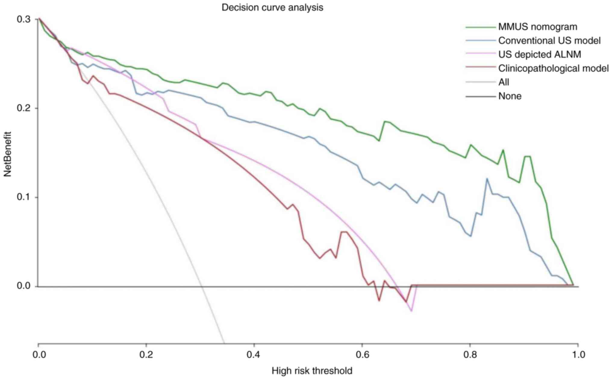
cohort. DCA demonstrated that the MMUS nomogram achieved an
improved benefit and clinical utility in predicting ALNM for
patients with early-stage IBC when high-risk threshold probability
was 0.0–1.0 (Fig. 4).
Discussion
SLN biopsy is known to have multiple complications
owing to its invasive process and false-negative rates of 7.8–27.3%
(27,28). Therefore, there have been efforts to
establish a non-invasive mathematical model to replace SLN biopsy
in determining preoperative ALN status. Previous studies have
reported that conventional US, SE, SWE US or CEUS methods serve an
important role in prediction of tumor growth or metastasis
(13,29–31).
However, to the best of our knowledge, the present study is the
first to construct a MMUS nomogram based on a set of comprehensive
multimodal US features of primary breast tumor to predict ALNM. The
MMUS nomogram achieved the most favorable performance compared with
the clinicopathological model, US-depicted ALN status and
conventional US model (AUC of 0.922 vs. 0.670, 0.716 and 0.894 in
the validation cohort). Furthermore, MMUS nomogram was a useful
predictive model for calibration and DCA.
It is known that clinical and pathological
characteristics serve an important role in prognosis prediction
(32,33). A study by The Memorial
Sloan-Kettering Cancer Center (MSKCC) developed a
clinicopathological model (MSKCC nomogram) to predict SLN
metastasis; however, AUC of the MSKCC nomogram was relatively low
at 0.754 (32). Previous studies
have reported that age, ER, PR and HER2 status have no effect in
predicting ALNM in patients with early-stage BC (33–35).
The present study demonstrated that older age, clinical T2 stage
and high tumor grade were more likely to predict ALNM in the
univariate analysis, but they were not independent risk factors
associated with ALNM in the multivariate analysis. The
clinicopathological model based on these three variables displayed
low predictive probability with AUCs of 0.681 and 0.670 in the
training and validation cohorts, respectively. These results
indicated that the clinicopathological factors may not provide
sufficient value in predicting ALN status, which is consistent with
previous studies (33–36).
Conventional US features of breast tumor showed that
larger tumor size, heterogeneous echogenicity, calcification, high
vascularity and BI-RADS category 5 were potential risk factors
associated with ALNM in the univariate analysis. These variables
formed the conventional US predictive model, whereas calcification
and high vascularity were not significantly associated with ALNM in
the multivariate analysis and were excluded from the final MMUS
nomogram model. Larger tumor size, heterogeneous echogenicity and
US BI-RADS category 5, as independent risk factors associated with
ALNM, indicated that rapidly proliferating tumor cells with a
larger tumor size tend to propagate within the regional lymph nodes
(37,38). Heterogeneous echogenicity may be the
result of genomic heterogeneity, which is a potential biomarker to
predict metastasis (39). The more
malignant US features the lesion has, the higher the BI-RADS
category is, and the worse the prognosis may be (40,41).
US elastography enables differentiation of tissues
to be made on the basis of their stiffness. The stiff rim sign in
SWE, which represents increased stiffness at the lesion margin, is
regarded as a sign of malignancy (12,25).
It has been explained as either a desmoplastic reaction or
infiltration of cancer cells into the peritumoral stroma (25,41).
The results of the present study demonstrated that stiff rim sign
was an independent prognostic factor associated with ALNM,
consistent with previous studies (41,42).
CEUS is a promising technology that can reflect the
micro-circulation perfusion of breast lesions (30,31).
In the present study, perfusion defect and radial vessel at the
tumor margin were the best predictive factors associated with ALNM.
When the SWE and CEUS features were combined with the conventional
US variables to form the MMUS nomogram, the incremental value was
demonstrated, with AUC improved from 0.894 to 0.922 and with NRI
improved 0.288 in comparison with the conventional US model in the
validation cohort. The results indicated that aggressive breast
tumors require a higher level of angiogenesis to maintain growth
and infiltration, peritumoral vessels can lead to local ALN or
distant metastasis and intrinsic necrosis may be due to the
relatively insufficient nutrition supply, which is manifested as a
perfusion defect (31,37,38,43).
The present study demonstrated that the SE features
and SWE (Emean, Emax, Emin and
ESD) and CEUS quantitative parameters (AT, TTP, PI, AS,
DS and AUTIC) had no significant predictive value for ALNM. These
results were different from the results obtained by Zhang et
al (41) who reported that
Emax and Emean of primary tumors are higher
in ALNM cases, and Wan et al (30) who reported that ALNM is associated
with higher PI and larger AUTIC values. These discrepancies may be
due to inclusion criteria or different US equipment and settings.
Different US instruments have different sensitivities for detection
of SWE and CEUS quantitative parameters (25,31,41,43),
which may account for the inconsistent results. Further studies
with larger and multicenter data are required to assess possible
associations.
Certain limitations in the present study have to be
addressed. The present study is limited by a lack of multicenter
external validation patients. Although the nomogram achieved
favorable predictive ability, selection bias was inevitable due to
the retrospective nature of the study. Moreover, to analyze the
association between tumor features and ALN status, patients with
bilateral, multicentric or multifocal tumors were excluded and only
one US equipment was used, which may also have caused selection
bias. The present study did not distinguish the number of
metastatic ALNs. Another study by our team in collaboration with
external institutions was initiated to predict the burden of ALNM
(44). Although the variability
study was favorable in terms of features and parameters evaluated
from the same images at different times, full analysis of
variability, such as the acquirement of multiple images at
different times and from different operators, is needed. Internal
mammary lymph nodes were not evaluated. For patients with internal
mammary lymph node metastasis, radiotherapy is necessary following
surgery and the final pathological node status is difficult to
determine (45). In addition, US is
the most frequently used imaging modality in determining lymph node
metastasis in daily clinical practice (29–31).
Nomograms are a simple, intuitive and easy to understand risk
scoring tool represented by graphs (32). MMUS nomogram in the present study
achieved favorable diagnostic performance and may facilitate
clinicians in appropriate preoperative decision-making. Previous
researchers have reported that standard breast magnetic resonance
imaging (MRI) is comparable with dedicated axillary US for
evaluation of axillary nodal status in patients with BC (46,47).
However, the comparison of MMUS nomogram with enhanced MRI in
determining lymph node metastasis was not performed in the present
study and further studies are required to address this issue in the
future. However, despite the aforementioned limitations, the
present study had adequate sample size and a well-characterized
cohort of patients with early-stage IBC with a large series of
combined multimodal US features of each breast tumor and the
nomogram represented an easy-to-use predictive tool with favorable
diagnostic performance. Moreover, application of the MMUS nomogram
in everyday clinical practice may be simpler compared with other
types of radiomics and deep learning analyses.
In conclusion, the present study demonstrated that
the MMUS nomogram was superior to US-depicted ALN status and
clinicopathological and conventional US model for the prediction of
ALNM. As a non-invasive, convenient, comprehensive and reliable
predictive tool, the MMUS nomogram may facilitate appropriate
preoperative decision-making for patients with early-stage IBC.
Acknowledgements
Not applicable.
Funding
Funding: No funding was received.
Availability of data and materials
The data generated in the present study may be
requested from the corresponding author.
Authors' contributions
JJY, WZ and WWZ were responsible for study
conception and design. WZ contributed to manuscript preparation and
editing. JJY and YZ performed ultrasonographic data analysis and
interpretation. XSC collected and interpreted clinicopathological
data. JQZ contributed to the statistical analysis. WZ and WWZ
confirm the authenticity of all the raw data. All authors have read
and approved the final manuscript.
Ethics approval and consent to
participate
The present study was approved by and conducted in
accordance with the Declaration of Helsinki and approved by the
Ethics Committee of Ruijin Hospital, School of Medicine, Shanghai
Jiao Tong University (Shanghai, China; approval no. 2020-309; July
15, 2020). Written informed consent were obtained from patients to
participate in the study.
Patient consent for publication
Not applicable.
Competing interests
The authors declare that they have no competing
interests.
Glossary
Abbreviations
Abbreviations:
|
ALNM
|
axillary lymph node metastasis
|
|
AUC
|
area under the curve
|
|
BI-RADS
|
Breast Imaging and Reporting Data
System
|
|
CEUS
|
contrast-enhanced ultrasound
|
|
DCA
|
decision curve analysis
|
|
ER
|
estrogen receptor
|
|
HER2
|
human epidermal growth factor receptor
2
|
|
IBC
|
invasive breast cancer
|
|
MM
|
multimodal
|
|
NRI
|
net reclassification improvement
|
|
PR
|
progesterone receptor
|
|
ROC
|
receiver operating characteristic
|
|
SE
|
strain elastography
|
|
SLN
|
sentinel lymph node
|
|
SWE
|
shear wave elastography
|
References
|
1
|
Sung H, Ferlay J, Siegel RL, Laversanne M,
Soerjomataram I, Jemal A and Bray F: Global cancer statistics 2020:
GLOBOCAN estimates of incidence and mortality worldwide for 36
cancers in 185 countries. CA Cancer J Clin. 71:209–249. 2021.
View Article : Google Scholar : PubMed/NCBI
|
|
2
|
Giuliano AE, Ballman KV, McCall L, Beitsch
PD, Brennan MB, Kelemen PR, Ollila DW, Hansen NM, Whitworth PW,
Blumencranz PW, et al: Effect of axillary dissection vs. no
axillary dissection on 10-year overall survival among women with
invasive breast cancer and sentinel node metastasis: The ACOSOG
Z0011 (alliance) randomized clinical trial. JAMA. 318:918–926.
2017. View Article : Google Scholar : PubMed/NCBI
|
|
3
|
Lyman GH, Somerfield MR, Bosserman LD,
Perkins CL, Weaver DL and Giuliano AE: Sentinel lymph node biopsy
for patients with early-stage breast cancer: American society of
clinical oncology clinical practice guideline update. J Clin Oncol.
35:561–564. 2017. View Article : Google Scholar : PubMed/NCBI
|
|
4
|
Wilke LG, McCall LM, Posther KE, Whitworth
PW, Reintgen DS, Leitch AM, Gabram SG, Lucci A, Cox CE, Hunt KK, et
al: Surgical complications associated with sentinel lymph node
biopsy: Results from a prospective international cooperative group
trial. Ann Surg Oncol. 13:491–500. 2006. View Article : Google Scholar : PubMed/NCBI
|
|
5
|
Manca G, Rubello D, Tardelli E, Giammarile
F, Mazzarri S, Boni G, Chondrogiannis S, Marzola MC, Chiacchio S,
Ghilli M, et al: Sentinel lymph node biopsy in breast cancer:
Indications, contraindications, and controversies. Clin Nucl Med.
41:126–133. 2016. View Article : Google Scholar : PubMed/NCBI
|
|
6
|
Zhang YN, Wang CJ, Xu Y, Zhu QL, Zhou YD,
Zhang J, Mao F, Jiang YX and Sun Q: Sensitivity, specificity and
accuracy of ultrasound in diagnosis of breast cancer metastasis to
the axillary lymph nodes in Chinese patients. Ultrasound Med Biol.
41:1835–1841. 2015. View Article : Google Scholar : PubMed/NCBI
|
|
7
|
Hotton J, Salleron J, Henrot P, Buhler J,
Leufflen L, Rauch P and Marchal F: Pre-operative axillary
ultrasound with fine-needle aspiration cytology performance and
predictive factors of false negatives in axillary lymph node
involvement in early breast cancer. Breast Cancer Res Treat.
183:639–647. 2020. View Article : Google Scholar : PubMed/NCBI
|
|
8
|
Yu FH, Wang JX, Ye XH, Deng J, Hang J and
Yang B: Ultrasound-based radiomics nomogram: A potential biomarker
to predict axillary lymph node metastasis in early-stage invasive
breast cancer. Eur J Radiol. 119:1086582019. View Article : Google Scholar : PubMed/NCBI
|
|
9
|
Qiu X, Jiang Y, Zhao Q, Yan C, Huang M and
Jiang T: Could ultrasound-based radiomics noninvasively predict
axillary lymph node metastasis in breast cancer? J Ultrasound Med.
39:1897–1905. 2020. View Article : Google Scholar : PubMed/NCBI
|
|
10
|
Jiang M, Li CL, Luo XM, Chuan ZR, Chen RX,
Tang SC, Lv WZ, Cui XW and Dietrich CF: Radiomics model based on
shear-wave elastography in the assessment of axillary lymph node
status in early-stage breast cancer. Eur Radiol. 32:2313–2325.
2022. View Article : Google Scholar : PubMed/NCBI
|
|
11
|
Lee S, Jung Y and Bae Y: Clinical
application of a color map pattern on shear-wave elastography for
invasive breast cancer. Surg Oncol. 25:44–48. 2016. View Article : Google Scholar : PubMed/NCBI
|
|
12
|
Tozaki M and Fukuma E: Pattern
classification of ShearWave™ elastography images for differential
diagnosis between benign and malignant solid breast masses. Acta
Radiol. 52:1069–1075. 2011. View Article : Google Scholar : PubMed/NCBI
|
|
13
|
Evans A, Whelehan P, Thomson K, McLean D,
Brauer K, Purdie C, Baker L, Jordan L, Rauchhaus P and Thompson A:
Invasive breast cancer: Relationship between shear-wave
elastographic findings and histologic prognostic factors.
Radiology. 263:673–677. 2012. View Article : Google Scholar : PubMed/NCBI
|
|
14
|
Huang R, Lin Z, Dou H, Wang J, Miao J,
Zhou G, Jia X, Xu W, Mei Z, Dong Y, et al: AW3M: An auto-weighting
and recovery framework for breast cancer diagnosis using
multi-modal ultrasound. Med Image Anal. 72:1021372021. View Article : Google Scholar : PubMed/NCBI
|
|
15
|
Zheng X, Yao Z, Huang Y, Yu Y, Wang Y, Liu
Y, Mao R, Li F, Xiao Y, Wang Y, et al: Deep learning radiomics can
predict axillary lymph node status in early-stage breast cancer. J
Nat Commun. 11:12362020. View Article : Google Scholar : PubMed/NCBI
|
|
16
|
Guo X, Liu Z, Sun C, Zhang L, Wang Y, Li
Z, Shi J, Wu T, Cui H, Zhang J, et al: Deep learning radiomics of
ultrasonography: Identifying the risk of axillary non-sentinel
lymph node involvement in primary breast cancer. EBioMedicine.
60:1030182020. View Article : Google Scholar : PubMed/NCBI
|
|
17
|
Liu D, Wu J, Lin C, Ding S, Lu S, Fang Y,
Huang J, Hong J, Gao W, Zhu S, et al: The comparative safety of
epirubicin and cyclophosphamide versus docetaxel and
cyclophosphamide in lymph node-negative, HR-positive, HER2-negative
breast cancer (ELEGANT): A randomized trial. Cancers (Basel).
14:32212022. View Article : Google Scholar : PubMed/NCBI
|
|
18
|
Collins GS, Reitsma JB, Altman DG and
Moons KG: Transparent reporting of a multivariable prediction model
for individual prognosis or diagnosis (TRIPOD): The TRIPOD
statement. Br J Surg. 102:148–158. 2015. View Article : Google Scholar : PubMed/NCBI
|
|
19
|
Penault-Llorca F, André F, Sagan C,
Lacroix-Triki M, Denoux Y, Verriele V, Jacquemier J, Baranzelli MC,
Bibeau F, Antoine M, et al: Ki67 expression and docetaxel efficacy
in patients with estrogen receptor-positive breast cancer. J Clin
Oncol. 27:2809–2815. 2009. View Article : Google Scholar : PubMed/NCBI
|
|
20
|
Coates AS, Winer EP, Goldhirsch A, Gelber
RD, Gnant M, Piccart-Gebhart M, Thürlimann B and Senn HJ; Panel
Members, : Tailoring therapies-improving the management of early
breast cancer: St Gallen international expert consensus on the
primary therapy of early breast cancer 2015. Ann Oncol.
26:1533–1546. 2015. View Article : Google Scholar : PubMed/NCBI
|
|
21
|
Mercado CL: BI-RADS update. Radiol Clin
North Am. 52:481–487. 2014. View Article : Google Scholar : PubMed/NCBI
|
|
22
|
Bedi DG, Krishnamurthy R, Krishnamurthy S,
Edeiken BS, Le-Petross H, Fornage BD, Bassett RL Jr and Hunt KK:
Cortical morphologic features of axillary lymph nodes as a
predictor of metastasis in breast cancer: In vitro sonographic
study. AJR Am J Roentgenol. 191:646–652. 2008. View Article : Google Scholar : PubMed/NCBI
|
|
23
|
Koelliker SL, Chung MA, Mainiero MB,
Steinhoff MM and Cady B: Axillary lymph nodes: US-guided
fine-needle aspiration for initial staging of breast
cancer-correlation with primary tumor size. Radiology. 246:81–89.
2008. View Article : Google Scholar : PubMed/NCBI
|
|
24
|
Itoh A, Ueno E, Tohno E, Kamma H,
Takahashi H, Shiina T, Yamakawa M and Matsumura T: Breast disease:
Clinical application of US elastography for diagnosis. Radiology.
239:341–350. 2006. View Article : Google Scholar : PubMed/NCBI
|
|
25
|
Zhou J, Zhan W, Chang C, Zhang X, Jia Y,
Dong Y, Zhou C, Sun J and Grant EG: Breast lesions: Evaluation with
shear wave elastography, with special emphasis on the ‘stiff rim’
sign. Radiology. 272:63–72. 2014. View Article : Google Scholar : PubMed/NCBI
|
|
26
|
Sidhu PS, Cantisani V, Dietrich CF, Gilja
OH, Saftoiu A, Bartels E, Bertolotto M, Calliada F, Clevert DA,
Cosgrove D, et al: The EFSUMB guidelines and recommendations for
the clinical practice of contrast-enhanced ultrasound (CEUS) in
non-hepatic applications: Update 2017 (long version). Ultraschall
Med. 39:e2–e44. 2018. View Article : Google Scholar : PubMed/NCBI
|
|
27
|
Li H, Jun Z, Zhi-Cheng G and Xiang Q:
Factors that affect the false negative rate of sentinel lymph node
mapping with methylene blue dye alone in breast cancer. J Int Med
Res. 47:4841–4853. 2019. View Article : Google Scholar : PubMed/NCBI
|
|
28
|
Pesek S, Ashikaga T, Krag LE and Krag D:
The false-negative rate of sentinel node biopsy in patients with
breast cancer: A meta-analysis. World J Surg. 36:2239–2251. 2012.
View Article : Google Scholar : PubMed/NCBI
|
|
29
|
Fang J, Zhao W, Li Q, Zhang B, Pu C and
Wang H: Correlation analysis of conventional ultrasound
characteristics and strain elastography with ki-67 status in breast
cancer. Ultrasound Med Biol. 46:2972–2978. 2020. View Article : Google Scholar : PubMed/NCBI
|
|
30
|
Wan CF, Du J, Fang H, Li FH, Zhu JS and
Liu Q: Enhancement patterns and parameters of breast cancers at
contrast-enhanced US: Correlation with prognostic factors.
Radiology. 262:450–459. 2012. View Article : Google Scholar : PubMed/NCBI
|
|
31
|
Zhu AQ, Li XL, An LW, Guo LH, Fu HJ, Sun
LP and Xu HX: Predicting axillary lymph node metastasis in patients
with breast invasive ductal carcinoma with negative axillary
ultrasound results using conventional ultrasound and
contrast-enhanced ultrasound. J Ultrasound Med. 39:2059–2070. 2020.
View Article : Google Scholar : PubMed/NCBI
|
|
32
|
Bevilacqua JL, Kattan MW, Fey JV, Cody HS
III, Borgen PI and Van Zee KJ: Doctor, what are my chances of
having a positive sentinel node? A validated nomogram for risk
estimation. J Clin Oncol. 25:3670–3679. 2007. View Article : Google Scholar : PubMed/NCBI
|
|
33
|
Yoshihara E, Smeets A, Laenen A, Reynders
A, Soens J, Van Ongeval C, Moerman P, Paridaens R, Wildiers H,
Neven P and Christiaens MR: Predictors of axillary lymph node
metastases in early breast cancer and their applicability in
clinical practice. Breast. 22:357–361. 2013. View Article : Google Scholar : PubMed/NCBI
|
|
34
|
Xiong J, Zuo W, Wu Y, Wang X, Li W, Wang
Q, Zhou H, Xie M and Qin X: Ultrasonography and clinicopathological
features of breast cancer in predicting axillary lymph node
metastases. BMC Cancer. 22:11552022. View Article : Google Scholar : PubMed/NCBI
|
|
35
|
Yu Y, Tan Y, Xie C, Hu Q, Ouyang J, Chen
Y, Gu Y, Li A, Lu N, He Z, et al: Development and validation of a
preoperative magnetic resonance imaging radiomics-based signature
to predict axillary lymph node metastasis and disease-free survival
in patients with early-stage breast cancer. JAMA Netw Open.
3:e20280862020. View Article : Google Scholar : PubMed/NCBI
|
|
36
|
Dong Y, Feng Q, Yang W, Lu Z, Deng C,
Zhang L, Lian Z, Liu J, Luo X, Pei S, et al: Preoperative
prediction of sentinel lymph node metastasis in breast cancer based
on radiomics of T2-weighted fat-suppression and diffusion-weighted
MRI. Eur Radiol. 28:582–591. 2018. View Article : Google Scholar : PubMed/NCBI
|
|
37
|
Fujii T, Yajima R, Tatsuki H, Suto T,
Morita H, Tsutsumi S and Kuwano H: Significance of lymphatic
invasion combined with size of primary tumor for predicting
sentinel lymph node metastasis in patients with breast cancer.
Anticancer Res. 35:3581–3584. 2015.PubMed/NCBI
|
|
38
|
Auvinen P, Tammi R, Parkkinen J, Tammi M,
Agren U, Johansson R, Hirvikoski P, Eskelinen M and Kosma VM:
Hyaluronan in peritumoral stroma and malignant cells associates
with breast cancer spreading and predicts survival. Am J Pathol.
156:529–536. 2000. View Article : Google Scholar : PubMed/NCBI
|
|
39
|
Bae MS, Shin SU, Song SE, Ryu HS, Han W
and Moon WK: Association between US features of primary tumor and
axillary lymph node metastasis in patients with clinical T1-T2N0
breast cancer. Acta Radiol. 59:402–408. 2018. View Article : Google Scholar : PubMed/NCBI
|
|
40
|
Sheng DL, Shen XG, Shi ZT, Chang C and Li
JW: Survival outcome assessment for triple-negative breast cancer:
A nomogram analysis based on integrated clinicopathological,
sonographic, and mammographic characteristics. Eur Radiol.
32:6575–6587. 2022. View Article : Google Scholar : PubMed/NCBI
|
|
41
|
Zhang H, Dong Y, Jia X, Zhang J, Li Z,
Chuan Z, Xu Y, Hu B, Huang Y, Chang C, et al: Comprehensive risk
system based on shear wave elastography and BI-RADS categories in
assessing axillary lymph node metastasis of invasive breast
cancer-A multi-center study. Front Oncol. 12:8309102022. View Article : Google Scholar : PubMed/NCBI
|
|
42
|
Evans A, Rauchhaus P, Whelehan P, Thomson
K, Purdie CA, Jordan LB, Michie CO, Thompson A and Vinnicombe S:
Does shear wave ultrasound independently predict axillary lymph
node metastasis in women with invasive breast cancer? Breast Cancer
Res Treat. 143:153–157. 2014. View Article : Google Scholar : PubMed/NCBI
|
|
43
|
Matsubayashi R, Matsuo Y, Edakuni G, Satoh
T, Tokunaga O and Kudo S: Breast masses with peripheral rim
enhancement on dynamic contrast-enhanced MR images: Correlation of
MR findings with histologic features and expression of growth
factors. Radiology. 217:841–848. 2000. View Article : Google Scholar : PubMed/NCBI
|
|
44
|
Yao J, Zhou W, Xu S, Jia X, Zhou J, Chen X
and Zhan W: Machine learning-based breast tumor ultrasound
radiomics for pre-operative prediction of axillary sentinel lymph
node metastasis burden in early-stage invasive breast cancer.
Ultrasound Med Biol. 50:229–236. 2024. View Article : Google Scholar : PubMed/NCBI
|
|
45
|
Yang K, Kim H, Choi DH, Park W, Noh JM and
Cho WK: Optimal radiotherapy for patients with internal mammary
lymph node metastasis from breast cancer. Radiat Oncol. 15:162020.
View Article : Google Scholar : PubMed/NCBI
|
|
46
|
van Nijnatten TJA, Ploumen EH, Schipper
RJ, Goorts B, Andriessen EH, Vanwetswinkel S, Schavemaker M,
Nelemans P, de Vries B, Beets-Tan RGH, et al: Routine use of
standard breast MRI compared to axillary ultrasound for
differentiating between no, limited and advanced axillary nodal
disease in newly diagnosed breast cancer patients. Eur J Radiol.
85:2288–2294. 2016. View Article : Google Scholar : PubMed/NCBI
|
|
47
|
Samiei S, van Nijnatten TJA, van Beek HC,
Polak MPJ, Maaskant-Braat AJG, Heuts EM, van Kuijk SMJ, Schipper
RJ, Lobbes MBI and Smidt ML: Diagnostic performance of axillary
ultrasound and standard breast MRI for differentiation between
limited and advanced axillary nodal disease in clinically
node-positive breast cancer patients. Sci Rep. 9:174762019.
View Article : Google Scholar : PubMed/NCBI
|