Spandidos Publications Logo
  • About
    • About Spandidos
    • Aims and Scopes
    • Abstracting and Indexing
    • Editorial Policies
    • Reprints and Permissions
    • Job Opportunities
    • Terms and Conditions
    • Contact
  • Journals
    • All Journals
    • Oncology Letters
      • Oncology Letters
      • Information for Authors
      • Editorial Policies
      • Editorial Board
      • Aims and Scope
      • Abstracting and Indexing
      • Bibliographic Information
      • Archive
    • International Journal of Oncology
      • International Journal of Oncology
      • Information for Authors
      • Editorial Policies
      • Editorial Board
      • Aims and Scope
      • Abstracting and Indexing
      • Bibliographic Information
      • Archive
    • Molecular and Clinical Oncology
      • Molecular and Clinical Oncology
      • Information for Authors
      • Editorial Policies
      • Editorial Board
      • Aims and Scope
      • Abstracting and Indexing
      • Bibliographic Information
      • Archive
    • Experimental and Therapeutic Medicine
      • Experimental and Therapeutic Medicine
      • Information for Authors
      • Editorial Policies
      • Editorial Board
      • Aims and Scope
      • Abstracting and Indexing
      • Bibliographic Information
      • Archive
    • International Journal of Molecular Medicine
      • International Journal of Molecular Medicine
      • Information for Authors
      • Editorial Policies
      • Editorial Board
      • Aims and Scope
      • Abstracting and Indexing
      • Bibliographic Information
      • Archive
    • Biomedical Reports
      • Biomedical Reports
      • Information for Authors
      • Editorial Policies
      • Editorial Board
      • Aims and Scope
      • Abstracting and Indexing
      • Bibliographic Information
      • Archive
    • Oncology Reports
      • Oncology Reports
      • Information for Authors
      • Editorial Policies
      • Editorial Board
      • Aims and Scope
      • Abstracting and Indexing
      • Bibliographic Information
      • Archive
    • Molecular Medicine Reports
      • Molecular Medicine Reports
      • Information for Authors
      • Editorial Policies
      • Editorial Board
      • Aims and Scope
      • Abstracting and Indexing
      • Bibliographic Information
      • Archive
    • World Academy of Sciences Journal
      • World Academy of Sciences Journal
      • Information for Authors
      • Editorial Policies
      • Editorial Board
      • Aims and Scope
      • Abstracting and Indexing
      • Bibliographic Information
      • Archive
    • International Journal of Functional Nutrition
      • International Journal of Functional Nutrition
      • Information for Authors
      • Editorial Policies
      • Editorial Board
      • Aims and Scope
      • Abstracting and Indexing
      • Bibliographic Information
      • Archive
    • International Journal of Epigenetics
      • International Journal of Epigenetics
      • Information for Authors
      • Editorial Policies
      • Editorial Board
      • Aims and Scope
      • Abstracting and Indexing
      • Bibliographic Information
      • Archive
    • Medicine International
      • Medicine International
      • Information for Authors
      • Editorial Policies
      • Editorial Board
      • Aims and Scope
      • Abstracting and Indexing
      • Bibliographic Information
      • Archive
  • Articles
  • Information
    • Information for Authors
    • Information for Reviewers
    • Information for Librarians
    • Information for Advertisers
    • Conferences
  • Language Editing
Spandidos Publications Logo
  • About
    • About Spandidos
    • Aims and Scopes
    • Abstracting and Indexing
    • Editorial Policies
    • Reprints and Permissions
    • Job Opportunities
    • Terms and Conditions
    • Contact
  • Journals
    • All Journals
    • Biomedical Reports
      • Information for Authors
      • Editorial Policies
      • Editorial Board
      • Aims and Scope
      • Abstracting and Indexing
      • Bibliographic Information
      • Archive
    • Experimental and Therapeutic Medicine
      • Information for Authors
      • Editorial Policies
      • Editorial Board
      • Aims and Scope
      • Abstracting and Indexing
      • Bibliographic Information
      • Archive
    • International Journal of Epigenetics
      • Information for Authors
      • Editorial Policies
      • Editorial Board
      • Aims and Scope
      • Abstracting and Indexing
      • Bibliographic Information
      • Archive
    • International Journal of Functional Nutrition
      • Information for Authors
      • Editorial Policies
      • Editorial Board
      • Aims and Scope
      • Abstracting and Indexing
      • Bibliographic Information
      • Archive
    • International Journal of Molecular Medicine
      • Information for Authors
      • Editorial Policies
      • Editorial Board
      • Aims and Scope
      • Abstracting and Indexing
      • Bibliographic Information
      • Archive
    • International Journal of Oncology
      • Information for Authors
      • Editorial Policies
      • Editorial Board
      • Aims and Scope
      • Abstracting and Indexing
      • Bibliographic Information
      • Archive
    • Medicine International
      • Information for Authors
      • Editorial Policies
      • Editorial Board
      • Aims and Scope
      • Abstracting and Indexing
      • Bibliographic Information
      • Archive
    • Molecular and Clinical Oncology
      • Information for Authors
      • Editorial Policies
      • Editorial Board
      • Aims and Scope
      • Abstracting and Indexing
      • Bibliographic Information
      • Archive
    • Molecular Medicine Reports
      • Information for Authors
      • Editorial Policies
      • Editorial Board
      • Aims and Scope
      • Abstracting and Indexing
      • Bibliographic Information
      • Archive
    • Oncology Letters
      • Information for Authors
      • Editorial Policies
      • Editorial Board
      • Aims and Scope
      • Abstracting and Indexing
      • Bibliographic Information
      • Archive
    • Oncology Reports
      • Information for Authors
      • Editorial Policies
      • Editorial Board
      • Aims and Scope
      • Abstracting and Indexing
      • Bibliographic Information
      • Archive
    • World Academy of Sciences Journal
      • Information for Authors
      • Editorial Policies
      • Editorial Board
      • Aims and Scope
      • Abstracting and Indexing
      • Bibliographic Information
      • Archive
  • Articles
  • Information
    • For Authors
    • For Reviewers
    • For Librarians
    • For Advertisers
    • Conferences
  • Language Editing
Login Register Submit
  • This site uses cookies
  • You can change your cookie settings at any time by following the instructions in our Cookie Policy. To find out more, you may read our Privacy Policy.

    I agree
Search articles by DOI, keyword, author or affiliation
Search
Advanced Search
presentation
Oncology Letters
Join Editorial Board Propose a Special Issue
Print ISSN: 1792-1074 Online ISSN: 1792-1082
Journal Cover
May-2026 Volume 31 Issue 5

Full Size Image

Sign up for eToc alerts
Recommend to Library

Journals

International Journal of Molecular Medicine

International Journal of Molecular Medicine

International Journal of Molecular Medicine is an international journal devoted to molecular mechanisms of human disease.

International Journal of Oncology

International Journal of Oncology

International Journal of Oncology is an international journal devoted to oncology research and cancer treatment.

Molecular Medicine Reports

Molecular Medicine Reports

Covers molecular medicine topics such as pharmacology, pathology, genetics, neuroscience, infectious diseases, molecular cardiology, and molecular surgery.

Oncology Reports

Oncology Reports

Oncology Reports is an international journal devoted to fundamental and applied research in Oncology.

Experimental and Therapeutic Medicine

Experimental and Therapeutic Medicine

Experimental and Therapeutic Medicine is an international journal devoted to laboratory and clinical medicine.

Oncology Letters

Oncology Letters

Oncology Letters is an international journal devoted to Experimental and Clinical Oncology.

Biomedical Reports

Biomedical Reports

Explores a wide range of biological and medical fields, including pharmacology, genetics, microbiology, neuroscience, and molecular cardiology.

Molecular and Clinical Oncology

Molecular and Clinical Oncology

International journal addressing all aspects of oncology research, from tumorigenesis and oncogenes to chemotherapy and metastasis.

World Academy of Sciences Journal

World Academy of Sciences Journal

Multidisciplinary open-access journal spanning biochemistry, genetics, neuroscience, environmental health, and synthetic biology.

International Journal of Functional Nutrition

International Journal of Functional Nutrition

Open-access journal combining biochemistry, pharmacology, immunology, and genetics to advance health through functional nutrition.

International Journal of Epigenetics

International Journal of Epigenetics

Publishes open-access research on using epigenetics to advance understanding and treatment of human disease.

Medicine International

Medicine International

An International Open Access Journal Devoted to General Medicine.

Journal Cover
May-2026 Volume 31 Issue 5

Full Size Image

Sign up for eToc alerts
Recommend to Library

  • Article
  • Citations
    • Cite This Article
    • Download Citation
    • Create Citation Alert
    • Remove Citation Alert
    • Cited By
  • Similar Articles
    • Related Articles (in Spandidos Publications)
    • Similar Articles (Google Scholar)
    • Similar Articles (PubMed)
  • Download PDF
  • Download XML
  • View XML

  • Supplementary Files
    • Supplementary_Data.pdf
Case Report

Diagnostic and surgical challenges of a lymph node metastasis from occult gastric cancer mimicking a pancreatic head tumor: A case report

  • Authors:
    • Takuya Ando
    • Yuki Hirose
    • Kazuyasu Takizawa
    • Jun Sakata
    • Hirosuke Ishikawa
    • Shun Abe
    • Yusuke Kawachi
    • Mae Nakano
    • Hiroshi Ichikawa
    • Yoshifumi Shimada
    • Toshifumi Wakai
  • View Affiliations / Copyright

    Affiliations: Division of Digestive and General Surgery, Niigata University Graduate School of Medical and Dental Sciences, Chuo-ku, Niigata 951‑8510, Japan
  • Article Number: 162
    |
    Published online on: March 5, 2026
       https://doi.org/10.3892/ol.2026.15515
  • Expand metrics +
Metrics: Total Views: 0 (Spandidos Publications: | PMC Statistics: )
Metrics: Total PDF Downloads: 0 (Spandidos Publications: | PMC Statistics: )
Cited By (CrossRef): 0 citations Loading Articles...

This article is mentioned in:


Abstract

Pancreatic head tumors are usually regarded as primary pancreatic neoplasms; however, metastatic lymph nodes from gastrointestinal malignancies may occasionally mimic pancreatic tumors and lead to diagnostic and therapeutic challenges, particularly when the primary lesion cannot be identified preoperatively. In the present case report, a 76‑year‑old man was referred for evaluation of a pancreatic head mass with elevated serum carbohydrate antigen 19‑9 levels. Contrast‑enhanced computed tomography and positron emission tomography‑computed tomography demonstrated a protruding lesion in the pancreatic head and an enlarged infrapyloric lymph node, without evidence of a primary gastrointestinal tumor. Esophagogastroduodenoscopy was unremarkable and primary pancreatic cancer was suspected. Endoscopic ultrasound‑guided biopsy of the infrapyloric lymph node revealed adenocarcinoma; however, the primary site could not be determined. Pancreaticoduodenectomy was planned. Intraoperatively, the lesion appeared to represent a bulky metastatic lymph node rather than a primary pancreatic tumor, and distal gastrectomy was additionally performed. Histopathological examination confirmed metastatic lymph node involvement from occult gastric cancer. The patient remains alive without evidence of recurrence 2 years after surgery. In conclusion, occult gastric cancer metastasis can masquerade as a pancreatic head tumor. Awareness of this diagnostic and surgical challenge may facilitate appropriate intraoperative reassessment when pancreatic head lesions are accompanied by suspicious perigastric lymphadenopathy.

Introduction

Pancreatic head tumors are most commonly diagnosed as primary pancreatic neoplasms, particularly pancreatic ductal adenocarcinoma, which remains one of the leading causes of cancer-related mortality worldwide (1). Despite advances in systemic therapy, surgical resection by pancreaticoduodenectomy remains the only potentially curative treatment for primary pancreatic head malignancies and is therefore frequently performed when imaging suggests a primary pancreatic head tumor (2–4).

Gastric cancer is also a prevalent malignancy globally and is characterized by well-defined lymphatic drainage pathways, including the infrapyloric lymph nodes (station no. 6), which are anatomically adjacent to the pancreatic head (5,6). Although gastric cancer typically presents with identifiable mucosal lesions, poorly differentiated tumors may infiltrate and spread into the submucosa and metastasize to regional lymph nodes without forming an obvious mass (7,8).

Nevertheless, metastatic lymph nodes from gastrointestinal malignancies, particularly gastric cancer, may occasionally present as pancreatic head masses and pose a significant diagnostic challenge (9–11). Such cases are especially problematic when no primary lesion is detected on preoperative endoscopic evaluation (7,8,12). We report a case of occult gastric cancer in which metastatic infrapyloric lymph node involvement closely mimicked a pancreatic head tumor, highlighting a clinically relevant diagnostic pitfall in the surgical evaluation of pancreatic head lesions and its implications for preoperative assessment and intraoperative decision-making.

Case report

A 76-year-old man presented to Niigata University Medical and Dental Hospital (Niigata, Japan) with occasional back pain and elevated serum carbohydrate antigen 19-9 (CA 19-9) levels (462 U/ml). Preoperative serum carcinoembryonic antigen (CEA) level was within normal range. Contrast-enhanced computed tomography at a referring hospital revealed a mass in the pancreatic head, and the patient was referred to our institution for further evaluation.

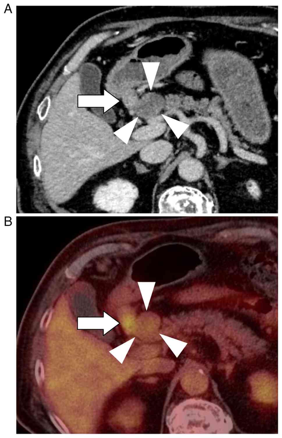
Subsequent imaging demonstrated a 30-mm protruding mass in the pancreatic head, characterized by hypodensity in the arterial phase and gradual enhancement in the portal and venous phases, along with an enlarged lymph node in the infrapyloric region (Fig. 1A). Positron emission tomography-computed tomography demonstrated increased FDG uptake in both the pancreatic head lesion (SUVmax, 2.74) and the infrapyloric lymph node (station No. 6; SUVmax, 6.28), whereas no abnormal uptake was observed in the gastric wall (Fig. 1B). Esophagogastroduodenoscopy was performed with careful inspection of the entire stomach, including the antrum and pyloric region. No endoscopically identifiable lesion suggestive of primary gastric cancer was detected (Fig. S1). Endoscopic ultrasound (EUS)-guided biopsy of the enlarged infrapyloric lymph node demonstrated adenocarcinoma; however, the primary site of origin could not be determined. EUS-guided sampling of the pancreatic head lesion itself was not performed, as a safe and reliable puncture route could not be secured due to intervening vascular structures.

Radiologic imaging of a tumor in the
pancreatic head. (A) Abdominal contrast-enhanced computed
tomography shows 30-mm protruding mass in the pancreatic head with
hypodensity (arrowheads), along with enhanced lymph nodes in the
infrapyloric region (arrow). (B) Positron emission
tomography-computed tomography reveals an abnormal uptake in both
the pancreatic head lesion (arrowheads) and lymph nodes in the
infrapyloric region (arrow).

Figure 1.

Radiologic imaging of a tumor in the pancreatic head. (A) Abdominal contrast-enhanced computed tomography shows 30-mm protruding mass in the pancreatic head with hypodensity (arrowheads), along with enhanced lymph nodes in the infrapyloric region (arrow). (B) Positron emission tomography-computed tomography reveals an abnormal uptake in both the pancreatic head lesion (arrowheads) and lymph nodes in the infrapyloric region (arrow).

Preoperatively, the differential diagnoses included primary pancreatic ductal adenocarcinoma, pancreatic neuroendocrine neoplasm, malignant lymphoma, and bulky metastatic lymphadenopathy from an occult gastrointestinal primary. Primary pancreatic ductal adenocarcinoma was considered; however, the absence of pancreatic ductal dilatation and the atypical protruding morphology of the lesion made this diagnosis less likely. Pancreatic neuroendocrine neoplasm was also considered, but was not strongly supported due to the lack of typical hypervascular features on contrast-enhanced imaging and normal serum neuroendocrine markers, including neuron-specific enolase. Malignant lymphoma was included in the differential diagnosis; however, the presence of adenocarcinoma on biopsy of the infrapyloric lymph node and normal serum soluble interleukin-2 receptor levels argued against this possibility. Given the coexistence of infrapyloric lymphadenopathy and adenocarcinoma on biopsy, bulky metastatic lymphadenopathy from an occult gastrointestinal primary was also considered, although no primary lesion was identified on preoperative endoscopic evaluation.

Based on these findings, primary pancreatic cancer including pancreatic ductal adenocarcinoma or neuroendocrine carcinoma, which frequently includes adenocarcinoma component, with lymph node metastasis was suspected, and pancreaticoduodenectomy 
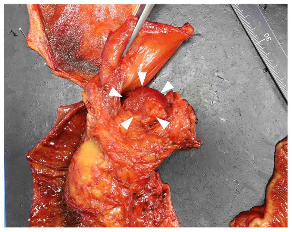
was planned. Intraoperatively, the pancreatic head lesion appeared as a markedly protruding mass, raising suspicion of a bulky metastatic lymph node. Consequently, the surgical procedure was modified to include distal gastrectomy (Fig. 2).

Surgically resected specimen. The
resected specimen shows a 30-mm protruding mass in the pancreatic
head (arrowheads).

Figure 2.

Surgically resected specimen. The resected specimen shows a 30-mm protruding mass in the pancreatic head (arrowheads).

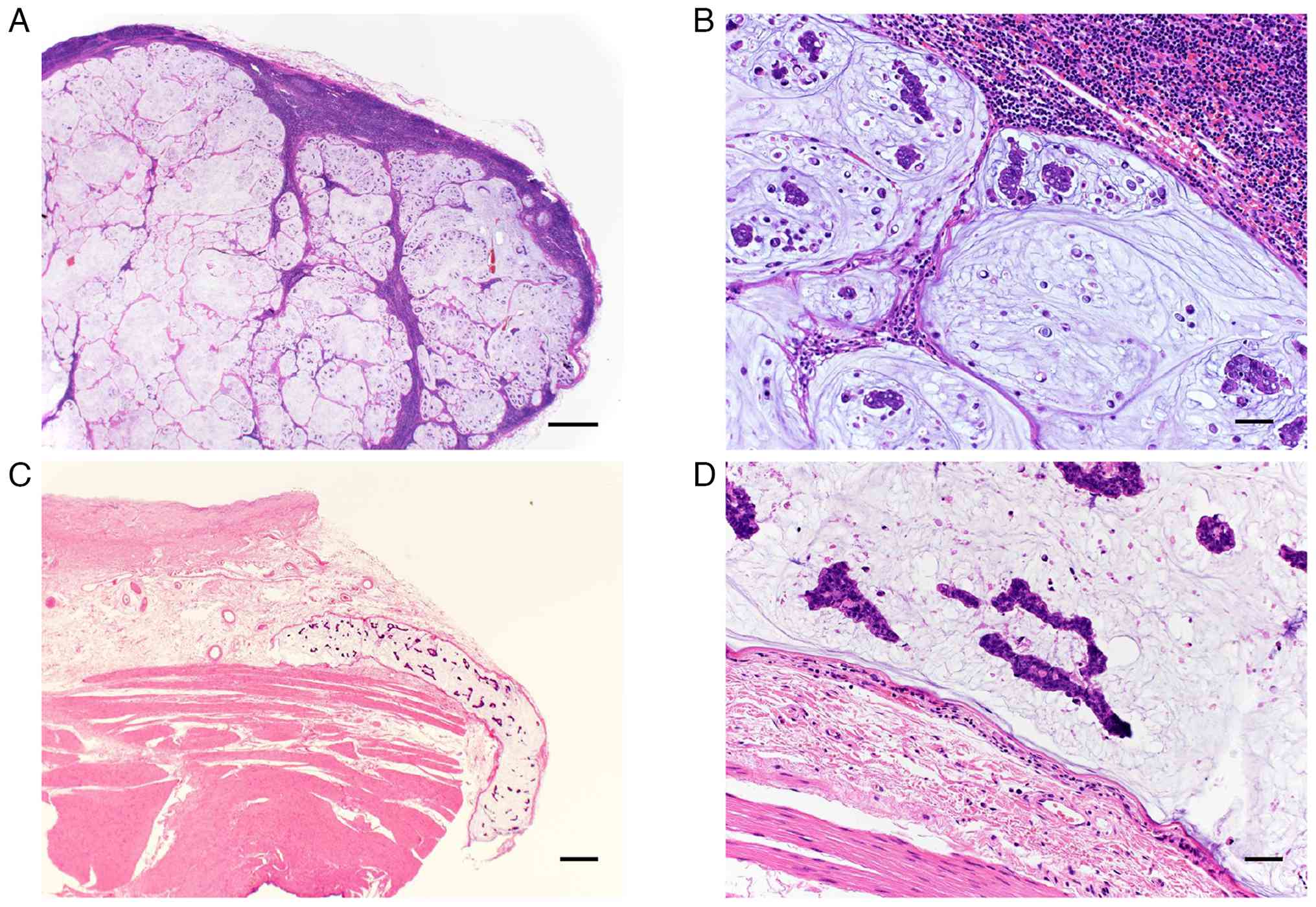
Histopathological examination revealed that the pancreatic head lesion represented a metastatic lymph node composed of poorly differentiated adenocarcinoma with signet ring cell features (Fig. 3A and B). Postoperative immunohistochemical analyses were performed on the resected specimens, including the metastatic lymph node located at the pancreatic head. The tumor cells were negative for synaptophysin and chromogranin A, whereas they were positive for cytokeratin 7, cytokeratin 20, CDX2, HNF4α, and mucin markers (MUC2, MUC5AC, and MUC6) (data not shown), supporting a gastrointestinal origin of the metastatic lesion.

Hematoxylin and eosin staining. (A) A
tumor in the pancreatic head was identified as a metastatic lymph
node. (B) The metastatic lymph node contained signet ring cells and
poorly differentiated adenocarcinoma with a large amount of
mucinous component. (C) Microscopic vascular invasion was detected
within the submucosa of the resected stomach. (D) Tumor cells
within the submucosa of the resected stomach showed same
histological features as metastatic lymph nodes. Scale bars
indicate 1,000, 50, 500 and 50 µm, respectively.

Figure 3.

Hematoxylin and eosin staining. (A) A tumor in the pancreatic head was identified as a metastatic lymph node. (B) The metastatic lymph node contained signet ring cells and poorly differentiated adenocarcinoma with a large amount of mucinous component. (C) Microscopic vascular invasion was detected within the submucosa of the resected stomach. (D) Tumor cells within the submucosa of the resected stomach showed same histological features as metastatic lymph nodes. Scale bars indicate 1,000, 50, 500 and 50 µm, respectively.

No macroscopically identifiable primary gastric tumor was detected in the resected stomach. However, microscopic examination demonstrated lymphatic and vascular invasion composed of poorly differentiated adenocarcinoma with signet ring cell features in the submucosa of the gastric antrum along the lesser curvature (Fig. 3C and D), supporting the diagnosis of occult gastric cancer.

Metastatic lymph nodes were identified in the infrapyloric region (station no. 6), the peripancreatic region adjacent to the pancreatic head, and station no. 5. A total of 27 lymph nodes were retrieved, and metastatic involvement was confirmed in these stations. All surgical margins were negative. According to the UICC TNM classification (8th edition) (13), the final pathological stage was pTxN2M0.

Postoperatively, the patient received adjuvant chemotherapy with oral S-1 for one year. Follow-up consisted of clinical evaluation every two months and contrast-enhanced computed tomography every six months. Esophagogastroduodenoscopy performed 10 months after surgery revealed no remarkable findings. Serum tumor marker levels, including CEA and CA19-9, remained within the normal range during follow-up. The patient remains alive without evidence of recurrence two years after surgery.

Discussion

Pancreatic head tumors are usually regarded as primary pancreatic neoplasms and often prompt surgeons to proceed directly to pancreaticoduodenectomy. However, bulky metastatic lymph nodes from gastrointestinal malignancies may anatomically and radiologically mimic pancreatic tumors, resulting in an important oncologic diagnostic pitfall (9–11). Metastatic involvement of the pancreas or peripancreatic region from gastric cancer has been reported, although it remains uncommon (11,14–18). The present case illustrates how occult gastric cancer metastasis can present as a pancreatic head tumor despite comprehensive preoperative imaging and endoscopic evaluation (7,8,12,19). From a practical standpoint, this case suggests that pancreatic head masses presenting as protruding lesions extending outside the pancreas and differing from the typical appearance of primary pancreatic neoplasms should prompt consideration of metastatic lymph node involvement, even when esophagogastroduodenoscopy reveals no remarkable findings.

Bulky metastatic lymph nodes located in the infrapyloric or peripancreatic region may anatomically overlap with the pancreatic head, leading to imaging findings indistinguishable from primary pancreatic tumors (11,15–19). In particular, the infrapyloric lymph node (station No.6) is situated adjacent to the pancreatic head and represents a major lymphatic drainage pathway for distal gastric cancer (5,6). Consequently, metastatic enlargement of these lymph nodes may masquerade as a pancreatic head mass on computed tomography, positron emission tomography-computed tomography, or EUS, even when multimodal imaging is performed (10,11,20). although adenocarcinoma was identified by preoperative biopsy of the infrapyloric lymph node, the primary site could not be determined, highlighting the limitation of tissue sampling in distinguishing primary pancreatic cancer from metastatic disease (17).

Occult gastric cancer is characterized by the absence of an identifiable primary lesion on endoscopic examination despite the presence of metastatic disease (7,8,19). Poorly differentiated adenocarcinoma, including signet ring cell carcinoma, may infiltrate the submucosa and lymphovascular structures without forming an obvious mucosal lesion (7,8). In the present case, microscopic vascular invasion with histological features identical to those of the metastatic lymph nodes was detected in the gastric submucosa, strongly suggesting a gastric origin. This biological behavior explains why careful esophagogastroduodenoscopy failed to detect a primary tumor and underscores the limitation of endoscopic evaluation alone in excluding gastric cancer. Furthermore, when postoperative pathological examination reveals lymph node metastasis without identification of a primary tumor, the possibility of a gastric origin should be kept in mind, and close postoperative follow-up with esophagogastroduodenoscopy may be appropriate.

From a surgical perspective, recognition of this diagnostic pitfall has important implications for procedure selection. When a protruding lesion is encountered in the pancreatic head in association with suspicious infrapyloric lymphadenopathy, metastatic lymph node involvement from occult gastric cancer should be included in the differential diagnosis (5,6). Previous studies have demonstrated that pancreatic resection for isolated metastatic disease can be performed safely in selected patients at high-volume centers (17,21–23). In the present case, intraoperative recognition of the protruding nature of the lesion prompted modification of the surgical strategy to include distal gastrectomy considering the possibility of the lymph node metastasis from occult gastric cancer, enabling oncologically appropriate en bloc resection.

This case underscores that, even when comprehensive imaging and endoscopic evaluation suggest primary pancreatic cancer, metastatic lymph nodes from occult gastric cancer may represent an alternative diagnosis, particularly in the presence of infrapyloric lymphadenopathy. Awareness of this diagnostic and surgical pitfall is essential for appropriate surgical decision-making when evaluating pancreatic head lesions accompanied by perigastric lymphadenopathy.

Supplementary Material

Supporting Data

Acknowledgements

Not applicable.

Funding

Funding: No funding was received.

Availability of data and materials

The data generated in the present study may be requested from the corresponding author.

Authors' contributions

TA, YH, KT, JS, HIs, SA, YK, MN, HIc, YS and TW participated in the conception and design of this report. TA and YH drafted and revised the manuscript, and are responsible for the paper. JS, HIs, SA and HIc provided advice on imaging findings, interpreted the imaging data and contributed the writing of the manuscript. KT, MN, YS and TW provided advice on histological findings and contributed to the writing of the manuscript. KT, JS and TW critically revised the paper. TA and YH confirm the authenticity of all the raw data. All authors read and approved the final manuscript.

Ethics approval and consent to participate

Not applicable.

Patient consent for publication

The patient provided written informed consent for the publication of the data.

Competing interests

The authors declare that they have no competing interests.

References

1 

Siegel RL, Giaquinto AN and Jemal A: Cancer statics, 2024. CA Cancer J Clin. 74:12–49. 2024.PubMed/NCBI

2 

Oba A, Del Chiaro M, Fujii T, Okano K, Stoop TF, Wu YHA, Maekawa A, Yoshida Y, Hashimoto D, Sugawara T, et al: ‘Conversion surgery’ for locally advanced pancreatic cancer: A position paper by the study group at the joint meeting of the International Association of Pancreatology (IAP) & Japan Pancreas Society (JPS) 2022. Pancreatology. 23:712–720. 2023. View Article : Google Scholar : PubMed/NCBI

3 

Perri G, Marchegiani G, Frigerio I, Dervenis CG, Conlon KC, Bassi C and Salvia R: Management of pancreatic cystic lesions. Dig Surg. 37:1–9. 2020. View Article : Google Scholar : PubMed/NCBI

4 

Stoop TF, Javed AA, Oba A, Koerkamp BG, Seufferlein T, Wilmink JW and Besselink MG: Pancreatic cancer. Lancet. 405:1182–1202. 2025. View Article : Google Scholar : PubMed/NCBI

5 

Rosa F, Costamagna G, Doglietto GB and Alfieri S: Classification of nodal stations in gastric cancer. Transl Gastroenterol Hepatol. 2:22017. View Article : Google Scholar : PubMed/NCBI

6 

Tokunaga M, Ohyama S, Hiki N, Fukunaga T, Yamada K, Sano T and Yamagucji T: Investigation of the lymphatic stream of the stomach in gastric cancer with solitary lymph node metastasis. World J Surg. 33:1235–1241. 2009. View Article : Google Scholar : PubMed/NCBI

7 

Lee SH, Lim KH, Song SY, Lee HY, Park SC, Kang CD, Lee SJ, Choi DW, Park SB and Ryu YJ: Occult gastric cancer with distant metastasis proven by random gastric biopsy. World J Gastroenterol. 22:4270–4274. 2016. View Article : Google Scholar : PubMed/NCBI

8 

Kawakami N, Moriya T, Kato R, Nakamura K, Saito H, Wakai Y, Saito K and Sakashita M: Pulmonary tumor thrombotic microangiopathy in occult early gastric cancer that was undetectable on upper endoscopy: A case report and review of similar cases. BMC Gastroenterol. 21:4232021. View Article : Google Scholar : PubMed/NCBI

9 

Hammond NA, Miller FH, Day K and Nikolaidis P: Imaging features of the less common pancreatic masses. Abdom Imaging. 38:561–572. 2013. View Article : Google Scholar : PubMed/NCBI

10 

AI-Hawary MM, Kaza RK, Azar SF, Ruma JA and Francis IR: Mimics of pancreatic ductal adenocarcinoma. Cancer Imaging. 13:342–349. 2013. View Article : Google Scholar : PubMed/NCBI

11 

Duan Q, Huang Y and Li H: Large metastatic lymph nodes misdiagnosed as a pancreatic tumor: A case report. J Surg Case Rep. 2025:rjaf5832025. View Article : Google Scholar : PubMed/NCBI

12 

Rabenstein T, May A, Gossner L, Manner H, Pech O, Günter E, Huijmans J, Vieth M, Stolte M and Ell C: Invisible gastric carcinoma detected by random biopsy: Long-term results after photodynamic therapy. Endoscopy. 40:899–904. 2008. View Article : Google Scholar : PubMed/NCBI

13 

Brierley JD, Gospodarowicz MK and Wittekind C: TNM Classification of Malignant Tumours. 8th edtion. Wiley Blackwell; Chichester: pp. 93–95. 2017

14 

Robbins EG, Franceschi D and Barkin JS: Solitary metastatic tumors to the pancreas: A case report and review of the literature. Am J Gastroenterol. 91:2414–2417. 1996.PubMed/NCBI

15 

Adsay NV, Andea A, Basturk O, Kilinc N, Nassar H and Cheng JD: Secondary tumors of the pancreas: An analysis of a surgical and autopsy database and review of the literature. Virchows Arch. 444:527–535. 2004. View Article : Google Scholar : PubMed/NCBI

16 

Roalsø MT, Lucocq J, Pandanaboyama S and Søreide K: Surgery for metastasis to the pancreas: Systematic review of observational cohort studies. HPB (Oxford). 27:1290–1300. 2025. View Article : Google Scholar : PubMed/NCBI

17 

Z'graggen K, Fernandez-del Castillo C, Rattner DW and Warshaw AL: Metastases to the pancreas and their surgical extirpation. Arch Surg. 133:413–417. 1998. View Article : Google Scholar : PubMed/NCBI

18 

Ma J, Kimura W, Takeshita A, Hirai I, Moriya T and Mitzutani M: Neuroendocrine carcinoma of the stomach with peripancreatic lymph node metastases successfully treated with pancreaticoduodenectomy. Hepatogastroenterology. 54:1945–1950. 2007.PubMed/NCBI

19 

Biswas M, Smith JC and Davies JS: Bilateral adrenal enlargement and non-suppressible hypercortisolism as a presenting feature of gastric cancer. Ann Clin Biochem. 41:494–497. 2004. View Article : Google Scholar : PubMed/NCBI

20 

Tang Y, Sun X and Xu L: A slowly growing solitary skip lymph nodule of occult gastric cancer: A case report. Surg Case Rep. 6:1352020. View Article : Google Scholar : PubMed/NCBI

21 

Reddy S and Wolfgang CL: The role of surgery in the management of isolated metastases to the pancreas. Lancet Oncol. 10:287–293. 2009. View Article : Google Scholar : PubMed/NCBI

22 

Hiotis SP, Klimsta DS, Conlon KC and Brennan MF: Results after pancreatic resection for metastatic lesions. Ann Surg Oncol. 9:675–679. 2002. View Article : Google Scholar : PubMed/NCBI

23 

Wente MN, Bergmann F, Fröhlich BE, Schirmacher P, Bücheler MW and Freess H: Pancreatic metastasis from gastric carcinoma: A case report. World J Surg Oncol. 2:432004. View Article : Google Scholar : PubMed/NCBI

Related Articles

  • Abstract
  • View
  • Download
  • Twitter
Copy and paste a formatted citation
Spandidos Publications style
Ando T, Hirose Y, Takizawa K, Sakata J, Ishikawa H, Abe S, Kawachi Y, Nakano M, Ichikawa H, Shimada Y, Shimada Y, et al: Diagnostic and surgical challenges of a lymph node metastasis from occult gastric cancer mimicking a pancreatic head tumor: A case report. Oncol Lett 31: 162, 2026.
APA
Ando, T., Hirose, Y., Takizawa, K., Sakata, J., Ishikawa, H., Abe, S. ... Wakai, T. (2026). Diagnostic and surgical challenges of a lymph node metastasis from occult gastric cancer mimicking a pancreatic head tumor: A case report. Oncology Letters, 31, 162. https://doi.org/10.3892/ol.2026.15515
MLA
Ando, T., Hirose, Y., Takizawa, K., Sakata, J., Ishikawa, H., Abe, S., Kawachi, Y., Nakano, M., Ichikawa, H., Shimada, Y., Wakai, T."Diagnostic and surgical challenges of a lymph node metastasis from occult gastric cancer mimicking a pancreatic head tumor: A case report". Oncology Letters 31.5 (2026): 162.
Chicago
Ando, T., Hirose, Y., Takizawa, K., Sakata, J., Ishikawa, H., Abe, S., Kawachi, Y., Nakano, M., Ichikawa, H., Shimada, Y., Wakai, T."Diagnostic and surgical challenges of a lymph node metastasis from occult gastric cancer mimicking a pancreatic head tumor: A case report". Oncology Letters 31, no. 5 (2026): 162. https://doi.org/10.3892/ol.2026.15515
Copy and paste a formatted citation
x
Spandidos Publications style
Ando T, Hirose Y, Takizawa K, Sakata J, Ishikawa H, Abe S, Kawachi Y, Nakano M, Ichikawa H, Shimada Y, Shimada Y, et al: Diagnostic and surgical challenges of a lymph node metastasis from occult gastric cancer mimicking a pancreatic head tumor: A case report. Oncol Lett 31: 162, 2026.
APA
Ando, T., Hirose, Y., Takizawa, K., Sakata, J., Ishikawa, H., Abe, S. ... Wakai, T. (2026). Diagnostic and surgical challenges of a lymph node metastasis from occult gastric cancer mimicking a pancreatic head tumor: A case report. Oncology Letters, 31, 162. https://doi.org/10.3892/ol.2026.15515
MLA
Ando, T., Hirose, Y., Takizawa, K., Sakata, J., Ishikawa, H., Abe, S., Kawachi, Y., Nakano, M., Ichikawa, H., Shimada, Y., Wakai, T."Diagnostic and surgical challenges of a lymph node metastasis from occult gastric cancer mimicking a pancreatic head tumor: A case report". Oncology Letters 31.5 (2026): 162.
Chicago
Ando, T., Hirose, Y., Takizawa, K., Sakata, J., Ishikawa, H., Abe, S., Kawachi, Y., Nakano, M., Ichikawa, H., Shimada, Y., Wakai, T."Diagnostic and surgical challenges of a lymph node metastasis from occult gastric cancer mimicking a pancreatic head tumor: A case report". Oncology Letters 31, no. 5 (2026): 162. https://doi.org/10.3892/ol.2026.15515
Follow us
  • Twitter
  • LinkedIn
  • Facebook
About
  • Spandidos Publications
  • Careers
  • Cookie Policy
  • Privacy Policy
How can we help?
  • Help
  • Live Chat
  • Contact
  • Email to our Support Team