Introduction
Lung cancer is one of the leading causes of
cancer-related mortality worldwide, with non-small cell lung cancer
(NSCLC) accounting for approximately 80% of all cases (1–3).
Immune checkpoint inhibitors (ICIs) that restore antitumor immunity
by blocking inhibitory pathways, such as the programmed death-1
(PD-1) and PD-ligand 1 (PD-L1) pathways, have emerged as major
therapeutic strategies. In NSCLC, PD-1/PD-L1 blockade enhances
T-cell activation and improves patient prognosis (3). Among the ICIs, nivolumab, an anti-PD-1
antibody, has demonstrated clinical efficacy in patients with
advanced NSCLC (4). Nevertheless,
currently available biomarkers remain inadequate for accurately
predicting the sensitivity and clinical outcomes of ICIs, leading
to suboptimal patient stratification. Consequently, a substantial
proportion of patients do not benefit from treatment and may
develop immune-related adverse events or hyperprogression,
underscoring the urgent need for reliable predictive biomarkers for
advanced NSCLC (5). Several
tissue-based markers, such as tumoral PD-L1 expression, tumor
mutation burden, and interferon-γ signatures, have been proposed to
predict response to ICI and prognosis (3). However, their evaluation requires
invasive tumor tissue sampling. Blood-based biomarkers are highly
attractive because they can be obtained repeatedly via a minimally
invasive method and may offer a more accurate and dynamic
reflection of disease status.
Aberrant glycosylation is a common feature observed
in many human malignancies, including NSCLC, and serves as a marker
for distinguishing tumor cells from surrounding normal tissues
(6). Clinical tumor markers, such
as carbohydrate antigen 19-9 and carcinoembryonic antigen, which
are widely used in practice to indicate tumor presence and disease
progression, reflect cancer-specific glycan structures and are
detected in the blood using antibody-based immunoassays (7). Lectins are glycan-binding proteins
with high selectivity for specific glycan structures. To
distinguish subtle differences in glycan composition, lectins have
been widely used in biomarker studies to detect cancer-associated
glycan alterations (8). Recent
studies have shown that glycosylation of cancer cell surface
proteins affects immune evasion and the efficacy of ICIs,
highlighting its important role in regulating antitumor immune
responses (9).
This study focused on high-mannose glycans, a
subclass of tumor-associated glycans that selectively accumulate in
certain cancers (10). Notably, a
lectibody is a fusion molecule composed of a lectin domain that
selectively binds to specific glycan structures and an antibody Fc
region that mediates immune effector functions. One such lectibody,
constructed by fusing the high-mannose-binding lectin avaren to the
human immunoglobulin G1 Fc region, binds specifically to lung
cancer cell lines and tumor regions in clinical lung cancer tissues
(10). These findings demonstrate
the presence of high-mannose glycans in lung cancer cells and the
tumor microenvironment.
MicroRNAs (miRNAs) are small non-coding RNAs, 18–25
nucleotides in length, that regulate gene expression by binding to
the 3′-untranslated regions of target mRNAs, leading to mRNA
degradation or translational repression (11,12).
In lung cancer, miR-335, miR-17, miR-146a, and the miR-320 family
are candidate regulators of immune checkpoint molecules such as
PD-1, PD-L1, CD155, CD28, and cytotoxic T-lymphocyte antigen 4
(13). In this study, we focused on
high-mannose glycans, a class of cancer-associated glycan
structures, and attempted to identify tumor-specific circulating
miR markers by enriching cancer-derived components in the plasma
using OAA1, recombinant Oscillatoria agardhii agglutinin
(OAA), a lectin that selectively binds to high-mannose structures
(14,15). Notably, previous studies have
reported that miR-320b is significantly upregulated in plasma
exosomes from patients with a progressive disease (PD) compared
with those with a partial response (PR) at baseline before
PD-1/PD-L1 inhibitor treatment (16). Although miR-320a suppresses PD-L1
expression (17), no study has
reported an association between its circulating levels and ICI
sensitivity in patients with lung cancer. Moreover, to date, no
study has demonstrated the involvement of miR-3613-5p in the
response to ICIs. Moreover, although lectin-based methods have
previously been used to detect and enrich tumor-specific glycan
structures in the blood (15), no
studies have combined this approach with miRNA profiling to
evaluate its potential as a predictive biomarker strategy for
response to ICI. Therefore, this study aimed to determine whether
OAA1 enrichment of miRNAs could enhance their utility as predictive
biomarkers of response to nivolumab treatment and post-treatment
survival.
In this study, we developed a novel lectin-based
enrichment strategy using an OAA1 lectin column to selectively
capture glycan-associated circulating miRNAs from plasma. By
combining this approach with circulating miRNA profiling, we aimed
to identify biomarkers predictive of response to nivolumab in
patients with NSCLC. This strategy enables the enrichment of
glycan-associated miRNAs that are not readily detectable using
conventional plasma miRNA analyses and may improve the
identification of ICI responders and non-responders. Our findings
suggest that lectin-captured plasma miRNAs represent a promising
and previously unexplored class of predictive biomarkers for
immunotherapy response.
Materials and methods
Patients
A total of 48 patients with recurrent or advanced
NSCLC who initiated treatment with nivolumab at Gunma University
Hospital (Maebashi, Japan), Hidaka Hospital (Takasaki, Japan), and
National Hospital Organization Shibukawa Medical Center (Shibukawa,
Japan) between February 2016 and November 2017 were included in
this study. The inclusion criteria were i) pathologically confirmed
NSCLC, ii) recurrent or advanced stage, iii) eligibility for
nivolumab treatment following initial chemotherapy, and iv) Eastern
Cooperative Oncology Group performance status of 0–2. Plasma
samples and clinical data from patients included in a previous
study were used (18). Plasma
samples from 11 healthy volunteers were used as the controls.
The follow-up period for censored cases ranged from
1.3 to 36.5 months (median: 12.4 months). miRNA microarray analysis
was performed using total RNA extracted from OAA1-enriched plasma
samples collected before and 1 month post-treatment from two
individuals who developed PD despite receiving nivolumab. Patient
consent was obtained using the opt-out method. Healthy volunteers
were hospital staff aged ≥20 years who provided written informed
consent after receiving an explanation of the study. Individuals
with a history of malignant disease, acute infection, or regular
use of immunosuppressive medications were excluded. Additional
exclusion criteria included a bleeding tendency, severe anemia,
coagulation disorders, current use of anticoagulant or antiplatelet
drugs, pregnancy or breastfeeding, poor physical condition on the
day of blood sampling (e.g., fever or presyncope), or any other
condition judged inappropriate by the investigators.
This study was conducted in accordance with the
Declaration of Helsinki and was approved by the Institutional
Review Board for Clinical Research at Gunma University Hospital
(Maebashi, Japan; approval no. HS2023-094).
Preparation of OAA1 and
OAA1-immobilized column
OAA1, which contained two amino acid substitutions
of OAA and an additional linker sequence at the C-terminal region
for covalent immobilization, and OAA1-immobilized columns on
monolithic silica were prepared according to our previous report
(19).
Histochemical analysis of lectin for
tumor-specific high-mannose glycans using OAA1
Histochemical analysis of lectin was performed on
formalin-fixed paraffin-embedded sections of lung cancer tissue to
detect tumor-specific high-mannose glycans using OAA1. OAA1 was
labeled with biotin using the biotin-labeling kit-NH2
(final concentration: 1.3 mg/ml) [Dojindo Molecular Technologies,
Kumamoto, Japan]. Non-specific binding sites were blocked by
incubating the sections with 1% bovine serum albumin
(Sigma-Aldrich)/phosphate-buffered saline (PBS) for 1 h at room
temperature. The sections were then incubated with biotinylated
OAA1 diluted in PBS (×800) for 1 h at room temperature. After
washing, the slides were treated with the VECTASTAIN Elite ABC Kit
(Vector Laboratories, Inc.) for 30 min. The color was developed
using a diaminobenzidine (DAB) substrate solution, and the sections
were lightly counterstained with hematoxylin and mounted. Negative
controls were prepared by omitting the lectin incubation step.
Immunohistochemistry
We obtained 28 sections consisting of resected
specimens (n=20) and needle biopsies (n=8) from patients for whom
clinical samples were obtained.
For immunohistochemistry, 4-µm sections were cut
from the paraffin blocks of each sample. Each section was mounted
on a silane-coated glass slide, deparaffinized in xylene,
rehydrated through graded ethanol to water, and incubated with 0.3%
hydrogen peroxide for 30 min at room temperature to block
endogenous peroxidase activity. After rehydration through a graded
series of the ethanol treatments (90% for 1 min, 80% for 1 min, and
60% for 1 min), the sections for CD8 staining were heated in
boiling water using Immunosaver (Nisshin EM Co., Ltd.) for 45 min
at 98–100°C for antigen retrieval. For PD-L1 staining, Universal
HIER antigen retrieval reagent (cat. no. ab208572; Abcam) at 120°C
for 20 min in an autoclave. Non-specific binding sites were blocked
by incubating the sections with Protein Block Serum-Free (Agilent
Technologies) for 30 min at room temperature. Samples were
incubated overnight at 4°C with the following primary antibodies:
PD-L1 (E1L3N Rabbit mAb 1:200; cat. no. 13684; Cell Signaling
Technology, Inc., Danvers, MA) and CD8 (cat. no. ab4055; 1:1,000;
Abcam). The primary antibody was visualized using the Histofine
Simple Stain MAX-PO (Multi) Kit (Nichirei Biosciences, Inc.).
Chromogen 3,3-diaminobenzidine tetrahydrochloride was used as a
0.02% solution in 50 mM citric acid-ammonium acetate buffer (pH 6)
containing 0.005% hydrogen peroxide. The sections were lightly
counterstained with hematoxylin and mounted.
Tissue sections were examined by two independent
evaluators who were blinded to the patient data. The expression of
PD-L1 was evaluated using a semiquantitative scoring method based
on the percentage of stained cells: 1, ≤1; 2, 1–5; 3, 5–10; 4,
10–50; and 5, ≥50%. Tumors with a score >3 were graded as
positive. CD8 expression was semi-quantitatively evaluated based on
the extent of positive lymphocyte infiltration in the tumor
specimens, and patients with >5% positive lymphocytes were
defined as positive, based on previous studies (20–22).
MicroRNA extraction from OAA1-captured
plasma samples
To enrich for tumor-specific high-mannose
glycan-containing molecules, plasma samples were subjected to
lectin affinity capture using an OAA1 column. OAA1 was immobilized
on a silica monolith matrix in the column. Briefly, each plasma
sample was centrifuged at 10,000 × g for 10 min, and 50 µl of
plasma was applied onto the pre-equilibrated OAA1 column. The
column was centrifuged at 3,000 × g for 1 min to allow the sample
to pass through. After binding, unbound plasma components were
removed by washing the column with 400 µl of 10X D-PBS (−)
(FUJIFILM Wako Pure Chemical Corporation) and D-PBS (−) (FUJIFILM
Wako Pure Chemical Corporation). Glycan-containing molecules
specifically bound to OAA1 were then eluted using 200 µl of QIAzol
Lysis Reagent (QIAGEN), which was applied to a column and allowed
to stand for 5 min at room temperature by centrifugation at 3,000 ×
g for 1 min.
Total RNA, including miRNA, was extracted from
glycan-containing molecules in the plasma samples as follows: 40 µl
of chloroform was added to each sample, and the tubes were shaken
vigorously for 15 sec. The tubes were incubated at room temperature
for 2 min, followed by centrifugation at 12,000 × g for 5 min. The
upper aqueous phase was carefully transferred to a new tube, and an
equal volume of isopropanol was added. After thorough vortex
mixing, the miRNA was purified using the NucleoSpin miRNA Plasma
kit (Macherey-Nagel, Germany) with DNA digestion treatment,
according to the manufacturer's protocol. Without the OAA1
enrichment group, miRNA was directly extracted from plasma samples
using the NucleoSpin miRNA Plasma kit with DNA digestion treatment,
according to the manufacturer's protocol. The concentration and
purity of the extracted RNA were assessed using a NanoDrop
spectrophotometer (Thermo Fisher Scientific), and miRNA samples
were stored at −80°C until further analysis. The amount of RNA
obtained from the plasma-derived, lectin-captured fraction was
extremely low and consisted primarily of short RNAs such as miRNAs;
therefore, reliable quantification using NanoDrop and quality
assessment based on the RNA integrity number were difficult. In
this study, RNA was extracted from 50 µl of plasma from each
sample, and RNA quality was indirectly evaluated based on the
reproducibility and stability of Ct values in the TaqMan miRNA
assay.
miRNA microarray
For miRNA microarray analysis, to enrich
tumor-specific high-mannose glycan-containing molecules, plasma
samples were subjected to lectin affinity capture using OAA1 lectin
columns, as previously described, with modifications to the sample
volume and extraction protocol. Briefly, 1 ml of plasma was divided
equally among five OAA1 columns (200 µl plasma per column), each
containing OAA1 immobilized on a silica monolith matrix. After
centrifugation of each plasma sample at 10,000 × g for 10 min, the
plasma was applied to pre-equilibrated columns. The columns were
centrifuged at 3,000 × g for 1 min to allow the samples to pass
through. After washing, glycan-containing molecules bound to OAA1
were eluted by adding 200 µl of QIAzol Lysis Reagent to each column
and incubating at room temperature for 5 min, followed by
centrifugation at 3,000 × g for 1 min. The eluates from each column
were combined, and total RNA, including miRNA, was extracted as
follows: chloroform was added to the QIAzol eluate, and the mixture
was vigorously vortexed. After phase separation, the upper aqueous
phase was collected, and an equal volume of isopropanol was added.
miRNA was purified using the NucleoSpin miRNA Plasma kit with DNA
digestion treatment, according to the manufacturer's protocol, for
subsequent miRNA microarray analysis.
Microarray analysis was performed using the
GeneChip™ miRNA 4.0 Array (Thermo Fisher Scientific). RNA labeling
was performed with the FlashTag™ Biotin HSR RNA Labeling Kit
(Thermo Fisher Scientific) according to the manufacturer's
protocol, with a minor modification: instead of adjusting the RNA
input based on concentration, the maximum recommended sample volume
(8 µl per sample) was used, due to variability in RNA yield from
the upstream lectin-based procedure. Subsequent steps, including
hybridization, washing, staining, and scanning, were performed
according to the manufacturer's instructions.
miRNA microarray analysis and
candidate selection (discovery phase)
miRNA microarray analysis was conducted as an
exploratory discovery step. Only miRNAs classified as ‘T’ (true
detection) by the microarray analysis software were included,
whereas those flagged as ‘F’ (not reliably detected) were excluded
from subsequent analyses. Candidate miRNAs were selected based on
differential abundance between paired pre-treatment and
post-treatment plasma samples from the same patients.
miRNAs exhibiting a fold change >2 and a nominal
P-value <0.05 were considered eligible for further validation.
To minimize potential sex-related bias, miRNAs encoded on sex
chromosomes (X or Y) were excluded from candidate selection.
Reverse transcription-quantitative
polymerase chain reaction (RT-qPCR) for miRNA
For miR-320a (assay ID 002277), miR-320b (assay ID
002844), and miR-3613-5p (assay ID 463197_mat; Thermo Fisher
Scientific), RT-qPCR was performed, and cDNA was synthesized from
total miRNA using the TaqMan™ MicroRNA Reverse Transcription Kit
(Thermo Fisher Scientific) and specific stem-loop reverse
transcription primers (Thermo Fisher Scientific) according to the
manufacturer's protocol. The exact primer and probe sequences used
in the TaqMan MicroRNA Assays are proprietary and were not
disclosed by the manufacturer; therefore, the assay identification
numbers are provided to ensure reproducibility. The 15 µl reaction
volumes were incubated in 0.2 ml tubes, and the following
temperature profile was used: 16°C for 30 min, followed by 42°C for
30 min, and 85°C for 5 min. PCR was performed using a QuantStudio™
5 System (Thermo Fisher Scientific). The 10 µl PCR mix, including
the TaqMan™ Fast Advanced Master Mix for RT-qPCR (Thermo Fisher
Scientific), was incubated in a 384-well optical plate at 95°C for
20 s, followed by 40 cycles of 95°C for 1 sec and 60°C for 20 sec.
Levels of miR-320a, miR-320b, and miR-3613-5p were normalized to
that of miR-16-5p (used as an internal control; assay ID 000391)
and analyzed using the 2−∆Cq method. miR-16-5p was
chosen as a suitable reference gene, as described previously
(23–25). The fold change in target miRNA
expression relative to that in healthy volunteers was determined
using the 2−ΔΔCq method (26,27).
Statistical analysis
Statistical analyses were performed using the
Mann-Whitney U test for continuous variables and χ2 test
or Fisher's exact test for categorical variables. ROC curve
analysis was performed to compare the prognostic utility of
OAA1-captured plasma miRNAs with that of plasma miRNAs without OAA1
treatment. Kaplan-Meier curves were generated for overall survival,
and statistical significance was examined using the log-rank test.
Univariate analyses were performed using logistic regression or the
Cox proportional hazards model. P<0.05 was considered to
indicate a statistically significant difference. All statistical
analyses were performed using IBM SPSS Statistics, version 31 (IBM
Corp., Armonk, NY) and GraphPad Prism version 10 (GraphPad
Software, San Diego, CA). Figures were generated using GraphPad
Prism version 10.
The Cancer Genome Atlas (TCGA) data
analysis using UALCAN
TCGA RNA-seq data were analyzed using the UALCAN web
portal (http://ualcan.path.uab.edu). For lung
adenocarcinoma and lung squamous cell carcinoma cohorts, mRNA
expression levels (transcripts per million) of N-glycan
biosynthesis and processing enzymes, including ALG3
α-1,3-mannosyltransferase (ALG3), mannosidase α class 1A member 1
(MAN1A1), mannosidase α class 1C member 1 (MAN1C1),
α-1,3-mannosyl-glycoprotein 2-β-N-acetylglucosaminyltransferase
(MGAT1) and β-1,4-mannosyl-glycoprotein
4-β-N-acetylglucosaminyltransferase (MGAT3), were retrieved and
compared between primary tumor and normal tissues using TCGA module
of UALCAN. Statistical significance of expression differences was
assessed using an unpaired two sample t-test implemented in
UALCAN.
Results
Identification of circulating
OAA1-captured miRs associated with lung cancer and resistance to
nivolumab
Fig. 1A shows the
histochemical results for OAA1 in surgically resected lung cancer
specimens. This analysis revealed that high-mannose glycan
structures bound by OAA1 were more abundantly expressed in lung
cancer cells than in adjacent non-tumorous tissues (Fig. 1A). Similarly, TCGA RNA-seq data
analysis via UALCAN showed a higher expression of ALG3, with an
early addition of mannose during N-glycan assembly in the
endoplasmic reticulum, and lower expression of mannosidase MAN1A1
and MAN1C1, resulting in trimmed mannose residues in the Golgi to
prepare glycans for further processing, as well as lower expression
of MGAT1, which initiates complex N-glycan branching with the
addition of GlcNAc, and MGAT3, resulting in a ‘bisecting’ GlcNAc
that modulates later branching (28–30).
These expression patterns were consistent with the attenuated
trimming and maturation of N-glycans in tumors (Figs. S1 and S2).
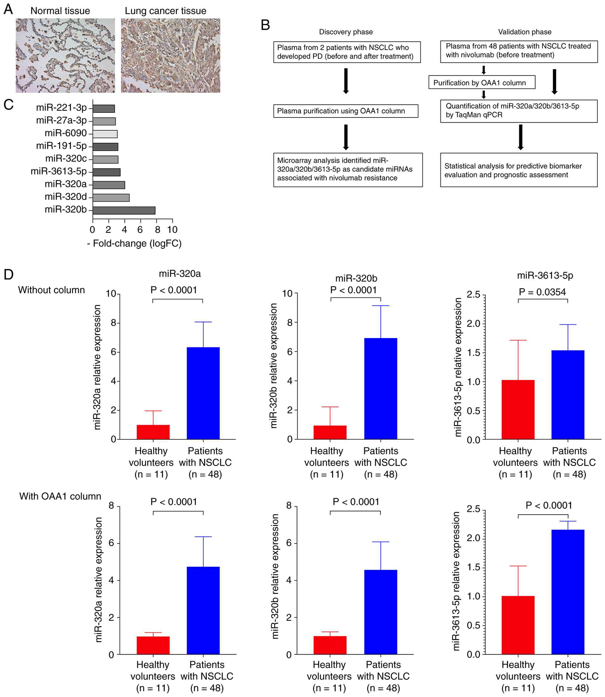 | Figure 1.Identification of circulating
OAA1-captured miRNAs associated with lung cancer and resistance to
nivolumab. (A) Lectin histochemistry using OAA1 on surgically
resected lung cancer tissues (×200 magnification). The left panel
shows OAA1 lectin staining in normal lung tissue, whereas the right
panel shows OAA1 lectin staining in lung cancer tissue. (B) In the
discovery phase, plasma samples from two patients with NSCLC with
PD were collected before and after nivolumab treatment. After
plasma purification using an OAA1 column, microarray analysis was
performed to identify candidate miRNAs (miR-320a, miR-320b, and
miR-3613-5p) associated with nivolumab resistance. In the
validation phase, plasma samples from 48 patients with NSCLC
(before nivolumab treatment) were analyzed with and without OAA1
column purification. The levels of the identified miRNAs were
quantified using TaqMan qPCR, and statistical analyses were
conducted to evaluate their predictive and prognostic values as
biomarkers for nivolumab resistance. (C) Microarray analysis of
OAA1-captured plasma miRNAs in patients with PD after nivolumab
treatment reveals several upregulated candidate miRNAs, including
miR-320a, miR-320b, and miR-3613-5p. (D) Relative levels of
miR-320a, miR-320b, and miR-3613-5p in pre-treatment plasma from
patients with NSCLC (n=48) and healthy controls (n=11) with and
without OAA1 enrichment. Plasma miRNA levels were quantified by
reverse transcription-qPCR. Relative miRNA levels were normalized
to miR-16-5p and calculated using the 2−ΔΔCq method,
with the mean level of healthy volunteers serving as the
calibrator. Statistical significance was determined using the
Mann-Whitney U test. Data are presented as median with 95%
confidence intervals. miRNA/miR, microRNA; NSCLC, non-small cell
lung cancer; OAA1, Oscillatoria agardhii agglutinin 1; PD,
progressive disease; qPCR, quantitative polymerase chain
reaction. |
Based on this finding, we used OAA1 to selectively
capture glycan-containing molecules associated with tumors from the
plasma of patients with lung cancer and conducted miRNA microarray
analysis to identify circulating miRNAs that were elevated during
treatment in patients who exhibited tumor progression to PD despite
nivolumab therapy (Fig. 1B). As
shown in Fig. 1C, several candidate
miRNAs were identified. Among them, hsa-miR-320a, hsa-miR-320b, and
hsa-miR-3613-5p were significantly upregulated in OAA1-captured
plasma samples after treatment compared with their pre-treatment
levels. Furthermore, we quantified these OAA1-captured miRNAs in
pre-treatment plasma samples from patients with advanced or
recurrent NSCLC (n=48) and healthy volunteers (n=11). As shown in
Fig. 1D, the levels of these miRNAs
were significantly higher in patients with NSCLC than in healthy
controls.
Differential expression of miR-320a,
miR-320b, and miR-3613-5p with and without OAA1 enrichment in the
plasma samples of patients with PR vs. those with stable disease
(SD) + PD
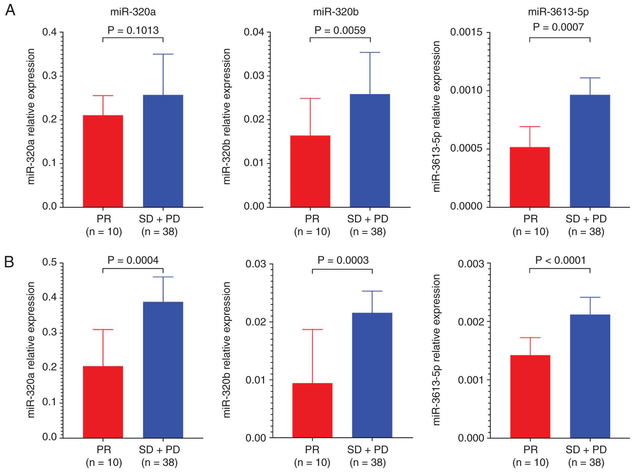
Next, we measured the levels of miR-320a, miR-320b,
and miR-3613-5p in plasma samples collected before nivolumab
treatment from patients who exhibited PR (n=10) and SD + PD (n=38).
This analysis was conducted under two conditions: With and without
OAA1 enrichment. Without OAA1 enrichment, there was no significant
difference in the levels of miR-320a between the PR and SD + PD
groups. However, both miR-320b and miR-3613-5p were expressed at
significantly higher levels in the SD + PD group than in the PR
group (Fig. 2 A). In contrast, with
OAA1 enrichment, the levels of all three miRNAs (miR-320a,
miR-320b, and miR-3613-5p) were significantly higher in the SD + PD
group than in the PR group (Fig. 2
B), suggesting that OAA1 enrichment enhances the sensitivity of
circulating miRNA-based detection for predicting the response to
nivolumab.
OAA1 enrichment enhances the
predictive power of circulating miRNAs for response to nivolumab in
NSCLC
Univariate logistic regression analysis was
performed to evaluate the association between circulating miRNA
levels and response to nivolumab (SD or PD) in patients with NSCLC
(Table I). Without OAA1 enrichment,
the odds ratios for predicting SD/PD were 3.208 for miR-320a,
12.444 for miR-320b, and 19.5 for miR-3613-5p. In contrast, with
OAA1 enrichment, the odds ratios increased to 12.889 for miR-320a,
27.222 for miR-320b, and 39.857 for miR-3613-5p, representing a
substantial improvement compared with the values obtained without
OAA1 enrichment (Table I). These
results suggest that OAA1 enrichment improves the predictive
utility of circulating miRNAs for identifying patients with poor
response to nivolumab. This finding is consistent with an earlier
comparison between the PR and SD + PD groups and supports the
potential of OAA1-captured plasma miRNAs as predictive biomarkers
in NSCLC.
 | Table I.Univariate logistic regression
analysis of clinicopathological factors and circulating
pre-treatment miRNA levels for predicting SD + PD. |
Table I.
Univariate logistic regression
analysis of clinicopathological factors and circulating
pre-treatment miRNA levels for predicting SD + PD.
| Clinicopathological
characteristic | Odds ratio (95%
confidence interval) | P-value |
|---|
| Age, years |
|
|
|
≤65 | 1 | 0.331 |
|
>65 | 2.100
(0.471–9.364) |
|
| Sex |
|
|
|
Male | 1 | 0.942 |
|
Female | 1.067
(0.188–6.045) |
|
| Histology |
|
|
|
SQC | 1 | 0.395 |
|
ADC | 0.481
(0.089–2.601) |
|
| Recurrent
disease |
|
|
|
Negative | 1 | 1 |
|
Positive | 1 (0.248–4.03) |
|
| Levels of tumor
markers |
|
|
|
Low | 1 | 0.686 |
|
High | 0.745
(0.179–3.096) |
|
| PD-L1 status |
|
|
|
Negative | 1 | 0.658 |
|
Positive | 0.658
(0.131–3.610) |
|
| CD8 expression |
|
|
|
Negative | 1 | 0.374 |
|
Positive | 0.350
(0.035–3.548) |
|
| Levels of
miR-320a |
|
|
|
Low | 1 | 0.127 |
|
High | 3.208
(0.717–14.35) |
|
| Levels of OAA1
captured miR-320a |
|
|
|
Low | 1 | 0.004a |
|
High | 12.889
(2.307–72.02) |
|
| Levels of
miR-320b |
|
|
|
Low | 1 | 0.002a |
|
High | 12.444
(2.489–62.21) |
|
| Levels of OAA1
captured miR-320b |
|
|
|
Low | 1 |
<0.001a |
|
High | 27.222
(4.53–163.75) |
|
| Levels of
miR-3613-5p |
|
|
|
Low | 1 | 0.007a |
|
High | 19.50
(2.213–171.86) |
|
| Levels of OAA1
captured miR-3613-5p |
|
|
|
Low | 1 | 0.001a |
|
High | 39.857
(4.317–368.02) |
|
OAA1 enrichment enhances the
predictive accuracy of circulating miRNAs for response to nivolumab
in patients with NSCLC
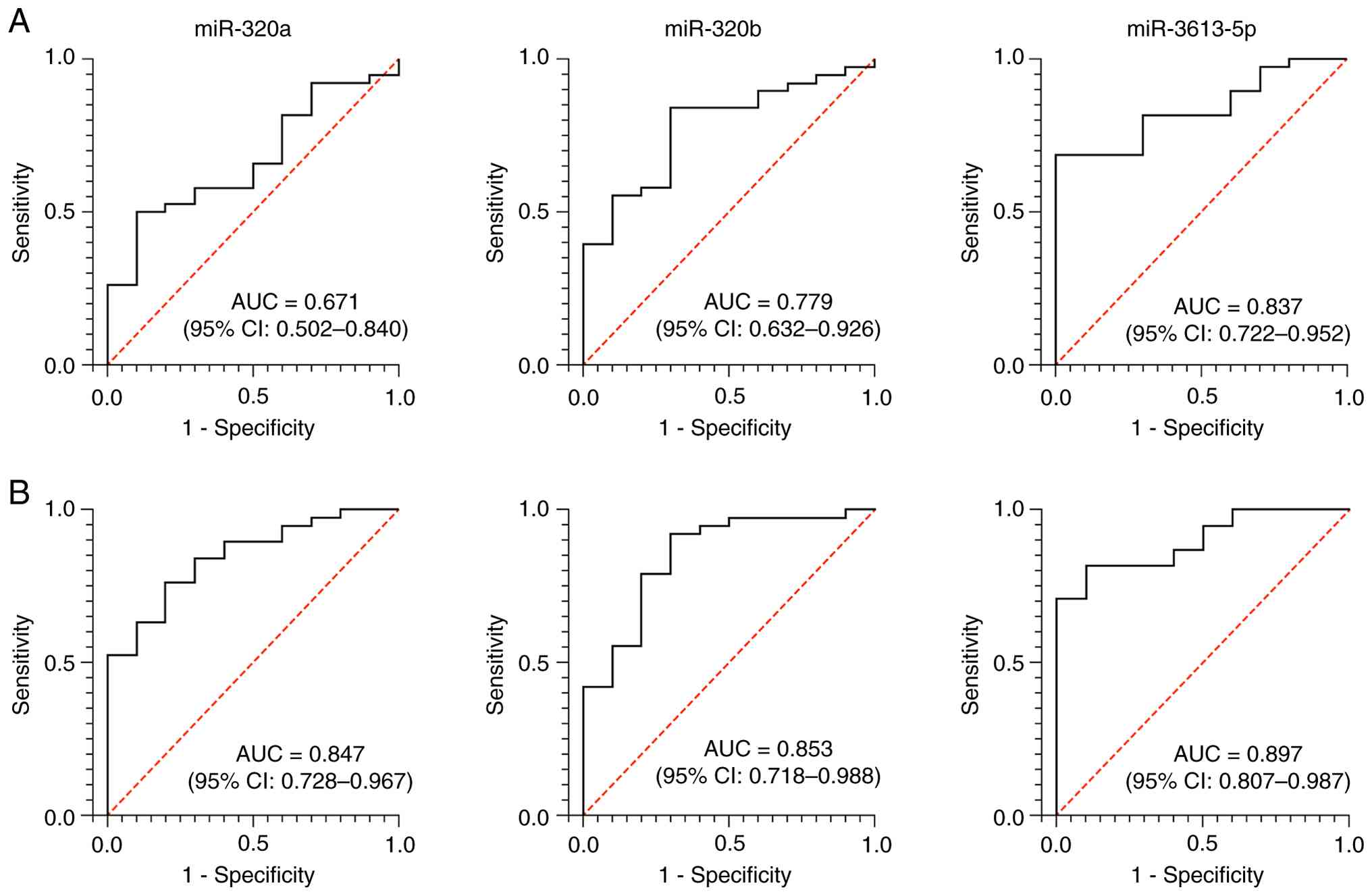
To evaluate the prognostic value of circulating
miR-320a, miR-320b, and miR-3613-5p in patients who responded to
nivolumab, receiver operating characteristic (ROC) curve analyses
were performed with and without OAA1 enrichment. Without OAA1
enrichment, the area under the curve (AUC) values for miR-320a,
miR-320b, and miR-3613-5p were 0.671 [95% confidence interval (CI):
0.502–0.840], 0.779 (95% CI: 0.632–0.926) and 0.837 (95% CI:
0.722–0.952), respectively (Fig.
3A). In contrast, with OAA1 enrichment, the AUCs increased to
0.847 (95% CI: 0.723–0.967) for miR-320a, 0.853 (0.718–0.988) for
miR-320b, and 0.897 (95% CI: 0.807–0.987) for miR-3613-5p, showing
improved performance compared with the corresponding values
obtained without OAA1 enrichment (Fig.
3B). These results suggest that OAA1 enrichment improves the
prognostic performance of these circulating miRNAs for overall
survival in patients with NSCLC treated with nivolumab.
The optimal cutoff values (2−ΔCq) were
determined using the ROC analysis. Without OAA1 enrichment, the
cutoff values were 0.2335 for miR-320a, 0.01662 for miR-320b, and
6.947×10⁻⁴ for miR-3613-5p, whereas with OAA1
enrichment, the corresponding values were 0.2610, 0.01164, and
1.740×10⁻³, respectively.
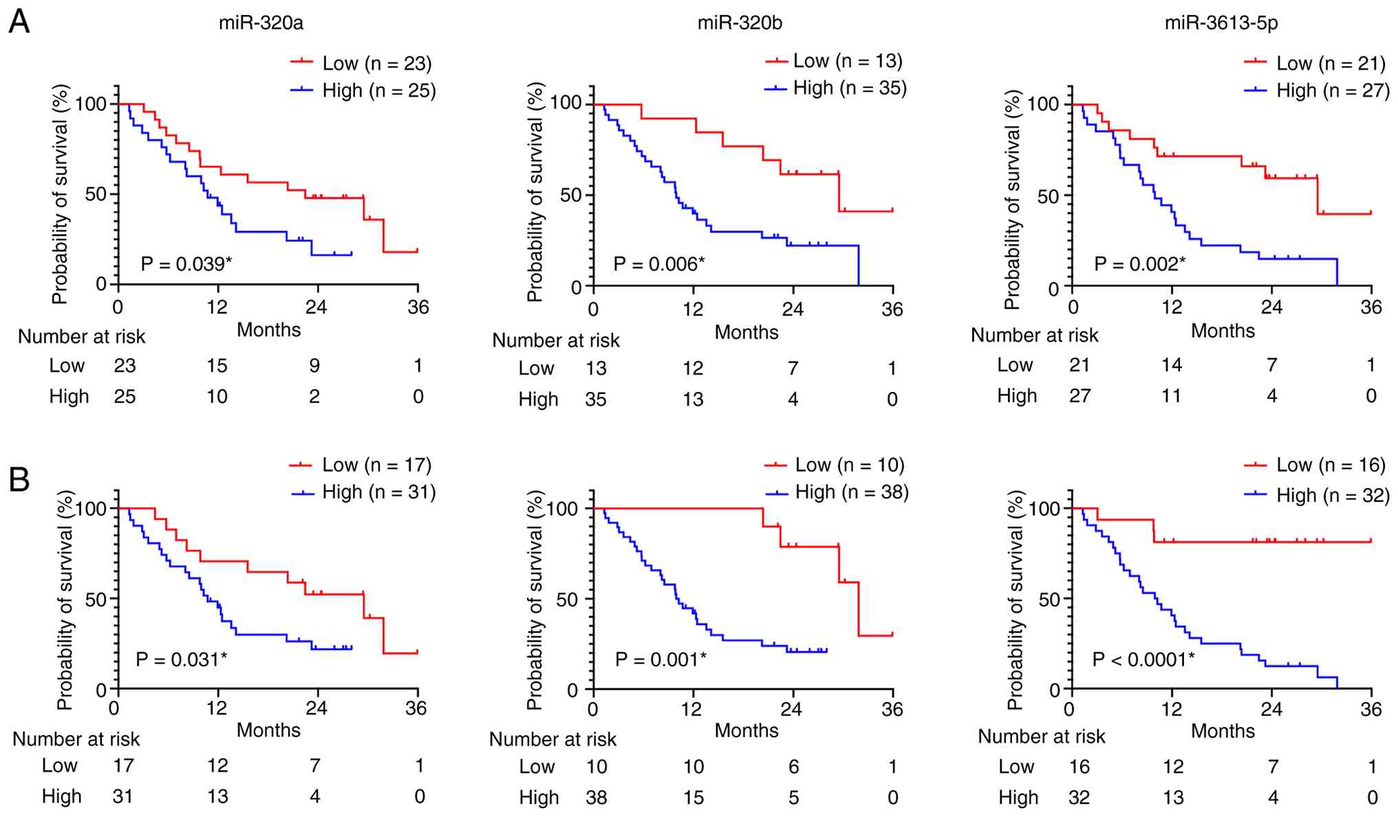
Association between OAA1-enriched
circulating miRNAs and survival outcomes in patients with NSCLC
treated with nivolumab
Circulating miRNA profiles with and without OAA1
enrichment were categorized into high- and low-expression groups
based on the cutoff values determined by the ROC curves for
patients who responded to nivolumab, as shown in Fig. 3. Based on each ROC-based threshold,
48 patients with NSCLC were stratified into high or low miRNA
expression groups. Table SI
summarizes the association between the levels of the target miRNAs
and clinicopathological features without OAA1 enrichment. In the
group without OAA1, high levels of miR-320b and miR-3613-5p were
significantly associated with low sensitivity to nivolumab (SD and
PD); however, no other clinical features were significantly
correlated (Table SI). In
contrast, with OAA1 enrichment, a high expression of all three
target miRNAs was significantly associated with SD and PD (Table SII).
Kaplan-Meier survival analysis with log-rank testing
was performed to assess the prognostic relevance of circulating
miR-320a, miR-320b, and miR-3613-5p in patients with NSCLC treated
with nivolumab with and without OAA1 enrichment. High plasma levels
of these miRNAs were significantly associated with poor overall
survival, regardless of the OAA1 enrichment status (Fig. 4).
Cox regression analysis was used to evaluate the
prognostic relevance of circulating miRNA levels for overall
survival in patients with NSCLC based on univariate modeling
(Table II). Without OAA1
enrichment, the hazard ratios (HRs) for predicting poor survival
were 2.136 for miR-320a, 3.578 for miR-320b, and 3.386 for
miR-3613-5p. In contrast, with OAA1 enrichment, the corresponding
HRs increased to 2.389 for miR-320a, 7.922 for miR-320b, and 7.815
for miR-3613-5p (Table II). These
results indicate that OAA1 enrichment enhances the prognostic
utility of circulating miRNAs and improves their ability to
stratify patients according to overall survival following nivolumab
treatment. Multivariable Cox regression analysis, including CD8
expression and the miRNAs identified as significant in the
univariate analysis, further supported these findings. In the
analysis without OAA1 enrichment, CD8 expression (P=0.008,
HR=0.149), miR-320b (P=0.024, HR=5.505) and miR-3613-5p (P=0.048,
HR=3.152) were identified as independent prognostic factors. In
contrast, in the analysis with OAA1 enrichment, only miR-3613-5p
remained an independent prognostic factor (P=0.010, HR=7.462)
(Tables SIII and SIV). Notably, the HR of miR-3613-5p was
higher with OAA1 enrichment than without enrichment.
| Table II.Univariate Cox regression analysis of
clinicopathological factors and circulating pre-treatment miRNA
levels of patients with non-small cell lung cancer treated with
nivolumab. |
Table II.
Univariate Cox regression analysis of
clinicopathological factors and circulating pre-treatment miRNA
levels of patients with non-small cell lung cancer treated with
nivolumab.
| Clinicopathological
characteristic | Hazard ratio (95%
confidence interval) | P-value |
|---|
| Age, years |
|
|
|
≤65 | 1 | 0.787 |
|
>65 | 0.908
(0.448–1.837) |
|
| Sex |
|
|
|
Male | 1 | 0.33 |
|
Female | 1.224
(0.815–1.838) |
|
| Smoking |
|
|
| No | 1 | 0.385 |
|
Yes | 1.238
(0.767–2.004) |
|
| Histology |
|
|
|
SQC | 1 | 0.7 |
|
ADC | 0.929
(0.637–1.354) |
|
| Recurrent
disease |
|
|
|
Negative | 1 | 0.587 |
|
Positive | 0.822
(0.406–1.666) |
|
| PD-L1 status |
|
|
|
Negative | 1 | 0.725 |
|
Positive | 0.827
(0.286–2.388) |
|
| CD8 expression |
|
|
|
Negative | 1 | 0.026a |
|
Positive | 0.283
(0.094–0.858) |
|
| Levels of tumor
markers |
|
|
|
Low | 1 | 0.185 |
|
High | 1.634
(0.791–3.377) |
|
| Levels of
miR-320a |
|
|
|
Low | 1 | 0.043a |
|
High | 2.136
(1.022–4.461) |
|
| Levels of OAA1
captured miR-320a |
|
|
|
Low | 1 | 0.036a |
|
High | 2.389
(1.059–5.390) |
|
| Levels of
miR-320b |
|
|
|
Low | 1 | 0.01a |
|
High | 3.578
(1.357–9.435) |
|
| Levels of OAA1
captured miR-320b |
|
|
|
Low | 1 | 0.005a |
|
High | 7.922
(1.868–33.59) |
|
| Levels of
miR-3613-5p |
|
|
|
Low | 1 | 0.003a |
|
High | 3.386
(1.499–7.649) |
|
| Levels of OAA1
captured miR-3613-5p |
|
|
|
Low | 1 |
<0.001a |
|
High | 7.815
(2.359–25.89) |
|
Discussion
In this study, we identified circulating miR-320a,
miR-320b, and miR-3613-5p as miRNAs that were upregulated after
treatment compared with pre-treatment levels in the plasma of
patients with NSCLC with PD despite nivolumab therapy. We further
demonstrated that these miRNAs were significantly associated with
disease progression and poor prognosis in patients with NSCLC
treated with nivolumab. Histochemical analysis of lectin using OAA1
showed that high-mannose glycan structures were abundantly
expressed in lung cancer tissues compared with adjacent
non-tumorous areas.
Supporting the presence of high-mannose-type glycans
indicated by lectin staining, gene expression analysis of the TCGA
lung cancer cohort using the UALCAN database (http://ualcan.path.uab.edu) also revealed
characteristic changes in glycan enzyme expression. Specifically,
in lung cancer tissues, the expression of the high-mannose-type
glycosyltransferase ALG3 was significantly elevated compared with
that in normal lung tissues. Conversely, the expression of Golgi
α-mannosidases I (MAN1A1 and MAN1C1), which are responsible for
removing and processing high-mannose-type N-glycans, and
N-acetylglucosamine transferases (MGAT1 and MGAT3) was
significantly reduced [Figs. S1
and S2] (28). MGAT1 initiates the conversion of
high-mannose to hybrid/complex N-glycans, whereas MGAT3 adds
bisecting GlcNAc, which modulates branching. The reduced expression
of these enzymes is expected to impair maturation and favor the
retention of high-mannose glycans (29). Indeed, a relative increase in
N-glycans with high-mannose structures has been reported in the
tissues of patients with lung cancer compared with normal tissues,
which is consistent with the gene expression patterns revealed in
this analysis (30). These results
provide molecular-level support for the possibility that
high-mannose-type glycans are produced and accumulate due to
abnormalities in the glycan synthesis pathway in tumor cells.
In addition, extracellular vesicles (EVs) reflect
the molecular characteristics of their cells of origin, including
glycosylation patterns. Therefore, EVs released from tumor cells
with aberrant glycan biosynthesis are expected to carry
high-mannose-type glycans on their surface. Consistent with this
notion, the OAA1 lectin column selectively captured EV-associated
particles bearing high-mannose glycans, as evidenced by the
enrichment of EV markers and tumor-associated miRNAs in the
captured fraction (19). Overall,
these findings support the interpretation that a substantial
proportion of the lectin-captured EVs originate from tumor cells
and retain tumor-specific glycosylation features.
Based on this finding, we established a strategy to
selectively enrich tumor-derived glycan-associated molecules from
plasma using an OAA1 lectin column, followed by miRNA profiling.
Circulating miRNAs captured by OAA1 showed significantly higher
expression in patients with SD or PD than in those with PR.
Logistic regression analysis revealed that the odds ratios for
predicting SD/PD were markedly increased in the OAA1-enrichment
group, indicating an improved predictive power. Furthermore, ROC
curve and Kaplan-Meier analyses showed that high expression of
these miRNAs was significantly associated with poor overall
survival, regardless of OAA1 enrichment status. Notably, both AUC
and HRs consistently improved with OAA1 enrichment, further
indicating its prognostic performance. To the best of our
knowledge, this is the first study to demonstrate that plasma
enrichment of tumor-specific glycans using lectin combined with
circulating miRNA profiling improves the prediction of resistance
to ICIs and poor prognosis in NSCLC.
In addition to OAA1, several other lectins that bind
high-mannose-type glycans, such as Concanavalin A (ConA),
Galanthus nivalis lectin (GNL), and Narcissus
pseudonarcissus agglutinin (NPA), have long been used in
glycomic analyses and cancer biomarker studies. However, these
lectins generally exhibit broad binding affinities for high-mannose
and other glycans that may be present in non-tumor tissues, thereby
limiting their tumor specificity (8). In contrast, OAA, a novel lectin
derived from the cyanobacterium Oscillatoria agardhii,
selectively recognizes and binds to high-mannose structures present
in tumor cell-derived exosomes (15). OAA1 (a recombinant OAA) shows
minimal binding to secreted factors derived from noncancerous cells
and exhibits a unique capacity to enrich tumor-specific components
(19). In addition to its high
specificity and strong binding affinity, OAA exhibits remarkable
physicochemical stability. Previous studies have demonstrated that
its activity is maintained over a wide pH range (pH 4–11) and under
high-temperature conditions (31).
Furthermore, its activity is independent of divalent cations, as
neither EDTA treatment nor the addition of Ca2+,
Mg2+, or Mn2+ affects its function. Based on
these characteristics and the specific binding of OAA1 to tumor
cells, we considered OAA1 to be superior to other mannose-binding
lectins for selectively enriching tumor-associated glycans with
miRNA biomarkers. Therefore, we employed OAA1 in this study to
identify predictive markers of nivolumab sensitivity.
The association between high levels of circulating
miR-320a, miR-320b, and miR-3613-5p with poor response to nivolumab
in patients with NSCLC suggests that these miRNAs may be involved
in the mechanisms of immune evasion or resistance to PD-1 blockade.
Previous studies have reported that members of the miR-320 family
suppress PD-L1 expression and modulate T-cell activation and
cytokine signaling by regulating immune checkpoint molecules
(13). In contrast, although high
levels of PD-L1 protein expression in tumor tissues are generally
associated with poor prognosis (32), tumors that simultaneously exhibit
abundant CD8+ cytotoxic T-cell infiltration are considered ‘hot
tumors’ and are well known to respond favorably to ICI therapy
(33). Particularly, miR-320 family
members target PD-L1 mRNA, promoting its degradation and thereby
reducing PD-L1 protein levels. Therefore, in our cohort with high
plasma levels of miR-320a and miR-320b, it is plausible that the
intratumoral expression of these miRNAs leads to the suppression of
PD-L1 protein, thereby shifting the immune landscape toward a ‘cold
tumor’ phenotype with reduced immunogenicity and ICI sensitivity.
Although miR-320a has been reported to function as a tumor
suppressor in NSCLC by inhibiting tumor cell proliferation and
invasion (34,35), its immunomodulatory effects,
particularly PD-L1 suppression, may paradoxically contribute to an
immune-cold tumor microenvironment with limited responsiveness to
immune checkpoint blockade.
Furthermore, a recent study on immune reconstitution
after allogeneic hematopoietic stem cell transplantation in
patients with acute myeloid leukemia reported that elevated plasma
levels of miR-3613-5p were inversely correlated with the number of
peripheral CD8+ cytotoxic T cells (36). This suggests that miR-3613-5p may
reflect systemic immunosuppression. Collectively, these findings
indicate that high levels of circulating miRNAs may mark a tumor
microenvironment characterized by low PD-L1 expression and impaired
CD8+ T-cell activity, which is consistent with a cold tumor state.
Such patients may be unable to mount an effective immune response
despite nivolumab administration, resulting in therapeutic
resistance and a poor prognosis. Therefore, the OAA1-based plasma
miRNA assay may serve as a noninvasive tool for assessing the
immune status of nivolumab-targeted lesions. In our cohort, we
observed no correlation between PD-L1 and miRNA levels. This null
finding may reflect limited PD-L1 availability (assessed in only 28
of 48 patients) and temporal discordance because PD-L1 was measured
in archival surgical or biopsy specimens rather than immediately
before nivolumab initiation. Further studies are warranted to
investigate the relationship between OAA1-captured circulating
miRNAs and the intratumoral expression of immune checkpoint
proteins and immune cell infiltration.
The evaluation of circulating miRNAs captured from
the plasma using OAA1 holds promise as a blood-based biomarker for
predicting therapeutic responses and stratifying prognosis in the
context of ICI treatment, particularly in patients with NSCLC.
Although current biomarkers, such as PD-L1 expression and tumor
mutational burden, are widely used to guide ICI therapy (6), they are tissue-based and thus limited
by their invasiveness and challenges associated with repeated
assessment. The strategy investigated in this study, which involves
the enrichment of cancer-derived glycans from plasma using OAA1,
followed by miRNA quantification, provides a minimally invasive and
repeatable alternative. This approach may be particularly useful in
cases where tumor tissue is difficult to obtain or where
longitudinal monitoring during treatment is necessary. Importantly,
the identified miRNAs were associated with nivolumab sensitivity
and clinical outcomes, highlighting their potential as biomarkers
for treatment selection and disease monitoring in patients with
advanced NSCLC. Taken together, these OAA1-based assays may
complement existing tissue-derived markers and support personalized
approaches for ICI therapy.
This study had some limitations. First, the
relatively small sample size might have limited the statistical
power to detect significant associations. In addition, the cutoff
values used to classify miRNA levels into high and low groups were
derived from ROC analysis based on nivolumab response status.
However, validation in large independent external cohorts is
required to ensure the generalizability of these findings. Second,
PD-L1 and CD8 immunohistochemistry could only be evaluated in 28 of
the 48 patients because of the limited availability of tissue
samples. This partial ascertainment may introduce a selection bias
and limit the statistical power, and the combination of resected
specimens and needle biopsies may also increase sampling
variability.
Third, although the enrichment of plasma components
using OAA1 improved the performance of miRNA biomarkers, the
underlying biological mechanisms remain unclear. The coexistence of
high-mannose glycans with miRNAs and their contribution to
nivolumab sensitivity and patient prognosis remain unclear.
Finally, the analysis was limited to patients with NSCLC who were
treated with nivolumab. Therefore, it remains uncertain whether the
identified miRNAs or the OAA1-based enrichment method is applicable
to other ICIs or different tumor types. In addition, because the
amount of RNA obtained from the plasma-derived, lectin-captured
fraction was extremely low, spectrophotometric RNA quality
assessment using NanoDrop was performed near the lower detection
limit of the instrument, making it difficult to obtain reliable
absorbance spectra or purity ratios. Therefore, in this study, RNA
quality was primarily evaluated based on the stability and
reproducibility of Ct values of the endogenous control miRNA
(miR-16-5p), as measured using the TaqMan miRNA assay.
In conclusion, this study identified circulating
miR-320a, miR-320b, and miR-3613-5p as promising biomarkers of
nivolumab resistance and poor prognosis in NSCLC. By employing the
tumor-specific high-mannose glycan-binding lectin OAA1, we
successfully established a novel strategy to capture and enrich
glycan-associated molecules from the plasma. This approach enables
a more sensitive detection of clinically relevant miRNAs and
improves their predictive and prognostic performance. The
consistent enhancement of AUCs, odds ratios, and HRs following
OAA1-based enrichment highlights the utility of glycan-focused
sample processing in liquid biopsy strategies. Importantly, this is
the first study to demonstrate that lectin-mediated glycan capture
can augment the clinical value of circulating miRNAs in ICI
therapy. These findings offer a promising foundation for developing
precise oncology tools that integrate glycomics and miRNA-based
profiling to optimize therapeutic decision-making in NSCLC and
other malignancies.
Supplementary Material
Supporting Data
Supporting Data
Acknowledgements
The authors would like to thank Ms. Mariko Nakamura
(Department of General Surgical Science, Graduate School of
Medicine, Maebashi, Gunma, Japan), Ms. Kao Abe (Division of Gene
Therapy Science, Gunma University, Initiative for Advanced
Research, Maebashi, Gunma, Japan) and Ms. Yukiko Suto (Core
Facility Management and Technical Collaboration Center, Maebashi,
Gunma, Japan) for technical and administrative assistance.
Funding
This study was supported by Grants-in-Aid for Scientific
Research from the Japan Society for the Promotion of Science (JSPS;
grant no. 23K08288); the Japan Agency for Medical Research and
Development (AMED; grant no. JP256f0137008); the Gunma Foundation
for Medicine and Health Science Research; and the Takeda Science
Foundation.
Availability of data and materials
The miRNA microarray data generated in the present
study may be found in the Gene Expression Omnibus under accession
number GSE310370 or at the following URL: https://www.ncbi.nlm.nih.gov/geo/query/acc.cgi?acc=GSE310370.
The other data generated in the present study may be requested from
the corresponding author.
Authors' contributions
EN, YN, SN and TYo contributed to the study concept
and design. EN was responsible for data and statistical analyses,
and drafting and revision of the manuscript. YN performed sample
processing, data collection, data interpretation, and drafting and
revision of the manuscript. KS and HS contributed to data analysis
and interpretation, and assisted in the preparation of the
manuscript. KH, YO, NK, TYa, RY, KN, RK, HK, YT, SMMZ, HO, TS, NN,
TI and KK contributed to data collection, analysis and
interpretation, and critically reviewed the manuscript. EN, YN and
TYo confirm the authenticity of all the raw data. All authors read
and approved the final manuscript.
Ethics approval and consent to
participate
The present study was conducted in accordance with
The Declaration of Helsinki and was approved by the Institutional
Review Board for Clinical Research at Gunma University Hospital
(Maebashi, Japan; approval no. HS2023-094). Informed consent to
participate was obtained from patients using an opt-out method.
Written informed consent for participation in this study was
obtained from all the healthy volunteers.
Patient consent for publication
Not applicable.
Competing interests
YO, NK, TYa, RY, KN, RK, SMMZ, HO, TS, NN, TI, KK,
HS and KS declare no competing interests. EN, YN, SN, KH, HK, YT,
KS, and TYo are co-inventors on a pending patent application
related to a lectin-column formulation and its applications, titled
‘Method for Predicting Response to Immune Checkpoint Inhibitor
Therapy and Kit’ (filed September 11, 2025). The patent applicant
(assignee) is The IT Lab Co., Ltd. KH and HK are employees, YN is
an employee and shareholder, and YT is a co-founder and board
member of The IT Lab Co., Ltd. TYo received research grants from
The IT Lab. Co., Ltd., which also provided the study materials.
References
|
1
|
Bray F, Laversanne M, Sung H, Ferlay J,
Siegel RL, Soerjomataram I and Jemal A: Global cancer statistics
2022: GLOBOCAN estimates of incidence and mortality worldwide for
36 cancers in 185 countries. CA Cancer J Clin. 74:229–263.
2024.PubMed/NCBI
|
|
2
|
Siegel RL, Kratzer TB, Giaquinto AN, Sung
H and Jemal A: Cancer statistics, 2025. CA Cancer J Clin. 75:10–45.
2025.PubMed/NCBI
|
|
3
|
Zhang J, Song Z, Zhang Y, Zhang C, Xue Q,
Zhang G and Tan F: Recent advances in biomarkers for predicting the
efficacy of immunotherapy in non-small cell lung cancer. Front
Immunol. 16:15548712025. View Article : Google Scholar : PubMed/NCBI
|
|
4
|
Brahmer JR, Lee JS, Ciuleanu TE, Bernabe
Caro R, Nishio M, Urban L, Audigier-Valette C, Lupinacci L, Sangha
R, Pluzanski A, et al: Five-year survival outcomes with nivolumab
plus ipilimumab versus chemotherapy as first-line treatment for
metastatic non-small-cell lung cancer in CheckMate 227. J Clin
Oncol. 41:1200–1212. 2023. View Article : Google Scholar : PubMed/NCBI
|
|
5
|
Li Y, Chen T, Nie TY, Han J, He Y, Tang X
and Zhang L: Hyperprogressive disease in non-small cell lung cancer
after PD-1/PD-L1 inhibitors immunotherapy: Underlying killer. Front
Immunol. 14:12008752023. View Article : Google Scholar : PubMed/NCBI
|
|
6
|
Giurini EF, Pappas SG and Gupta KH: Sweet
surprises: Decoding tumor-associated glycosylation in cancer
progression and therapeutic potential. Cells. 15:2332026.
View Article : Google Scholar : PubMed/NCBI
|
|
7
|
Lee T, Teng TZJ and Shelat VG:
Carbohydrate antigen 19-9-tumor marker: Past, present, and future.
World J Gastrointest Surg. 12:468–490. 2020. View Article : Google Scholar : PubMed/NCBI
|
|
8
|
Islam MK, Khan M, Gidwani K, Witwer KW,
Lamminmäki U and Leivo J: Lectins as potential tools for cancer
biomarker discovery from extracellular vesicles. Biomark Res.
11:852023. View Article : Google Scholar : PubMed/NCBI
|
|
9
|
Ren X, Lin S, Guan F and Kang H:
Glycosylation targeting: A paradigm shift in cancer immunotherapy.
Int J Biol Sci. 20:2607–2621. 2024. View Article : Google Scholar : PubMed/NCBI
|
|
10
|
Oh YJ, Dent MW, Freels AR, Zhou Q,
Lebrilla CB, Merchant ML and Matoba N: Antitumor activity of a
lectibody targeting cancer-associated high-mannose glycans. Mol
Ther. 30:1523–1535. 2022. View Article : Google Scholar : PubMed/NCBI
|
|
11
|
Valencia-Sanchez MA, Liu J, Hannon GJ and
Parker R: Control of translation and mRNA degradation by miRNAs and
siRNAs. Genes Dev. 20:515–524. 2006. View Article : Google Scholar : PubMed/NCBI
|
|
12
|
Smolarz B, Durczyński A, Romanowicz H,
Szyłło K and Hogendorf P: miRNAs in cancer (review of literature).
Int J Mol Sci. 23:28052022. View Article : Google Scholar : PubMed/NCBI
|
|
13
|
Cánovas-Cervera I, Nacher-Sendra E, Suay
G, Lahoz A, García-Giménez JL and Mena-Mollá S: Role of miRNAs as
epigenetic regulators of immune checkpoints in lung cancer
immunity. Int Rev Cell Mol Biol. 390:109–139. 2025. View Article : Google Scholar : PubMed/NCBI
|
|
14
|
Sato Y, Okuyama S and Hori K: Primary
structure and carbohydrate binding specificity of a potent anti-HIV
lectin isolated from the filamentous cyanobacterium Oscillatoria
agardhii. J Biol Chem. 282:11021–11029. 2007. View Article : Google Scholar : PubMed/NCBI
|
|
15
|
Yamamoto M, Harada Y, Suzuki T, Fukushige
T, Yamakuchi M, Kanekura T, Dohmae N, Hori K and Maruyama I:
Application of high-mannose-type glycan-specific lectin from
Oscillatoria agardhii for affinity isolation of tumor-derived
extracellular vesicles. Anal Biochem. 580:21–29. 2019. View Article : Google Scholar : PubMed/NCBI
|
|
16
|
Peng XX, Yu R, Wu X, Wu SY, Pi C, Chen ZH,
Zhang XC, Gao CY, Shao YW, Liu L, et al: Correlation of plasma
exosomal microRNAs with the efficacy of immunotherapy in EGFR/ALK
wild-type advanced non-small cell lung cancer. J Immunother Cancer.
8:e0003762020. View Article : Google Scholar : PubMed/NCBI
|
|
17
|
Costa C, Indovina P, Mattioli E, Forte IM,
Iannuzzi CA, Luzzi L, Bellan C, De Summa S, Bucci E, Di Marzo D, et
al: P53-regulated miR-320a targets PDL1 and is downregulated in
malignant mesothelioma. Cell Death Dis. 11:7482020. View Article : Google Scholar : PubMed/NCBI
|
|
18
|
Yokobori T, Yazawa S, Asao T, Nakazawa N,
Mogi A, Sano R, Kuwano H, Kaira K and Shirabe K: Fucosylated
α1-acid glycoprotein as a biomarker to predict prognosis following
tumor immunotherapy of patients with lung cancer. Sci Rep.
9:145032019. View Article : Google Scholar : PubMed/NCBI
|
|
19
|
Nakayama S, Umeda M, Kobayashi K, Nakano
Y, Hori K, Umemura T and Kurokawa H: MicroRNAs in circulating
extracellular vesicles as biomarkers of early colorectal cancer
captured using high-mannose N-glycan-specific lectin from
Oscillatoria agardhii. Front Oncol. 15:16194602025. View Article : Google Scholar : PubMed/NCBI
|
|
20
|
Nakazawa N, Yokobori T, Kaira K, Turtoi A,
Baatar S, Gombodorj N, Handa T, Tsukagoshi M, Ubukata Y, Kimura A,
et al: High stromal TGFBI in lung cancer and intratumoral
CD8-positive T cells were associated with poor prognosis and
therapeutic resistance to immune checkpoint inhibitors. Ann Surg
Oncol. 27:933–942. 2020. View Article : Google Scholar : PubMed/NCBI
|
|
21
|
Kuriyama K, Yokobori T, Sohda M, Nakazawa
N, Yajima T, Naruse I, Kuwano H, Shirabe K, Kaira K and Saeki H:
Plasma Plastin-3: A tumor marker in patients with non-small-cell
lung cancer treated with nivolumab. Oncol Lett.
21:112021.PubMed/NCBI
|
|
22
|
Kaira K, Higuchi T, Naruse I, Arisaka Y,
Tokue A, Altan B, Suda S, Mogi A, Shimizu K, Sunaga N, et al:
Metabolic activity by 18F-FDG-PET/CT is predictive of early
response after nivolumab in previously treated NSCLC. Eur J Nucl
Med Mol Imaging. 45:56–66. 2018. View Article : Google Scholar : PubMed/NCBI
|
|
23
|
Yoruker EE, Terzioglu D, Teksoz S, Uslu
FE, Gezer U and Dalay N: MicroRNA expression profiles in papillary
thyroid carcinoma, benign thyroid nodules and healthy controls. J
Cancer. 7:803–809. 2016. View Article : Google Scholar : PubMed/NCBI
|
|
24
|
McDermott AM, Kerin MJ and Miller N:
Identification and validation of miRNAs as endogenous controls for
RQ-PCR in blood specimens for breast cancer studies. PLoS One.
8:e837182013. View Article : Google Scholar : PubMed/NCBI
|
|
25
|
Dezfuli NK, Alipoor SD, Dalil Roofchayee
N, Seyfi S, Salimi B, Adcock IM and Mortaz E: Evaluation expression
of miR-146a and miR-155 in non-small-cell lung cancer patients.
Front Oncol. 11:7156772021. View Article : Google Scholar : PubMed/NCBI
|
|
26
|
Schmittgen TD and Livak KJ: Analyzing
real-time PCR data by the comparative C(T) method. Nat Protoc.
3:1101–1108. 2008. View Article : Google Scholar : PubMed/NCBI
|
|
27
|
Monastirioti A, Papadaki C, Kalapanida D,
Rounis K, Michaelidou K, Papadaki MA, Mavroudis D and Agelaki S:
Plasma-based microRNA expression analysis in advanced stage NSCLC
patients treated with nivolumab. Cancers (Basel). 14:47392022.
View Article : Google Scholar : PubMed/NCBI
|
|
28
|
Chatterjee S, Ugonotti J, Lee LY,
Everest-Dass A, Kawahara R and Thaysen-Andersen M: Trends in
oligomannosylation and α1,2-mannosidase expression in human
cancers. Oncotarget. 12:2188–2205. 2021. View Article : Google Scholar : PubMed/NCBI
|
|
29
|
Ruhaak LR, Taylor SL, Stroble C, Nguyen
UT, Parker EA, Song T, Lebrilla CB, Rom WN, Pass H, Kim K, et al:
Differential N-glycosylation patterns in lung adenocarcinoma
tissue. J Proteome Res. 14:4538–4549. 2015. View Article : Google Scholar : PubMed/NCBI
|
|
30
|
Lattova E, Skrickova J, Hausnerova J,
Krystofova K, Zdrahal Z, Kren L and Popovic M: N-glycans in lung
tissue specimens: A prospective target for enhanced cancer
diagnosis and prognosis. J Transl Med. 23:9182025. View Article : Google Scholar : PubMed/NCBI
|
|
31
|
Sato Y, Murakami M, Miyazawa K and Hori K:
Purification and characterization of a novel lectin from a
freshwater cyanobacterium, Oscillatoria agardhii. Comp Biochem
Physiol B Biochem Mol Biol. 125:169–177. 2000. View Article : Google Scholar : PubMed/NCBI
|
|
32
|
Wang A, Wang HY, Liu Y, Zhao MC, Zhang HJ,
Lu ZY, Fang YC, Chen XF and Liu GT: The prognostic value of PD-L1
expression for non-small cell lung cancer patients: A
meta-analysis. Eur J Surg Oncol. 41:450–456. 2015. View Article : Google Scholar : PubMed/NCBI
|
|
33
|
Tumeh PC, Harview CL, Yearley JH, Shintaku
IP, Taylor EJM, Robert L, Chmielowski B, Spasic M, Henry G, Ciobanu
V, et al: PD-1 blockade induces responses by inhibiting adaptive
immune resistance. Nature. 515:568–571. 2014. View Article : Google Scholar : PubMed/NCBI
|
|
34
|
Khandelwal A, Sharma U, Barwal TS, Seam
RK, Gupta M, Rana MK, Vasquez KM and Jain A: Circulating miR-320a
acts as a tumor suppressor and prognostic factor in non-small cell
lung cancer. Front Oncol. 11:6454752021. View Article : Google Scholar : PubMed/NCBI
|
|
35
|
Yadav R, Khatkar R, Yap KC, Kang CY, Lyu
J, Singh RK, Mandal S, Mohanta A, Lam HY, Okina E, et al: The miRNA
and PD-1/PD-L1 signaling axis: An arsenal of immunotherapeutic
targets against lung cancer. Cell Death Discov. 10:4142024.
View Article : Google Scholar : PubMed/NCBI
|
|
36
|
Izadifard M, Ahmadvand M, Chahardouli B,
Vaezi M, Janbabai G, Seghatoleslami G, Bahrami M, Yaghmaie M and
Barkhordar M: Plasma-circulating miR-638, miR-6511b-5p,
miR-3613-5p, miR-455-3p, miR-5787, and miR-548a-3p as noninvasive
biomarkers of immune reconstitution post-allogeneic hematopoietic
stem cell transplantation in acute myeloid leukemia patients.
Transpl Immunol. 91:1022402025. View Article : Google Scholar : PubMed/NCBI
|