Introduction
Colon cancer represents the second leading cause of
global cancer-related mortality, imposing a notable economic and
healthcare burden (1). In China, it
ranks among the top five most prevalent types of cancer, alongside
lung, liver, thyroid and gastric cancers (2,3).
Worldwide, the incidence rate increased from 3.96 (95% UI:
3.69–4.21) per 100,000 individuals in 1990 to 5.37 (95% UI:
4.91–5.86) per 100,000 in 2021, while the mortality rate decreased
from 2.19 (95% UI: 2.01–2.36) per 100,000 in 1990 to 2.01 (95% UI:
1.84–2.19) per 100,000 in 2021 (4),
affecting not only the elderly but also adolescents and young
adults (5,6). The absence of early symptoms and
specific biomarkers often leads to diagnosis at advanced stages
(7,8). Despite advances in treatment
modalities such as surgery and targeted therapies, the prognosis
for colorectal cancer (CRC) remains unfavorable, with a 5-year
mortality rate of ~12.8% (9).
Metastatic disease confers a worse prognosis, with 5-year survival
rates falling <50% even with surgical intervention (10). The pathogenesis of colon cancer is
multifactorial, involving genetic predisposition, environmental
factors and chronic inflammatory conditions (11). Emerging evidence further indicates
that immune dysregulation plays a key role in driving
carcinogenesis (12,13).
Dysregulation of the complement and coagulation
cascade (CCC) pathway has been implicated in various types of
cancer (14–17). Activation of this pathway promotes a
procoagulant state, which can compromise vascular integrity and
enhance leukocyte infiltration (18). Furthermore, specific CCC genes
contribute to complement-mediated endothelial damage (19), a process involved in tumorigenesis.
Despite its established relevance in pre-cancers, the role of the
CCC pathway in colon cancer remains insufficiently explored
(17).
To systematically investigate the role of CCC
pathway proteins in colon cancer, a set of 88 genes associated with
this pathway was first retrieved from the Kyoto Encyclopedia of
Genes and Genomes (KEGG) database. Their expression patterns were
subsequently analyzed using the GEPIA2 cancer database through the
‘Expression DIY’ and ‘Survival Analysis’ modules. This approach led
to the identification of four key genes: Complement C3 (C3),
factor VIII (F8), serpin family A member 1 (SERPINA1)
and SERPING1, which are implicated in colon cancer
progression. Among these, SERPINA1 was chosen for subsequent
validation. The expression and clinical relevance of
SERPINA1 were further assessed through a meta-analysis and
immunohistochemistry (IHC) performed on tissue microarrays.
Additionally, the relationship between SERPINA1 expression
levels and overall patient survival was evaluated using available
microarray datasets.
Materials and methods
Ethics approval
The present study received approval from Shanghai
Biotechnology Co., Ltd. (approval no. SHYJS-CP-1707004) and adhered
to the ethical standards outlined in the 2013 Declaration of
Helsinki. Written informed consent was obtained from every
participant included in the present study. From July 2006 to March
2007, tumor samples, along with matched normal colon tissue from
adjacent sites, were collected from residual clinical material
remaining after routine pathological assessment. Relevant clinical
and pathological parameters, including demographic details, were
retrospectively gathered from the institution's electronic health
record system.
Screening of differentially expressed
genes (DEGs) within the CCC pathway
To identify DEGs related to the CCC pathway in colon
cancer, a two-step screening approach was employed. First, the gene
list defining the CCC pathway was obtained from the KEGG database
(http://www.genome.jp/kegg/mapper.html). Subsequently,
the gene expression data for colon adenocarcinoma (COAD) were
acquired from the GEPIA2 portal (http://gepia2.cancer-pku.cn/) to analyze gene
expression differences. DEGs were identified using thresholds of
absolute log2 fold change (|log2FC|)>1 and
an adjusted q-value<0.01. Finally, the overlap between these
COAD-associated DEGs and the 88 CCC pathway genes was determined
using R software (version 4.4.2; Posit Software, PBC).
Bioinformatics analysis for functional
enrichment and hub gene identification
To investigate the biological relevance of the
identified CCC-related DEGs, a protein-protein interaction (PPI)
network was constructed using the Search Tool for the Retrieval of
Interacting Genes and proteins (STRING) database (version 12.0;
http://www.string-db.org/). Interaction
confidence was set at a minimum score of 0.7 and statistical
significance was defined by a false discovery rate <0.05. The
resulting network was visualized and analyzed in Cytoscape
(v3.10.0) (20). Key functional
submodules within the PPI network were detected using the Molecular
Complex Detection (MCODE) plugin (21), with parameters set as follows:
Degree cutoff=2, node score cutoff=0.2, k-core=2, maximum depth=100
and a minimum module size of 4 genes. Hub genes were subsequently
identified via the cytoHubba plugin (22), applying the maximal clique
centrality algorithm to rank nodes by their topological importance
within the network.
Analysis of the relationship between
gene expression and patient survival
The association between gene expression and patient
survival was analyzed using the GEPIA2 online database (http://gepia2.cancer-pku.cn/#survival).
Overall survival data for all 32 DEGs in COAD were assessed using
both median (50% cutoff) or quartile (75% cutoff-High) options,
with a 95% confidence interval (CI) and the hazard ratio (HR)
applied. Following plot generation, the log-rank P-value and HR
(high) were extracted from the image. After comparing the median
and quartile group cutoffs, the plots showing genes with lower
log-rank P-values were selected for further study. Additionally,
the relationship between four specific genes (SERPINA1, C3,
F8 and SERPING1) and patient survival was examined
across 31 other tumor types available in the GEPIA2 database.
Potential drug target analyses
The therapeutic potential of SERPINA1, C3, F8
and SERPING1 was evaluated for druggability using the Open
Targets Platform (23), which
provided data on associated diseases and known targeting compounds.
Further investigation into SERPINA1 was conducted via the
Cancer Therapeutics Response Portal (CTRP) (https://portals.broadinstitute.org), where candidate
compounds were prioritized based on the correlation between their
sensitivity profiles and SERPINA1 expression levels,
applying an interquartile multiplier cutoff.
Meta-analysis
SERPINA1 expression data were obtained from
articles indexed in PubMed (https://pubmed.ncbi.nlm.nih.gov/) using the keywords
‘SERPINA1’ and ‘cancer’. The inclusion criteria were as
follows: i) Research conducted on adult human samples; ii)
comparison between cancer patients and healthy controls; and iii)
primary data clearly presenting differential groups, with DEGs that
could be extracted or recalculated. Studies focusing solely on the
effects of specific treatments in cancer patients or only including
samples from treated patients were excluded. Additionally,
preprints, reanalyses of data from databases, and reviews were
excluded. Retrieved articles were managed using Endnote (version
21). Two reviewers (DL and DXH) independently extracted the data
following PRISMA guidelines (24),
and discrepancies were resolved through discussion. For the
meta-analysis of SERPINA1 expression data, a random-effects
model was implemented in Stata 14.0 (StataCorp LP). The key metrics
extracted per study were the log2FC, reflecting the
cancer vs. control expression difference and the negative log
P-value (−logP) as a measure of statistical strength. The overall
effect size was calculated as a weighted average of the
log2FC values, with the weight of each study determined
by the-logP-value to emphasize more reliable findings. The
random-effects model was used for weight assessment. The 95% CIs
for the pooled estimate were derived from this weighting scheme.
Results were visualized as forest plots and the between-study
heterogeneity was evaluated using the I2 statistic.
Significance was established if the 95% CI of the combined estimate
did not include zero.
IHC validation of SERPINA1 expression
using tissue microarray
IHC analysis was performed using a colon cancer
tissue microarray (cat. no. HCo1A180Su16; Shanghai Outdo Biotech
Co., Ltd.), which included 180 samples [tumor (n=104) and adjacent
normal tissues (n=76)] from 104 patients: 6 stage I–II, 1 stage
I–III, 51 stage II, 33 stage II–III and 13 stage III (according to
the 7th edition of the AJCC TNM staging system (25). The patient cohort consisted of 59
men and 45 women, with a mean age of 68.2±10.8 years (range, 24–90
years). Samples had been collected between July 2006 and May 2007.
By July 2015, 41 patients remained alive, while 63 were deceased.
Tissue sections were incubated with an undiluted rabbit anti-α-1
antitrypsin (AAT; SERPINA1) primary antibody (cat. no.
ZA-0007; Beijing Zhongshan Jinqiao Biotechnology Co., Ltd.)
followed by goat anti-rabbit IgG H&L (HRP) diluted 1:1,000
(cat. no. ab6721; Abcam). The sections were then stained by
diaminobenzidine (DAB). Staining and slide imaging were automated
using the BOND RX Research Stainer (Leica Biosystems) and the
TissueFAXS 200 system (TissueFAXS Viewer software v6.0.6245.146;
TissueGnostics GmbH), respectively. SERPINA1 expression was
quantified via optical density measurement with HistoQuest software
(v6.0.1.114; TissueGnostics GmbH). Prior to quantification, the
analysis pipeline was optimized by refining three key parameters:
Total tissue area segmentation, positive cell detection and
background signal correction. The final expression level was
calculated as the percentage of positive cells within a total
analyzed tissue volume of 3.16 mm3.
Statistical analysis
Statistical analyses and graphical presentations
were performed with GraphPad Prism (v9.0.0; Dotmatics) or R Studio
(v4.4.2). Values are expressed as the mean ± standard deviation.
Group comparisons were made by Student's unpaired t-test. P<0.05
was considered to indicate a statistically significant
difference.
Results
Identification and network analysis of
complement and coagulation cascade (CCC)-related DEGs in COAD
A total of 32 genes from the 88-gene CCC pathway
were identified as DEGs (|log2FC|>1 and q-value
cutoff of 0.01) in COAD based on the GEPIA2 database. Among these,
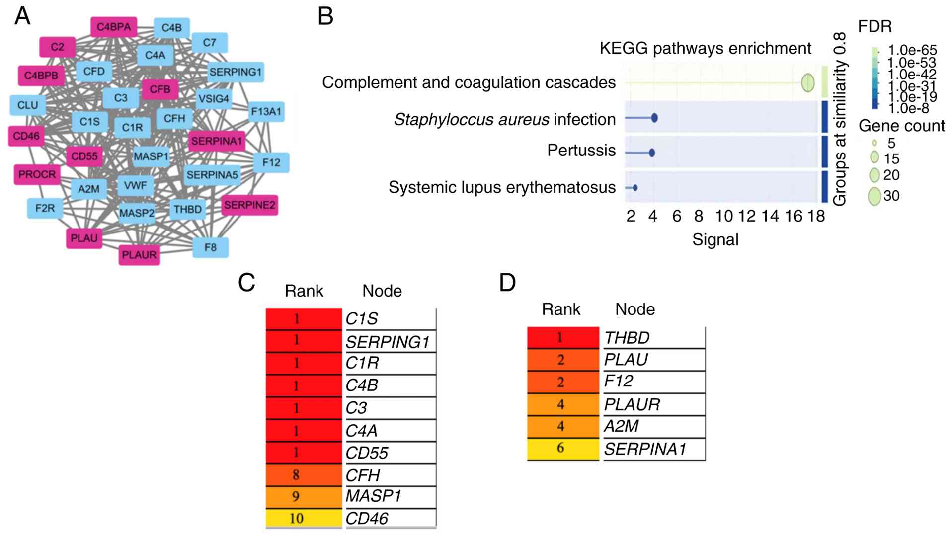
13 genes were upregulated (P<0.05) and 19 were downregulated
(P<0.05) (Fig. 1A and Table I), with broad representation across
the intrinsic, classical, lectin and alternative complement
pathways (Fig. S1). Enrichment
analysis performed in the STRING database indicated that the ‘CCC
pathway’ was the most significantly overrepresented KEGG term,
followed by pathways linked to Staphylococcus aureus
infection, pertussis and systemic lupus erythematosus (Fig. 1B). PPI interaction analysis revealed
a densely interconnected network. Further clustering of this PPI
network via the MCODE plugin in Cytoscape delineated two functional
modules. Module 1, comprising 17 nodes and 121 edges, included
complement C1s, SERPING1 and C3. Module 2, consisting
of 6 nodes and 12 edges, encompassed the hub genes thrombomodulin
and SERPINA1 (Fig. 1C and
D).
 | Table I.Differentially expressed genes in
colon adenocarcinoma from GEPIA2 database. |
Table I.
Differentially expressed genes in
colon adenocarcinoma from GEPIA2 database.
| Genes | Median (tumor) | Median
(normal) |
log2FC | P-value |
|---|
| C7 | 1.67 | 111.39 | −5.40 | 6.80×10-112 |
| MASP1 | 0.30 | 23.52 | −4.24 | 7.16×10-73 |
| CFD | 9.65 | 162.01 | −3.94 | 6.45×10-97 |
| CLU | 56.28 | 744.12 | −3.70 | 3.50×10-65 |
| F13A1 | 1.65 | 17.67 | −2.82 | 2.72×10-69 |
| VSIG4 | 4.15 | 22.13 | −2.17 | 2.24×10-41 |
|
SERPING1 | 67.62 | 261.89 | −1.94 | 2.52×10-36 |
| C1S | 70.60 | 241.45 | −1.76 | 5.43×10⁻³⁵ |
| C3 | 68.83 | 234.82 | −1.76 | 4.95×10⁻²¹ |
| C4A | 18.13 | 59.54 | −1.66 | 2.46×10⁻²⁶ |
| C4B | 18.15 | 59.09 | −1.65 | 1.28×10⁻²⁵ |
| CFH | 6.62 | 19.90 | −1.46 | 1.91×10⁻³⁵ |
| SERPINA5 | 0.34 | 2.64 | −1.44 | 8.93×10-60 |
| MASP2 | 0.24 | 1.92 | −1.24 | 3.64×10-97 |
| A2M | 58.60 | 139.21 | −1.23 | 1.56×10⁻²⁸ |
| C1R | 77.29 | 176.88 | −1.18 | 5.10×10⁻²³ |
| THBD | 3.53 | 9.01 | −1.14 | 1.64×10⁻²⁷ |
| F8 | 1.72 | 4.88 | −1.11 | 3.70×10⁻²⁵ |
| VWF | 16.77 | 36.48 | −1.08 | 2.30×10⁻²³ |
| C4BPA | 1.78 | 0.18 | 1.24 | 5.55×10⁻²⁷ |
| CD46 | 174.82 | 71.39 | 1.28 | 2.42×10-78 |
| F2R | 12.27 | 4.27 | 1.33 | 1.45×10-47 |
| PROCR | 35.05 | 12.79 | 1.39 | 2.04×10⁻³⁵ |
| CD55 | 65.26 | 22.58 | 1.49 | 9.23×10-42 |
| CFB | 69.48 | 21.30 | 1.66 | 3.87×10-45 |
| C4BPB | 7.37 | 1.57 | 1.70 | 7.87×10⁻²⁷ |
| PLAU | 29.05 | 6.56 | 1.99 | 5.20×10-68 |
| SERPINE2 | 47.67 | 10.86 | 2.04 | 5.04×10-41 |
| PLAUR | 59.00 | 11.74 | 2.24 | 1.58×10-66 |
| C2 | 57.80 | 9.82 | 2.44 | 1.14×10-86 |
| SERPINA1 | 120.77 | 17.31 | 2.73 | 6.53×10⁻³³ |
| F12 | 18.78 | 1.94 | 2.75 | 3.72×10-96 |
Four genes related to the survival of
patients with pan-cancers
Overall survival analysis using a median group
cutoff demonstrated an association between four genes from the CCC
pathway (SERPINA1, C3, F8 and SERPING1) and patient
survival in COAD (Fig. 2). This
association remained significant for SERPINA1 alone when a
more stringent quartile cutoff with Cutoff-high of 75% was applied.
Extending the analysis to other types of cancer revealed broader
prognostic relevance. SERPINA1 expression was associated
with patient survival in brain lower grade glioma (LGG), skin
cutaneous melanoma (SKCM) and breast invasive carcinoma. The other
three genes also showed significant associations across multiple
types of cancer, with C3 linked to adrenocortical carcinoma,
COAD, LGG and SKCM; F8 to COAD and kidney renal clear cell
carcinoma; and SERPING1 to COAD, LGG and SKCM (Table II).
| Table II.Survival analysis of four genes in
pan-cancer panel from GEPIA2 database. |
Table II.
Survival analysis of four genes in
pan-cancer panel from GEPIA2 database.
|
|
Type of
cancer |
|---|
|
|
|
|---|
| Genes | COAD | LGG | SKCM | BRCA | ACC | CHOD | KICH | KRIC | MESO | PCPG | CESC | THYM | LUSC | SARC |
|---|
| SERPINA1 |
| Log
rank P-value | 0.043 | 2.7×10⁻⁶ | 0.00026 | 9.3×10⁻¹² | - | - | - | - | - | - | - | - | - | - |
| HR
(high) | 0.51 | 2.4 | 0.61 | 0.61 | - | - | - | - | - | - | - | - | - | - |
| C3 |
| Log
rank P-value | 0.033 | 0.0031 | 0.017 | - | 9.5×10⁻⁵ | 0.033 | 0.032 | 4.1×10⁻⁵ | 0.0025 | 0.013 | - | - | - | - |
| HR
(high) | 1.7 | 1.7 | 0.72 | - | 0.19 | 1.7 | 0.14 | 1.9 | 0.47 | 1.6×10⁻⁹ | - | - | - | - |
| F8 |
| Log
rank P-value | 0.015 | - | - | - | 0.019 | - | - | 0.0003 | - | - | 0.0026 | 0.036 | - | - |
| HR
(high) | 1.8 | - | - | - | 0.39 | - | - | 0.57 | - | - | 0.49 | 6.9 | - | - |
| SERPING1 |
| Log
rank P-value | 0.032 | 3.3×10⁻⁷ | 0.00052 | - | - | - | - | - | 0.0003 | - | - | - | 0.037 | 3.9×10⁻⁵ |
| HR
(high) | 1.7 | 2.6 | 0.62 | - | - | - | - | - | 0.42 | - | - | - | 1.3 | 0.43 |
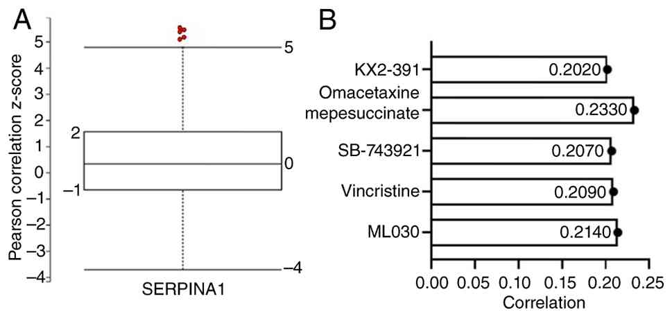
Evaluating the druggability profile of
SERPINA1, C3, F8 and SERPING1
To investigate the potential druggability of the
four identified genes (SERPINA1, C3, F8 and
SERPING1), established disease associations were analyzed
using the Open Targets Platform (Table
II). All genes displayed associations with various types of
cancer, with strength of evidence varying. Gene-cancer association
scores were obtained from the GeneCards Suite, which integrates
multi-omics and clinical evidence to generate a normalized ALIscore
(0–1) for each gene-disease pair, with higher scores indicating
stronger association. For instance, SERPINA1 showed strong
associations with gastric and breast cancer, whereas the link
between SERPING1 and types of cancer, such as breast cancer
and prostate cancer, was comparatively weak. Subsequent review of
approved targeted therapies revealed that F8, C3 and
SERPING1 are already targeted by existing drugs (Table III) (26–28).
Notably, however, no clinically approved drugs currently target
SERPINA1, suggesting its potential as a novel therapeutic
candidate (https://www.genecards.org/card/SERPINA1).
| Table III.Summary of disease associations and
approved or investigational drugs targeting the four genes. |
Table III.
Summary of disease associations and
approved or investigational drugs targeting the four genes.
| Genes | Type of cancer
(scorea) | Known drugs | Main treatment | Earliest
approved | (Refs.) |
|---|
|
SERPINA1 | Gastric cancer | No | / | / | N/A |
|
| (0.82); |
|
|
|
|
|
| Pancancer
(0.81) |
|
|
|
|
|
| Breast cancer |
|
|
|
|
|
| (0.75); |
|
|
|
|
|
| Colorectal
cancer |
|
|
|
|
|
| (0.59) |
|
|
|
|
| C3 | Ovarian cancer | Pegcetacoplan | Paroxysmal | 2021 | (26) |
|
| (0.83); |
| nocturnal |
|
|
|
| Pancancer
(0.81); |
| hemoglobinuria |
|
|
|
| Gastric cancer |
| and immune |
|
|
|
| (0.72);
Colorectal |
| system disease |
|
|
|
| cancer (0.36) |
|
|
|
|
| F8 | Pancancer
(0.67); | Moroctocog
alfa | Hemophilia a | 1999 | (27) |
|
| Esophageal |
|
|
|
|
|
| cancer (0.39); |
|
|
|
|
|
| Breast cancer |
|
|
|
|
|
| (0.20); Lung |
|
|
|
|
|
| cancer (0.14) |
|
|
|
|
|
SERPING1 | Breast cancer | Conestat alfa | Hereditary | 2010 | (28) |
|
| (0.24);
Pancancer |
| angioedema |
|
|
|
| (0.18);
Prostate |
|
|
|
|
|
| cancer (0.15); |
|
|
|
|
|
| Pituitary
cancer |
|
|
|
|
|
| (0.13) |
|
|
|
|
In the absence of approved drugs targeting
SERPINA1, the CTRP database was used to identify potential
compounds interacting with this gene. As presented in Fig. 3, five compounds showed a correlation
with SERPINA1 expression, listed in order of decreasing
correlation score: Omacetaxine mepesuccinate (0.2330), ML030
(0.2140), vincristine (0.2090), SB-743921 (0.2070) and KX2-391
(0.2020). These agents may represent potential therapeutic
candidates for colon cancer via modulation of SERPINA1
activity.
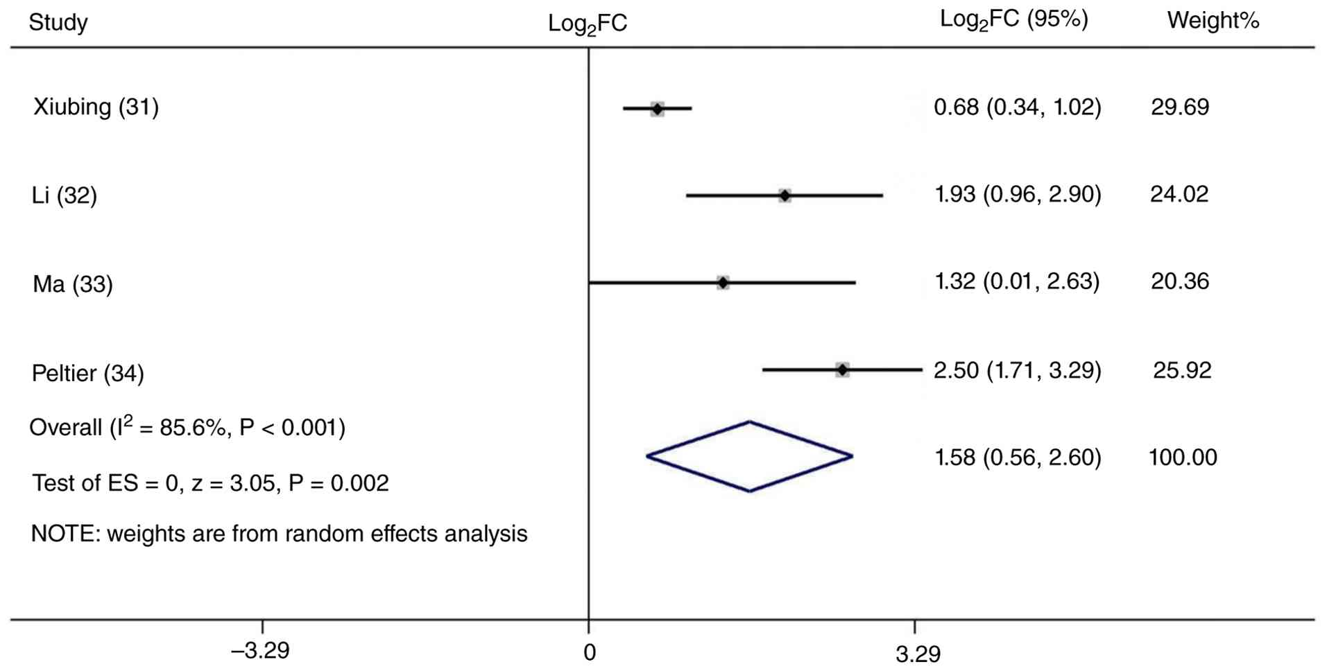
Meta-analysis of SERPINA1
expression
Based on multiple criteria, including prognostic
relevance, hub gene status and the absence of approved targeted
therapies, SERPINA1 was prioritized for further
investigation. Following a literature review in PubMed (https://pubmed.ncbi.nlm.nih.gov/) using the
keywords ‘cancer’ and ‘SERPINA1’, four eligible studies
(29–32) were included (Table SI). As shown in Fig. 4, the meta-analysis demonstrated that
SERPINA1 was consistently upregulated in cancer tissues
compared with healthy controls, with a pooled log2FC of
1.58 (95% CI: 0.56, 2.60). However, high heterogeneity was observed
across the included studies (I2=85.6%; P<0.001). This
may be attributed to the limited reference data (derived from only
four articles) and inherent data heterogeneity. In the present
study, the data came from two types of cancer: Colon cancer (n=3)
and pancreatic ductal adenocarcinoma (n=1). In addition, different
sample types were involved, including tissue (n=2), plasma exosomes
(n=1) and serum (n=1).
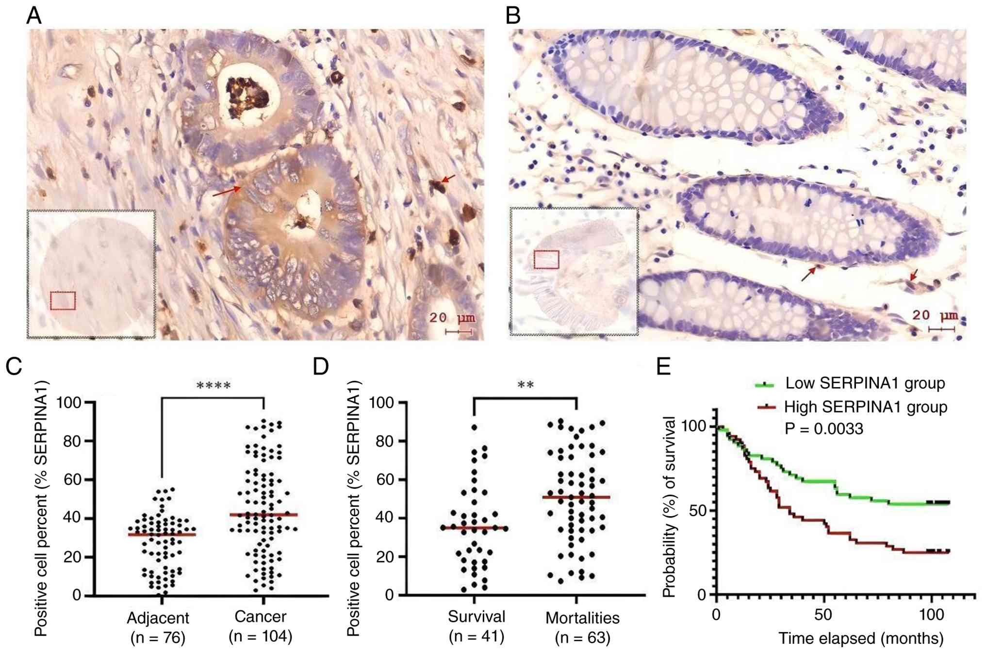
SERPINA1 validation by IHC
analysis
To validate SERPINA1 expression at the
protein level, IHC analysis was performed. Although previous data
from the GEPIA2 database and meta-analysis had indicated
upregulation of SERPINA1 in colon cancer, direct protein
validation using established methods such as IHC, western blot or
ELISA remained necessary. To address this, IHC was conducted on a
tissue microarray containing 180 spots of colon tumor and matched
adjacent normal tissues. Anti-AAT staining demonstrated markedly
stronger AAT accumulation in tumor tissues (Fig. 5A) compared with adjacent normal
tissues (Fig. 5B). Quantitative
optical density analysis further demonstrated that AAT was
upregulated by 1.84-fold in tumors (P<0.0001; Fig. 5C). When comparing AAT expression
between surviving and deceased patient groups, higher AAT levels
(P<0.05) were observed in the deceased group (Fig. 5D). Consistently, survival analysis
revealed significantly shorter overall survival in patients with
higher SERPINA1 expression compared with those with lower
expression (Fig. 5E).
Discussion
CRC is a highly prevalent malignancy of the
digestive system, whose development is influenced by a combination
of genetic predisposition, environmental factors, chronic
inflammation and gut microbiota dysbiosis. Prior research has
highlighted the involvement of the complement system, a central
element of innate immunity, in CRC pathogenesis (33,34),
as it contributes to host defense against pathogens and modulates
intestinal inflammatory responses during cancer progression.
Furthermore, complement signaling has been implicated across
multiple stages of tumorigenesis, including initiation,
proliferation, metastasis and response to therapy (34–36).
Dysregulation of the CCC pathway has been documented in several
malignancies, such as metastatic urothelial carcinoma (14), acute lymphoblastic leukemia
(37), lower-grade glioma (15) and bladder cancer (38). Nevertheless, the role of the CCC
pathway in CRC remains largely unexplored. In the present study,
the expression profiles of 88 CCC genes in COAD were evaluated
using the GEPIA2 database and 32 differentially expressed CCC genes
(13 upregulated and 19 downregulated) were identified, suggesting
that widespread dysregulation may be implicated in clinical
outcomes for patients with CRC.
Following PPI and survival analyses, four hub genes
associated with patient survival were identified: SERPINA1
(upregulated), C3 (downregulated), F8 (downregulated)
and SERPING1 (downregulated). SERPINA1 is a serine
protease inhibitor primarily targeting elastase, and is capable of
irreversibly inhibiting trypsin, chymotrypsin and plasminogen
activator. The aberrant isoform of SERPINA1 inhibits
insulin-induced nitric oxide synthesis in platelets, shortens
coagulation time and exhibits proteolytic activity against insulin
and plasmin (39). SERPINA1
has been implicated in colon cancer (29–32)
and in combination with fibrinogen demonstrates superior diagnostic
efficacy compared with conventional markers such as
carcinoembryonic antigen and carbohydrate antigen 19-9 (30). In CRC, SERPINA1 is highly
expressed, is associated with unfavorable clinical outcomes and
promotes CRC cell proliferation and migration via activation of the
STAT3 pathway (31). C3
serves as a precursor for non-enzymatic constituents of the
classical, alternative, lectin and granzyme K complement pathways.
These pathways comprise a proteolytic cascade that drives pathogen
phagocytosis and degradation while enhancing adaptive immune
signaling. C3 deficiency exacerbates inflammatory responses
in the colon (40). Although
downregulated in colon cancer, elevated C3 expression is
associated with worse overall survival in gastric cancer (34). F8, in the presence of calcium
and phospholipids, functions as a cofactor for factor IXa during
the conversion of factor X to its active form, factor Xa.
Deficiency in coagulation factor VIII underlies hemophilia A, for
which recombinant or plasma-derived factor VIII remains first-line
therapy (41). SERPING1,
another serine protease inhibitor, regulates the classical
complement pathway. SERPING1 is downregulated in prostate
cancer and reduced SERPING1 expression is associated with
higher Gleason scores, advanced pathological grade and more
progressive tumor stages (42). In
summary, SERPINA1, C3, F8 and SERPING1 are four
cancer-related genes likely to play notable roles in colon cancer,
despite the limited number of studies specifically focused on their
functions in this malignancy.
The druggability of SERPINA1, C3, F8 and
SERPING1 was further evaluated based on reports from the
Open Targets Platform (23). Among
these, only SERPINA1 has not yet been established as a known
drug target, to the best of our knowledge. However, based on
predictive screening using the CTRP database, omacetaxine
mepesuccinate was identified as a potential compound targeting
SERPINA1. This agent is an approved anticancer drug
currently used in the treatment of chronic myeloid leukemia
(43,44). Taken together, these findings
suggest that SERPINA1, C3, F8 and SERPING1 may
represent promising candidate targets for therapeutic
intervention.
SERPINA1 was selected for meta-analysis and
IHC validation using tissue microarrays. While prior studies have
examined the expression and function of SERPINA1 in colon
cancer (30–32), the present approach distinguishes
itself from these earlier works by employing different specimens
and different validation methods. For instance, Li et al
(30) and Peltier et al
(32) employed plasma samples from
patients with CRC and validated SERPINA1 via ELISA, whereas
Ma et al (31) utilized a
mouse CRC model. The present study, based on tissue microarray
analysis, thus serves as a methodological complement to existing
research. Furthermore, the expression pattern of SERPINA1 in
colon cancer remains contentious. The cProSite database, which
incorporates proteomic and phosphoproteomic data from the National
Cancer Institute's Clinical Proteomic Tumor Analysis Consortium and
International Cancer Proteogenome Consortium (45), indicates that SERPINA1 is
downregulated in colon cancer (Fig.
S2). By contrast, the GEPIA2 database, which performs gene
expression analysis based on tumor and normal samples from the TCGA
and GTEx databases (46), reports
its upregulation. In the present study, SERPINA1 expression
was elevated in tumor tissues compared with adjacent normal colon
tissues, and was higher in deceased patients compared with
surviving patients. Furthermore, in the GEPIA2 database cohort,
patients with higher SERPINA1 expression exhibited longer
survival. However, in the present study, increased SERPINA1
expression was associated with shortened survival in patients with
colon cancer, a finding consistent with observations in pancreatic
ductal adenocarcinoma (30). This
discrepancy in survival outcomes between the GEPIA2 database and
the present study may be attributed to the difference between
RNA-sequencing data (from GEPIA2) and protein expression data (from
the present study). The post-transcriptional and post-translational
modifications of SERPINA1 may lead to inconsistencies between its
mRNA and protein expression levels, which also highlights the
necessity of protein-level experimental validation for
bioinformatics and omics findings. Additionally, a review of
published literature indicated that SERPINA1 was commonly
upregulated across multiple types of cancer (30–32).
Thus, the present study provides experimental evidence that helps
to clarify the discrepant findings between the cProSite and GEPIA2
database cohorts.
The present study has several limitations that need
to be addressed. First, the protein expression levels of the other
three candidate genes (C3, F8 and SERPING1) were not
experimentally validated. Since mRNA expression levels do not
always correlate with protein abundance due to post-transcriptional
regulation, future studies should therefore employ
immunohistochemistry or western blot analysis to confirm their
protein expression in colon cancer tissues. Second, the
meta-analysis was performed solely for SERPINA1 expression.
Consequently, the diagnostic or prognostic value of the other three
genes (C3, F8, SERPING1) across different cohorts remains elusive.
Further meta-analyses integrating multiple independent datasets are
needed to evaluate their clinical relevance. Third, no experiments
were conducted to validate potential compounds targeting
SERPINA1. Thus, the druggability of SERPINA1 suggested by
the Open Targets Platform is purely computational and lacks
experimental support. Future research should perform in
vitro or in vivo assays (e.g., viability, apoptosis or
targeted inhibition assays) to test candidate compounds. Finally,
the precise functional role of SERPINA1 in patients with
colon cancer has yet to be experimentally elucidated. This is a
critical limitation because without functional validation (e.g.,
via knockdown or overexpression models), the observed expression
differences cannot be causally linked to tumor progression or
patient outcomes. Future investigations using gain- or
loss-of-function approaches in colon cancer cell lines or animal
models are required to define its mechanistic role.
In summary, the present study analyzed the
expression of 88 genes in the CCC pathway and identified 32 DEGs.
Among these, four hub genes were associated with the survival of
patients with colon cancer. SERPINA1, an upregulated gene,
was further validated in colon tissue microarrays via IHC analysis,
showing that its upregulation was associated with worse survival
outcomes. These findings suggest that SERPINA1 may serve as
a potential diagnostic biomarker for colon cancer and represents a
promising candidate for therapeutic targeting.
Supplementary Material
Supporting Data
Supporting Data
Acknowledgements
Not applicable.
Funding
This research received financial support from University Student
Innovation and Entrepreneurship Training Plan of Heilongjiang
Province (grant no. S202510222084).
Availability of data and materials
The data generated in the present study may be
requested from the corresponding author.
Authors' contributions
PX conceived and designed the study and revised the
manuscript. DL collected, analyzed and interpreted the data, and
wrote the manuscript. DH designed the differential expression
analysis pipeline and interpreted the transcriptome data. KL
performed the functional enrichment analysis and curated all public
datasets used in this study. HJ conducted statistical analysis of
bioinformatics data and generated all related figures. DH, KL and
HJ critically revised the bioinformatics sections of the manuscript
for important intellectual content. PX and DL confirm the
authenticity of all the raw data. All authors read and approved the
final manuscript.
Ethics approval and consent to
participate
The research complied with the principles outlined
in the Declaration of Helsinki and received formal approval from
Shanghai Biotechnology Co., Ltd. (approval no. SHYJS-CP-1707004).
Written informed consent was secured from all participants prior to
their involvement.
Patient consent for publication
Not applicable.
Competing interests
The authors declare that they have no competing
interests.
Use of artificial intelligence tools
During the preparation of this work, DeepSeek-V3.1
(https://chat.deepseek.com/) was used to
improve the readability and language of the manuscript or to
generate images, and subsequently, the authors revised and edited
the content produced by the artificial intelligence tools as
necessary, taking full responsibility for the ultimate content of
the present manuscript.
Glossary
Abbreviations
Abbreviations:
|
CCC pathway
|
complement and coagulation cascades
pathway
|
|
DEGs
|
differentially expressed genes
|
|
COAD
|
colon cancer
|
|
KEGG
|
Kyoto Encyclopedia of Genes and
Genomes
|
|
FDR
|
false discovery rate
|
|
CI
|
confidence interval
|
|
MCODE
|
Molecular Complex Detection
|
|
FC
|
fold change
|
|
PPI
|
protein-protein interaction
|
|
SERPINA1
|
serpin family A member 1
|
|
SERPING1
|
serpin family G member 1
|
|
F8
|
factor VIII
|
|
C3
|
complement C3
|
|
IHC
|
immunohistochemistry
|
|
CRC
|
colorectal cancer
|
|
CTRP
|
Cancer Therapeutics Response
Portal
|
|
ACC
|
adrenocortical carcinoma
|
|
LGG
|
brain lower grade glioma
|
|
SKCM
|
skin cutaneous melanoma
|
|
KIRC
|
kidney renal clear cell carcinoma
|
|
BRCA
|
breast invasive carcinoma
|
|
AAT
|
anti-α-1 antitrypsin
|
|
AJCC
|
American Joint Committee on
Cancer
|
References
|
1
|
Lukic M, Licaj I, Laaksonen MA, Weiderpass
E, Borch KB and Rylander C: The burden of colon cancer attributable
to modifiable factors-The Norwegian women and cancer study. Int J
Cancer. 152:195–202. 2023. View Article : Google Scholar : PubMed/NCBI
|
|
2
|
Han B, Zheng R, Zeng H, Wang S, Sun K,
Chen R, Li L, Wei W and He J: Cancer incidence and mortality in
China, 2022. J Natl Cancer Cent. 4:47–53. 2024.PubMed/NCBI
|
|
3
|
He S, Xia C, Li H, Cao M, Yang F, Yan X,
Zhang S, Teng Y, Li Q and Chen W: Cancer profiles in China and
comparisons with the USA: A comprehensive analysis in the
incidence, mortality, survival, staging, and attribution to risk
factors. Sci China Life Sci. 67:122–131. 2024. View Article : Google Scholar : PubMed/NCBI
|
|
4
|
Zhang J, Ou D, Xie A, Chen D and Li X:
Global burden and cross-country health inequalities of early-onset
colorectal cancer and its risk factors from 1990 to 2021 and its
projection until 2036. BMC Public Health. 24:31242024. View Article : Google Scholar : PubMed/NCBI
|
|
5
|
Bleyer A, Ries LAG, Cameron DB, Mansfield
SA, Siegel SE and Barr RD: Colon, colorectal, and all cancer
incidence increase in the young due to appendix reclassification. J
Natl Cancer Inst. 117:1340–1349. 2025. View Article : Google Scholar : PubMed/NCBI
|
|
6
|
Aggarwal S, Lavingiya V, Krishna V,
Chitalkar P, Ostwal V and Parikh PM: Young onset colorectal cancer.
South Asian J Cancer. 13:225–228. 2024. View Article : Google Scholar : PubMed/NCBI
|
|
7
|
Lo Nigro C, Ricci V, Vivenza D, Granetto
C, Fabozzi T, Miraglio E and Merlano MC: Prognostic and predictive
biomarkers in metastatic colorectal cancer anti-EGFR therapy. World
J Gastroenterol. 22:6944–6954. 2016. View Article : Google Scholar : PubMed/NCBI
|
|
8
|
Alaiyan B, Ilyayev N, Stojadinovic A,
Izadjoo M, Roistacher M, Pavlov V, Tzivin V, Halle D, Pan H, Trink
B, et al: Differential expression of colon cancer associated
transcript1 (CCAT1) along the colonic adenoma-carcinoma sequence.
BMC Cancer. 13:1962013. View Article : Google Scholar : PubMed/NCBI
|
|
9
|
Sahli H, Dahlbäck C, Lydrup ML and
Buchwald P: Impact of previous diverticulitis on 5-year survival
and recurrence rates in patients with colorectal cancer. Scand J
Gastroenterol. 58:1280–1285. 2023. View Article : Google Scholar : PubMed/NCBI
|
|
10
|
Stewart CL, Warner S, Ito K, Raoof M, Wu
GX, Kessler J, Kim JY and Fong Y: Cytoreduction for colorectal
metastases: Liver, lung, peritoneum, lymph nodes, bone, brain. When
does it palliate, prolong survival, and potentially cure? Curr
Probl Surg. 55:330–379. 2018.PubMed/NCBI
|
|
11
|
Jahanafrooz Z, Mosafer J, Akbari M,
Hashemzaei M, Mokhtarzadeh A and Baradaran B: Colon cancer therapy
by focusing on colon cancer stem cells and their tumor
microenvironment. J Cell Physiol. 235:4153–4166. 2020. View Article : Google Scholar : PubMed/NCBI
|
|
12
|
Yang WW, Zhou X and He G: Patients with
colorectal cancer combined with HIV had a worse overall survival
after surgery: A meta-analysis. Front Oncol. 15:14401052025.
View Article : Google Scholar : PubMed/NCBI
|
|
13
|
Fidelle M, Yonekura S, Picard M, Cogdill
A, Hollebecque A, Roberti MP and Zitvogel L: Resolving the paradox
of colon cancer through the integration of genetics, immunology,
and the microbiota. Front Immunol. 11:6008862020. View Article : Google Scholar : PubMed/NCBI
|
|
14
|
Gong Z, He Y, Mi X, Li C, Sun X, Wang G,
Li L, Han Y, Xu C, Wang W, et al: Complement and coagulation
cascades pathway-related signature as a predictor of immunotherapy
in metastatic urothelial cancer. Aging (Albany NY). 15:9479–9498.
2023. View Article : Google Scholar : PubMed/NCBI
|
|
15
|
Yang J, Shen L, Yang J, Qu Y, Gong C, Zhou
F, Liu Y, Luo M and Zhao L: Complement and coagulation cascades are
associated with prognosis and the immune microenvironment of
lower-grade glioma. Transl Cancer Res. 13:112–136. 2024. View Article : Google Scholar : PubMed/NCBI
|
|
16
|
Conway EM: Reincarnation of ancient links
between coagulation and complement. J Thromb Haemost. 13 (Suppl
1):S121–S132. 2015. View Article : Google Scholar : PubMed/NCBI
|
|
17
|
Bonetto A, Aydogdu T, Kunzevitzky N,
Guttridge DC, Khuri S, Koniaris LG and Zimmers TA: STAT3 activation
in skeletal muscle links muscle wasting and the acute phase
response in cancer cachexia. PLoS One. 6:e225382011. View Article : Google Scholar : PubMed/NCBI
|
|
18
|
Gavriilaki E, Ho VT, Schwaeble W, Dudler
T, Daha M, Fujita T and Jodele S: Role of the lectin pathway of
complement in hematopoietic stem cell transplantation-associated
endothelial injury and thrombotic microangiopathy. Exp Hematol
Oncol. 10:572021. View Article : Google Scholar : PubMed/NCBI
|
|
19
|
Ray A, Winter KAK, Naik DSL and Okorie C:
Prognostic significance of the coagulation and complement systems
in critical COVID-19 infection. Prague Med Rep. 124:77–93. 2023.
View Article : Google Scholar : PubMed/NCBI
|
|
20
|
Shannon P, Markiel A, Ozier O, Baliga NS,
Wang JT, Ramage D, Amin N, Schwikowski B and Ideker T: Cytoscape: A
software environment for integrated models of biomolecular
interaction networks. Genome Res. 13:2498–2504. 2003. View Article : Google Scholar : PubMed/NCBI
|
|
21
|
Bader GD and Hogue CWV: An automated
method for finding molecular complexes in large protein interaction
networks. BMC Bioinformatics. 4:22003. View Article : Google Scholar : PubMed/NCBI
|
|
22
|
Chin CH, Chen SH, Wu HH, Ho CW, Ko MT and
Lin CY: cytoHubba: Identifying hub objects and sub-networks from
complex interactome. BMC Syst Biol. 8 (Suppl 4):S112014. View Article : Google Scholar : PubMed/NCBI
|
|
23
|
Ochoa D, Hercules A, Carmona M, Suveges D,
Baker J, Malangone C, Lopez I, Miranda A, Cruz-Castillo C, Fumis L,
et al: The next-generation open targets platform: Reimagined,
redesigned, rebuilt. Nucleic Acids Res. 51(D1): D1353–D1359. 2023.
View Article : Google Scholar : PubMed/NCBI
|
|
24
|
Page MJ, McKenzie JE, Bossuyt PM, Boutron
I, Hoffmann TC, Mulrow CD, Shamseer L, Tetzlaff JM, Akl EA, Brennan
SE, et al: The PRISMA 2020 statement: An updated guideline for
reporting systematic reviews. Rev Esp Cardiol (Engl Ed).
74:790–799. 2021.(In English, Spanish). View Article : Google Scholar : PubMed/NCBI
|
|
25
|
Edge SB and Compton CC: The American joint
committee on cancer: The 7th edition of the AJCC cancer staging
manual and the future of TNM. Ann Surg Oncol. 17:1471–1474. 2010.
View Article : Google Scholar : PubMed/NCBI
|
|
26
|
U.S. Food and Drug Administration (FDA), .
FDA approves new treatment for adults with serious rare blood
disease (EB/OL). (2021-05-18). FDA; Silver Spring, MD: https://www.fda.gov/drugs/news-events-human-drugs/fda-approves-new-treatment-adults-serious-rare-blood-disease
|
|
27
|
European Medicines Agency, . ReFacto AF
(moroctocog alfa) EPAR[EB/OL]. (1999-04-13). European Medicines
Agency; Amsterdam: 2016, https://www.ema.europa.eu/en/medicines/human/EPAR/refacto-af
|
|
28
|
European Medicines Agency, . Ruconest
(conestat alfa) EPAR[EB/OL]. (2010 10 28). European Medicines
Agency; Amsterdam: 2017, https://www.ema.europa.eu/en/medicines/human/EPAR/ruconest
|
|
29
|
Xiubing C, Huazhen L, Xueyan W, Jing N,
Qing L, Haixing J, Shanyu Q and Jiefu L: SERPINA1 promotes the
invasion, metastasis, and proliferation of pancreatic ductal
adenocarcinoma via the PI3K/Akt/NF-κB pathway. Biochem Pharmacol.
230:1165802024. View Article : Google Scholar : PubMed/NCBI
|
|
30
|
Li L, Song X, Chen G, Zhang Z, Zheng B,
Zhang Q, Wang S and Xie L: Plasma exosomal protein PLG and SERPINA1
in colorectal cancer diagnosis and coagulation abnormalities. J
Cancer Res Clin Oncol. 149:8507–8519. 2023. View Article : Google Scholar : PubMed/NCBI
|
|
31
|
Ma Y, Chen Y, Zhan L, Dong Q, Wang Y, Li
X, He L and Zhang J: CEBPB-mediated upregulation of SERPINA1
promotes colorectal cancer progression by enhancing STAT3
signaling. Cell Death Discov. 10:2192024. View Article : Google Scholar : PubMed/NCBI
|
|
32
|
Peltier J, Roperch JP, Audebert S, Borg JP
and Camoin L: Quantitative proteomic analysis exploring progression
of colorectal cancer: Modulation of the serpin family. J
Proteomics. 148:139–148. 2016. View Article : Google Scholar : PubMed/NCBI
|
|
33
|
Ding P, Xu Y, Li L, Lv X, Li L, Chen J,
Zhou D, Wang X, Wang Q, Zhang W, et al: Intracellular complement
C5a/C5aR1 stabilizes β-catenin to promote colorectal tumorigenesis.
Cell Rep. 39:1108512022. View Article : Google Scholar : PubMed/NCBI
|
|
34
|
Krieg C and Guglietta S: The complement
system in intestinal inflammation and cancer. J Clin Invest.
135:e1883482025. View Article : Google Scholar : PubMed/NCBI
|
|
35
|
Bao D, Zhang C, Li L, Wang H, Li Q, Ni L,
Lin Y, Huang R, Yang Z, Zhang Y and Hu Y: Integrative analysis of
complement system to prognosis and immune infiltrating in colon
cancer and gastric cancer. Front Oncol. 10:5532972021. View Article : Google Scholar : PubMed/NCBI
|
|
36
|
Roumenina LT, Daugan MV, Petitprez F,
Sautès-Fridman C and Fridman WH: Context-dependent roles of
complement in cancer. Nat Rev Cancer. 19:698–715. 2019. View Article : Google Scholar : PubMed/NCBI
|
|
37
|
Tang Y, Chen L, Xiao Y, Ran Q, Li Z and
Chen M: Clinical significance of complement and coagulation
cascades genes for patients with acute lymphoblastic leukemia. Int
J Lab Hematol. 47:266–275. 2025. View Article : Google Scholar : PubMed/NCBI
|
|
38
|
Liu Y, Xiong S, Liu S, Chen J, Yang H, Liu
G and Li G: Analysis of gene expression in bladder cancer: Possible
involvement of mitosis and complement and coagulation cascades
signaling pathway. J Comput Biol. 27:987–998. 2020. View Article : Google Scholar : PubMed/NCBI
|
|
39
|
Long GL, Chandra T, Woo SL, Davie EW and
Kurachi K: Complete sequence of the cDNA for human alpha
1-antitrypsin and the gene for the S variant. Biochemistry.
23:4828–4837. 1984. View Article : Google Scholar : PubMed/NCBI
|
|
40
|
Choi YJ, Kim JE, Lee SJ, Gong JE, Jin YJ,
Lee H and Hwang DY: Promotion of the inflammatory response in mid
colon of complement component 3 knockout mice. Sci Rep.
12:17002022. View Article : Google Scholar : PubMed/NCBI
|
|
41
|
Gualtierotti R, Solimeno LP and Peyvandi
F: Hemophilic arthropathy: Current knowledge and future
perspectives. J Thromb Haemost. 19:2112–2121. 2021. View Article : Google Scholar : PubMed/NCBI
|
|
42
|
Peng S, Du T, Wu W, Chen X, Lai Y, Zhu D,
Wang Q, Ma X, Lin C, Li Z, et al: Decreased expression of serine
protease inhibitor family G1 (SERPING1) in prostate cancer can help
distinguish high-risk prostate cancer and predicts malignant
progression. Urol Oncol. 36:366.e1–366.e9. 2018. View Article : Google Scholar : PubMed/NCBI
|
|
43
|
Damlaj M, Lipton JH and Assouline SE: A
safety evaluation of omacetaxine mepesuccinate for the treatment of
chronic myeloid leukemia. Expert Opin Drug Saf. 15:1279–1286. 2016.
View Article : Google Scholar : PubMed/NCBI
|
|
44
|
Rosshandler Y, Shen AQ, Cortes J and
Khoury HJ: Omacetaxine mepesuccinate for chronic myeloid leukemia.
Expert Rev Hematol. 9:419–424. 2016. View Article : Google Scholar : PubMed/NCBI
|
|
45
|
Wang D, Qian X, Du YCN, Sanchez-Solana B,
Chen K, Kanigicherla M, Jenkins LM, Luo J, Eng S, Park B, et al:
cProSite: A web based interactive platform for online proteomics,
phosphoproteomics, and genomics data analysis. J Biotechnol Biomed.
6:573–578. 2023. View Article : Google Scholar : PubMed/NCBI
|
|
46
|
Tang Z, Kang B, Li C, Chen T and Zhang Z:
GEPIA2: An enhanced web server for large-scale expression profiling
and interactive analysis. Nucleic Acids Res. 47((W1)): W556–W560.
2019. View Article : Google Scholar : PubMed/NCBI
|