Introduction
Neuroendocrine differentiation (NED) is a term used
in clinical pathology to describe a number of tumor types with
distinct biological implications. According to the World Health
Organisation (WHO) classification and recent research, tumors
exhibiting NED can be categorized into neuroendocrine neoplasms
(NENs), mixed neuroendocrine non-NENs (MiNEN), amphicrine-like
carcinoma (ALC) and conventional carcinoma with NED (1,2).
Differentiating these tumors poses numerous challenges. First,
distinguishing between conventional carcinomas with NED and ALC is
difficult. Second, there is notable variability in applying
diagnostic criteria among observers. Third, the evolving WHO
classification criteria adds further complexity. Current TNM
staging systems utilize adenocarcinoma of the respective organ for
staging (2), potentially
underestimating the adverse biological behavior of conventional
carcinomas with NED. Therefore, a comprehensive understanding of
the biological features of conventional carcinomas with NED is key
in developing precise personalized treatment. Gastrointestinal
conventional carcinomas with NED are prevalent. The present review
therefore aimed to elucidate the definition, diagnosis, clinical
characteristics and pathogenesis of these carcinomas and discuss
future directions from the perspectives of basic mechanisms,
molecular subtyping and clinical translation.
Definition and diagnosis of MiNEN, ALC and
conventional carcinoma with NED
Classification of tumors with
neuroendocrine characteristics
MiNEN neoplasms are characterized by distinct
nodules or regions comprising both neuroendocrine and
non-neuroendocrine components. Each component constitutes at least
30% of the tumor volume (1). ALC is
defined by two possible patterns. In one pattern, individual tumor
cells demonstrate both exocrine and neuroendocrine features,
exhibiting exocrine glandular morphology (such as mucin secretion)
alongside NED traits. In the other pattern, neuroendocrine and
non-neuroendocrine cells are intimately admixed and dispersed
throughout the tumor (2).
Conventional carcinoma with NED is defined as a conventional
carcinoma that lacks neuroendocrine morphological characteristics,
such as trabecular, gyriform, solid-nesting or
solid-paraganglioma-like patterns, but exhibits expression of
neuroendocrine markers, including CgA, Syn or insulinoma-associated
protein 1 (INSM1), detectable by immunohistochemistry (IHC). The
positive cells typically appear as scattered single cells or small
clusters, accounting for >30% of tumor cells. However, this
threshold has not reached universal consensus (1). This threshold is borrowed from the WHO
diagnostic criteria for MiNEN, but its direct application to
conventional carcinoma without neuroendocrine morphology remains
debated. Diagnostic thresholds vary widely across studies (3–7), from
‘any positive cell’ to >2, >5, >10 and 30%, with some
evidence suggesting that even minor neuroendocrine components
(<30%) may carry prognostic significance. These discrepancies
highlight the need for further validation of the optimal
cutoff.
Evolution and application of
neuroendocrine markers
NED is typically determined through
immunohistochemical detection of specific biomarkers, with the
standard combination being CgA and Syn. INSM1 has further emerged
as a promising marker for diagnosing neuroendocrine tumors, known
for its high specificity. Originally identified from the insulinoma
glucagon spheroid depletion library, INSM1 is a zinc finger
transcription factor located on 20p11.2 without introns (8). A study by McHugh et al
(9) demonstrated that while INSM1
has lower sensitivity (80.9%) compared with Syn (99.1%), its
specificity (95.7%) surpasses that of traditional biomarkers such
as Syn (86.0%), CgA (87.3%) and CD56 (86.0%). Litmeyer et al
(3) further highlighted the utility
of INSM1 in distinguishing MiNEN, neuroendocrine carcinoma (NEC)
and conventional colorectal carcinomas (CRCs), particularly in
cases with diffuse Syn expression. Notably, INSM1 is typically
absent or expressed at low levels in conventional CRCs, aiding in
the differentiation from neuroendocrine tumors. Previous
investigations regarding biomarkers such as CD56, neuron-specific
enolase (NSE) and pro-gastrin-releasing peptide (proGRP) for
neuroendocrine tumors have been conducted, but due to their lack of
specificity, they are no longer recommended as histological
diagnostic markers. However, NSE and proGRP do retain importance in
serological testing (10,11).
Diagnostic algorithm
To aid in diagnostic workflow, a practical algorithm
for distinguishing conventional carcinomas with NED from other NENs
is presented in Fig. 1. The
following rationale is provided for each node of diagnostic
algorithm and the literature summarized in the present review: i)
Node 1 (morphological assessment of neuroendocrine features), where
the first step is to evaluate H&E sections for typical
neuroendocrine architecture, such as nested, trabecular, insular or
rosette-like growth patterns. If clear neuroendocrine morphology is
present, the tumor may be a NEC, MiNEN or ALC. If such morphology
is absent, the tumor is classified as a conventional carcinoma;
however, NED can still be detected by IHC; ii) Node 2
(neuroendocrine marker expression), where IHC for at least two of
the three validated markers (CgA, Syn and INSM1) is performed.
According to the WHO classification, determination of NED requires
positivity for at least two of these markers. This is because no
single marker is perfectly sensitive and specific: Syn is highly
sensitive but may be expressed in non-neuroendocrine tumors, CgA is
specific but often lost in high-grade NENs and INSM1 combines good
specificity with relatively high sensitivity; iii) Node 3 (presence
of a non-neuroendocrine component), whereby if the tumor exhibits
both neuroendocrine and non-neuroendocrine morphology, such as
conventional adenocarcinoma or squamous cell carcinoma, it is not a
pure NEN. The next distinction concerns whether the two components
are spatially distinct, meaning they form separate nodules or
regions or intimately admixed; iv) Node 4 (spatially distinct
components, each representing at least 30% of the tumor), whereby
when a tumor exhibits neuroendocrine morphology on H&E and
contains both neuroendocrine and non-neuroendocrine components that
form separate, recognizable areas and each accounts for at least
30% of the tumor volume, the diagnosis is MiNEN. If the
neuroendocrine component comprises <30%, the tumor is classified
as conventional carcinoma with a minor neuroendocrine component.
Conversely, if the non-neuroendocrine component comprises <30%,
the tumor is classified as NEN with a minor non-neuroendocrine
component; v) Node 5 (intimate admixture with diffuse
neuroendocrine marker expression involving at least 30% of cells),
whereby if the tumor demonstrates neuroendocrine morphology on
H&E and the neuroendocrine and non-neuroendocrine cells are
intermingled throughout the tumor without separate regions and the
proportion of neuroendocrine-positive cells reaches at least 30%,
the tumor falls into the ALC category. In ALC, the dual
differentiation is evenly distributed and the tumor is staged
according to its non-neuroendocrine counterpart. The Ki-67
proliferative index should be reported for the whole neoplasm; and
vi) Node 6 (neuroendocrine marker expression in scattered, isolated
cells), the main focus of the present review: Conventional
carcinoma with NED. Here, the tumor lacks neuroendocrine morphology
on H&E. However, it exhibits widespread expression of
neuroendocrine markers in scattered or isolated cells, with
positive cells accounting for >30% of the tumor cell population.
If the proportion of positive cells is <30%, the tumor is
classified as conventional carcinoma without NED. Despite this, the
cut-off has not yet reached universal consensus and further studies
are needed to validate or refine the appropriate threshold.
 | Figure 1.Diagnostic algorithm for
distinguishing conventional carcinomas with NED from other NE
neoplasms in the gastrointestinal tract. The diagnostic algorithm
follows a stepwise approach based on World Health Organisation
recommendations. First, the tumor is examined for NE morphology
using H&E. If NE morphology is present, the case enters the NEN
pathway. If absent, immunohistochemistry for CgA, Syn and INSM1 is
performed and at least two of these three markers must be positive
to determine NED. When a non-NE component coexists, the key
discriminating factors are: i) Whether the components are spatially
distinct and each constitutes at least 30% of the tumor, leading to
a diagnosis of MiNEN; ii) whether NED is diffuse (≥30% of cells)
but intimately admixed, resulting in a diagnosis of ALC; or iii)
whether the tumor lacks NE morphology but shows NE markers
expressed in scattered, isolated cells comprising >30% of the
tumor, in which case the tumor is classified as conventional
carcinoma with NED. If the proportion of positive cells is <30%,
the tumor is classified as conventional carcinoma without NED.
Tumors are categorized into six groups: NEC, ALC, NEN with non-NE
carcinoma or conventional carcinoma with minor NEN, MiNEN,
conventional carcinoma and conventional carcinoma with NED. NE
markers include CgA, Syn and INSM1. NE, neuroendocrine; NED, NE
differentiation; NEN, NE neoplasms; INSM1; insulinoma-associated
protein 1; MiNEN, mixed NE-non-NE neoplasm; ALC, amphicrine-like
carcinoma; NEC, NE carcinoma. |
Controversies and challenges
surrounding diagnostic thresholds
Diagnoses of conventional carcinomas with NED
presents challenges regarding inconsistent positive thresholds and
differentiation from ALC. Currently, research utilizes a number of
positive thresholds, ranging from ‘any positive cell’ to thresholds
of ‘>2’, ‘>5’, ‘>10’ and ‘30%’ (7,12,13).
This lack of uniformity hinders study comparisons and the
establishment of standardized clinical guidelines. In addition,
distinguishing between conventional carcinomas with NED and ALC is
complex, as the latter necessitates determination of both exocrine
and neuroendocrine characteristics, relying on the expertise and
techniques of the pathologist, including electron microscopy, which
are not routine in clinical practice. The clinical and pathological
distinctions of ALC and its prognostic variances from conventional
carcinoma with NED will be further discussed in subsequent sections
regarding clinical features and cross-organ mechanisms.
Prognostic importance of conventional
carcinomas with NED in organs of the digestive tract
Notable variations exist in the prevalence of
conventional carcinomas with NED across different organs of the
digestive tract, reflecting the diverse tumor biology among these
organs. Discrepancies in detection rates can be attributed to
variations in diagnostic criteria, positivity thresholds and study
populations. Research has primarily focused on the stomach and
colorectum, leaving other areas underexplored. Furthermore,
divergent findings regarding the prognostic relevance of NED exist,
with certain studies indicating independent prognostic value while
others suggest collinearity with established high-risk factors.
Table I consolidates
epidemiological and prognostic data from key studies spanning
numerous digestive tract organs.
 | Table I.Key research data on conventional
carcinomas with NED in organs of the digestive tract. |
Table I.
Key research data on conventional
carcinomas with NED in organs of the digestive tract.
| A, Stomach |
|---|
|
|---|
| First author,
year | Study
conclusion | Sample size | NED positivity
rate | Diagnostic criteria
(biomarkers/thresholds) | Key findings | (Refs.) |
|---|
| Li et al,
2026 | Support
(subgroup) | 309 | 22.3% | CgA, Syn and CD56
(1%) | Patients with GC
and NED had a higher incidence of PNI compared with those without
NED (29.0 vs. 15.8%). No significant difference in OS or RFS was
observed overall, but a poorer survival trend was noted in the
differentiated GC subgroup with NED (P=0.015) | (13) |
| Xu et al,
2016 | Support
(subgroup) | 240 | 29.1% | CgA, Syn and SCGN
(5%) | Syn positivity was
associated with deeper tumor invasion, whereas SCGN positivity was
associated with lymph node metastasis. Patients with NED and
diffuse or stage II GC exhibited lower cumulative survival rates
(P=0.006) | (5) |
| Blumenfeld et
al, 1996 | Descriptive
research | 48 | 75.0% | CgA, Syn, NSE,
gastrin and serotonin (any positive result) | No prognostic data
reported | (14) |
|
| B, Colon and
rectum |
|
| First author,
year | Study
conclusion | Sample
size | NED positivity
rate | Diagnostic
criteria (biomarkers/thresholds) | Key
findings | (Refs.) |
|
| Syversen et
al, 1995 | Support | 91 | 40.0% | NSE and CgA (any
positive result) | CRC patients with
NED exhibited a decreasing trend in median OS compared with those
without NED (P=0.100) | (15) |
| Grabowski et
al, 2001 | Support | 116 | 17.2% | CgA and Syn
(2%) | No observed
association between neuroendocrine differentiation and clinical
pathological parameters. The median OS was 18.6 months in patients
with NED compared with 48.9 months in those without NED
(P<0.001) | (12) |
| Atasoy et
al, 2003 | Support | 50 | 38.0% | CgA, Syn and NSE
(any positive result) | Among the three
markers, only CgA was positivity associated with tumor grade and
stage and it was an independent adverse prognostic factor
(P<0.05) | (25) |
| Liu et al,
2014 | Support | 171 | 41.5% | Syn and CgA (any
positive result) | NED was more common
in women, the 5-year OS rate was 68% in patients with NED compared
with 90% in those without NED (P=0.018). The median OS was 38.6
months in patients with NED vs. 53.2 months in those without
NED | (16) |
| Guo et al,
2020 | Support | 94,291 | Not reported | Not reported | The presence of NED
was significantly associated with lymph node metastasis. In
multivariable and propensity score analyses, NED remained an
independent adverse prognostic factor, with a more pronounced
effect observed in lymph node-positive patients | (17) |
| Fassan et
al, 2021 | Support | 159 | 22.0% | Syn (20%) | Metastatic lesions
showed a higher rate of Syn positivity compared with primary
lesions. Syn positivity was independently associated with shorter
OS and PFS. The Syn-positive group had a poorer treatment response
compared with the Syn-negative group (66.7 vs. 36.8%) | (6) |
| Liang et al,
2025 | Support | 3,441 | 8.3% (total
population) and 5.5% (phase II) | CgA and Syn
(2%) | Patients with NED
had a worse prognosis and were identified as an independent risk
factor (P=0.002). In stage II patients, NED positivity was equally
important as traditional high-risk factors | (4) |
| Sabella et
al, 2025 | Support | 663 | 4.1% | Syn (30%) | Syn positivity was
associated with right colon, G2 grade, mild TILs and BRAF
mutations. The presence of Syn expression in ≥30% of tumor cells
forming glands independently predicted poor OS and DFS | (7) |
| Chen et al,
2017 | Opposition | 70 | 51.4% | CgA and Syn (any
positive result) | In multivariate
analysis, only lymph node metastasis was identified as an
independent prognostic factor (P<0.001). NED positivity was
associated with lymph node metastasis (P=0.006) | (19) |
| Cho et al,
2010 | Opposition | 89 | 30.3% (CgA) and
77.5% (Syn) | CgA and Syn
(2%) | No significant
association was found between NED and any clinicopathological
parameters except preoperative CEA. NED positivity was not
associated with DFS | (20) |
| Konukiewitz et
al, 2021 | Opposition | 1,002 | 23.8% | Syn (1%) | Syn positivity was
associated with lymph node metastasis, UICC stage and lymphatic
infiltration, but not with prognosis. Only MiNEN and NEC were
associated with poor survival | (21) |
| Litmeyer et
al, 2023 | Opposition | 1,033 | 14% (INSM1), 24%
(Syn) and 11% (CgA) | Syn, INSM1 and CgA
(any positive result) | The presence of NED
showed no association with prognosis | (3) |
Stomach
Reported prevalence rates of NED in gastric cancer
vary markedly, ranging from 22.3 to 75.0%, largely depending on the
diagnostic threshold applied. Early studies (14–16)
employing the ‘any detectable positive cell’ criterion reported
positivity rates as high as 75%. By contrast, studies adopting a
threshold of ≥1 or 5% tumor cells reported rates of ~20-30%
(5,13). NED-positive cells were found in
diffuse-type and poorly differentiated adenocarcinomas. NED has
been associated with deeper tumor invasion and perineural invasion.
Evidence supporting the prognostic significance of NED in gastric
cancer is primarily subgroup-specific. Li et al (13) identified an association between NED
and unfavorable survival trends in well-differentiated gastric
cancer. Similarly, Xu et al (5) reported that patients with NED and
Lauren diffuse-type or stage II gastric cancer exhibited lower
cumulative survival rates. However, the independent prognostic
value of NED in the overall gastric cancer cohort remains uncertain
and no consensus has been reached.
Colon and rectum
NED is more commonly observed in right-sided and
mucinous adenocarcinomas of colon and rectum. The positive cells
are usually scattered singly or in small foci. The prognostic
importance of NED in colorectal cancer has been the subject of
notable debate, with studies reporting conflicting findings. A
number of large-scale studies have demonstrated that NED serves as
an independent adverse prognostic factor, particularly in stage II
colorectal cancer (4,7,17).
Furthermore, two retrospective studies evaluated the prognostic
value of NED in stage II colorectal cancer. Liu et al
(18) analyzed 151 stage II
patients and reported a NED incidence of 34.44% (51/151). The
5-year overall survival rate was 68% for NED-positive patients
compared with 90% for NED-negative patients (P=0.001), with NED
positivity identified as an independent prognostic factor (P=0.014)
alongside age ≥65 years (P=0.007). Liang et al (4) examined 3,441 patients with stage
II/III colorectal cancer and found that NED-positive stage II
patients exhibited a significantly poorer prognosis (P=0.001), with
NED independently predicting worse outcome (P=0.002) along with
tumor differentiation (P=0.018) and nerve invasion (P<0.001).
Notably, the prognosis of NED-positive stage II patients was
comparable with that of patients with traditional high-risk factors
(P=0.639). However, the limitations of retrospective studies should
be acknowledged. It should be noted that the presence of NED does
not alter the extent of surgical resection, lymph node dissection
or the urgency of surgery. The decision to offer adjuvant
chemotherapy in stage II NED-positive colorectal cancer might be
made on a case-by-case basis within a multidisciplinary team,
considering other conventional high-risk factors, including T4
disease, poor histological differentiation, lymphovascular
invasion, perineural invasion, intestinal obstruction, localized
perforation, indeterminate or positive resection margins, and fewer
than 12 lymph nodes examined.
In addition, among BRAF V600E-mutant tumors, Fassan
et al (6) showed that Syn
positivity was associated with shorter OS (HR=2.27; 95% CI:
1.35–3.85; P=0.001) and PFS (HR=2.00; 95% CI: 1.21–3.33; P=0.006),
suggesting that the prognostic impact of NED may be further
modified by molecular background. Conflicting results have also
been reported. Chen et al (19) performed multivariate Cox analysis in
poorly differentiated colorectal cancer and found that only lymph
node metastasis retained independent prognostic importance, while
the effect of NED was closely associated with lymph node
metastasis. Cho et al (20)
observed no association between NED and disease-free survival in
lymph node-negative patients. Furthermore, two large cohort studies
reported no notable association between the expression of
neuroendocrine markers and prognosis in conventional cancers after
rigorous exclusion of MiNEN and NEC cases (3,21).
Esophagus, small intestine, appendix
and anal canal
Data regarding conventional carcinomas with NED in
the esophagus, small intestine, appendix and anal canal are
limited. For esophageal and anal canal carcinomas, only specific
case reports are available (22–24).
Similarly, large-scale studies are lacking for the small intestine
and appendix. Consequently, the clinical importance of NED in these
sites remains to be elucidated.
Sources of prognostic divergence
A number of factors contribute to the divergent
prognostic findings reported in the literature. These can be
broadly categorized into differences in diagnostic criteria,
heterogeneity in study populations and variability in study
design.
First, diagnostic criteria vary markedly across
studies. Both the choice of neuroendocrine markers and the
positivity thresholds differ. Regarding marker selection, early
studies often employed numerous markers, including CgA, Syn, NSE
and GRP (14,15,25),
whereas more recent studies have favored Syn or INSM1 based on
their higher specificity (3,7,21).
The sensitivity-specificity trade-off between markers means that
studies using different marker combinations may capture distinct
patient populations. Regarding positivity thresholds, the reported
thresholds range from ‘any detectable positive cell’ to ≥30%
(7,13,16,19,26).
Studies using lower thresholds may inadvertently include
‘background noise’, potentially diluting the true prognostic effect
of NED. Conversely, studies using higher thresholds may select
cases that more closely resemble MiNEN, thereby overestimating the
adverse impact of NED.
Second, heterogeneity in study populations
contributes to conflicting findings. Key differences include
disease stage, histological subtype, molecular background and
treatment exposure. In colorectal cancer, NED consistently predicts
poor outcome in stage II disease (4,16), but
its independent prognostic value in stage III–IV disease remains
uncertain, with some studies showing an effect (12,17)
and others suggesting collinearity with lymph node metastasis
(19,21). In lymph node-negative patients, NED
has shown no association with disease-free survival (20). Regarding histological subtype,
studies focusing on poorly differentiated cancers report higher NED
prevalence (13,19,25),
but the poor prognosis of these cancers may confound the effect of
NED. Regarding molecular background, Fassan et al (6) demonstrated that Syn positivity
predicts worse survival specifically in the BRAF V600E-mutant
subgroup. suggesting that the prognostic impact of NED may be
restricted to certain molecular contexts. Finally, treatment
exposure is also important: Studies incorporating patients who
received neoadjuvant therapy report higher NED rates and stronger
associations with poor survival (27,28),
whereas those limited to treatment-naïve surgical cohorts may
underestimate the prognostic impact.
Third, study design and statistical power further
influence results. Small single-center studies (25) may lack statistical power to detect
modest effects, whereas large database analyses (17) provide greater precision but may lack
detailed pathological data, including the specific diagnostic
thresholds used. A previous meta-analysis attempted to synthesize
available evidence (29), but
heterogeneity in inclusion criteria limits the generalizability of
their conclusions.
Fourth, diagnostic confusion with other
neuroendocrine entities further complicates interpretation. On the
one hand, when conventional carcinomas with NED are not rigorously
distinguished from MiNEN, the poor prognosis associated with MiNEN
may be erroneously attributed to NED. A number of studies (3,21,30)
have emphasized that only MiNEN and NEC are associated with poor
survival, whereas the expression of neuroendocrine markers in
conventional carcinomas does not independently impact survival
(3). On the other hand, the rare
entity of ALC adds another layer of complexity. Although
large-scale survival data for this entity are lacking, available
case series suggest a grade-dependent prognosis: High-grade tumors
exhibit shorter survival times and higher metastatic rates, whereas
low-grade tumors demonstrate improved outcomes (31–33).
The exclusion of ALC from the majority of analyses further
contributes to the uncertainty surrounding the prognostic role of
NED in conventional carcinomas.
Collectively, the divergent prognostic findings
reflect not a single issue but a confluence of factors, including
differences in diagnostic criteria, heterogeneity in study
populations, variability in study design and diagnostic confusion
with related entities. While numerous large-sample studies support
the independent prognostic value of NED in stage II colorectal
cancer (4,7,17), the
effect in other settings remains contested and its importance may
be confounded by the aforementioned factors.
Treatment response characteristics of
conventional carcinomas with NED
Chemosensitivity
Bozkaya et al (34) conducted a comparative analysis of
the response to the modified docetaxel, cisplatin and fluorouracil
regimen in 35 cases of gastric adenocarcinoma with NED and 356
cases without NED. The objective response rates were 50.0 and 41.0%
for the two groups, respectively. The median progression-free
survival durations were 7.6 and 7.5 months, respectively,
indicating no significant difference. These findings suggest that
conventional chemotherapy remains effective for gastric
adenocarcinoma with NED.
Changes in therapeutic efficacy of
anti-angiogenic therapy
Dost Gunay et al (35) analyzed 123 cases of advanced
colorectal cancer and found that the efficacy of bevacizumab
combined with chemotherapy may be diminished in patients with NED,
an effect potentially attributable to increased infiltration of
tumor-associated macrophages (TAMs). This finding suggests that the
presence of NED may be associated with altered responsiveness to
anti-angiogenic therapy. Notably, a previous study demonstrated
that gastric cancer with NED remains responsive to conventional
chemotherapy (26), though whether
this holds true for colorectal cancer with NED requires further
investigation.
Treatment-induced NED
Enhanced expression of neuroendocrine markers
following neoadjuvant therapy, particularly in colorectal cancer
and gastroesophageal adenocarcinoma, has been observed (27,28). A
study by Dodington et al (28) involving 218 samples of gastric and
esophageal adenocarcinoma resection post-neoadjuvant therapy
revealed NED in 59% of residual tumors. Patients with NED exhibited
a significantly shorter median OS compared with those without (22.5
vs. 48.8 months). NED has been identified as an independent
prognostic risk factor. This observation prompts key clinical
inquiries: Whether this phenotype arises from clonal selection due
to treatment pressure, undetected pre-existing elements or
therapy-induced lineage transformation. It is important to note
that currently there are no reliable preoperative imaging features
or serum biomarkers (including CgA or proGRP) that can predict the
presence of NED before surgery. Thus, NED status can only be
determined postoperatively on resected specimens or biopsies.
Surgeons should not alter their preoperative planning based on
suspicion of NED, as it cannot be reliably diagnosed prior to
pathological examination. Further investigation is therefore
warranted.
Integration of commonalities and mechanisms
across organs
Common origin: Conventional carcinomas
with NED arise from differentiation plasticity rather than
independent clonal evolution
Zhang et al (36) and Wang et al (37) conducted clonal analyses in
colorectal and gastric cancers, respectively. Their findings
indicated that scattered NED cells shared consistent genetic
alterations with adjacent adenocarcinoma cells, suggesting a common
clonal origin. By contrast to MiNEN, where neuroendocrine
components exhibit additional genetic changes (38,39),
the genetic profiles of NED cells and adjacent adenocarcinoma cells
in conventional carcinomas are more homogeneous.
Lineage tracing studies have provided insights into
the cellular origin of neuroendocrine cells in colorectal neoplasia
(40,41). Humphries et al (41) used mitochondrial DNA mutation
analysis and immunofluorescence staining to study human colorectal
adenomas. This study established that pluripotent stem cells within
a single adenoma crypt can give rise to both mucinous secretory
cells and neuroendocrine cells. This finding suggests that, at the
premalignant stage, neuroendocrine cells and epithelial cells may
share a common stem cell origin (41). Whether this lineage association is
maintained during malignant transformation remains to be
elucidated, but it offers a plausible explanation for the shared
clonal origin observed in conventional carcinomas with NED.
The discrepancy indicates that conventional
carcinomas with NED primarily demonstrate the plasticity of tumor
cell differentiation rather than distinct clonal evolution events.
Factors within the tumor microenvironment, such as inflammation,
hypoxia and therapeutic stress, may trigger the transient
expression of neuroendocrine markers in certain adenocarcinoma
cells without enduring genetic alterations. This mechanism of
differentiation plasticity also offers a theoretical rationale for
‘treatment-induced NED’, where therapeutic interventions including
chemotherapy and radiotherapy may prompt some adenocarcinoma cells
to acquire neuroendocrine characteristics in response to
microenvironmental cues.
Molecular regulatory mechanisms and
tumor microenvironment remodeling of NED
Chen et al (42) proposed that colorectal cancer with
NED releases neuroendocrine granules, which influence neighboring
colorectal cancer cells through paracrine signaling, activating the
PI3K/Akt pathway and thereby promoting tumor invasion and
metastasis. Ladaika et al (43) elucidated an epigenetic regulatory
mechanism underlying NED development in mucinous colorectal cancer,
whereby lysine-specific demethylase 1 (LSD1) and REST corepressor 2
(CoREST2) facilitate STAT3 demethylation, enhancing its chromatin
binding capacity and promoting intestinal endocrine cell
differentiation (43,44). In vivo experiments
demonstrated that CoREST2 knockdown reduces the growth and lung
metastasis of transplanted tumors. This mechanism was identified
specifically in mucinous adenocarcinoma. Whether similar or
distinct pathways operate in other histological subtypes warrants
further investigation.
In parallel with these intrinsic signaling pathways,
accumulating evidence has implicated the tumor microenvironment in
NED-associated progression. Zeng et al (45) and Zou et al (46) independently determined an
association between NED and increased TAMs infiltration, fostering
an immunosuppressive tumor microenvironment in colorectal cancer
and gastric cancer, respectively. Zeng et al (45) showed that neuroendocrine-like cells
recruit TAMs through C-X-C motif chemokine ligand (CXCL)-10 and
CXCL11 chemokines, a finding supported by experiments
overexpressing CgA and Syn in colon cancer cell lines. Li et
al (47) demonstrated that
colon cancer cells with NED overexpressing CgA transmit
lnc-HOXB8-1:2 through exosomes, acting as a competing endogenous
RNA to sequester hsa-miR-6825-5p, upregulate C-X-C motif chemokine
receptor 3 (CXCR3) expression, induce M2 polarization and TAMs
infiltration, thus forming a feedback loop that promotes tumor
progression.
Zou et al (46) conducted a comprehensive analysis of
the immune microenvironment in gastric cancer with NED. This study
observed that gastric cancer with NED exhibits more pronounced
immunosuppressive characteristics compared with gastric cancer
without NED. Specifically, there is an increase in tertiary
lymphoid structures (TLS) but with reduced maturity, higher
densities of immature B cells and follicular regulatory T cells
within TLS, elevated numbers of regulatory T cells outside TLS and
a higher proportion of T cell exhaustion. These findings suggest
that similar immune microenvironment alterations may occur in
adenocarcinomas of the colon, pancreas, lungs and prostate. This
indicates that NED may have a universal impact on tumor
microenvironment interactions across different organs. This shared
mechanism may contribute to the prognostic implications of
conventional carcinomas with NED, as schematically summarized in
Fig. 2.
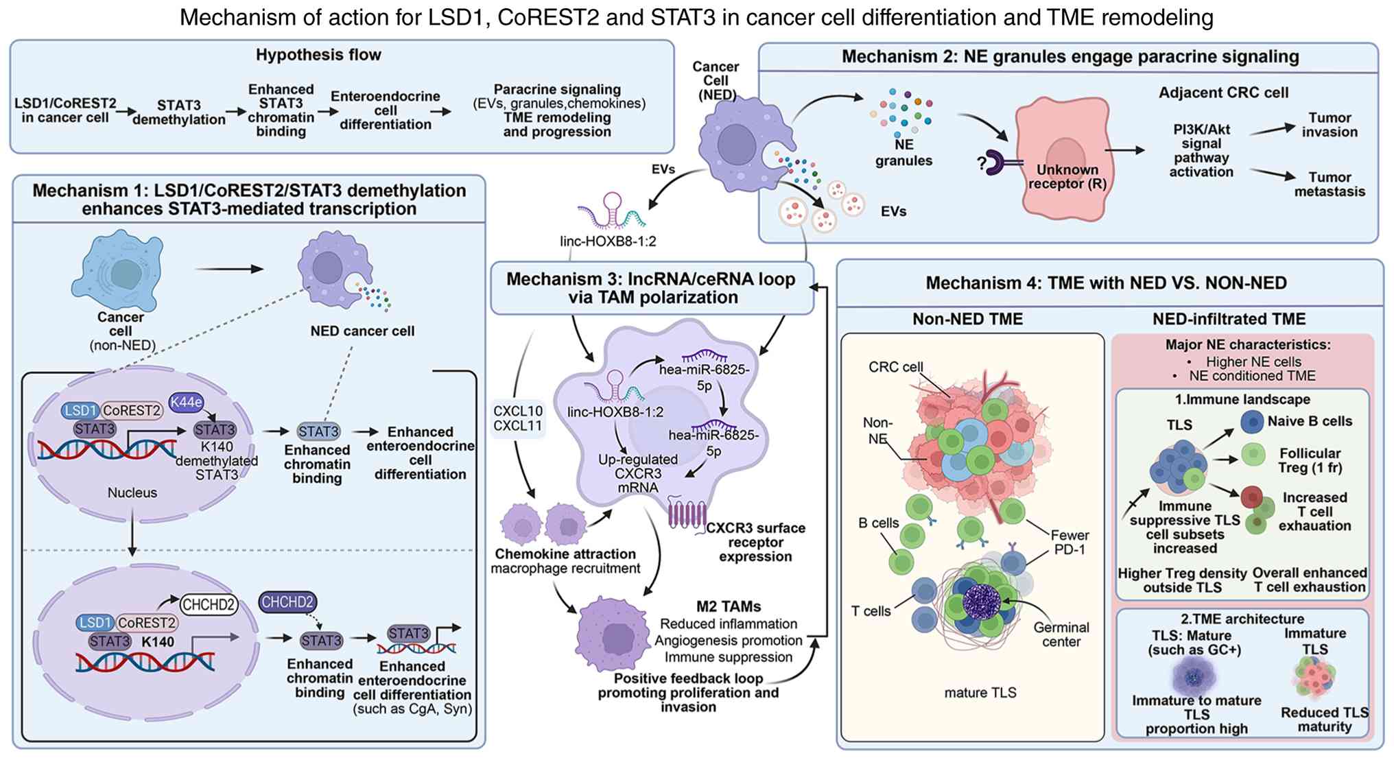 | Figure 2.Molecular mechanisms and TME
regulation in conventional carcinoma with NED. The LSD1/CoREST2
complex mediates STAT3 demethylation, which induces NED in cancer
cells. These differentiated cells promote invasion and metastasis
by releasing NE granules that activate the PI3K/Akt pathway in
adjacent cancer cells through paracrine signaling. Concurrently,
cells recruit macrophages through CXCL10 and CXCL11 and deliver
exosomal lnc-HOXB8-1:2, which acts as a ceRNA for miR-6825-5p,
upregulates CXCR3 and induces M2 polarization of macrophages,
thereby establishing a pro-tumorigenic positive feedback loop.
Furthermore, this differentiation markedly remodels the immune
repertoire, characterized by increased but immature TLS, enrichment
of naïve B cells and regulatory T cells and exacerbated T cell
exhaustion, ultimately resulting in a highly immunosuppressive
microenvironment. TME, tumor microenvironment; NE, neuroendocrine;
NED, NE differentiation; EVs, extracellular vesicles; TLS, tertiary
lymphoid structures; CRC, colorectal cancer; lnc/lncRNA, long
non-coding RNA; miR, micro-RNA; LSD1, lysine-specific demethylase
1; CoREST2, REST corepressor 2; CXCL, C-X-C motif chemokine ligand;
CXCR, C-X-C chemokine receptor; ceRNA, competing endogenous RNA;
TAMS, tumor associated macrophages; CHCHD2, coiled-coil domain
helix 2; Treg, regulatory T cell. |
Future directions and clinical
translation
Unresolved core scientific
questions
A number of key questions regarding conventional
carcinomas with NED remain unanswered despite decades of research:
i) What are the fundamental transcription factors and epigenetic
mechanisms responsible for inducing neuroendocrine phenotypes in
adenocarcinoma cells?; ii) how do microenvironmental cues such as
hypoxia, inflammation and therapeutic stress trigger NED through
intracellular signaling pathways?; iii) what is the molecular basis
and clinical implications of the discordance in NED status between
primary tumors and metastatic sites?; iv) are there distinct
metabolic features associated with NED and can these be
therapeutically targeted?; and v) how can one effectively
differentiate between conventional carcinoma with NED and ALC and
do their molecular distinctions support varied treatment
approaches?.
Key directions for future
research
Basic research should aim to focus on elucidating
the molecular switches governing lineage plasticity. Systematic
investigation of neuroendocrine lineage determinants such as
achaete-scute Family BHLH transcription factor 1, neurogenic
differentiation factor 1 and POU class 2 homeobox 3 in NED, using
single-cell transcriptome sequencing and organoid models, will help
clarify their roles. The LSD1/CoREST2/STAT3 axis represents a
promising target and further exploration of DNA methylation,
histone modification and chromatin remodeling in NED development is
warranted.
Clinical translation should prioritize three areas.
First, standardization of diagnostic criteria, including unified
positivity thresholds and observer consistency, is important in
enabling meaningful comparisons across studies. Second, prospective
clinical trials are needed to validate the independent prognostic
value of NED in stage II colorectal cancer and to determine whether
the presence of NED should guide adjuvant therapy decisions. Third,
given the association between NED and increased TAM infiltration,
combination strategies targeting both angiogenesis and TAMs warrant
investigation in patients with advanced colorectal cancer and NED.
In addition, although prospective data are lacking, a cautious
approach may be to consider closer postoperative surveillance for
NED-positive patients with stage II colorectal cancer or for those
who develop NED after neoadjuvant therapy. Such surveillance could
include more frequent imaging (for example, every 3–4 months for
the first 2 years) and serial monitoring of serum tumor markers
such as CgA or proGRP, recognizing that the clinical utility of
these markers in this setting remains unproven.
Conclusions
Gastrointestinal conventional carcinomas with NED
represent a distinct entity characterized by the absence of
neuroendocrine morphology but detectable expression of
neuroendocrine markers. Accumulating evidence has indicated that
NED arises from differentiation plasticity rather than independent
clonal evolution, with the PI3K/Akt and LSD1/CoREST2/STAT3 pathways
serving as key regulatory axes. While NED holds independent
prognostic importance in stage II colorectal cancer, its role in
other settings remains controversial, largely due to heterogeneity
in diagnostic criteria, study populations and diagnostic confusion
with associated entities.
Clinical practice faces a number of challenges,
including distinguishing NED from ALC, observer variability in
diagnostic thresholds and evolving classification concepts.
Addressing these issues requires standardized diagnostic
guidelines, wider adoption of high-specificity markers such as
INSM1 and prospective trials to validate the prognostic and
therapeutic implications of NED. Ultimately, unifying diagnostic
criteria will be key in transforming NED from a pathological
observation into a clinically actionable biomarker, enabling a
shift from mere identification to active intervention.
Acknowledgements
Not applicable.
Funding
The present study was supported by the National Natural Science
Foundation of China (grant no. 82404030).
Availability of data and materials
Not applicable.
Authors' contributions
TF conceived the idea of the present review. CZ, ST,
DZ and WW prepared the figures and tables. CZ, ST, DZ, YP, JX, SL,
WW and TF performed the literature search, wrote the manuscript and
approved the submission of the article. Data authentication is not
applicable. All authors read and approved the final version of the
manuscript.
Ethics approval and consent to
participate
Not applicable.
Patient consent for publication
Not applicable.
Competing interests
The authors declare that they have no competing
interests.
References
|
1
|
Jesinghaus M, Schmitt MP, Foersch S and
Konukiewitz B: MiNEN, amphicrine carcinomas, and conventional
carcinomas with neuroendocrine differentiation: Diagnostic
criteria, open questions, and future perspectives. Virchows Arch.
488:33–43. 2026. View Article : Google Scholar : PubMed/NCBI
|
|
2
|
Arends MJ, Esposito I, Gill AJ, Hruban RH,
Khoury JD, Kojima M, Montgomery EA, Abdulkareem F, Carneiro F,
Costamagna G, et al: Changes in the 6th edition of the World Health
Organization classification of tumours of the digestive system.
Histopathology. 88:1295–1314. 2026. View Article : Google Scholar : PubMed/NCBI
|
|
3
|
Litmeyer AS, Konukiewitz B, Kasajima A,
Foersch S, Schicktanz F, Schmitt M, Kellers F, Grass A, Jank P,
Lehman B, et al: High expression of insulinoma-associated protein 1
(INSM1) distinguishes colorectal mixed and pure neuroendocrine
carcinomas from conventional adenocarcinomas with diffuse
expression of synaptophysin. J Pathol Clin Res. 9:498–509. 2023.
View Article : Google Scholar : PubMed/NCBI
|
|
4
|
Liang Y, Li Y, Guo R, Zhao Y, Miao H,
Chang H and Chen Y: Identification and initial validation of
neuroendocrine differentiation as a novel prognostic factor in
stage II colorectal cancer patients. Oncology. 103:156–166.
2025.PubMed/NCBI
|
|
5
|
Xu F, Liu Q, Han F, Xu E, Lai M and Zhao
Z: Association of neuroendocrine differentiation with progression
and prognosis of gastric adenocarcinoma. Zhejiang Da Xue Xue Bao Yi
Xue Ban. 45:24–30. 2016.(In Chinese). PubMed/NCBI
|
|
6
|
Fassan M, Milione M, Maddalena G,
Cremolini C, Schirripa M, Pietrantonio F, Pella N, Dell'Aquila E,
Sperti E, Zichi C, et al: Synaptophysin expression in
V600EBRAF−mutated advanced colorectal cancers identifies
a new subgroup of tumours with worse prognosis. Eur J Cancer.
146:145–154. 2021. View Article : Google Scholar : PubMed/NCBI
|
|
7
|
Sabella G, Centonze G, Lagano V, Scardino
A, Belli F, Garzone G, Pardo C, Galbiati D, Pusceddu S, Mangogna A,
et al: Unveiling the prognostic role of synaptophysin in
conventional colorectal carcinomas. Neuroendocrinology.
115:632–647. 2025. View Article : Google Scholar : PubMed/NCBI
|
|
8
|
Li Q, Notkins AL and Lan MS: Molecular
characterization of the promoter region of a neuroendocrine tumor
marker, IA-1. Biochem Biophys Res Commun. 236:776–781. 1997.
View Article : Google Scholar : PubMed/NCBI
|
|
9
|
McHugh KE, Mukhopadhyay S, Doxtader EE,
Lanigan C and Allende DS: INSM1 is a highly specific marker of
neuroendocrine differentiation in primary neoplasms of the
gastrointestinal tract, appendix, and pancreas. Am J Clin Pathol.
153:811–820. 2020. View Article : Google Scholar : PubMed/NCBI
|
|
10
|
Pavel M, Oberg K, Falconi M, Krenning EP,
Sundin A, Perren A and Berruti A; ESMO Guidelines Committee.
Electronic address, : simpleclinicalguidelines@esmo.org:
Gastroenteropancreatic neuroendocrine neoplasms: ESMO Clinical
Practice Guidelines for diagnosis, treatment and follow-up. Ann
Oncol. 31:844–860. 2020. View Article : Google Scholar : PubMed/NCBI
|
|
11
|
Sorbye H, Grande E, Pavel M, Tesselaar M,
Fazio N, Reed NS, Knigge U, Christ E, Ambrosini V, Couvelard A and
Tiensuu Janson E: European Neuroendocrine Tumor Society (ENETS)
2023 guidance paper for digestive neuroendocrine carcinoma. J
Neuroendocrinol. 35:e132492023. View Article : Google Scholar : PubMed/NCBI
|
|
12
|
Grabowski P, Schindler I, Anagnostopoulos
I, Foss HD, Riecken EO, Mansmann U, Stein H, Berger G, Buhr HJ and
Scherübl H: Neuroendocrine differentiation is a relevant prognostic
factor in stage III–IV colorectal cancer. Eur J Gastroenterol
Hepatol. 13:405–411. 2001. View Article : Google Scholar : PubMed/NCBI
|
|
13
|
Li J, Wang C, Shen Y, Huang Z, Zhang J,
Rao S, Cao Y and Min J: Survival outcomes and prognostic factors of
gastric carcinoma with neuroendocrine differentiation. Int J
Cancer. 158:2209–2218. 2026. View Article : Google Scholar : PubMed/NCBI
|
|
14
|
Blumenfeld W, Chandhoke DK, Sagerman P and
Turi GK: Neuroendocrine differentiation in gastric adenocarcinomas.
An immunohistochemical study. Arch Pathol Lab Med. 120:478–481.
1996.PubMed/NCBI
|
|
15
|
Syversen U, Halvorsen T, Mårvik R and
Waldum HL: Neuroendocrine differentiation in colorectal carcinomas.
Eur J Gastroenterol Hepatol. 7:667–674. 1995.PubMed/NCBI
|
|
16
|
Liu Y, Xu J, Jiao Y, Hu Y, Yi C, Li Q,
Tong Z, Wang X, Hu L, Xiao Q, et al: Neuroendocrine differentiation
is a prognostic factor for stage II poorly differentiated
colorectal cancer. Biomed Res Int. 2014:7895752014.PubMed/NCBI
|
|
17
|
Guo J, Zheng P, Wang R, Tan X, Yang W,
Xiao S and Chen Z: Prognostic relevance of neuroendocrine
differentiation in colorectal cancer: A population-based,
propensity score matching study. Int J Colorectal Dis.
35:2185–2195. 2020. View Article : Google Scholar : PubMed/NCBI
|
|
18
|
Liu Y, He J, Xu J, Li J, Jiao Y, Bei D, Hu
Y, Chen H, Xiao Q and Ding K: Neuroendocrine differentiation is
predictive of poor survival in patients with stage II colorectal
cancer. Oncol Lett. 13:2230–2236. 2017. View Article : Google Scholar : PubMed/NCBI
|
|
19
|
Chen Y, Liu F, Meng Q and Ma S: Is
neuroendocrine differentiation a prognostic factor in poorly
differentiated colorectal cancer? World J Surg Oncol. 15:712017.
View Article : Google Scholar : PubMed/NCBI
|
|
20
|
Cho YB, Yang SS, Lee WY, Song SY, Kim SH,
Shin HJ, Yun SH and Chun HK: The clinical significance of
neuroendocrine differentiation in T3-T4 node-negative colorectal
cancer. Int J Surg Pathol. 18:201–206. 2010. View Article : Google Scholar : PubMed/NCBI
|
|
21
|
Konukiewitz B, Kasajima A, Schmitt M,
Schwamborn K, Groll T, Schicktanz F, Delbridge C, Schütze LM,
Wilhelm D, Lang C, et al: Neuroendocrine differentiation in
conventional colorectal adenocarcinomas: Incidental finding or
prognostic biomarker? Cancers (Basel). 13:51112021. View Article : Google Scholar : PubMed/NCBI
|
|
22
|
Takii M, Takemura M, Oshima T, Yamada M,
Inazu D, Miyamoto H and Yamaguchi H: A rare case of poorly
differentiated lower esophageal cancer with neuroendocrine
differentiation and α-fetoprotein production. Surg Case Rep.
12:25–0619. 2026. View Article : Google Scholar : PubMed/NCBI
|
|
23
|
Geng X, Liu J, Sun H, Song Z, Qin S, Li Y,
Zhang Y, Hao F and Cai Y: Subcutaneous soft tissue metastases from
esophageal squamous cell carcinoma with neuroendocrine
differentiation: Case report and literature review. Front Oncol.
12:8951892022. View Article : Google Scholar : PubMed/NCBI
|
|
24
|
Yamaura M, Yamada T, Watanabe R, Kawai H,
Hirose S, Tajima H, Sato M, Uchida Y, Suganuma D, Yamamoto Y, et
al: Anal canal adenocarcinoma with neuroendocrine features
accompanying secondary extramammary Paget disease, successfully
treated with modified FOLFOX6: A case report. BMC Cancer.
18:11422018. View Article : Google Scholar : PubMed/NCBI
|
|
25
|
Atasoy P, Ensari A, Demirci S and Kurşun
N: Neuroendocrine differentiation in colorectal carcinomas:
Assessing its prognostic significance. Tumori. 89:49–53. 2003.
View Article : Google Scholar : PubMed/NCBI
|
|
26
|
Centonze G, Maisonneuve P, Prinzi N,
Pusceddu S, Albarello L, Pisa E, Barberis M, Vanoli A, Spaggiari P,
Bossi P, et al: Prognostic factors across poorly differentiated
neuroendocrine neoplasms: A pooled analysis. Neuroendocrinology.
113:457–469. 2023. View Article : Google Scholar : PubMed/NCBI
|
|
27
|
Shia J, Tickoo SK, Guillem JG, Qin J,
Nissan A, Hoos A, Stojadinovic A, Ruo L, Wong WD, Paty PB, et al:
Increased endocrine cells in treated rectal adenocarcinomas: A
possible reflection of endocrine differentiation in tumor cells
induced by chemotherapy and radiotherapy. Am J Surg Pathol.
26:863–872. 2002. View Article : Google Scholar : PubMed/NCBI
|
|
28
|
Dodington DW, Serra S, Bracey T, Chetty R
and Nowak KM: Neuroendocrine differentiation and serotonin
expression in oesophageal adenocarcinomas after neoadjuvant
therapy: Correlation with clinicopathological features and outcome.
Histopathology. 86:559–570. 2025. View Article : Google Scholar : PubMed/NCBI
|
|
29
|
Zeng YJ, Lai W, Liu L, Wu H, Luo XX, Wang
J and Chu ZH: Prognostic significance of neuroendocrine
differentiation in colorectal adenocarcinoma after radical
operation: A meta-analysis. J Gastrointest Surg. 18:968–976. 2014.
View Article : Google Scholar : PubMed/NCBI
|
|
30
|
Albayrak NE, Liu BL, Mehrotra M,
Houldsworth J, Polydorides AD and Ward SC: Clinicopathologic and
molecular analysis of colorectal carcinomas with spectrum of
neuroendocrine carcinoma components. Mod Pathol. 38:1008552025.
View Article : Google Scholar : PubMed/NCBI
|
|
31
|
Huang D, Ren F, Ni S, Tan C, Weng W, Zhang
M, Xu M, Wang L, Xu Q and Sheng W: Amphicrine carcinoma of the
stomach and intestine: A clinicopathologic and pan-cancer
transcriptome analysis of a distinct entity. Cancer Cell Int.
19:3102019. View Article : Google Scholar : PubMed/NCBI
|
|
32
|
Li D, Lin X and Huang W:
Clinicopathological characterization of gastric amphicrine
carcinoma: A case series. Ann Diagn Pathol. 81:1525842026.
View Article : Google Scholar : PubMed/NCBI
|
|
33
|
Chen H, Chen J and Ge R: Gastrointestinal
amphicrine carcinoma: A clinicopathologic study of five patients.
Int J Surg Pathol. 33:1713–1718. 2025. View Article : Google Scholar : PubMed/NCBI
|
|
34
|
Bozkaya Y, Ozdemir N, Colak A and Zengin
N: Gastric adenocarcinoma with neuroendocrine differentiation:
Clinicopathological features and efficacy of modified DCF
chemotherapy. J BUON. 22:919–925. 2017.PubMed/NCBI
|
|
35
|
Dost Gunay FS, Kırmızı BA, Ensari A, İcli
F and Akbulut H: Tumor-associated macrophages and neuroendocrine
differentiation decrease the efficacy of bevacizumab plus
chemotherapy in patients with advanced colorectal cancer. Clin
Colorectal Cancer. 18:e244–e250. 2019.PubMed/NCBI
|
|
36
|
Zhang HY, Wei XL, Wang LL, Zhang BY, Zhao
ZS, Lü YB and Yao GY: Detection and clinicopathologic significance
of microsatellite alteration and p53 mutation of neuroendocrine
cells in colorectal adenocarcinoma. Zhonghua Bing Li Xue Za Zhi.
42:10–14. 2013.(In Chinese). PubMed/NCBI
|
|
37
|
Wang LL, Yao GY, Zhao ZS, Wei XL and Xu
RJ: Clonality analysis of neuroendocrine cells in gastric
adenocarcinoma. World J Gastroenterol. 19:5340–5346. 2013.
View Article : Google Scholar : PubMed/NCBI
|
|
38
|
Kim KM, Kim MJ, Cho BK, Choi SW and Rhyu
MG: Genetic evidence for the multi-step progression of mixed
glandular-neuroendocrine gastric carcinomas. Virchows Arch.
440:85–93. 2002. View Article : Google Scholar : PubMed/NCBI
|
|
39
|
Sun L, Zhang J, Wang C, Zhao S, Shao B,
Guo Y, Liu Y and Sun Y: Chromosomal and molecular pathway
alterations in the neuroendocrine carcinoma and adenocarcinoma
components of gastric mixed neuroendocrine-nonneuroendocrine
neoplasm. Mod Pathol. 33:2602–2613. 2020. View Article : Google Scholar : PubMed/NCBI
|
|
40
|
Sipos F and Műzes G: Injury-associated
reacquiring of intestinal stem cell function. World J
Gastroenterol. 21:2005–2010. 2015. View Article : Google Scholar : PubMed/NCBI
|
|
41
|
Humphries A, Cereser B, Gay LJ, Miller DS,
Das B, Gutteridge A, Elia G, Nye E, Jeffery R, Poulsom R, et al:
Lineage tracing reveals multipotent stem cells maintain human
adenomas and the pattern of clonal expansion in tumor evolution.
Proc Natl Acad Sci USA. 110:E2490–E2499. 2013. View Article : Google Scholar : PubMed/NCBI
|
|
42
|
Chen Y, Liang Y, Cao L, Dong X and Sun D:
Neuroendocrine differentiation: A risk fellow in colorectal cancer.
World J Surg Oncol. 21:892023. View Article : Google Scholar : PubMed/NCBI
|
|
43
|
Ladaika CA, Ghobashi AH, Boulton WC,
Miller SA and O'Hagan HM: LSD1 and CoREST2 potentiate STAT3
activity to promote enteroendocrine cell differentiation in
mucinous colorectal cancer. Cancer Res. 85:52–68. 2025. View Article : Google Scholar : PubMed/NCBI
|
|
44
|
Ladaika CA, Chakraborty A, Masood A,
Hostetter G, Yi JM and O'Hagan HM: LSD1 inhibition attenuates
targeted therapy-induced lineage plasticity in BRAF mutant
colorectal cancer. Mol Cancer. 24:1222025. View Article : Google Scholar : PubMed/NCBI
|
|
45
|
Zeng YJ, Lai W, Wu H, Liu L, Xu HY, Wang J
and Chu ZH: Neuroendocrine-like cells -derived CXCL10 and CXCL11
induce the infiltration of tumor-associated macrophage leading to
the poor prognosis of colorectal cancer. Oncotarget. 7:27394–27407.
2016. View Article : Google Scholar : PubMed/NCBI
|
|
46
|
Zou Y, Li D, Yu X, Zhou C, Zhu C and Yuan
Y: Correlation of neuroendocrine differentiation with a
distinctively suppressive immune microenvironment in gastric
cancer. Neuroendocrinology. 114:192–206. 2024. View Article : Google Scholar : PubMed/NCBI
|
|
47
|
Li X, Lan Q, Lai W, Wu H, Xu H, Fang K,
Chu Z and Zeng Y: Exosome-derived lnc-HOXB8-1:2 induces
tumor-associated macrophage infiltration to promote neuroendocrine
differentiated colorectal cancer progression by sponging
hsa-miR-6825-5p. BMC Cancer. 22:9282022. View Article : Google Scholar : PubMed/NCBI
|

















