Introduction
Cholangiocarcinoma (CCA) is a highly heterogeneous
epithelial carcinoma with variable degrees of differentiation
(1,2). Based on anatomical location, CCA is
classified into intrahepatic CCA and extrahepatic
cholangiocarcinoma (eCCA). According to the location relative to
the cystic duct, eCCA is further subdivided into perihilar
cholangiocarcinoma and distal cholangiocarcinoma (3,4). eCCA
accounts for 20–30% of all biliary tract cancers (5). Established risk factors mainly include
non-alcoholic fatty liver disease, liver cirrhosis and primary
sclerosing cholangitis (6). Over
recent decades, the incidence of eCCA has increased steadily.
However, the prognosis remains poor, with a 5-year survival rate of
~11% (7,8). Surgery is still the only curative
treatment for non-metastatic eCCA. For patients with advanced
disease, the primary treatment strategy is chemotherapy combined
with immunotherapy, although the therapeutic efficacy remains
limited. Therefore, investigation of the pathogenesis of eCCA and
development of more effective therapies are of great
importance.
Unlike tumor cell lines, organoids are
three-dimensional (3D) structures derived from pluripotent stem
cells or isolated organ progenitor cells. These structures can
self-organize into ‘mini-organs’ that recapitulate the architecture
of native tissues (9) and reproduce
the pathophysiological characteristics of the parental tumors
(10). Organoids closely mimic the
structure and function of primary tissues while retaining high
genetic fidelity and donor-specific therapeutic responses (11). Furthermore, the establishment of
organoids requires substantially less time and tissue than the
generation of patient-derived xenograft models (12). Importantly, compared to animal-based
models, organoid systems offer a significant ethical advantage by
reducing or replacing the need for animal experimentation, thereby
addressing animal welfare concerns. While the use of human-derived
cells for in vitro culture involves its own distinct ethical
considerations, it bypasses many of the complex ethical
implications inherent in animal testing.
This study aimed to explore the feasibility of
establishing eCCA organoids using suspension culture, with the goal
of providing a potentially more economical and rapid alternative
construction platform for basic research on this malignancy.
Materials and methods
Tissue sources
For organoid culture, tumor tissues were collected
from five patients with a preoperative diagnosis of eCCA who
underwent surgical resection at the Second Hospital of Lanzhou
University (Lanzhou, China) between February 2023 and December
2024. These tissues were used to establish patient-derived organoid
models. None of the patients had received radiotherapy or
chemotherapy before surgery. This study was conducted in accordance
with the Declaration of Helsinki. The study protocol was approved
by the Ethics Committee of the Second Hospital of Lanzhou
University (approval no. 2023A-381) and informed consent was
obtained from all patients and/or their legal guardians, as
appropriate.
Primary reagents, instruments and
consumables
DMEM/F12 medium, trypsin and fetal bovine serum were
purchased from Biological Industries. GlutaMAX and Dispase II were
from Invitrogen (Thermo Fisher Scientific, Inc.). Collagenase II
was purchased from Gibco (Thermo Fisher Scientific, Inc.).
Serum-free cell cryopreservation medium was obtained from NCM
Biotechnology Co., Ltd. Antibodies against cytokeratin (CK)7 (cat.
no. MAB-0828; dilution, 1:100), CK19 (cat. no. MAB-0829; dilution,
1:100) and Ki-67 (cat. no. RMA-0542; dilution, 1:100) were
purchased from Fuzhou Maixin Biotechnology Co., Ltd. The
horseradish peroxidase-conjugated universal secondary antibody
(cat. no. KIT-5020) was purchased from Fuzhou Maixin Biotechnology
Co., Ltd. Penicillin-streptomycin solution and
penicillin-streptomycin-amphotericin B solution were obtained from
Shanghai Dotehill Biological Technology Co., Ltd. PBS was purchased
from Wuhan Servicebio Technology Co., Ltd. Gemcitabine
hydrochloride for injection was obtained from Jiangsu Hansoh
Pharmaceutical Group Co., Ltd., Oxaliplatin for injection from
Jiangsu Hengrui Pharmaceuticals Co., Ltd., 5-Fluorouracil (5-FU)
injection from Shanghai Xudong Haipu Pharmaceutical Co., Ltd. and
albumin-bound paclitaxel from Wuxi Zishan Pharmaceutical Co.,
Ltd.
The major instruments used in this study included an
inverted microscope (Nikon Corp.) and a paraffin microtome (Leica
Microsystems GmbH).
Primary consumables included ultra-low attachment
plates, 50-ml centrifuge tubes, 1,000-µl pipette tips, pasteur
pipettes and 60-mm culture dishes purchased from NEST Biotechnology
Co., Ltd. Furthermore, 15-ml centrifuge tubes and microcentrifuge
tubes were obtained from Wuhan Servicebio Technology Co., Ltd.
Establishment of organoid models
Fresh tumor tissues were collected under aseptic
conditions and processed as follows. The organoid culture protocol
was established based on our previously described method with minor
modifications (13). A tissue block
of ~1×1×1 cm was excised for organoid culture and placed in a 15-ml
centrifuge tube containing 10 ml of transport medium, namely
complete organoid culture medium consisting of DMEM/F12
supplemented with 1X GlutaMAX, 10% fetal bovine serum and 1%
penicillin-streptomycin solution. In addition, a tumor tissue
fragment measuring ~0.5×0.5×0.5 cm was placed in a 2-ml
microcentrifuge tube and stored at −80°C for subsequent
experiments. The centrifuge tube was immediately transported to the
laboratory in a specimen transport box containing crushed ice.
Upon arrival at the laboratory, the tumor tissue was
transferred to a 60-mm culture dish and rinsed with 5 ml of PBS to
remove blood clots and necrotic debris. The tissue was then
carefully minced with a sterile No. 23 surgical scalpel.
Subsequently, 5 ml of mixed digestive enzyme solution (consisting
of Collagenase II at 1 mg/ml and Dispase II at 2 mg/ml in DMEM/F12)
was added at room temperature and the resulting tissue suspension
was transferred into a 15-ml centrifuge tube using a Pasteur
pipette. The tube was placed on a shaker and digested at 37°C for
~45 min. During this period, the digestion process was closely
monitored. When the tissue volume had decreased by approximately
half, the tube was allowed to stand undisturbed for 10 min. The
supernatant was then carefully transferred into a new centrifuge
tube using a Pasteur pipette and centrifuged at 150 × g for 5 min
at 4°C. After removal of the supernatant, this washing step was
repeated twice to remove residual digestive enzymes and cellular
debris. The final pellet was resuspended in complete organoid
culture medium consisting of DMEM/F12 supplemented with 1X
GlutaMAX, 10% fetal bovine serum and 1% penicillin-streptomycin
solution. The cell suspension was then evenly seeded into a 12-well
ultra-low-attachment plate, and the volume in each well was
adjusted to 3 ml with complete organoid culture medium, to initiate
organoid culture. The cells were cultured at 37°C in a 5%
CO2 humidified incubator. Visible cell aggregates
typically formed within 2–3 days, and organoids reached ~100 µm in
diameter within 5–7 days after seeding.
Passaging, freezing and thawing of
organoids
Passaging
During organoid culture, the morphology of organoids
in each well was photographed every 2 days. Passaging was performed
when the diameter of the eCCA organoids reached ~100 µm. The
organoid suspension from each well was collected into a 50-ml
centrifuge tube using a Pasteur pipette. Approximately 10 ml of PBS
was added and the suspension was gently mixed by pipetting with a
1,000-µl pipette tip, followed by centrifugation at 340 × g for 5
min at 4°C. The supernatant was discarded. Next, 3 ml of 0.25%
trypsin was added to the organoid pellet and mixed gently.
Digestion was carried out at 37°C and monitored under a light
microscope using a 4× objective lens (total magnification, ×40).
When the organoid clusters had dissociated into small fragments
containing <10 cells, digestion was terminated by adding 6 ml of
complete organoid medium. The suspension was gently pipetted again
with a 1,000-µl pipette tip, then centrifuged at 340 × g for 5 min
at 4°C, and the supernatant was removed. For passaging at a split
ratio of 1:2 to 1:3, 12 ml of complete organoid medium was added to
the centrifuge tube and the pellet was gently resuspended.
Subsequently, one-half or one-third of the organoid suspension was
evenly seeded into a new 12-well ultra-low attachment plate and the
volume in each well was adjusted to 3 ml with complete medium.
Culture was subsequently continued as described above.
Cryopreservation
The remaining organoids in the centrifuge tube were
centrifuged at 340 × g for 5 min at 4°C. According to the pellet
size, 1–2 ml of cryopreservation medium was added and the pellet
was gently resuspended by pipetting. A commercial cell-freezing
medium was also acceptable. The suspension was then transferred to
a 2-ml cryovial and stored in liquid nitrogen for long-term
preservation.
Thawing
For organoid recovery, the cryovial was rapidly
thawed in a 37°C water bath. The organoid suspension was then
immediately transferred to a 15-ml centrifuge tube using a 1,000-µl
pipette tip. Next, 5 ml of complete organoid culture medium was
added and the suspension was gently mixed by pipetting. After
centrifugation at 340 × g for 5 min at 4°C, the supernatant was
discarded. The pellet was then resuspended in 12 ml of complete
organoid culture medium, gently mixed again and evenly seeded into
a 12-well ultra-low-attachment plate. Organoid culture was
continued as described above.
Organoid embedding and sectioning
After centrifugation of the organoid suspension at
340 × g at 4°C for 5 min, 5 ml of 4% paraformaldehyde fixative was
added to the centrifuge tube and fixation was carried out at 37°C
for 24 h. The samples were then dehydrated through a graded ethanol
series (50, 70, 80, 95 and 100% ×2), with each step lasting 30 min.
Subsequently, the fixed organoid blocks were immersed in a
xylene-ethanol mixture (1:1) for 30 min, followed by two
incubations in pure xylene for 1 h each. The organoid blocks were
then embedded in paraffin and the paraffin blocks were sectioned
into slices ~5 µm thick using a microtome. The sections were
mounted on glass slides, air-dried and stored for subsequent
use.
Hematoxylin and eosin (H&E)
staining of organoids and primary tumor tissue
Sections from paraffin-embedded organoid blocks and
primary tumor tissue blocks were mounted on glass slides and placed
on staining racks. Deparaffinization was performed in xylene (3×2
min), followed by rehydration through a graded ethanol series
consisting of 100% ethanol (3×2 min), 95% ethanol (2 min) and 70%
ethanol (2 min). The slides were then rinsed under running tap
water at room temperature for at least 5 min.
Staining was performed with eosin solution for 2
min, followed by differentiation in 95% ethanol with ~20 dips. The
sections were then dehydrated sequentially in 95% ethanol for 2 min
and 100% ethanol (2×2 min). Clearing was performed in xylene (3×2
min). Finally, the sections were mounted with neutral balsam and
covered with coverslips. Slides that passed microscopic quality
control were subjected to image acquisition and analysis.
Immunohistochemistry of organoids
Cryopreserved eCCA tumor tissues stored at −80°C
were fixed in 4% paraformaldehyde and embedded in paraffin to
prepare 5 µm-thick sections, following the protocol previously
described for organoids. To characterize the tissue origin and
proliferative activity, both organoid sections and tumor tissue
sections were subjected to standard chromogenic
immunohistochemistry. After deparaffinization and rehydration
through a graded ethanol series, antigen retrieval was performed in
citrate buffer using a microwave oven, and endogenous peroxidase
activity was blocked with 3% H2O2. The
sections were then incubated overnight at 4°C with primary
antibodies against CK7, CK19 and Ki-67, each at a dilution of
1:100. On the following day, a horseradish peroxidase-conjugated
universal secondary antibody (ready-to-use) was applied for 30 min
at room temperature, followed by visualization with
3,3′-diaminobenzidine. Positive signals were identified as
brownish-yellow precipitates under a light microscope. Finally, the
slides were counterstained with hematoxylin and mounted. Image
acquisition and analysis were performed using optical microscopy.
For Ki-67 quantification, two pathologists independently and
blindly evaluated five randomly selected high-power fields per
slide using ImageJ software (version 1.54f; National Institutes of
Health). The mean percentage of positive nuclei relative to the
total cell count was then calculated to determine the final
labeling index.
Drug sensitivity testing of
organoids
Organoids were collected into a 15-ml centrifuge
tube using a Pasteur pipette, centrifuged at 97 × g for 5 min at
room temperature and the supernatant was discarded. The organoid
pellet was then digested with trypsin to obtain a single-cell
suspension. After cell counting with a hemocytometer, 100 µl of
cell suspension adjusted to a cell density of 1.2×10⁵
cells/ml was evenly added into each well of a 96-well plate (NEST
Biotechnology Co., Ltd). After 24 h, four antitumor drugs,
gemcitabine, oxaliplatin, 5-FU and albumin-bound paclitaxel, were
diluted in complete medium (RPMI-1640 supplemented with 10% fetal
bovine serum and 1% penicillin-streptomycin) to the following
concentrations. These concentration ranges were established based
on our preliminary dose-finding experiments. Gemcitabine (600, 150,
30, 6, 1.5, 0.3, 0.06, 0.015, 0.003, 0.0006, 0.00015, 0.00003,
0.000006 and 0.0000012 µmol/l), oxaliplatin (400, 200, 100, 40, 20,
10, 5, 2.5, 1.25, 0.625, 0.3125, 0.15625, 0.078 and 0.039 µmol/l),
5-FU (7,680, 1,920, 384, 96, 24, 6, 1.5, 0.3, 0.06, 0.015, 0.003,
0.0006, 0.00015 and 0.00003 µmol/l) and albumin-bound paclitaxel
(40, 20, 10, 5, 2.5, 0.5, 0.1, 0.025, 0.005, 0.001, 0.00025,
0.00005, 0.00001 and 0.0000025 µmol/l). After removal of the spent
medium, 170 µl of the corresponding drug solution was added to each
well. The plate included a control group and drug treatment groups.
The control group received 170 µl of complete medium, whereas the
treatment groups received an equal volume of the corresponding drug
solution. The control group and each drug concentration group were
set up in quadruplicate. After 72 h of culture, the medium was
removed and 100 µl of Cell Counting Kit (CCK)-8 (APExBIO Technology
LLC) diluted in RPMI-1640 at a 1:9 ratio was added to each well.
Following incubation at 37°C for 3 h, the absorbance at 450 nm was
measured using a microplate reader (Synergy H1; Biotek China). The
experiment was independently performed in quadruplicate. Drug
dose-response curves were generated and the half-maximal inhibitory
concentration (IC50) values were calculated using
GraphPad Prism software, version 9.5.1 (Dotmatics). IC50
values were determined by a nonlinear regression using the
following four-parameter logistic equation:
where X is the logarithm of the drug concentration,
Y is the normalized cell viability and HillSlope represents the
slope of the dose-response curve. In the four-parameter logistic
equation, Top represents the maximum response (cell viability at
the lowest drug concentration, typically approaching 100%
viability) and Bottom represents the minimum response (cell
viability at the highest drug concentration, where inhibition
reaches a plateau).
Results
Establishment of extrahepatic bile
duct cancer organoids
A total of five eCCA tumor tissue samples were
collected in this study and the clinical data of the patients are
summarized in Table I. Tumor
samples were obtained from five patients (age range: 58–68 years;
median age: 61 years; three males and two females), and three
organoid lines were successfully established, yielding an overall
success rate of 60%. These were designated as Organoid-P1,
Organoid-P2 and Organoid-P3, which were derived from Patient 1,
Patient 2 and Patient 5, respectively, as listed in Table I (numbered sequentially according to
successful establishment).
 | Table I.Clinical data of 5 patients with
cholangiocarcinoma. |
Table I.
Clinical data of 5 patients with
cholangiocarcinoma.
| Parameter | Patient 1 | Patient 2 | Patient 3 | Patient 4 | Patient 5 |
|---|
| Age, years | 58 | 58 | 66 | 68 | 61 |
| Sex | Male | Male | Female | Male | Female |
| Diagnosis date | 2023/02 | 2023/04 | 2023/08 | 2024/12 | 2024/01 |
| Survival
status | Alive | Alive | Alive | Alive | Alive |
| Sample site | Distal CBD | Mid CBD | Mid CBD | Mid CBD | Proximal CBD |
| Tumor size, mm | 30×20 | 20×15 | - | 20×18 | 35×30 |
| TNM | T2N1M0 | T1N0M0 | T2N0M0 | T2N0M0 | T3N1M0 |
| Tumor | Moderately | Poorly | Moderately | Moderately |
Moderately-poorly |
|
differentiation | differentiated | differentiated | differentiated | differentiated | differentiated |
| CEA, ng/ml | 2.8 | 0.54 | 2.97 | 0.95 | 1.15 |
| CA19-9, ng/ml | 344 | 36 | 327 | 890 | 91 |
The two unsuccessful cases were mainly attributed to
low initial cell viability and marked tissue necrosis in the
surgical specimens. After serial passaging, cryopreservation and
thawing, stable eCCA organoids were successfully maintained without
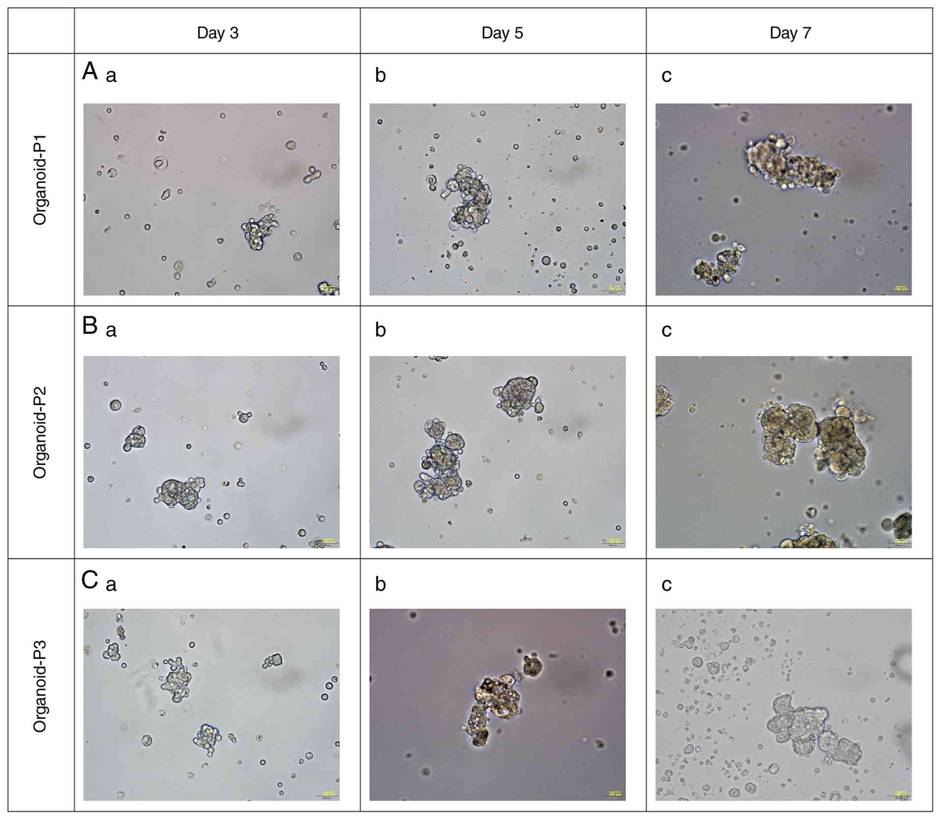
a significant reduction in viability. Under an inverted microscope,
the organoids exhibited irregular cystic, grape-like, lobulated or
solid morphologies. Some organoids retained central luminal
structures resembling biliary duct-like cavities, with
heterogeneous cyst sizes and irregular shapes (Fig. 1).
H&E staining results of eCCA
organoids and primary tumor tissue
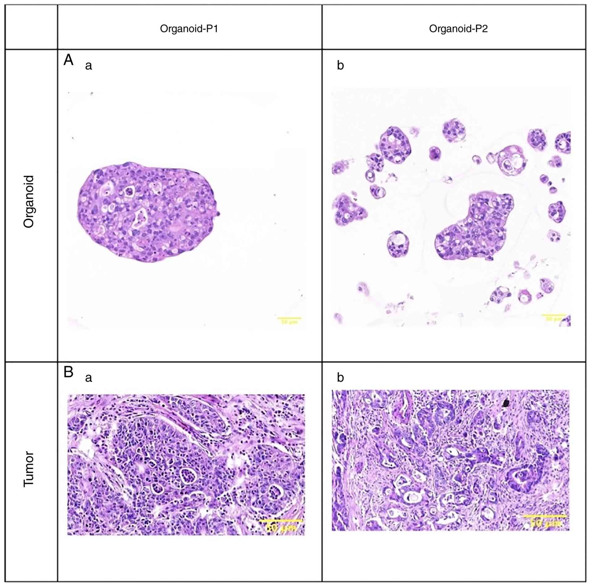
H&E staining was performed to evaluate the
histomorphological features of the established eCCA organoids. The
results showed that the organoids exhibited cystic or irregular
gland-like structures, accompanied by marked nuclear atypia and
pathological mitotic figures (Fig.
2). These features closely resembled the histoarchitectural
characteristics of the corresponding primary tumor tissues.
Immunohistochemical findings in eCCA
organoids
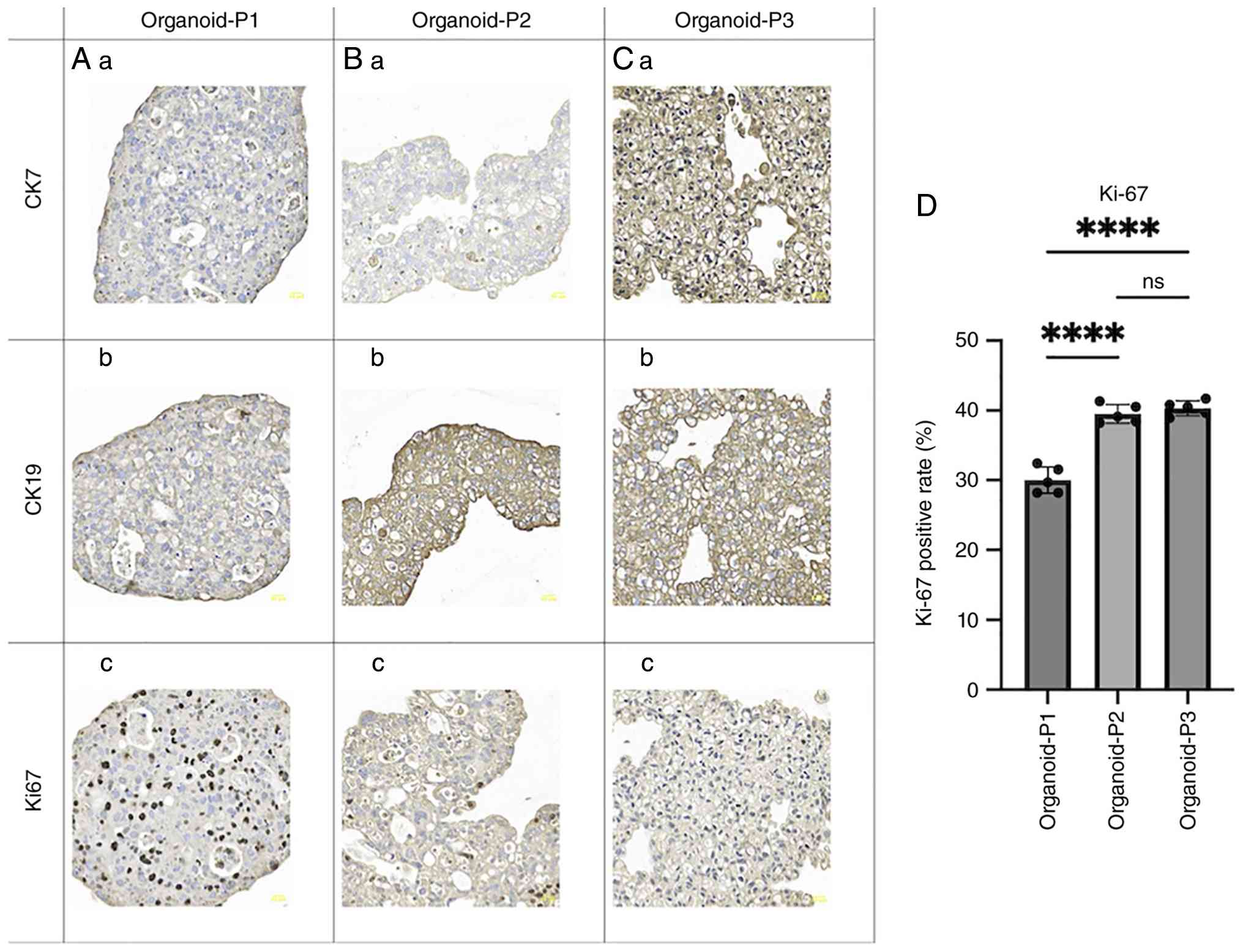
The protein expression levels of CK19, CK7 and Ki-67
in eCCA organoids were assessed by immunohistochemistry. The
results showed strong positive expression of CK7 and CK19 in all
organoid lines. This robust and uniform expression pattern further
supports the high fidelity of the organoids to the biliary
epithelial origin of the primary tumors. Positive staining was
mainly localized to the cytoplasm and cell membrane and showed a
homogeneous distribution throughout the organoid architecture.
Quantitative analysis based on double-blind counting in five
randomly selected high-power fields showed that all three organoid
lines maintained high proliferative activity. Specifically, the
Ki-67 labeling indices were 30.0±1.9% for Organoid-P1, 39.5±1.4%
for Organoid-P2 and 40.3±1.1% for Organoid-P3 (Fig. 3).
Drug sensitivity testing and screening
of eCCA organoids
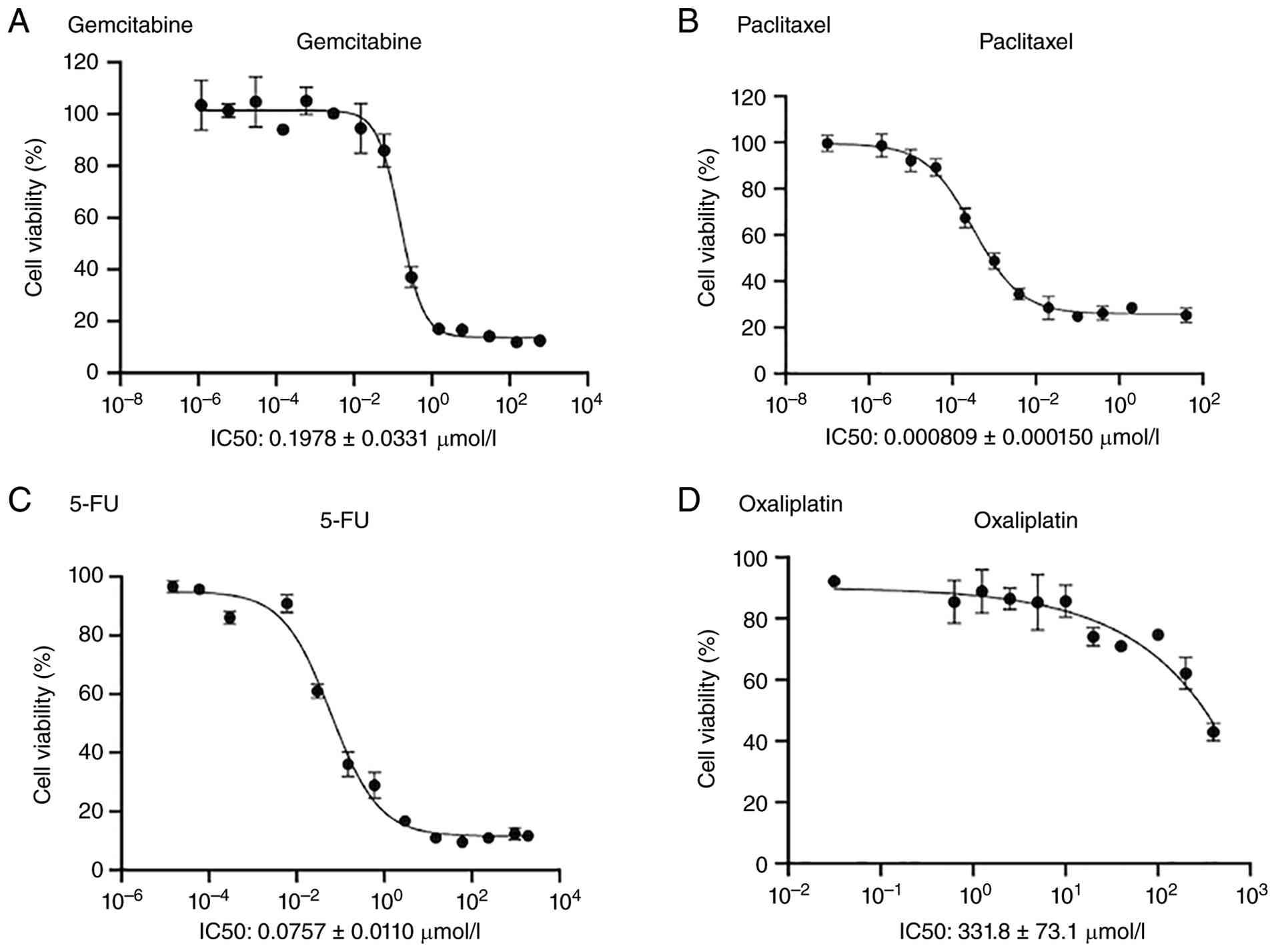
The drug-sensitivity testing results for Organoid-P1
(Fig. 4) showed differential
responses to gemcitabine, 5-FU, albumin-bound paclitaxel and
oxaliplatin. Organoid-P1 was sensitive to gemcitabine
(IC50=0.1978±0.0331 µmol/l), 5-FU
[IC50=(7.57±1.10) × 10−2 µmol/l] and
albumin-bound paclitaxel [IC50=(8.09±1.50) ×
10−4 µmol/l], but was resistant to oxaliplatin
(IC50=331.8±73.1 µmol/l). Patient 1, from whom
Organoid-P1 was derived, received seven cycles of postoperative
combination therapy with gemcitabine, tegafur-gimeracil-oteracil
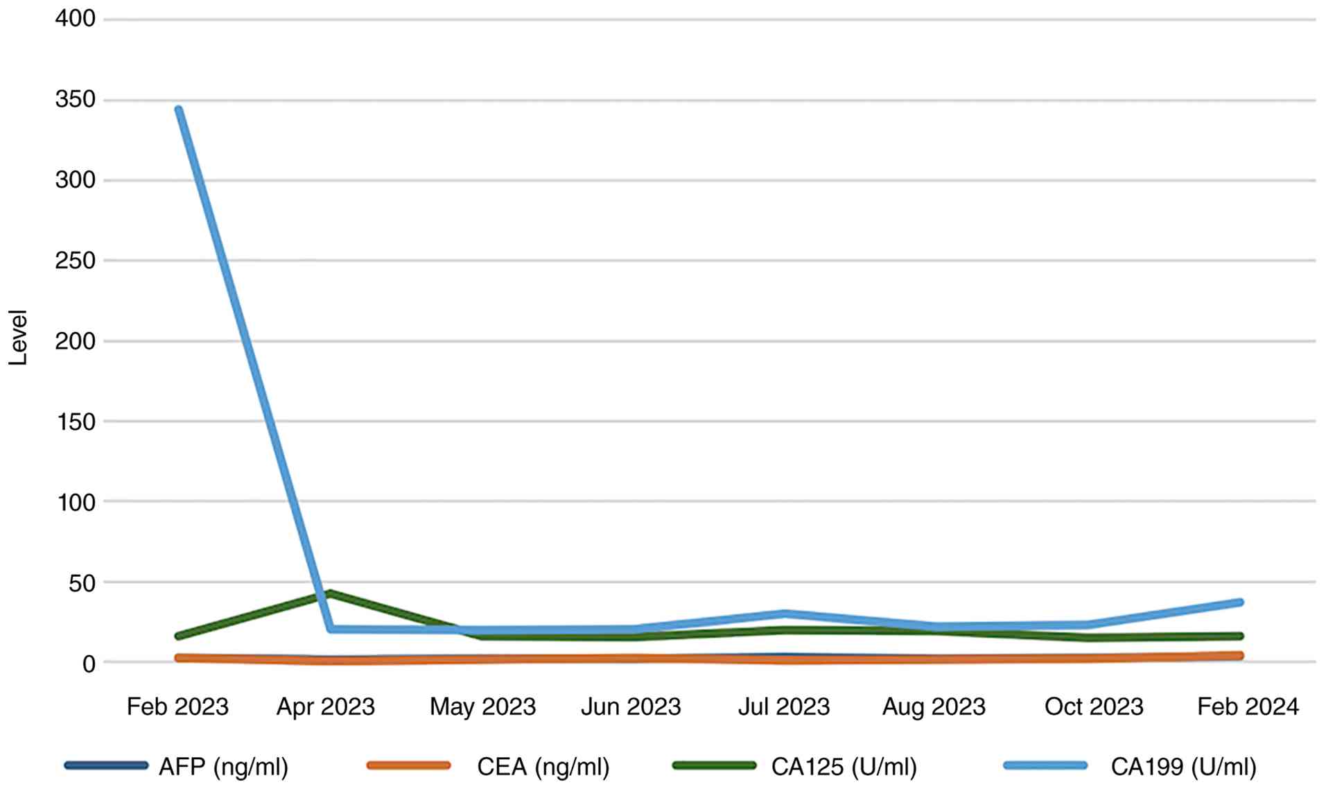
(S-1) and sintilimab. Regular follow-up examinations, including
serial measurements of serum tumor markers (AFP, CEA, CA19-9 and
CA-125) at different postoperative time-points (Fig. 5), showed no evidence of tumor
recurrence within 12 months after surgery. These findings provide
clinical support for the feasibility and predictive value of the
organoid-based drug-sensitivity testing platform. It should be
noted that comprehensive drug sensitivity testing was performed
only on Organoid-P1, as the primary goal of this study was to
provide proof-of-concept validation by comparing in vitro
drug responses with actual clinical outcomes. Patient 1 had
complete follow-up data and received standard postoperative
chemotherapy, making Organoid-P1 the appropriate line for
establishing clinical concordance.
Discussion
In the present study, a human eCCA organoid culture
system was successfully established using suspension culture and
its potential for clinical drug screening was preliminarily
validated. These findings provide a novel research model and
suggest a new direction for personalized therapy in this highly
aggressive malignancy with a poor prognosis. Although the
conventional Matrigel-based method remains the standard approach,
batch-to-batch variability associated with its animal-derived
origin may compromise reproducibility. In theory, the matrix-free
approach used here may offer advantages in scalability and
cost-effectiveness (14). It should
be clarified that while the present study is not the first to
establish CCA organoids, as Matrigel-based models for both
intrahepatic and extrahepatic CCA have been described previously
(15), it represents a novel
implementation of a matrix-free suspension culture system
specifically optimized for human eCCA. Currently, organoid
technology is a rapidly advancing field and has been successfully
established and utilized as ‘patient surrogates’ for various
malignancies beyond eCCA, including gastric cancer (13), primary liver cancer (16) and lung cancer (17), demonstrating its robust predictive
value for clinical drug responses.
In the present study, H&E staining confirmed
that suspension-cultured CCA organoids retained the histological
architecture of the primary tumors, as well as the characteristic
strong cytoplasmic expression of biliary markers (CK7 and CK19).
This high degree of resemblance may underlie the potential clinical
relevance of the experimental findings and supports the use of
these organoids as a promising alternative platform for drug
screening. Previous work by our research group successfully
established gastric cancer organoids using suspension culture, and
these organoids closely resembled the original tissues and served
as effective models for personalized drug screening (13). In addition, the matrix-free
suspension culture strategy adopted in the present study was based
on previous work on hepatic ductal organoids, in which efficient
organoid expansion was achieved using stirred-tank bioreactors
(18). Furthermore, a recent study
on polyisocyanopeptide-based hydrogel functionalized with invasin
hydrogel have shown that it can support the long-term
three-dimensional expansion of various epithelial organoids, with
efficacy comparable to that of Matrigel (19). Collectively, these studies help
reduce reliance on complex natural matrices and promote the
development of chemically defined and scalable organoid culture
systems.
In the context of precision medicine, drug
sensitivity testing using eCCA organoids show considerable
potential for advancing personalized therapy. Organoids derived
from different patients exhibit markedly different responses to
standard chemotherapy regimens, such as gemcitabine/cisplatin, and
these differences are closely associated with actual clinical
outcomes (16). This suggests that
prospective drug screening using patient-derived organoids before
treatment initiation may help identify effective therapeutic
strategies while avoiding the toxicity and treatment delays
associated with ineffective therapies (16,17).
This approach is particularly valuable for patients with eCCA, who
usually face limited treatment options and poor outcomes.
Conventional two-dimensional cell lines are often limited by low
culture success rates. In addition, they are prone to genetic drift
and loss of heterogeneity during in vitro passaging, which
markedly restricts their predictive value for clinical drug
responses (20). Multiple
prospective clinical studies have reported a high degree of
concordance between organoid drug sensitivity and patient outcomes,
supporting the role of organoids as promising in vitro
models for personalized medicine. Consistent with these findings,
the present study showed that the drug-sensitivity profile of
Organoid-P1 was in agreement with the clinical response of patient
1. Notably, Organoid-P1 exhibited marked resistance to oxaliplatin
(IC50=331.8±73.1 µmol/l), which is consistent with the
clinical selection of the GS regimen (gemcitabine and S-1) rather
than an oxaliplatin-based regimen for patient 1. This resistance is
also consistent with previous findings showing that a subset of
patients with eCCA derives limited benefit from platinum-based
therapies, further supporting the utility of organoids in
identifying potentially ineffective treatments (21,22).
However, despite the high biological fidelity of this model, this
single clinical correlation should be interpreted cautiously as
preliminary and hypothesis-generating. Validation in larger
prospective cohorts is still required to determine the definitive
predictive value of this platform in eCCA.
However, the translation of organoid-based drug
sensitivity testing into routine clinical practice still faces
several challenges. A major limitation of the current model is its
simplified composition, as it consists predominantly of tumor
epithelial cells and lacks critical components of the tumor
microenvironment, such as immune cells and fibroblasts (23). Although Patient 1 showed no
recurrence after receiving a regimen that included the programmed
cell death-1 inhibitor sintilimab, the current organoid model,
composed solely of tumor epithelial cells, cannot predict or
evaluate responses to immunotherapy. Therefore, the favorable
clinical outcome observed in this patient may mainly reflect
sensitivity to the chemotherapy components, gemcitabine and S-1,
rather than immune-mediated effects. Future studies should address
this limitation by establishing co-culture systems of organoids
with autologous immune cells from patients (24,25)
thereby enabling evaluation of the efficacy of immune checkpoint
inhibitors (17,26) and facilitating the development of
more predictive ‘all-in-one’ drug sensitivity models. In parallel,
the standardization and automation of culture protocols, together
with cost reduction, will be essential for large-scale clinical
application.
With regard to technical feasibility, the successful
establishment rate in this study was 60% (3/5), which reflects the
inherent difficulty of organoid culture in biliary tract cancer.
The failures in the remaining two cases may be attributable to both
biological and technical factors. First, marked intratumoral
heterogeneity and differences in the proportion of cancer stem
cells may lead to variation in malignant proliferative capacity
among samples. Second, the advanced age of the two patients in the
failed cases (66 and 68 years) compared to the successful cases
(58, 58 and 61 years) may have been associated with reduced
cellular proliferative potential. In addition, both failed cases
were moderately differentiated tumors, and their relatively lower
degree of malignancy and slower proliferative rate, compared to
poorly differentiated tissues, may have led to poor adaptation to
the matrix-free suspension environment, subsequently promoting
apoptosis. From a technical perspective, the high stromal content
of these specimens required prolonged enzymatic digestion, which
may have reduced primary cell yield and increased cellular damage.
Finally, the anatomical proximity of the bile duct to the
intestinal lumen increases the risk of microbial contamination.
This issue was encountered during the culture process and remains a
major obstacle to successful organoid establishment. In future
work, the organoid biobank will be expanded by incorporating a
broader range of sample types. In addition, the culture process
should prioritize the maintenance of cellular viability rather than
prolonged culture duration, and the methodology should be further
standardized accordingly.
In summary, this study successfully established eCCA
organoid models using suspension culture and preliminarily
demonstrated their potential for exploring personalized treatment
strategies. With continued technical optimization and further
clinical validation, suspension-cultured organoids may become a
valuable tool for guiding precision therapy in patients with
eCCA.
Acknowledgements
Not applicable.
Funding
This study was supported by the National Natural Science
Foundation of China (grant no. 82260555), Zhejiang Province
Traditional Chinese Medicine Science and Technology Plan Project
(grant no. 2025ZL290), Gansu Provincial Top-notch Talent Program
[grant no. (2023)9], Cuiying Science and Technology Innovation
Project of Lanzhou University Second Hospital (grant no.
CY2024-CQ-01), Department of Education of Gansu Province, Excellent
Graduate Student ‘Star of Innovation’ Project (grant no.
2025CXZX-020), Lanzhou University Innovation and Entrepreneurship
Cultivation Project (grant no. cxcy2024002) and the Cuiying
Scientific Training Program for Undergraduates of the Second
Hospital & Clinical Medical School, Lanzhou University (grant
no. CYXZPT2025-08).
Availability of data and materials
The data generated in the present study may be
requested from the corresponding author.
Authors' contributions
WH, YS, WZ and HX conceived and designed the study.
WH and CY collected the data. WH, CY and YS contributed data or
analysis tools. WH, CY and YZ performed the analysis. WH, WZ and HX
wrote the manuscript. HX, WZ, WH and CY have checked and confirm
the authenticity of all the raw data. All authors read and approved
the final version of the manuscript.
Ethics approval and consent to
participate
This study was conducted in accordance with the
Declaration of Helsinki. The research protocol was approved by the
Ethics Committee of the Second Hospital of Lanzhou University
(approval no. 2023A-381) and informed consent was obtained from all
patients and/or their legal guardians, as appropriate.
Patient consent for publication
Not applicable.
Competing interests
The authors declare that they have no competing
interests.
References
|
1
|
Banales JM, Cardinale V, Carpino G,
Marzioni M, Andersen JB, Invernizzi P, Lind GE, Folseraas T, Forbes
SJ, Fouassier L, et al: Expert consensus document:
Cholangiocarcinoma: Current knowledge and future perspectives
consensus statement from the European network for the study of
cholangiocarcinoma (ENS-CCA). Nat Rev Gastroenterol Hepatol.
13:261–280. 2016. View Article : Google Scholar : PubMed/NCBI
|
|
2
|
Ilyas SI and Gores GJ: Pathogenesis,
diagnosis, and management of cholangiocarcinoma. Gastroenterology.
145:1215–1229. 2013. View Article : Google Scholar : PubMed/NCBI
|
|
3
|
Kendall T, Verheij J, Gaudio E, Evert M,
Guido M, Goeppert B and Carpino G: Anatomical, histomorphological
and molecular classification of cholangiocarcinoma. Liver Int. 39
(Suppl 1):S7–S18. 2019. View Article : Google Scholar
|
|
4
|
Vij M, Puri Y, Rammohan A, G G, Rajalingam
R, Kaliamoorthy I and Rela M: Pathological, molecular, and clinical
characteristics of cholangiocarcinoma: A comprehensive review.
World J Gastrointest Oncol. 14:607–627. 2022. View Article : Google Scholar : PubMed/NCBI
|
|
5
|
Nakeeb A, Pitt HA, Sohn TA, Coleman J,
Abrams RA, Piantadosi S, Hruban RH, Lillemoe KD, Yeo CJ and Cameron
JL: Cholangiocarcinoma. A spectrum of intrahepatic, perihilar, and
distal tumors. Ann Surg. 224:463–73; discussion 473-5. 1996.
View Article : Google Scholar : PubMed/NCBI
|
|
6
|
Khan SA, Tavolari S and Brandi G:
Cholangiocarcinoma: Epidemiology and risk factors. Liver Int. 39
(Suppl 1):S19–S31. 2019. View Article : Google Scholar
|
|
7
|
Everhart JE and Ruhl CE: Burden of
digestive diseases in the united states part III: Liver, biliary
tract, and pancreas. Gastroenterology. 136:1134–1144. 2009.
View Article : Google Scholar : PubMed/NCBI
|
|
8
|
Banales JM, Marin JJG, Lamarca A,
Rodrigues PM, Khan SA, Roberts LR, Cardinale V, Carpino G, Andersen
JB, Braconi C, et al: Cholangiocarcinoma 2020: The next horizon in
mechanisms and management. Nat Rev Gastroenterol Hepatol.
17:557–588. 2020. View Article : Google Scholar : PubMed/NCBI
|
|
9
|
Sato K, Zhang W, Safarikia S, Isidan A,
Chen AM, Li P, Francis H, Kennedy L, Baiocchi L, Alvaro D, et al:
Organoids and spheroids as models for studying cholestatic liver
injury and cholangiocarcinoma. Hepatology. 74:491–502. 2021.
View Article : Google Scholar : PubMed/NCBI
|
|
10
|
Yoshida GJ: Applications of
patient-derived tumor xenograft models and tumor organoids. J
Hematol Oncol. 13:42020. View Article : Google Scholar : PubMed/NCBI
|
|
11
|
Drost J and Clevers H: Organoids in cancer
research. Nat Rev Cancer. 18:407–418. 2018. View Article : Google Scholar : PubMed/NCBI
|
|
12
|
Invrea F, Rovito R, Torchiaro E, Petti C,
Isella C and Medico E: Patient-derived xenografts (PDXs) as model
systems for human cancer. Curr Opin Biotechnol. 63:151–156. 2020.
View Article : Google Scholar : PubMed/NCBI
|
|
13
|
Miao X, Wang C, Chai C, Tang H, Hu J, Zhao
Z, Luo W, Zhang H, Zhu K, Zhou W and Xu H: Establishment of gastric
cancer organoid and its application in individualized therapy.
Oncol Lett. 24:4472022. View Article : Google Scholar : PubMed/NCBI
|
|
14
|
Hughes CS, Postovit LM and Lajoie GA:
Matrigel: A complex protein mixture required for optimal growth of
cell culture. Proteomics. 10:1886–1890. 2010. View Article : Google Scholar : PubMed/NCBI
|
|
15
|
Broutier L, Mastrogiovanni G, Verstegen
MM, Francies HE, Gavarró LM, Bradshaw CR, Allen GE, Arnes-Benito R,
Sidorova O, Gaspersz MP, et al: Human primary liver cancer -derived
organoid cultures for disease modelling and drug screening. Nat
Med. 23:1424–1435. 2017. View
Article : Google Scholar : PubMed/NCBI
|
|
16
|
Li L, Knutsdottir H, Hui K, Weiss MJ, He
J, Philosophe B, Cameron AM, Wolfgang CL, Pawlik TM, Ghiaur G, et
al: Human primary liver cancer organoids reveal intratumor and
interpatient drug response heterogeneity. JCI Insight.
4:e1214902019. View Article : Google Scholar : PubMed/NCBI
|
|
17
|
Li K, Liu C, Sui X, Li C, Zhang T, Zhao T,
Zhang D, Wu H, Liu Y, Wang S, et al: An organoid co-culture model
for probing systemic anti-tumor immunity in lung cancer. Cell Stem
Cell. 32:1218–1234.e7. 2025. View Article : Google Scholar : PubMed/NCBI
|
|
18
|
Gong S, He K, Liu Y, Luo X, Ashraf K, He
J, Li W, Yang L, Rehman TU, Shen M, et al: Scalable matrigel-free
suspension culture for generating high-quality human liver ductal
organoids. Cell Prolif. 58:e700332025. View Article : Google Scholar : PubMed/NCBI
|
|
19
|
Wijnakker JJAPM, Lim S, Schreurs R, Faria
JF, Korving J, Begthel H, Iyer KK, Kouwer PHJ and Clevers H:
Invasin-functionalized PIC hydrogels enable long-term 3D culture of
epithelial organoids. Proc Natl Acad Sci USA. 122:e25075001222025.
View Article : Google Scholar : PubMed/NCBI
|
|
20
|
Shamir ER and Ewald AJ: Three-dimensional
organotypic culture: Experimental models of mammalian biology and
disease. Nat Rev Mol Cell Biol. 15:647–664. 2014. View Article : Google Scholar : PubMed/NCBI
|
|
21
|
Lamarca A, Barriuso J, McNamara MG and
Valle JW: Biliary tract cancer: State of the art and potential role
of DNA damage repair. Cancer Treat Rev. 70:168–177. 2018.
View Article : Google Scholar : PubMed/NCBI
|
|
22
|
Valle J, Wasan H, Palmer DH, Cunningham D,
Anthoney A, Maraveyas A, Madhusudan S, Iveson T, Hughes S, Pereira
SP, et al: Cisplatin plus gemcitabine versus gemcitabine for
biliary tract cancer. N Engl J Med. 362:1273–1281. 2010. View Article : Google Scholar : PubMed/NCBI
|
|
23
|
Polak R, Zhang ET and Kuo CJ: Cancer
organoids 2.0: Modelling the complexity of the tumour immune
microenvironment. Nat Rev Cancer. 24:523–539. 2024. View Article : Google Scholar : PubMed/NCBI
|
|
24
|
Mu P, Zhou S, Lv T, Xia F, Shen L, Wan J,
Wang Y, Zhang H, Cai S, Peng J, et al: Newly developed 3D in vitro
models to study tumor-immune interaction. J Exp Clin Cancer Res.
42:812023. View Article : Google Scholar : PubMed/NCBI
|
|
25
|
Linde N, Gutschalk CM, Hoffmann C, Yilmaz
D and Mueller MM: Integrating macrophages into organotypic
co-cultures: A 3D in vitro model to study tumor-associated
macrophages. PLoS One. 7:e400582012. View Article : Google Scholar : PubMed/NCBI
|
|
26
|
Neal JT, Li X, Zhu J, Giangarra V,
Grzeskowiak CL, Ju J, Liu IH, Chiou SH, Salahudeen AA, Smith AR, et
al: Organoid modeling of the tumor immune microenvironment. Cell.
175:1972–1988.e16. 2018. View Article : Google Scholar : PubMed/NCBI
|