Introduction
Primary liver cancer (PLC) is a common malignancy
worldwide. According to the International Agency for Research on
Cancer of the World Health Organization, there were ~866,000 new
PLC cases and 759,000 PLC-related deaths globally in 2022, ranking
the disease at sixth in terms of incidence and third in terms of
mortality among all malignancies (1). In China, PLC is the fourth most common
malignancy and the second leading cause of cancer-related death,
and is thus considered a major public health burden (2). Hepatocellular carcinoma (HCC) accounts
for 75–85% of PLC cases and is characterized by marked
heterogeneity and frequent drug resistance (2–4).
According to the International Classification of Diseases, 10th
Revision, PLC is coded as C22, and HCC as C22.000 (5). Current treatment for advanced HCC
includes tyrosine kinase inhibitors, such as sorafenib, and immune
checkpoint inhibitors; however, limited efficacy and treatment
resistance remain major challenges (6,7).
Therefore, new therapeutic strategies that overcome treatment
resistance in HCC are urgently needed.
Ferroptosis, first described in 2012, is an
iron-dependent form of programmed cell death characterized by lipid
peroxidation and oxidative stress (8). Unlike apoptosis, ferroptosis does not
depend on caspase activation; instead, iron-mediated Fenton
reactions generate reactive oxygen species (ROS), leading to
membrane lipid peroxidation and cell death (9,10)
Evidence suggests that ferroptosis contributes to tumor biology in
several types of cancer, including HCC, and has therapeutic
potential, particularly in apoptosis-resistant tumors (11,12) In
HCC models, ferroptosis induction can enhance the efficacy of
existing drugs and may help overcome drug resistance (13,14).
Conversely, ferroptosis inhibitors have been investigated for their
therapeutic potential; for example, they have been shown to reduce
the toxicity of celastrol, a natural pentacyclic triterpenoid
isolated from Tripterygium wilfordii that has been reported
to promote ferroptosis in HCC, while preserving its
insulin-sensitizing effects in insulin-resistant HepG2 human liver
cancer cells (15).
Ferroptosis also interacts with other forms of
programmed cell death through complex cross-regulatory networks.
Cross-talk between ferroptosis and apoptosis is particularly
relevant: When tumor cells acquire apoptosis resistance through
mechanisms such as Bcl-2 upregulation or caspase mutations,
ferroptosis may function as an alternative cell-death pathway
(16); conversely, inhibition of
glutathione (GSH) peroxidase 4 (GPX4), a key ferroptosis regulator,
may activate apoptotic signaling pathways (17). GPX4 inhibition has been shown to
activate caspase-3-dependent apoptosis, and p53 regulates both
apoptosis (by Bcl-2 modulation) and ferroptosis [by inhibiting
solute carrier family 7 member 11 (SLC7A11)] (18–20).
Autophagy also has a dual role in ferroptosis: Nuclear receptor
coactivator 4-mediated ferritinophagy can release free iron by
degrading ferritin, thereby promoting Fenton reactions and lipid
peroxidation (21), whereas
excessive autophagy may limit ferroptosis by clearing damaged
mitochondria (22). Cuproptosis, a
recently described copper-dependent form of programmed cell death,
may interact with ferroptosis through shared links to metal
metabolism and mitochondrial dysfunction; although no direct
studies exist in HCC, to the best of our knowledge, both pathways
share mitochondrial dysfunction and metal ion homeostasis (for
example, ferredoxin 1 and lipoic acid synthetase), suggesting a
potential basis for combined therapeutic strategies in HCC
(23,24). Given these interactions, further
investigation of ferroptosis regulation in HCC may therefore
support the development of novel combination-treatment
strategies.
The present narrative review summarizes the
regulatory mechanisms and translational prospects of ferroptosis in
HCC treatment, with particular emphasis on its potential synergy
with targeted therapy, chemotherapy and immunotherapy. In addition,
current limitations and future directions are discussed, with the
aim of clarifying how ferroptosis-based strategies could inform
future HCC management.
Core molecular mechanisms and regulatory
networks of ferroptosis
Key molecular characteristics and
metabolic basis of ferroptosis
Ferroptosis depends on intracellular iron
metabolism, GSH depletion and dysregulated lipid metabolism
(8). The molecular mechanisms of
ferroptosis involve multiple metabolic pathways, among which the
System Xc−/GSH/GPX4 axis is a central regulatory
pathway. The cystine/glutamate antiporter System Xc−
consists of SLC7A11 and solute carrier family 3 member 2, which
mediate cystine uptake for GSH synthesis (25). GPX4 uses GSH to reduce lipid
peroxides to non-toxic lipid alcohols, thereby inhibiting
ferroptosis and preserving membrane integrity (26). Iron metabolism-related proteins,
including transferrin receptor (TFRC), ferroportin and ferritin
light chain, regulate intracellular iron levels and thereby
influence susceptibility to ferroptosis (27,28).
These aforementioned molecules are frequently
dysregulated in HCC cells. For example, SLC7A11 is highly
expressed in several HCC cell lines, and is positively associated
with tumor proliferation and drug resistance (29); GPX4 expression is markedly
associated with prognosis in patients with HCC (30); and TFRC, a ferroptosis-associated
marker, is highly expressed in HCC and promotes ferroptosis by
mediating iron uptake (27).
Excessive ROS production is another key molecular feature of
ferroptosis, and this oxidative stress response is primarily
mediated by iron-catalyzed Fenton reactions (28). Dysregulation of these molecules may
contribute to HCC initiation and progression, and influence
sensitivity to ferroptosis inducers. The core molecular mechanisms
and metabolic basis of ferroptosis are illustrated in Fig. 1, which depicts the System
Xc−/GSH/GPX4 axis and iron metabolism pathways.
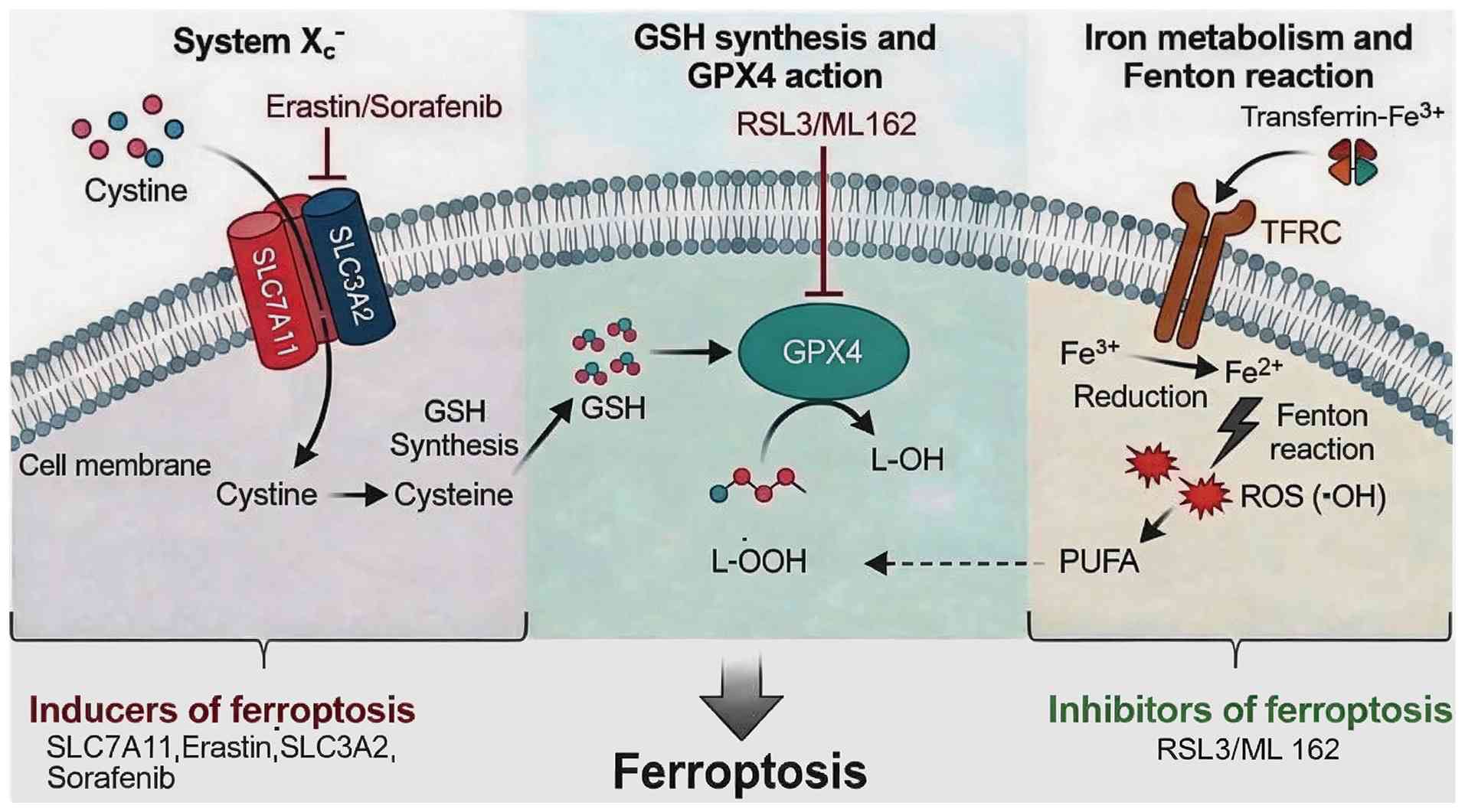 | Figure 1.Core molecular mechanisms and
metabolic regulation of ferroptosis. Ferroptosis is regulated
mainly by the System Xc−/GSH/GPX4 axis and iron
metabolism. System Xc−, composed of SLC7A11 and SLC3A2,
mediates cystine uptake for GSH synthesis and can be inhibited by
erastin or sorafenib. GPX4 uses GSH to reduce L-OOH to non-toxic
L-OH, whereas GPX4 inhibitors such as RSL3 and ML162 promote lipid
peroxide accumulation. TFRC-mediated uptake of transferrin-bound
Fe3+ increases intracellular Fe2+, which
promotes ROS generation through the Fenton reaction and drives
peroxidation of PUFAs, ultimately inducing ferroptosis. GPX, GSH
peroxidase; GSH, glutathione; L-OH, lipid alcohols; L-OOH, lipid
hydroperoxides; PUFA, polyunsaturated fatty acid; ROS, reactive
oxygen species; SLC3A2, solute carrier family 3 member 2; SLC7A11,
solute carrier family 7 member 11; TFRC, transferrin receptor. |
Induction pathways and regulatory
factors of ferroptosis
Ferroptosis can be induced through several
mechanisms, including: i) Inhibition of System Xc−
function, such as blockade of cystine uptake by erastin or
sorafenib, leading to GSH depletion (29,31);
ii) direct inhibition of GPX4 activity by agents such as RSL3 or
ML162 (26); and iii) modulation of
iron metabolism by iron chelators or iron carriers, thereby
affecting ROS production (28).
Zhang et al (31) showed
that, in addition to its kinase-inhibitory activity, sorafenib can
induce ferroptosis by inhibiting SLC7A11. Previous studies
have identified additional ferroptosis regulatory factors,
including ubiquitin-specific peptidase 22 (USP22), which inhibits
sorafenib-induced ferroptosis by deubiquitinating CDK1 (32); PNO1, which inhibits
autophagy-mediated ferroptosis by reprogramming GSH metabolism
(7); and m6A modification, which
influences ferroptosis sensitivity by regulating key molecules such
as GPX4 (33). Fig. 2 summarizes the three main
ferroptosis induction pathways (System Xc− inhibition,
direct GPX4 inhibition and iron metabolism dysregulation) along
with identified regulatory factors.
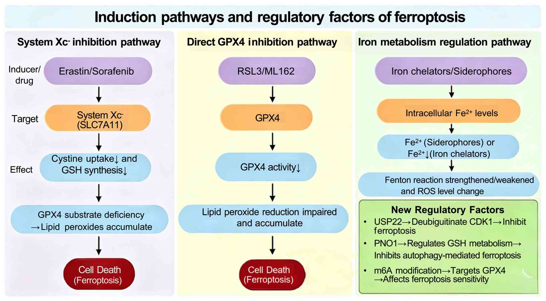 | Figure 2.Induction pathways and regulatory
factors of ferroptosis. Ferroptosis can be induced through three
main pathways: i) Inhibition of System Xc− (for example,
via erastin or sorafenib), leading to GSH depletion; ii) direct
inhibition of GPX4 (for example, by RSL3 or ML162), blocking lipid
peroxide reduction; and iii) regulation of iron metabolism,
affecting Fe2+ levels and the Fenton reaction. The lower
right corner lists identified regulatory factors, including USP22,
PNO1 and m6A modification, which regulate ferroptosis through
post-translational modification, metabolic reprogramming or
epigenetic mechanisms. GPX4, GSH peroxidase 4; GSH, glutathione;
ROS, reactive oxygen species; SLC7A11, solute carrier family 7
member 11; USP22, ubiquitin-specific peptidase 22. |
USP22 has been reported to inhibit sorafenib-induced
ferroptosis by deubiquitinating CDK1 (32). By contrast, erastin-induced
ferroptosis does not appear to be affected by USP22 in the same
cellular context. This discrepancy may arise from: i) Cell-type
heterogeneity, as different HCC cell lines have distinct genetic
backgrounds (4,6,13,14).
ii) Different mechanisms of ferroptosis inducers; for example,
sorafenib is a multi-kinase inhibitor that induces additional
cellular stress responses, such as autophagy, beyond GSH depletion
(31,33), whereas erastin is a more selective
System Xc− inhibitor (34); consequently, the USP22-CDK1 axis may
be engaged only under the broader metabolic perturbations caused by
sorafenib. iii) Experimental conditions, such as iron concentration
and oxidative stress baseline. Notably, RSL3 directly inhibits
GPX4, thereby bypassing both System Xc− and the
USP22-CDK1 axis, which explains why USP22 does not affect
RSL3-induced ferroptosis (35,36).
Currently, most evidence is derived from in vitro work
(37), whereas in vivo and
clinical validation is lacking. Resolving these contradictions will
require systematic side-by-side comparisons under standardized
conditions. These observations regarding USP22 expand the
ferroptosis regulatory network and identify potential targets for
HCC treatment.
Molecular determinants of ferroptosis
sensitivity in HCC cells
HCC cells exhibit marked heterogeneity in
ferroptosis sensitivity, which appears to be influenced by genetic
background, microenvironmental factors and epigenetic modifications
(38). For example, p53
status can influence ferroptosis sensitivity: Wild-type p53
promotes ferroptosis by inhibiting SLC7A11 expression,
whereas mutant p53 may exert the opposite effect (39). Additional regulatory factors, such
as PNO1, have also been implicated in modulating ferroptosis
sensitivity in HCC cells (7). Tumor
microenvironmental factors, such as hypoxia and acidosis, can
influence the expression of ferroptosis-related molecules by
regulating signaling pathways such as hypoxia-inducible factor-1α
and NF-κB (40).
Sorafenib-resistant HCC cells often exhibit reduced sensitivity to
ferroptosis, a process associated with GPX4 activation and
SLC7A11 upregulation (29,41).
Therefore, modulating these key molecules may help restore
ferroptosis sensitivity and reverse treatment resistance in
HCC.
The heterogeneity of HCC is reflected in diverse
patterns of ferroptosis regulation. In hepatitis B virus
(HBV)-associated HCC, HBV X protein (HBx) can suppress ferroptosis
through the protein arginine methyltransferase 9/heat shock protein
family A member 8/CD44 axis, thereby promoting disease onset and
progression (42). Conversely,
activation of protein inhibitor of activated STAT3 (PIAS3) promotes
ferroptosis in HBV-positive HCC cells by activating TGF-β signaling
and upregulating thioredoxin-interacting protein expression
(43). The balance between
HBx-mediated anti-ferroptotic signaling and PIAS3-mediated
pro-ferroptotic signaling may influence the sensitivity of
HBV-associated HCC to ferroptosis inducers. By contrast,
non-alcoholic steatohepatitis (NASH)-associated HCC arises in the
context of chronic lipid metabolic dysfunction and oxidative
stress, with elevated lipid peroxidation and abnormal iron
metabolism (44). Activating
transcription factor (ATF)4 and SLC7A11 expression are markedly
elevated in the livers of patients with NASH, suggesting that
NASH-associated HCC may possess compensatory ferroptosis-defense
mechanisms (45). Furthermore,
ferroptosis mediated by long-chain acyl-CoA synthetase 4 (ACSL4)
may have a dual role in non-alcoholic fatty liver disease
(NAFLD)-associated HCC: It can promote cancer cell death (46), but may also indirectly promote
carcinogenesis by exacerbating liver injury and fibrosis (47). Elucidating these subtype-specific
mechanisms is important for designing personalized
ferroptosis-targeted therapeutic strategies for patients with HCC
arising from different etiological backgrounds.
Key pathways involved in
ferroptosis-mediated sensitization in HCC therapy
Role of the ATF4/STAT3 signaling axis
in ferroptosis regulation
ATF4 is a central transcription factor in the
integrated stress response and has context-dependent effects on
ferroptosis regulation. Mechanistically, this dual role appears to
depend on stress intensity: Under mild oxidative stress,
ATF4 can upregulate SLC7A11 expression through the
PERK/eIF2 signaling axis, thereby enhancing antioxidant defenses
and suppressing ferroptosis (45).
Under sustained or intense ferroptosis-inducing stress, persistent
high PERK/eIF2α phosphorylation and ATF4 accumulation occur;
ATF4 then induces pro-death mediators including C/EBP homologous
protein (CHOP, also known as DNA damage-inducible transcript 3) and
damage-regulated autophagy-modulating protein (DRAM). CHOP inhibits
Bcl-2 and increases ROS production, whereas DRAM promotes
autophagic ferroptosis. Together with GSH depletion and lipid
peroxide accumulation, this shifts the cell towards ferroptotic
death (48). This biphasic pattern
suggests that ATF4 initially supports cellular adaptation
but may contribute to ferroptotic cell death when stress exceeds
the compensatory capacity of the cell.
STAT3 may suppress ferroptosis by
upregulating SLC7A11 and GPX4 expression, thereby
supporting HCC cell survival (49).
Preclinical studies have shown that STAT3 antisense
oligonucleotides can enhance sorafenib efficacy in HCC resistance
models, suggesting that STAT3 inhibition may help overcome
treatment resistance (50,51). Furthermore, ATF4 and
STAT3 may interact to regulate ferroptosis sensitivity
(52), although the mechanisms and
clinical relevance of this interaction in HCC require further
investigation. The stress intensity-dependent regulation of
ferroptosis by ATF4 and STAT3 is summarized in
Fig. 3.
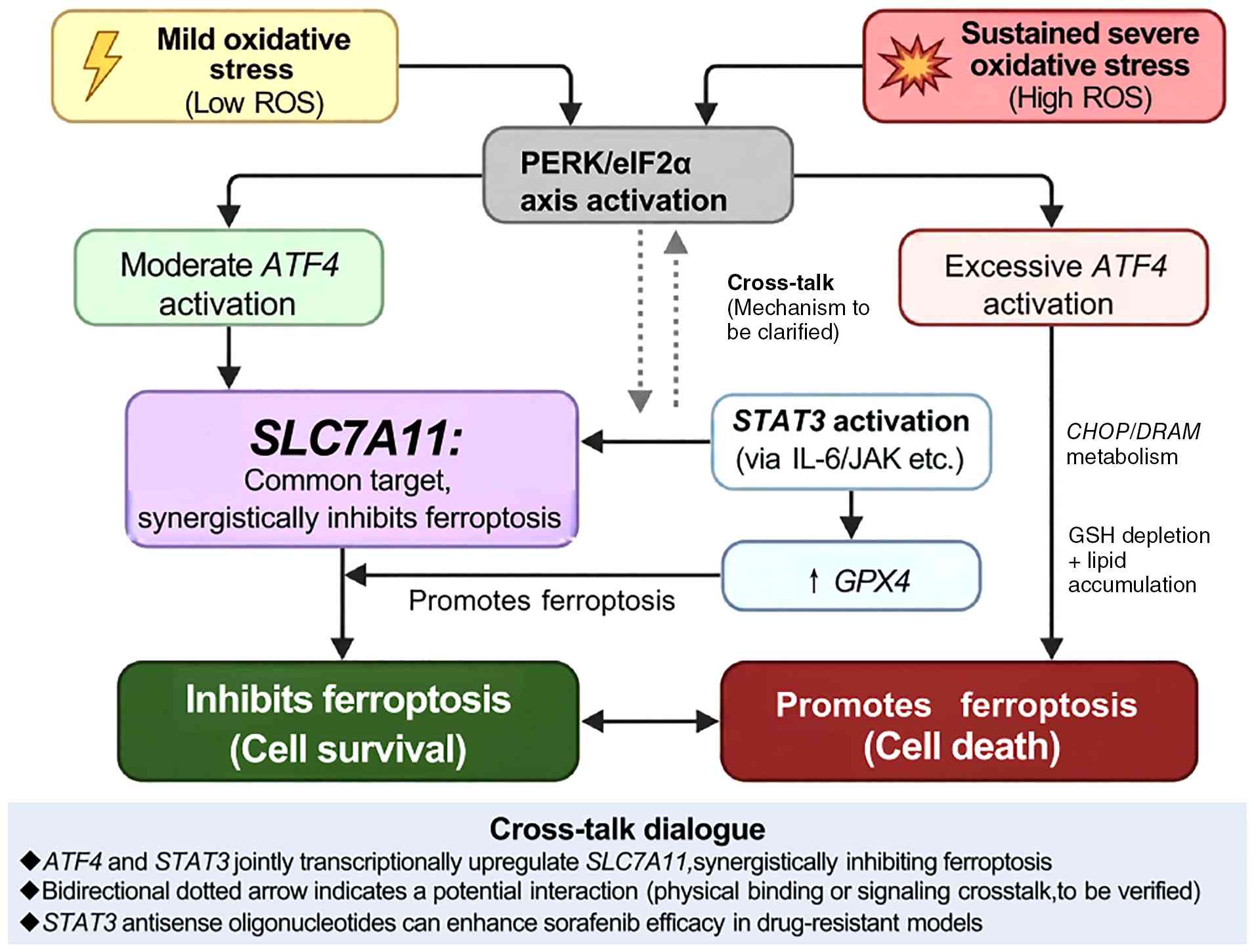 | Figure 3.Mechanisms by which ATF4 and
STAT3 signaling regulate ferroptosis. Under mild oxidative
stress, moderate activation of the PERK/eIF2α-ATF4 pathway can
upregulate SLC7A11, promote GSH synthesis and suppress
ferroptosis. Under sustained or severe oxidative stress, excessive
ATF4 activation may induce pro-death mediators such as CHOP and
DRAM; together with GSH depletion and lipid peroxide accumulation,
these changes promote ferroptotic cell death. STAT3 activation, for
example through IL-6/JAK signaling, may suppress ferroptosis by
upregulating SLC7A11 and GPX4 expression. The proposed ATF4-STAT3
cross-talk remains incompletely defined and requires further
mechanistic validation in hepatocellular carcinoma models. ATF4,
activating transcription factor 4; CHOP, C/EBP homologous protein;
DRAM, damage-regulated autophagy-modulating protein; GPX4, GSH
peroxidase 4; GSH, glutathione; ROS, reactive oxygen species;
SLC7A11, solute carrier family 7 member 11. |
Interaction between Wnt signaling and
ferroptosis
The Wnt/β-catenin signaling pathway serves a crucial
role in HCC development, progression and drug resistance. Recent
studies have suggested an association between Wnt/β-catenin
signaling and ferroptosis. Notably, the RNA helicase DEAD-box
helicase 5 (DDX5) can prevent escape from sorafenib-induced
ferroptosis by inhibiting Wnt/β-catenin signaling (53). In advanced HCC, DDX5 expression is
positively associated with patient survival. Multikinase inhibitors
such as sorafenib may reduce DDX5 expression, leading to activation
of Wnt/β-catenin signaling and altered ferroptosis sensitivity
(53,54). These findings suggest that targeting
the Wnt/β-catenin-ferroptosis axis may improve therapeutic
responses in HCC.
Mechanisms by which the p53 pathway
regulates ferroptosis in HCC cells
p53 is a key tumor suppressor. In HCC cells,
wild-type p53 can directly inhibit SLC7A11
transcription, thereby promoting lipid peroxidation (55); it can also enhance ferroptosis
sensitivity through transcription-independent mechanisms, such as
via interaction with spermidine/spermine
N1-acetyltransferase 1 (56). p53-mutant HCC cells are often
associated with abnormal activation of the Wnt/β-catenin pathway
(57). p53 restricts
cystine/glutamate exchange (20),
whereas Wnt signaling activation is typically associated with
metabolic reprogramming in the tumor microenvironment (58). These pathways may converge at
glutamine metabolism, which fuels GSH biosynthesis and maintains
cellular redox homeostasis, thereby influence ferroptosis
resistance in HCC cells (20,59).
Conversely, activation of the Wnt/β-catenin pathway
upregulates SLC7A11 expression, inhibiting ferroptosis
(53,54). These two pathways converge on
SLC7A11 regulation in an antagonistic manner. Moreover,
p53-mutant HCC frequently exhibits aberrant Wnt/β-catenin
activation, which may cooperatively suppress ferroptosis and
contribute to treatment resistance (20,57).
The interplay between p53 and Wnt signaling in the context of
ferroptosis represents a promising target for combination therapy,
although the precise molecular mechanisms require further
investigation.
Systemic regulation of ferroptosis by
non-coding RNAs (ncRNAs)
Previous studies have shown that long ncRNAs
(lncRNAs) and circular RNAs (circRNAs) regulate ferroptosis in HCC.
For example, the HCC-associated lncRNA HEPFAL is
downregulated in HCC tissues; HEPFAL overexpression via
transfection with a HEPFAL expression vector promotes
SLC7A11 ubiquitination and degradation, reduces SLC7A11 stability,
increases lipid ROS and iron levels, and enhances erastin-induced
ferroptosis sensitivity (60).
Conversely, the lncRNA HULC is highly expressed in HCC;
HULC knockdown leads to increased lipid ROS, elevated
malondialdehyde, GSH depletion and Fe2+ accumulation,
and enhances sensitivity to erastin-induced ferroptosis, thus
suggesting that HULC promotes HCC progression by inhibiting
ferroptosis (61). Among the
circRNAs, circTTC13 is highly expressed in HCC tissues and
is positively associated with tumor grade; in sorafenib-treated HCC
cells, silencing circTTC13 can increase lipid ROS, decrease
GPX4 and SLC7A11 expression, and enhance
sorafenib-induced ferroptosis, indicating that circTTC13
suppresses ferroptosis and mediates sorafenib resistance via the
microRNA-513a-5p/SLC7A11 axis (62). A circRNA associated with sorafenib
resistance, circRNA-SORE, binds ubiquilin-1 (UBQLN1) to
stabilize GPX4, thereby reducing intracellular lipid ROS
(specifically inhibiting lipid peroxidation product accumulation),
and enhances sorafenib resistance through the
circRNA-SORE/UBQLN1/GPX4 axis (63,64).
These findings expand the molecular network through which ncRNAs
regulate ferroptosis and identify potential targets for HCC
therapy.
Synergistic effects of ferroptosis inducers
and conventional treatments
Synergistic effects of ferroptosis
inducers and targeted therapies
Sorafenib is a first-line treatment for advanced
HCC, and its antitumor activity partly involves SLC7A11 inhibition
and ferroptosis induction (29).
However, acquired resistance after long-term treatment remains a
major obstacle. Preclinical studies have shown that sorafenib can
induce ferroptosis by inhibiting SLC7A11, and ferroptosis
resistance may contribute to sorafenib resistance in tumor cells
(29,65). In sorafenib-resistant HCC samples,
ferroptosis levels have been reported to be markedly lower than
those in sorafenib-sensitive samples (41). Long-term sorafenib exposure may
promote resistance through multiple mechanisms, including
USP22-mediated CDK1 deubiquitination and DDX5 downregulation
(32,54).
Several studies have therefore explored combining
sorafenib with other ferroptosis inducers to enhance its efficacy
(29,66–69).
For example, the combination of chelerythrine and berbamine with
sorafenib synergistically inhibits HCC cell proliferation, and has
demonstrated antitumor effects in both in vitro and in
vivo models (64). Furthermore,
nanocarrier-mediated co-delivery of sorafenib and other ferroptosis
inducers, such as salinomycin, may improve drug targeting and
efficacy (70).
Combined use of ferroptosis modulators
and chemotherapeutic agents
Combining ferroptosis inducers with chemotherapeutic
agents may produce synergistic antitumor effects. This synergy may
involve chemotherapy-induced oxidative stress and suppression of
tumor cell antioxidant defenses by ferroptosis inducers (71). For example, erastin combined with
cisplatin has been shown to markedly enhance cytotoxicity in HCC
cells (72), whereas sulfasalazine
promotes ferroptosis by activating the AMP-activated protein
kinase/sterol regulatory element-binding protein 1 pathway and
synergistically inhibits tumor growth when combined with
5-fluorouracil (71).
Branched-chain amino acid transaminase 2 (BCAT2) has also been
identified as a ferroptosis inhibitor, and BCAT2 targeting enhances
chemotherapy-induced ferroptosis (71). These findings suggest potential
therapeutic strategies for chemotherapy-resistant HCC, although
clinical validation remains limited.
Role of ferroptosis sensitizers in
overcoming drug resistance in HCC treatment
Ferroptosis sensitizers are compounds that enhance
tumor-cell sensitivity to ferroptosis inducers and may help
overcome treatment resistance in HCC. Emerging ferroptosis
sensitizers include small-molecule inhibitors targeting
ferroptosis-suppressive pathways, such as ferroptosis suppressor
protein 1 (FSP1) inhibitors (73);
compounds that modulate iron metabolism, including iron chelators
or iron carriers (28); and
nanomaterials, such as Fe3O4-PEI@HA-RSL3
nanocubes (74). Preclinical
studies have shown that targeting the ferroptosis-induced
inflammatory axis can enhance the in vivo efficacy of
sorafenib (75–78). In addition, an oral delivery
platform composed of butyrate-modified nanoparticles co-loaded with
sorafenib and salinomycin can increase sorafenib uptake in HCC and
induce ferroptosis, thereby improving therapeutic efficacy
(70). Modulation of the
USP22/H2BK120ub/TFRC axis also provides a potential target for
sensitizer development (32).
Together, these studies have identified several candidate
strategies for overcoming drug resistance in HCC treatment.
Preclinical studies have observed that programmed
death-ligand 1 (PD-L1) expression levels are associated with the
efficacy of combining ferroptosis inducers with immune checkpoint
inhibitors in HCC models (79,80).
For example, in a study using sorafenib and PD-L1 small interfering
RNA co-delivery systems, downregulation of PD-L1 was shown to
enhance ferroptosis-induced tumor cell death and improve antitumor
immunity (72). However, the direct
mechanistic link between PD-L1 signaling and ferroptosis
sensitivity remains incompletely defined. Possible hypotheses
include PD-L1-mediated regulation of glucose or lipid metabolism,
or indirect effects via tumor-immune crosstalk. Further studies are
required to assess this relationship.
Despite these advances, the clinical translation of
ferroptosis inducers remains at an early stage. Arsenic trioxide
(ATO), an approved treatment for acute promyelocytic leukemia, has
been investigated in mechanistic studies of ferroptosis in HCC. ATO
can induce ferroptosis in HCC cells, and this effect can be
reversed by the iron chelator desferrioxamine (81). ATO-induced ferroptosis may also
promote the release of tumor-associated antigens and enhance immune
responses. Notably, patients with low to moderate ferroptosis
activation in tumors exhibited the highest risk of recurrence
compared with those with no or high ferroptosis activation, and the
ferroptosis-elicited inflammatory axis was associated with
therapeutic resistance to sorafenib in HCC (78). However, the systemic toxicity of
ATO, particularly cardiotoxicity and hepatotoxicity, limits its use
as monotherapy for HCC (82).
ATO-based nanodelivery systems, such as ATO@SP94-TMV and LP@MnAS,
have demonstrated favorable targeting and biosafety in animal
models, offering possible opportunities for the clinical
translation of ATO (82,83). Among erastin derivatives, imidazole
ketone erastin (IKE), a metabolically more stable SLC7A11
inhibitor, inhibits tumor growth in animal models of HCC after
intraperitoneal administration, and dicoumarin can sensitize cells
to IKE-induced ferroptosis (34).
Although novel ferroptosis-targeting agents remain
largely preclinical in HCC, a small number of ferroptosis-related
agents or strategies have entered early-phase clinical trials in
other disease contexts. For example, eprenetapopt (APR-246), which
targets p53 mutations, has completed a phase II trial in
myeloid tumors (NCT03588078) (84),
and the iron-loaded nanocarbon formulation CNSI-Fe(II) has
completed a phase I dose-escalation study in advanced solid tumors
(NCT06048367) (85). In addition,
sorafenib combined with stereotactic body radiation therapy has
been evaluated in a phase II trial for colorectal cancer liver
metastases, with proposed efficacy and safety benefits partly
attributed to ferroptosis induction (86). PD-L1/BBζ chimeric switch receptor
(CSR)-modified dual-target chimeric antigen receptor T cells, in
which the CSR binds PD-L1 and converts the inhibitory signal into a
4–1BB costimulatory signal (87),
have also entered a phase I clinical trial for pleural or
peritoneal metastases (NCT04684459) (87), although this evidence remains
indirect for HCC. However, these agents and strategies generally
remain in the early stages of development. Clinical application of
ferroptosis inducers in HCC will require improved drug-delivery
efficiency, tumor-targeting specificity, toxicity control and
clearer indication selection.
Current status of research on
ferroptosis-related biomarkers
Biomarkers associated with ferroptosis
sensitivity
Research on biomarkers associated with ferroptosis
sensitivity provides an important basis for personalized treatment
of HCC. Multiple studies have suggested that ferroptosis-related
gene-expression profiles may have predictive value for treatment
response in HCC (88). Potential
biomarkers under investigation include ferroptosis regulatory
molecules, such as GPX4, SLC7A11 and FSP1 (30,73,88);
metabolism-related molecules, such as ACSL4 and
lysophosphatidylcholine acyltransferase 3 (88); signaling molecules, such as p53,
ATF4 and STAT3 (49,55);
and ncRNAs (33). Table I summarizes the evidence level and
main results for each biomarker based on available studies.
 | Table I.Summary of ferroptosis-related
biomarkers. |
Table I.
Summary of ferroptosis-related
biomarkers.
| Biomarker | Study type | Main results | (Refs.) |
|---|
| GPX4 | Patient HCC tissue
(n=106) | High expression is
associated with shorter overall survival; GPX4 is a negative
regulator of ferroptosis | (30) |
| SLC7A11 | Patient HCC tissue
and HCC cell lines | High expression
promotes sorafenib resistance and inhibits ferroptosis | (29,90) |
| FSP1 | HCC cell lines and
mouse models (KRAS-mutant) | Protects
KRAS-mutant cells from ferroptosis | (73) |
| ACSL4 | Patient HCC tissue
and HCC cell lines | Promotes
ferroptosis (PUFA substrate), but may also promote HCC in the
context of NASH | (46,47,88) |
| LPCAT3 | HCC cell lines | Promotes lipid
peroxidation and increases ferroptosis sensitivity | (88) |
| TP53 | HCC cell lines | Wild-type
p53 inhibits SLC7A11 transcription and promotes
ferroptosis | (55) |
| ATF4 | HCC cell lines and
mouse models | Dual role:
Suppresses ferroptosis in response to low/moderate stress, promotes
ferroptosis in response to high stress | (45) |
| STAT3 | HCC cell lines and
mouse models | Suppresses
ferroptosis via upregulation of SLC7A11/GPX4 | (49) |
| ncRNAs (for
example, HEPFAL and HULC) | HCC cell lines and
mouse models | Regulate
SLC7A11/GPX4 stability or expression | (33,60–64) |
In a HCC cohort (n=106), high GPX4 expression was
reported to be associated with shorter overall survival (HR=2.34,
P<0.01) and sorafenib resistance (30). In another cohort (n=89), high ACSL4
expression was revealed to be associated with microvascular
invasion, but also with increased sensitivity to ferroptosis
inducers (40); ACSL4 mRNA has been
detected in serum exosomes (89)
ACSL4 promotes ferroptosis by converting
polyunsaturated fatty acids into CoA esters; however, in
NAFLD/NASH-associated HCC it may exacerbate hepatocyte injury and
fibrosis, indirectly promoting HCC development (47). Thus, ACSL4 as a biomarker requires
careful interpretation depending on disease stage and etiology.
Clinical evidence for key
biomarkers
Among the aforementioned markers, GPX4, a key
negative regulator of ferroptosis, has been associated with
predictive or prognostic value in colorectal cancer and HCC
(30); FSP1 is highly expressed in
KRAS-mutated tumors (in models of pancreatic and lung
cancer) and is associated with ferroptosis resistance (73), although its role in HCC requires
further investigation; and abnormal expression of the
FTO/YTHDF2/GPX4 signaling axis is associated with ferroptosis
sensitivity in HCC (30). If
validated, these biomarkers may help identify patient subgroups
likely to benefit from ferroptosis-inducing therapies and support
the development of molecular subtyping systems based on ferroptosis
sensitivity. However, clinical-cohort validation of ferroptosis
biomarkers remains limited. Although immunohistochemical studies
have detected associations between GPX4 or SLC7A11 expression and
prognosis in small HCC tissue cohorts, most studies have been
retrospective, have included only several dozen to slightly more
than 100 cases, and lacked multicenter prospective validation
(90,91). Regarding detection feasibility, GPX4
and ACSL4 mRNA and protein levels can be measured in biopsy tissues
or blood exosomes using reverse transcription-quantitative PCR,
western blotting, enzyme-linked immunosorbent assay and
immunohistochemistry (89,92,93).
Use of biomarkers in personalized
treatment strategies
Personalized treatment strategies based on
ferroptosis-related biomarkers are being explored in HCC. Analysis
of ferroptosis-related gene expression profiles may help guide the
future selection of precision therapies for patients with HCC
(94). In immunotherapy, combined
analysis of ferroptosis markers and tumor immune microenvironment
characteristics may help optimize future combination regimens
involving immune checkpoint inhibitors and ferroptosis inducers
(95). The integration of
nanotechnology with ferroptosis-based therapy may also support
biomarker-guided personalized treatment. For example, in
photothermal-ferroptosis combination therapy, real-time monitoring
of iron metabolism-related molecules may enable dynamic adjustment
of treatment regimens (96).
Furthermore, interactions between lncRNAs and ferroptosis may
provide a novel framework for biomarker screening during the
development of nanomaterial-based combination therapies (97). As multi-omics technologies advance,
ferroptosis-susceptibility prediction models integrating genomics,
transcriptomics and metabolomics data may improve the precision of
personalized HCC treatment (96).
In parallel with ferroptosis-based biomarkers,
non-invasive approaches to evaluate the tumor immune
microenvironment have shown promise in predicting outcomes in HCC,
which may complement ferroptosis-based prognostic strategies
(98). Specifically, Wu et
al (98) developed a
radiomics-based non-invasive model [Radiomic Immunoscore (RIS)] to
evaluate the tumor immune microenvironment and predict prognosis in
patients with HCC. Using MRI-derived radiomics features, the RIS
model accurately predicted immune status [area under the curve
(AUC)=0.753] and showed potential in predicting anti-programmed
cell death protein 1 immunotherapy response (AUC=0.731) in patients
with advanced HCC. Such non-invasive strategies complement
ferroptosis-based prognostic strategies in several manners: i) They
provide information on the immune landscape, which influences
ferroptosis sensitivity (for example, PD-L1 expression is
associated with ferroptosis inducer efficacy); ii) they can be
combined with ferroptosis-related gene signatures to build
multi-dimensional prognostic models; and iii) they enable dynamic,
real-time monitoring of tumor evolution without repeated biopsies,
facilitating adaptive combination therapies that target both
ferroptosis and immune checkpoints.
Limitations and controversies in current
research
Unresolved issues in mechanistic
research
Although research into ferroptosis has advanced
substantially, several questions remain regarding its specific
regulatory mechanisms in HCC. First, the functions of
post-translational modifications of ferroptosis-associated
proteins, such as ubiquitination and phosphorylation of SLC7A11 and
GPX4, remain incompletely understood; these modifications may
influence ferroptosis sensitivity in HCC cells by modulating
protein stability and activity (99). Second, the mechanisms by which m6A
modifications regulate ferroptosis in HCC remain unclear.
Furthermore, ferroptosis sensitivity differs substantially across
HCC cell lines, but the molecular determinants of this
heterogeneity remain unclear.
Challenges in clinical
translation
Ferroptosis inducers face multiple barriers to
clinical translation in HCC treatment. A major issue is the
incomplete understanding of ferroptosis mechanisms in human HCC,
which limits rational drug development (40). Drug-delivery systems also remain
limited, as conventional delivery methods may not achieve
sufficient penetration or selective accumulation in HCC tissues
(100). Furthermore, most
ferroptosis inducers, such as ATO and erastin derivatives, remain
experimental in HCC (101), and
although nanomaterials such as
Fe3O4-PEI@HA-RSL3 have shown promise, they
remain distant from clinical application (74). Addressing these challenges will
require integration of basic research, drug development, biomarker
validation, toxicity assessment and clinical trial design.
Differences between animal models and
human studies
Important differences exist between animal models
and human HCC. Most mechanistic studies remain confined to cellular
or animal models, limiting direct clinical extrapolation. For
example, GPX4 has been identified as a major regulator of
ferroptosis in mouse HCC models, but its applicability to human HCC
requires verification (102). In
animal studies, USP22 promotes HCC growth and inhibits
sorafenib-induced ferroptosis; however, the complexity of the human
HCC microenvironment may produce different outcomes (32). In addition, the chronic hypoxic
microenvironment of HCC tissues is difficult to fully replicate in
animal models, which may affect the evaluation of ferroptosis
inducers (30). Therefore, animal
models that more closely recapitulate human HCC biology are needed,
along with stronger integration of mechanistic and clinical
research.
Targeted toxicity of GPX4
inhibitors
As a key negative regulator of ferroptosis, GPX4 is
a major target for ferroptosis-inducing strategies. However,
targeting GPX4 presents two major challenges.
Toxicity to normal tissues
Classic GPX4 inhibitors, such as RSL3 and ML210,
exert their inhibitory effects by covalently binding to the
selenocysteine active site of GPX4; however, these compounds have
poor selectivity and may damage normal tissues while inhibiting
GPX4 in tumor cells (103).
Non-selective ferroptosis induction may damage immune cells or
disrupt microenvironmental homeostasis (104). Because the liver is central to
iron metabolism and detoxification, it is particularly vulnerable
to GPX4 inhibition: Reduced GPX4 activity can render normal
hepatocytes susceptible to ferroptosis, leading to drug-induced
liver injury (105,106). This toxicological profile
substantially limits the clinical translation of GPX4
inhibitors.
Targeted delivery strategies
To address toxicity, researchers have developed
strategies to enhance the selectivity of GPX4 inhibition. One
approach involves nanodelivery systems that promote tumor-targeted
accumulation; for example, Fe3O4-PEI@HA-RSL3
nanocubes can release RSL3 in the acidic tumor microenvironment,
thereby reducing systemic exposure; in mouse HCC subcutaneous
xenograft models, this strategy achieved selective GPX4 inhibition,
ferroptosis induction and tumor growth inhibition without notable
hepatotoxicity (74). Another
approach involves GPX4 degraders, including proteolysis-targeting
chimeras, which uses E3 ligases that are highly expressed in tumor
cells to achieve cell-specific degradation; in mouse pancreatic
cancer models as a proof-of-concept study, tumor-specific E3 ligase
has been reported to achieve GPX4 degradation, reducing normal
tissue toxicity (107). A third
approach is screening for more selective GPX4 inhibitors, such as
the small-molecule compound N6F11, which can selectively induce
ferroptosis in tumor cells by triggering GPX4 ubiquitination and
degradation while causing no marked damage to immune cells; this
has been demonstrated in immunocompetent mouse models of pancreatic
cancer (108). However, most of
these strategies remain preclinical and their safety in humans
requires systematic evaluation.
Outlook
Although ferroptosis research has advanced
substantially, unresolved questions remain regarding its regulatory
mechanisms in HCC, including the role of post-translational
modifications, the mechanisms of m6A modification and the molecular
basis of cellular heterogeneity. For clinical translation,
ferroptosis inducers still face challenges, including limited
drug-delivery efficiency, insufficiently representative animal
models and inadequate validation in human studies. Future research
should focus on four priorities: i) Developing novel ferroptosis
inducers and nanotechnology-based targeted delivery systems to
improve drug selectivity and tumor accumulation; ii) elucidating
heterogeneity in ferroptosis regulatory networks and using
multi-omics technologies to construct ferroptosis-sensitivity
prediction models for personalized combination therapy; iii)
conducting biomarker-based prospective clinical trials to clarify
the efficacy and safety of ferroptosis inducers in patients with
advanced HCC; and iv) exploring synergistic mechanisms between
ferroptosis and other forms of cell death or treatment modalities,
such as cuproptosis and immunotherapy.
In summary, research on ferroptosis regulatory
networks offers a useful framework for understanding HCC drug
resistance and developing novel therapeutic strategies. However,
the clinical value of ferroptosis-based strategies in HCC will
depend on validated biomarkers, selective delivery systems,
toxicity control and prospective human trials.
Acknowledgements
Not applicable.
Funding
This work was supported by the Yichun Key Science and Technology
Project Platform Cultivation Plan (grant no. YCPT2022003) and the
Science and Technology Plan Project of Jiangxi Provincial Health
Commission (grant no. 202410935).
Availability of data and materials
Not applicable.
Authors' contributions
MZ contributed to the conception and design of the
review, and drafted and revised the manuscript. XH provided
conceptual guidance and critical input during manuscript revision,
and critically reviewed and edited the manuscript. Data
authentication is not applicable. Both authors read and approved
the final manuscript.
Ethics approval and consent to
participate
Not applicable.
Patient consent for publication
Not applicable.
Competing interests
The authors declare that they have no competing
interests.
References
|
1
|
Li Q, Ding C, Cao M, Yang F, Yan X, He S,
Cao M, Zhang S, Teng Y, Tan N, et al: Global epidemiology of liver
cancer 2022: An emphasis on geographic disparities. Chin Med J
(Engl). 137:2334–2342. 2024. View Article : Google Scholar : PubMed/NCBI
|
|
2
|
Zhou J, Sun H, Wang Z, Cong W, Zeng M,
Zhou W, Liu L, Wen T, Kuang M, Zhang B, et al: China liver cancer
guidelines for the diagnosis and treatment of hepatocellular
carcinoma (2024 Edition). Liver Cancer. 14:779–835. 2025.PubMed/NCBI
|
|
3
|
Wang Z, Zhu J, Wang Z, Mo Q, Du X, Ma Y,
Zhang H, Gao Y, Liu S, Tang C, et al: Intelligent identification
and targeted intervention of GRP75-caused drug resistant
hepatocellular carcinoma, a study based on radiomics, machine
learning, and molecular pharmacology. Int J Surg. Feb 19–2026.(Epub
ahead of print).
|
|
4
|
Naserkhaki R, Shokouhian B, Tahamtani Y,
Khosravi A, Iravani S, Zarrabi A and Vosough M: Revisiting
treatment strategies: Addressing epithelial-to-mesenchymal
transition-induced resistance in hepatocellular carcinoma. BME
Front. 6:01442025. View Article : Google Scholar : PubMed/NCBI
|
|
5
|
World Health Organization, . International
statistical classification of diseases and related health problems.
10th edition. 2019
|
|
6
|
Zou Y, Wan X, Zhou Q, Zhu G, Lin S, Tang
Q, Yang X and Wang S: Mechanisms of drug resistance in
hepatocellular carcinoma. Biol Proced Online. 27:192025. View Article : Google Scholar : PubMed/NCBI
|
|
7
|
Hu X, He Y, Han Z, Liu W, Liu D, Zhang X,
Chen L, Qi L, Chen L, Luo Y, et al: PNO1 inhibits
autophagy-mediated ferroptosis by GSH metabolic reprogramming in
hepatocellular carcinoma. Cell Death Dis. 13:10102022. View Article : Google Scholar : PubMed/NCBI
|
|
8
|
Dixon SJ and Stockwell BR: The role of
iron and reactive oxygen species in cell death. Nat Chem Biol.
10:9–17. 2014. View Article : Google Scholar : PubMed/NCBI
|
|
9
|
Dos Santos AF, Fazeli G, Xavier da Silva
TN and Friedmann Angeli JP: Ferroptosis: Mechanisms and
implications for cancer development and therapy response. Trends
Cell Biol. 33:1062–1076. 2023. View Article : Google Scholar : PubMed/NCBI
|
|
10
|
Hassannia B, Vandenabeele P and Vanden
Berghe T: Targeting ferroptosis to iron out cancer. Cancer Cell.
35:830–849. 2019. View Article : Google Scholar : PubMed/NCBI
|
|
11
|
Chen X, Kang R, Kroemer G and Tang D:
Broadening horizons: The role of ferroptosis in cancer. Nat Rev
Clin Oncol. 18:280–296. 2021. View Article : Google Scholar : PubMed/NCBI
|
|
12
|
Friedmann Angeli JP, Krysko DV and Conrad
M: Ferroptosis at the crossroads of cancer-acquired drug resistance
and immune evasion. Nat Rev Cancer. 19:405–414. 2019. View Article : Google Scholar : PubMed/NCBI
|
|
13
|
Li D, Zhang M, Liu J, Li Z and Ni B:
Potential therapies for HCC involving targeting the ferroptosis
pathway. Am J Cancer Res. 14:1446–1465. 2024. View Article : Google Scholar : PubMed/NCBI
|
|
14
|
Cong T, Luo Y, Fu Y, Liu Y, Li Y and Li X:
New perspectives on ferroptosis and its role in hepatocellular
carcinoma. Chin Med J (Engl). 135:2157–2166. 2022. View Article : Google Scholar : PubMed/NCBI
|
|
15
|
Liu JJ, Zhang X, Cai BL, Qi MM, Chi YB,
Peng B and Zhang DH: Ferroptosis inhibitors reduce celastrol
toxicity and preserve its insulin sensitizing effects in insulin
resistant HepG2 cells. J Integr Med. 22:286–294. 2024. View Article : Google Scholar : PubMed/NCBI
|
|
16
|
Yin W, Chang J, Sun J, Zhang T, Zhao Y, Li
Y and Dong H: Nanomedicine-mediated ferroptosis targeting
strategies for synergistic cancer therapy. J Mater Chem B.
11:1171–1190. 2023. View Article : Google Scholar : PubMed/NCBI
|
|
17
|
Ma F, Li Y, Cai M, Yang W, Wu Z, Dong J
and Qin JJ: ML162 derivatives incorporating a naphthoquinone unit
as ferroptosis/apoptosis inducers: Design, synthesis, anti-cancer
activity, and drug-resistance reversal evaluation. Eur J Med Chem.
270:1163872024. View Article : Google Scholar : PubMed/NCBI
|
|
18
|
Huang JQ, Jiang YY, Ren FZ and Lei XG:
Novel role and mechanism of glutathione peroxidase-4 in nutritional
pancreatic atrophy of chicks induced by dietary selenium
deficiency. Redox Biol. 57:1024822022. View Article : Google Scholar : PubMed/NCBI
|
|
19
|
Ni Y, Deng P, Yin R, Zhu Z, Ling C, Ma M,
Wang J, Li S and Liu R: Effect and mechanism of paclitaxel loaded
on magnetic FeO@mSiO-NH-FA nanocomposites to MCF-7 cells. Drug
Deliv. 30:64–82. 2023. View Article : Google Scholar : PubMed/NCBI
|
|
20
|
Shin D, Lee J and Roh JL: Pioneering the
future of cancer therapy: Deciphering the p53-ferroptosis nexus for
precision medicine. Cancer Lett. 585:2166452024. View Article : Google Scholar : PubMed/NCBI
|
|
21
|
Chen X, Tsvetkov AS, Shen HM, Isidoro C,
Ktistakis NT, Linkermann A, Koopman WJH, Simon HU, Galluzzi L, Luo
S, et al: International consensus guidelines for the definition,
detection, and interpretation of autophagy-dependent ferroptosis.
Autophagy. 20:1213–1246. 2024. View Article : Google Scholar : PubMed/NCBI
|
|
22
|
Zhu Y, Fujimaki M and Rubinsztein DC:
Autophagy-dependent versus autophagy-independent ferroptosis.
Trends Cell Biol. 35:745–760. 2025. View Article : Google Scholar : PubMed/NCBI
|
|
23
|
Mo JQ, Zhang SY, Li Q, Chen MX, Zheng YQ,
Xie X, Zhang R and Wang SS: Immunomodulation of cuproptosis and
ferroptosis in liver cancer. Cancer Cell Int. 24:222024. View Article : Google Scholar : PubMed/NCBI
|
|
24
|
Wang X, Liu Z and Lin C: Metal
ions-induced programmed cell death: How does oxidative stress
regulate cell death? Life Sci. 374:1236882025. View Article : Google Scholar : PubMed/NCBI
|
|
25
|
Jiang Y and Sun M: SLC7A11: The Achilles
heel of tumor? Front Immunol. 15:14388072024. View Article : Google Scholar : PubMed/NCBI
|
|
26
|
Qian B, Che L, Du ZB, Guo NJ, Wu XM, Yang
L, Zheng ZX, Gao YL, Wang MZ, Chen XX, et al: Protein phosphatase
2A-B55β mediated mitochondrial p-GPX4 dephosphorylation promoted
sorafenib-induced ferroptosis in hepatocellular carcinoma via
regulating p53 retrograde signaling. Theranostics. 13:4288–4302.
2023. View Article : Google Scholar : PubMed/NCBI
|
|
27
|
Zhou X, Wang Y, Li X, Zhou J, Yang W, Wang
X, Jiao S, Zuo W, You Z, Ying W, et al: O-GlcNAcylation regulates
the stability of transferrin receptor (TFRC) to control the
ferroptosis in hepatocellular carcinoma cells. Redox Biol.
73:1031822024. View Article : Google Scholar : PubMed/NCBI
|
|
28
|
Zhang S, Xin W, Anderson GJ, Li R, Gao L,
Chen S, Zhao J and Liu S: Double-edge sword roles of iron in
driving energy production versus instigating ferroptosis. Cell
Death Dis. 13:402022. View Article : Google Scholar : PubMed/NCBI
|
|
29
|
Xiao Y, Xu Z, Cheng Y, Huang R, Xie Y,
Tsai HI, Zha H, Xi L, Wang K, Cheng X, et al: Fe3+-binding
transferrin nanovesicles encapsulating sorafenib induce ferroptosis
in hepatocellular carcinoma. Biomater Res. 27:632023. View Article : Google Scholar : PubMed/NCBI
|
|
30
|
Zhang G, Mi W, Wang C, Li J, Zhang Y, Liu
N, Jiang M, Jia G, Wang F, Yang G, et al: Targeting AKT induced
Ferroptosis through FTO/YTHDF2-dependent GPX4 m6A methylation
up-regulating and degradating in colorectal cancer. Cell Death
Discov. 9:4572023. View Article : Google Scholar : PubMed/NCBI
|
|
31
|
Zhang L, Li XM, Shi XH, Ye K, Fu XL, Wang
X, Guo SM, Ma JQ, Xu FF, Sun HM, et al: Sorafenib triggers
ferroptosis via inhibition of HBXIP/SCD axis in hepatocellular
carcinoma. Acta Pharmacol Sin. 44:622–634. 2023. View Article : Google Scholar : PubMed/NCBI
|
|
32
|
Wang X, Su Y, Lan B, Li X, Zhang B, Zhang
L, Wang Y, Zhang C and Xuan C: USP22 promotes the proliferation and
Sorafenib resistance of hepatocellular carcinoma cells via its
deubiquitinase activity. Clin Transl Med. 15:e703242025. View Article : Google Scholar : PubMed/NCBI
|
|
33
|
Li Y, Guo M, Qiu Y, Li M, Wu Y, Shen M,
Wang Y, Zhang F, Shao J, Xu X, et al: Autophagy activation is
required for N6-methyladenosine modification to regulate
ferroptosis in hepatocellular carcinoma. Redox Biol. 69:1029712024.
View Article : Google Scholar : PubMed/NCBI
|
|
34
|
Yang Z, Han T, Yang R, Zhang Y, Qin Y, Hou
J, Huo F, Feng Z, Ding Y, Yang J, et al: Dicoumarol sensitizes
hepatocellular carcinoma cells to ferroptosis induced by imidazole
ketone erastin. Front Immunol. 16:15318742025. View Article : Google Scholar : PubMed/NCBI
|
|
35
|
Nguyen KA, Conilh L, Falson P, Dumontet C
and Boumendjel A: The first ADC bearing the ferroptosis inducer
RSL3 as a payload with conservation of the fragile electrophilic
warhead. Eur J Med Chem. 244:1148632022. View Article : Google Scholar : PubMed/NCBI
|
|
36
|
Yan Z, Wu S, Zhou Y and Li F:
Acid-Responsive micelles releasing cinnamaldehyde enhance
RSL3-induced ferroptosis in tumor cells. ACS Biomater Sci Eng.
8:2508–2517. 2022. View Article : Google Scholar : PubMed/NCBI
|
|
37
|
Cai X, Zhang Y, Qin W and Li X: Molecular
code of ferroptosis: Emerging multi-dimensional modifications and
therapeutic targets in hepatic disorders. Metabolism.
175:1564572025. View Article : Google Scholar : PubMed/NCBI
|
|
38
|
Yao F, Zhou S, Zhang R, Chen Y, Huang W,
Yu K, Yang N, Qian X, Tie X, Xu J, et al: CRISPR/Cas9 screen
reveals that targeting TRIM34 enhances ferroptosis sensitivity and
augments immunotherapy efficacy in hepatocellular carcinoma. Cancer
Lett. 593:2169352024. View Article : Google Scholar : PubMed/NCBI
|
|
39
|
Jin AL, Zhang CY, Zheng WJ, Xian JR, Yang
WJ, Liu T, Chen W, Li T, Wang BL, Pan BS, et al: CD155/SRC complex
promotes hepatocellular carcinoma progression via inhibiting the
p38 MAPK signalling pathway and correlates with poor prognosis.
Clin Transl Med. 12:e7942022. View Article : Google Scholar : PubMed/NCBI
|
|
40
|
Chen Y, Shang H, Wang C, Zeng J, Zhang S,
Wu B and Cheng W: RNA-Seq explores the mechanism of oxygen-boosted
sonodynamic therapy based on all-in-one nanobubbles to enhance
ferroptosis for the treatment of HCC. Int J Nanomedicine.
17:105–123. 2022. View Article : Google Scholar : PubMed/NCBI
|
|
41
|
Gao Y, Tong M, Wong TL, Ng KY, Xie YN,
Wang Z, Yu H, Loh JJ, Li M and Ma S: Long noncoding RNA
URB1-Antisense RNA 1 (AS1) Suppresses sorafenib-induced ferroptosis
in hepatocellular carcinoma by driving ferritin phase separation.
ACS Nano. 17:22240–22258. 2023. View Article : Google Scholar : PubMed/NCBI
|
|
42
|
Deng W, Ai J, Zhang W, Zhou Z, Li M, Yan
L, Zhang L, Huang Z, Wu Z, Ai J and Jiang H: Arginine methylation
of HSPA8 by PRMT9 inhibits ferroptosis to accelerate hepatitis B
virus-associated hepatocellular carcinoma progression. J Transl
Med. 21:6252023. View Article : Google Scholar : PubMed/NCBI
|
|
43
|
Bao W, Wang J, Fan K, Gao Y and Chen J:
PIAS3 promotes ferroptosis by regulating TXNIP via TGF-β signaling
pathway in hepatocellular carcinoma. Pharmacol Res. 196:1069152023.
View Article : Google Scholar : PubMed/NCBI
|
|
44
|
Gao H, Jin Z, Bandyopadhyay G, Wang G,
Zhang D, Rocha KCE, Liu X, Zhao H, Kisseleva T, Brenner DA, et al:
Aberrant iron distribution via hepatocyte-stellate cell axis drives
liver lipogenesis and fibrosis. Cell Metab. 34:1201–1213.e5. 2022.
View Article : Google Scholar : PubMed/NCBI
|
|
45
|
He F, Zhang P, Liu J, Wang R, Kaufman RJ,
Yaden BC and Karin M: ATF4 suppresses hepatocarcinogenesis by
inducing SLC7A11 (xCT) to block stress-related ferroptosis. J
Hepatol. 79:362–377. 2023. View Article : Google Scholar : PubMed/NCBI
|
|
46
|
Sun N, Wang J, Qin J, Ma S, Luan J, Hou G,
Zhang W and Gao M: Oncogenic RTKs sensitize cancer cells to
ferroptosis via c-Myc mediated upregulation of ACSL4. Cell Death
Dis. 15:8612024. View Article : Google Scholar : PubMed/NCBI
|
|
47
|
Grube J, Woitok MM, Mohs A, Erschfeld S,
Lynen C, Trautwein C and Otto T: ACSL4-dependent ferroptosis does
not represent a tumor-suppressive mechanism but ACSL4 rather
promotes liver cancer progression. Cell Death Dis. 13:7042022.
View Article : Google Scholar : PubMed/NCBI
|
|
48
|
Loong JH, Wong TL, Tong M, Sharma R, Zhou
L, Ng KY, Yu HJ, Li CH, Man K, Lo CM, et al: Glucose
deprivation-induced aberrant FUT1-mediated fucosylation drives
cancer stemness in hepatocellular carcinoma. J Clin Invest.
131:e1433772021. View Article : Google Scholar : PubMed/NCBI
|
|
49
|
Dai Y, Cui C, Jiao D and Zhu X: JAK/STAT
signaling as a key regulator of ferroptosis: Mechanisms and
therapeutic potentials in cancer and diseases. Cancer Cell Int.
25:832025. View Article : Google Scholar : PubMed/NCBI
|
|
50
|
Zhang QY, Ding W, Mo JS, Ou-Yang SM, Lin
ZY, Peng KR, Liu GP, Lu JJ, Yue PB, Lei JP, et al: Novel STAT3
oligonucleotide compounds suppress tumor growth and overcome the
acquired resistance to sorafenib in hepatocellular carcinoma. Acta
Pharmacol Sin. 45:1701–1714. 2024. View Article : Google Scholar : PubMed/NCBI
|
|
51
|
Wang X, Hu R, Song Z, Zhao H, Pan Z, Feng
Y, Yu Y, Han Q and Zhang J: Sorafenib combined with STAT3 knockdown
triggers ER stress-induced HCC apoptosis and cGAS-STING-mediated
anti-tumor immunity. Cancer Lett. 547:2158802022. View Article : Google Scholar : PubMed/NCBI
|
|
52
|
Li X, Hu S, Shi H, Bian P, Wang Z and Sun
J: Super-enhancer-driven KIAA1522 upregulation suppresses
ferroptosis in hepatocellular carcinoma. Clin Transl Med.
16:e707102026. View Article : Google Scholar : PubMed/NCBI
|
|
53
|
Li Z, Kim W, Utturkar S, Yan B, Lanman NA,
Elzey BD, Kazemian M, Yeo Y and Andrisani O: DDX5 deficiency drives
non-canonical NF-κB activation and NRF2 expression, influencing
sorafenib response and hepatocellular carcinoma progression. Cell
Death Dis. 15:5832024. View Article : Google Scholar : PubMed/NCBI
|
|
54
|
Li Z, Caron de Fromentel C, Kim W, Wang
WH, Sun J, Yan B, Utturkar S, Lanman NA, Elzey BD, Yeo Y, et al:
RNA helicase DDX5 modulates sorafenib sensitivity in hepatocellular
carcinoma via the Wnt/β-catenin-ferroptosis axis. Cell Death Dis.
14:7862023. View Article : Google Scholar : PubMed/NCBI
|
|
55
|
Yuan F, Sun Q, Zhang S, Ye L, Xu Y, Deng
G, Xu Z, Zhang S, Liu B and Chen Q: The dual role of p62 in
ferroptosis of glioblastoma according to p53 status. Cell Biosci.
12:202022. View Article : Google Scholar : PubMed/NCBI
|
|
56
|
Xia Z, Yang X, Samovich SN, Tyurina YY,
Tyurin VA, Kon N, Zhang J, Jiang X, Stockwell BR, Jin J, et al: A
GPX1-OSBPL8 axis mediates noncanonical in vivo ferroptosis and
cancer growth suppression. Cell. 189:1957–1974.e17. 2026.
View Article : Google Scholar : PubMed/NCBI
|
|
57
|
Xu C, Xu Z, Zhang Y, Evert M, Calvisi DF
and Chen X: β-Catenin signaling in hepatocellular carcinoma. J Clin
Invest. 132:e1545152022. View Article : Google Scholar : PubMed/NCBI
|
|
58
|
Lin B and Li M: Role of the Wnt/β-catenin
signaling pathway in the development of HCC. Front Immunol.
16:16912972025. View Article : Google Scholar : PubMed/NCBI
|
|
59
|
Ziki RA and Colnot S: Glutamine
metabolism, a double agent combating or fuelling hepatocellular
carcinoma. JHEP Rep. 6:1010772024. View Article : Google Scholar : PubMed/NCBI
|
|
60
|
Zhang B, Bao W, Zhang S, Chen B, Zhou X,
Zhao J, Shi Z, Zhang T, Chen Z, Wang L, et al: LncRNA HEPFAL
accelerates ferroptosis in hepatocellular carcinoma by regulating
SLC7A11 ubiquitination. Cell Death Dis. 13:7342022. View Article : Google Scholar : PubMed/NCBI
|
|
61
|
Guan L, Wang F, Wang M, Han S, Cui Z, Xi
S, Xu H and Li S: Downregulation of HULC induces ferroptosis in
hepatocellular carcinoma via targeting of the miR-3200-5p/ATF4
axis. Oxid Med Cell Longev. 2022:96130952022. View Article : Google Scholar : PubMed/NCBI
|
|
62
|
Zhang Y, Yao R, Li M, Fang C, Feng K, Chen
X, Wang J, Luo R, Shi H, Chen X, et al: CircTTC13 promotes
sorafenib resistance in hepatocellular carcinoma through the
inhibition of ferroptosis by targeting the miR-513a-5p/SLC7A11
axis. Mol Cancer. 24:322025. View Article : Google Scholar : PubMed/NCBI
|
|
63
|
Ji L, Ruan Y, Tong M, Chen T, Cai J, Ye Z,
Cai X and Xu J: circRNA-SORE/UBQLN1/GPX4 mediates the acquisition
of sorafenib resistance in hepatocellular carcinoma through
inhibition of ferroptosis. MedComm (2020). 6:e704882025. View Article : Google Scholar : PubMed/NCBI
|
|
64
|
Yang S, Yang S, Zhang H, Hua H, Kong Q,
Wang J and Jiang Y: Targeting Na+/K+ -ATPase by berbamine and
ouabain synergizes with sorafenib to inhibit hepatocellular
carcinoma. Br J Pharmacol. 178:4389–4407. 2021. View Article : Google Scholar : PubMed/NCBI
|
|
65
|
Chen Y, Feng X, Li Z, Wang X, Xiong W, Liu
J, Wang G, Xu W, Jin R, Zhang L and Qian Z: Targeting
ATF4-DDIT4/TXNIP induced mitochondrial dysfunction and ferroptosis:
ISRIB as novel therapy for septic cardiomyopathy. J Transl Med.
23:9382025. View Article : Google Scholar : PubMed/NCBI
|
|
66
|
Li Y, Yang W, Zheng Y, Dai W, Ji J, Wu L,
Cheng Z, Zhang J, Li J, Xu X, et al: Targeting fatty acid synthase
modulates sensitivity of hepatocellular carcinoma to sorafenib via
ferroptosis. J Exp Clin Cancer Res. 42:62023. View Article : Google Scholar : PubMed/NCBI
|
|
67
|
Luo H, Jin X, Gao C, Deng Q, Han L, Hu F,
Tong R, Li D, Yang H and Bian X: Rottlerin triggers dual
degradation of SLC7A11 and GPX4 to drive ferroptosis and
chemosensitization in hepatocellular carcinoma. Cell Death Discov.
12:892026. View Article : Google Scholar : PubMed/NCBI
|
|
68
|
Wang H, Zhou Y, Xu H, Wang X, Zhang Y,
Shang R, O'Farrell M, Roessler S, Sticht C, Stahl A, et al:
Therapeutic efficacy of FASN inhibition in preclinical models of
HCC. Hepatology. 76:951–966. 2022. View Article : Google Scholar : PubMed/NCBI
|
|
69
|
Hu X, Zhang P, Li S, Zhang J, Wang D, Wang
Z, Zhu L and Wang L: Mitochondrial GCN5L1 acts as a novel regulator
for iron homeostasis to promote sorafenib sensitivity in
hepatocellular carcinoma. J Transl Med. 22:5932024. View Article : Google Scholar : PubMed/NCBI
|
|
70
|
Yu Y, Shen X, Xiao X, Li L and Huang Y:
Butyrate modification promotes intestinal absorption and hepatic
cancer cells targeting of ferroptosis inducer loaded nanoparticle
for enhanced hepatocellular carcinoma therapy. Small.
19:e23011492023. View Article : Google Scholar : PubMed/NCBI
|
|
71
|
Wang K, Zhang Z, Tsai HI, Liu Y, Gao J,
Wang M, Song L, Cao X, Xu Z, Chen H, et al: Branched-chain amino
acid aminotransferase 2 regulates ferroptotic cell death in cancer
cells. Cell Death Differ. 28:1222–1236. 2021. View Article : Google Scholar : PubMed/NCBI
|
|
72
|
Li Z, Bu J, Zhu X, Zhou H, Ren K, Chu PK,
Li L, Hu X and Ding X: Anti-tumor immunity and ferroptosis of
hepatocellular carcinoma are enhanced by combined therapy of
sorafenib and delivering modified GO-based PD-L1 siRNAs. Biomater
Adv. 136:2127612022. View Article : Google Scholar : PubMed/NCBI
|
|
73
|
Müller F, Lim JKM, Bebber CM, Seidel E,
Tishina S, Dahlhaus A, Stroh J, Beck J, Yapici FI, Nakayama K, et
al: Elevated FSP1 protects KRAS-mutated cells from ferroptosis
during tumor initiation. Cell Death Differ. 30:442–456. 2023.
View Article : Google Scholar : PubMed/NCBI
|
|
74
|
Liang Z, Wang Y, Wang J, Xu T, Ma S, Liu
Q, Zhao L, Wei Y, Lian X and Huang D: Multifunctional
Fe3O4-PEI@HA nanoparticles in the ferroptosis
treatment of hepatocellular carcinoma through modulating reactive
oxygen species. Colloids Surf B Biointerfaces. 227:1133582023.
View Article : Google Scholar : PubMed/NCBI
|
|
75
|
Yousef EH, El Gayar AM and El-Magd NFA:
Insights into Sorafenib resistance in hepatocellular carcinoma:
Mechanisms and therapeutic aspects. Crit Rev Oncol Hematol.
212:1047652025. View Article : Google Scholar : PubMed/NCBI
|
|
76
|
Wang Z, Zhou C, Zhang Y, Tian X, Wang H,
Wu J and Jiang S: From synergy to resistance: Navigating the
complex relationship between sorafenib and ferroptosis in
hepatocellular carcinoma. Biomed Pharmacother. 170:1160742024.
View Article : Google Scholar : PubMed/NCBI
|
|
77
|
Che L, Zhu L, Zhou L and Zhou Y:
Deciphering sorafenib resistance in hepatocellular carcinoma via
ferroptotic mechanisms. Biochim Biophys Acta Rev Cancer.
1881:1895662026. View Article : Google Scholar : PubMed/NCBI
|
|
78
|
Mu M, Huang CX, Qu C, Li PL, Wu XN, Yao W,
Shen C, Huang R, Wan CC, Jian ZW, et al: Targeting
ferroptosis-elicited inflammation suppresses hepatocellular
carcinoma metastasis and enhances sorafenib efficacy. Cancer Res.
84:841–854. 2024. View Article : Google Scholar : PubMed/NCBI
|
|
79
|
Zhang N, Yang X, Piao M, Xun Z, Wang Y,
Ning C, Zhang X, Zhang L, Wang Y, Wang S, et al: Biomarkers and
prognostic factors of PD-1/PD-L1 inhibitor-based therapy in
patients with advanced hepatocellular carcinoma. Biomark Res.
12:262024. View Article : Google Scholar : PubMed/NCBI
|
|
80
|
Mo Y, Zou Z and Chen E: Targeting
ferroptosis in hepatocellular carcinoma. Hepatol Int. 18:32–49.
2024. View Article : Google Scholar : PubMed/NCBI
|
|
81
|
Liu J, Li X, Chen J, Zhang X, Guo J, Gu J,
Mei C, Xiao Y, Peng C, Liu J, et al: Arsenic-loaded biomimetic iron
oxide nanoparticles for enhanced ferroptosis-inducing therapy of
hepatocellular carcinoma. ACS Appl Mater Interfaces. 15:6260–6273.
2023. View Article : Google Scholar : PubMed/NCBI
|
|
82
|
Raza F, Zheng M, Zhong H, Su J, He B, Yuan
WE and Qiu M: Engineered tumor microvesicles modified by SP94
peptide for arsenic trioxide targeting drug delivery in liver
cancer therapy. Biomater Adv. 155:2136832023. View Article : Google Scholar : PubMed/NCBI
|
|
83
|
Jin Z, Yi X, Yang J, Zhou M, Wu P and Yan
G: Liposome-coated arsenic-manganese complex for magnetic resonance
imaging-guided synergistic therapy against carcinoma. Int J
Nanomedicine. 16:3775–3788. 2021. View Article : Google Scholar : PubMed/NCBI
|
|
84
|
Cluzeau T, Sebert M, Rahmé R, Cuzzubbo S,
Lehmann-Che J, Madelaine I, Peterlin P, Bève B, Attalah H, Chermat
F, et al: Eprenetapopt plus azacitidine in TP53-Mutated
myelodysplastic syndromes and acute myeloid leukemia: A phase II
study by the groupe francophone des myélodysplasies (GFM). J Clin
Oncol. 39:1575–1583. 2021. View Article : Google Scholar : PubMed/NCBI
|
|
85
|
Xie P, Huang Y, Wang Y, Shi H, Chen Y, Gou
Z, Lai L, Dang Q, Wu X, Yang ST and Tang X: First-in-human evidence
of multidrug resistance reversal in solid tumors: a cohort analysis
of carbon nanoparticles-Fe(II) complex trials. BMC Cancer. Jun
5–2026.(Epub ahead of print). View Article : Google Scholar
|
|
86
|
He J, Zhang Y, Luo S, Zhao Z, Mo T, Guan
H, Li H, Bian Z, Zhang X, Qiu S, et al: Targeting SLC7A11 with
sorafenib sensitizes stereotactic body radiotherapy in colorectal
cancer liver metastasis. Drug Resist Updat. 81:1012502025.
View Article : Google Scholar : PubMed/NCBI
|
|
87
|
Ma Q, He X, Zhang B, Guo F, Ou X, Yang Q,
Shu P, Chen Y, Li K, Gao G, et al: A PD-L1-targeting chimeric
switch receptor enhances efficacy of CAR-T cell for pleural and
peritoneal metastasis. Signal Transduct Target Ther. 7:3802022.
View Article : Google Scholar : PubMed/NCBI
|
|
88
|
Vinik Y, Maimon A, Dubey V, Raj H,
Abramovitch I, Malitsky S, Itkin M, Ma'ayan A, Westermann F,
Gottlieb E, et al: Programming a ferroptosis-to-apoptosis
transition landscape revealed ferroptosis biomarkers and repressors
for cancer therapy. Adv Sci (Weinh). 11:e23072632024. View Article : Google Scholar : PubMed/NCBI
|
|
89
|
Sha R, Xu Y, Yuan C, Sheng X, Wu Z, Peng
J, Wang Y, Lin Y, Zhou L, Xu S, et al: Predictive and prognostic
impact of ferroptosis-related genes ACSL4 and GPX4 on breast cancer
treated with neoadjuvant chemotherapy. EBioMedicine. 71:1035602021.
View Article : Google Scholar : PubMed/NCBI
|
|
90
|
Hu C, Xu J, Zhang Y, Zhang R, Pan S, Chen
J, Wang Y, Zhao Q, Wang Y, Zhu W, et al: Inhibition of glutathione
peroxidase 4 suppresses gastric cancer peritoneal metastasis via
regulation of RCC2 homeostasis. Redox Biol. 80:1035192025.
View Article : Google Scholar : PubMed/NCBI
|
|
91
|
He Q, Liu M, Huang W, Chen X, Zhang B,
Zhang T, Wang Y, Liu D, Xie M, Ji X, et al: IL-1β-Induced elevation
of solute carrier family 7 member 11 promotes hepatocellular
carcinoma metastasis through up-regulating programmed death ligand
1 and colony-stimulating factor 1. Hepatology. 74:3174–3193. 2021.
View Article : Google Scholar : PubMed/NCBI
|
|
92
|
Sun M, Chen J, Liu F, Li P, Lu J, Ge S,
Wang L, Zhang X and Wang X: Butylphthalide inhibits ferroptosis and
ameliorates cerebral Ischaemia-Reperfusion injury in rats by
activating the Nrf2/HO-1 signalling pathway. Neurotherapeutics.
21:e004442024. View Article : Google Scholar : PubMed/NCBI
|
|
93
|
Zeng Z, Deng J, Wang G, Luo Z, Xiao W, Xie
W, Liu J and Li K: Ferroptosis-related protein biomarkers for
diagnosis, differential diagnosis, and short-term mortality in
patients with sepsis in the intensive care unit. Front Immunol.
16:15289862025. View Article : Google Scholar : PubMed/NCBI
|
|
94
|
Shi Z, Li Z, Jin B, Ye W, Wang L, Zhang S,
Zheng J, Lin Z, Chen B, Liu F, et al: Loss of LncRNA DUXAP8
synergistically enhanced sorafenib induced ferroptosis in
hepatocellular carcinoma via SLC7A11 de-palmitoylation. Clin Transl
Med. 13:e13002023. View Article : Google Scholar : PubMed/NCBI
|
|
95
|
Mao Z, Hu Y, Zhao Y, Zhang X, Guo L, Wang
X, Zhang J and Miao M: The mutual regulatory role of ferroptosis
and immunotherapy in anti-tumor therapy. Apoptosis. 29:1291–1308.
2024. View Article : Google Scholar : PubMed/NCBI
|
|
96
|
Xie W, Gan Y, Wang L, Si Y, Li Q, Song T,
Wei P, Wu Z and Zhang G: Tumor microenvironment-activated
nanostructure to enhance MRI capability and nanozyme activity for
highly tumor-specific multimodal theranostics. Small.
20:e23064462024. View Article : Google Scholar : PubMed/NCBI
|
|
97
|
Ju Y, Lv Y, Liu X, Lu J, Shi Y, Guo H, Xu
S, Tian J, Yang J and Zhong J: Role of long non-coding RNAs in the
regulation of ferroptosis in tumors. Front Immunol. 16:15685672025.
View Article : Google Scholar : PubMed/NCBI
|
|
98
|
Wu J, Liu W, Qiu X, Li J, Song K, Shen S,
Huo L, Chen L, Xu M, Wang H, et al: A noninvasive approach to
evaluate tumor immune microenvironment and predict outcomes in
hepatocellular carcinoma. Phenomics. 3:549–564. 2023. View Article : Google Scholar : PubMed/NCBI
|
|
99
|
Xu Y, Xing Z, Abdalla Ibrahim Suliman R,
Liu Z and Tang F: Ferroptosis in liver cancer: A key role of
post-translational modifications. Front Immunol. 15:13755892024.
View Article : Google Scholar : PubMed/NCBI
|
|
100
|
Song Q, Liu Y, Ding X, Feng M, Li J, Liu
W, Wang B and Gu Z: A drug co-delivery platform made of
magnesium-based micromotors enhances combination therapy for
hepatoma carcinoma cells. Nanoscale. 15:15573–15582. 2023.
View Article : Google Scholar : PubMed/NCBI
|
|
101
|
Yang X, Luo W, Wang Y, Du Y and Yu R:
Recent advances in nanotechnology-based approaches for ferroptosis
therapy and imaging diagnosis in pancreatic cancer. Pharmaceutics.
17:9372025. View Article : Google Scholar : PubMed/NCBI
|
|
102
|
Conche C, Finkelmeier F, Pešić M, Nicolas
AM, Böttger TW, Kennel KB, Denk D, Ceteci F, Mohs K, Engel E, et
al: Combining ferroptosis induction with MDSC blockade renders
primary tumours and metastases in liver sensitive to immune
checkpoint blockade. Gut. 72:1774–1782. 2023. View Article : Google Scholar : PubMed/NCBI
|
|
103
|
Lee J and Roh JL: Targeting GPX4 in human
cancer: Implications of ferroptosis induction for tackling cancer
resilience. Cancer Lett. 559:2161192023. View Article : Google Scholar : PubMed/NCBI
|
|
104
|
Shang D, Zheng L, Chen J, Tan T, Yao M, Wu
H, Wu H, Cao C and Xu C: Ferroptosis: A new horizon in cancer
therapy. Chin Med J (Engl). 138:3351–3380. 2025. View Article : Google Scholar : PubMed/NCBI
|
|
105
|
Liu Y, Wan Y, Jiang Y, Zhang L and Cheng
W: GPX4: The hub of lipid oxidation, ferroptosis, disease and
treatment. Biochim Biophys Acta Rev Cancer. 1878:1888902023.
View Article : Google Scholar : PubMed/NCBI
|
|
106
|
Zhang W, Liu Y, Liao Y, Zhu C and Zou Z:
GPX4, ferroptosis, and diseases. Biomed Pharmacother.
174:1165122024. View Article : Google Scholar : PubMed/NCBI
|
|
107
|
Luo T, Zheng Q, Shao L, Ma T, Mao L and
Wang M: Intracellular delivery of glutathione peroxidase degrader
induces ferroptosis in vivo. Angew Chem Int Ed Engl.
61:e2022062772022. View Article : Google Scholar : PubMed/NCBI
|
|
108
|
Li J, Liu J, Zhou Z, Wu R, Chen X, Yu C,
Stockwell B, Kroemer G, Kang R and Tang D: Tumor-specific GPX4
degradation enhances ferroptosis-initiated antitumor immune
response in mouse models of pancreatic cancer. Sci Transl Med.
15:eadg30492023. View Article : Google Scholar : PubMed/NCBI
|

















