Introduction
Ewing sarcoma is a rare and aggressive cancer
primarily affecting children and adolescents. It typically arises
in the pelvis or long bones. Scapular involvement is observed in
only 3–4% of cases (1,2). Consequently, a few reports have
described the clinical characteristics and long-term outcomes.
These have indicated that scapular involvement is predominantly
observed in males (52–59%), with an average age at diagnosis of
16–17.6 years, and a 5-year survival rate of 68–71.4%, consistent
with previous studies for other sites (1,3).
Multimodal treatment has significantly increased
5-year overall survival in localized Ewing sarcoma from <10% in
the pre-chemotherapy era to approximately 70–80% today (4–6). To
achieve adequate local control, definitive surgery has been
performed for malignant tumors; however, total scapulectomy for
scapular lesions is associated with a loss of shoulder function
[Musculoskeletal Tumor Society (MSTS) scores 40–70%] (7). Ewing sarcoma is known to be
radiosensitive. However, the local failure rate for Ewing sarcoma
with radiotherapy alone is 15–35% which seems to be unsatisfactory
(8,9). In the past two decades, carbon ion
radiotherapy (CIRT) has been used for unresectable or
hard-to-excise bone and soft-tissue sarcomas because of its
physical and biological advantages over X-ray radiotherapy, in
terms of higher dose localization properties and higher
cell-killing effects (10,11). In addition, Bragg peak and high
biological effectiveness of CIRT allows for hypofractionated
schedule and a short treatment course compared to X-ray
radiotherapy (12). Given its
properties, CIRT appears to be a more suitable modality than
conventional radiotherapy for treating scapular Ewing sarcomas
located close to important neurovascular bundles, thereby offering
better disease control while minimizing side effects.
We report three cases of scapular Ewing sarcoma
treated with CIRT and evaluate their oncological and functional
outcomes.
Case report
All three patients were diagnosed with Ewing
sarcoma, received chemotherapy, were followed up at the Gunma
Prefectural Cancer Center (Ota, Japan), and were treated with CIRT
at the Gunma University Heavy Ion Medical Center (Maebashi, Japan).
The details of the CIRT procedure have been reported previously
(13). Briefly, the patients were
treated with CIRT [total dose: 67.2 Gy (relative biological
effectiveness)] in 16 fractions, once a day, 4 days a week. The
clinical target volume (CTV) included the gross tumor volume and
suspected microscopic extensions derived from planning computed
tomography (CT) with the assistance of Gd-enhanced diffusion
magnetic resonance imaging (MRI) and positron emission tomography
(PET)-CT. Subsequently, a 5-mm margin was added to the CTV to
define the Planning target volume. CIRT was performed at least four
weeks after the last dose of chemotherapy. Dose constraints were
not required for the ribs and lungs in the three cases because the
affected areas were small. A constraint was applied to the nerves,
limiting the dose to the most exposed 1 cm3 (D1
cm3) to ≤50 Gy (RBE).
Case 1
A 75-year-old man, without any significant medical
history, presented to our hospital with persistent right shoulder
pain in July 2023. On examination, he had severe shoulder pain with
swelling and slight local heat and could not actively move his
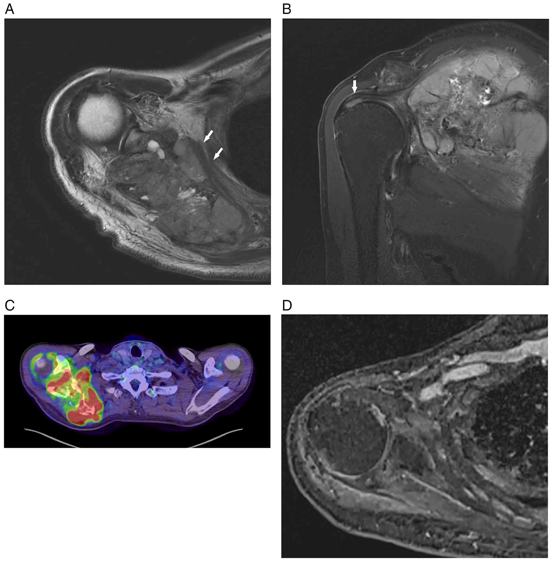
shoulder. MRI revealed a tumorous lesion in the right scapula,
infiltrating the anterior and posterior aspects and the subacromial
space (Fig. 1A and B). Positron
emission tomography-computed tomography (PET-CT) showed a high
maximum standardized uptake value (SUVmax) of 37.9 in the scapular
lesion (Fig. 1C) and multiple
pulmonary metastases. After being diagnosed with Ewing sarcoma, he
received the VDC-IE chemotherapy (vincristine, doxorubicin,
cyclophosphamide, ifosfamide, and etoposide). After three cycles,
the multiple lung metastases nearly disappeared, prompting us to
treat the scapular lesion with CIRT. The patient received three
additional chemotherapy cycles followed by CIRT. After two more
chemotherapy cycles, chemotherapy was halted due to prolonged
pancytopenia and severe febrile neutropenia. At the end of
treatment, PET-CT indicated a reduction in SUVmax to 3.59 in the
local lesion and complete disappearance of pulmonary metastases. At
two years post-treatment, there were no signs of local relapse or
metastasis on MRI (Fig. 1D) and
PET-CT, although scapular destruction persisted. He had a limited
range of motion of the right shoulder (flexion, 40°; extension,
10°; abduction, 20°; external rotation, 0°; MSTS score, 70%).
Case 2
A 40-year-old man presented to our hospital with a
tumor on his left scapula, with shoulder pain, and swelling 3
months prior to his first visit in August 2023. The patient was
diagnosed with Hodgkin's lymphoma at 25 years of age and treated
with chemotherapy that included doxorubicin. Shoulder motion was
not restricted, except for a slight disturbance of internal
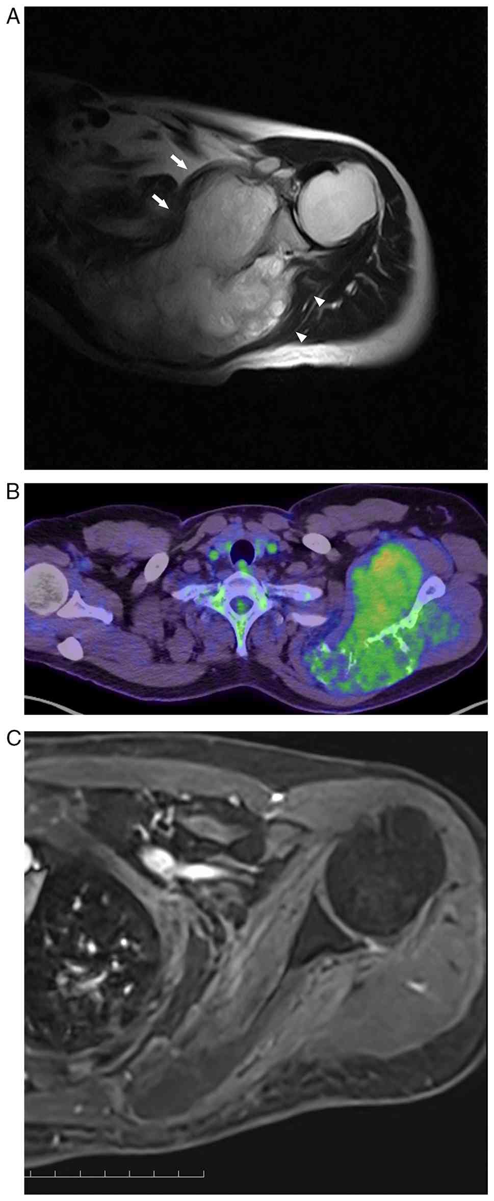
rotation. MRI and PET-CT revealed a tumor extending to both the
ventral and dorsal aspects of the left scapula (Fig. 2A and B). The patient was diagnosed
with Hodgkin lymphoma at 25 years of age, which was treated with
chemotherapy that included doxorubicin. To prevent
doxorubicin-associated cardiotoxicity, we replaced it with
actinomycin-D in the VDC-IE regimen. Two years after treatment,
PET-CT and MRI (Fig. 2C) revealed
no metastasis or local recurrence. The active range of motion of
the shoulder was as follows: forward flexion, 90°; extension, 30°;
abduction, 60°; external rotation, 10°; internal rotation, S1; and
MSTS score, 87%.
Case 3
An otherwise healthy 32-year-old woman was referred
to our hospital with a suspected malignant tumor of the right
scapula. She noticed a prominence around the shoulder 4 months
before visiting us and consulted several clinics; however, the
cause of the symptoms was not revealed. On the first visit to our
hospital in April 2023, she presented with pain in motion and
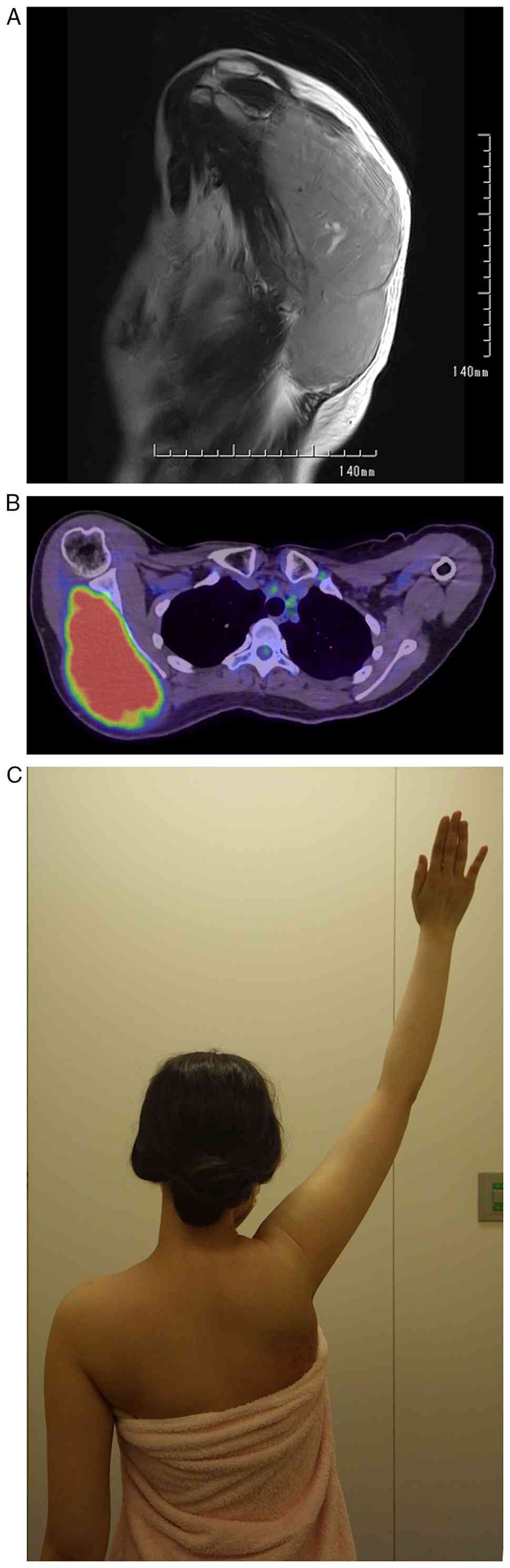
swelling, with slight redness around the shoulder. MRI revealed a
tumor with unclear borders and surrounding inflammation on the
scapula, eroding the dorsal cortex (Fig. 3A). PET-CT showed a high SUVmax in
the dorsal scapular lesion (Fig.
3B) and multiple pulmonary metastases. After four cycles of
VDC-IE, PET-CT revealed a reduced accumulation in her multiple lung
metastases. Based on the PET-CT results indicating that the
metastases were manageable, we chose to treat the scapular lesion
with CIRT. CIRT was administered, followed by two additional cycles
of VDC/IE. After the 12th of 17 scheduled cycles of chemotherapy
(10 months later from the initiation of VDC-IE regimen), the
recovery from myelosuppression was significantly prolonged, and
follow-up CT revealed that the pulmonary metastases had grown. The
chemotherapy regimen was modified to irinotecan (CPT-11) and
temozolomide (TMZ) due to these issues. After the fourth cycle of
CPT-11/TMZ, she developed fever, severe pain from the axilla to the
back, and an exacerbation of dermatitis. Based on the clinical
findings, radiation recall dermatitis was therefore suspected. One
month of steroid and narcotic medications alleviated her symptoms.
Due to the addition of prednisolone to prevent radiation recall,
the patient underwent a total of 20 cycles of CPT-11/TMZ, which
kept the growth of the patient's lung metastases gradual, even now,
three years after starting treatment. Two years after the
initiation of treatment, the patient's active range of motion in
the right shoulder was as follows: forward flexion, 150°;
extension, 30°; abduction, 150°; external rotation, 65°; internal
rotation, S1; and MSTS score, 87% (Fig.
3C).
The biopsy specimens from the three cases were fixed
in 10% neutral buffered formalin at room temperature for 24 h,
dehydrated in a graded ethanol series, cleared in xylene and
embedded in paraffin. For histological evaluation, 4-µm sections
were deparaffinized, rehydrated and stained with
Tissue-Tek® Gill's Hematoxylin (cat. no. 8648; Sakura
Finetek) and Tissue-Tek® Eosin (cat. no. 9134-4P; Sakura
Finetek) at room temperature using a Tissue-Tek Prisma®
Plus Automated Slide Stainer (Sakura Finetek) according to the
manufacturer's protocol.
For immunohistochemical analysis, 3-µm sections were
processed using the BOND Polymer Refine Detection system (Leica
Biosystems), including the secondary antibody, following the
manufacturer's instructions. Briefly, the sections were incubated
for 15 min at room temperature with the following ready-to-use
antibodies: Mouse monoclonal antibodies against CD99 (clone 12E7;
cat. no. IR057; Dako; Agilent Technologies, Inc.), CD3 (clone LN10;
cat. no. PA0553; Leica Biosystems), CD20 (clone L26; cat. no.
PA0200; Leica Biosystems), CD56 (clone CD564; cat. no. PA0191;
Leica Biosystems), CD138 (clone B-A38; code no. 413881; Histofine),
chromogranin A (CGA; clone 5H7; cat. no. PA0515; Leica Biosystems),
and synaptophysin (clone 27G12; cat. no. PA0299; Leica Biosystems);
and a rabbit monoclonal antibody against cyclin D1 (clone EP12;
cat. no. PA0046; Leica Biosystems). The sections were then
incubated with the pre-diluted secondary antibody for 10 min and
counterstained with hematoxylin for 5 min at room temperature.
Staining was visualized using a light microscope (Imager A2; Carl
Zeiss AG).
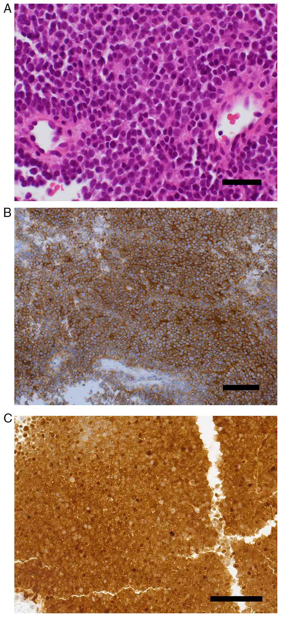
Histologically, the specimens showed a proliferation
of small round cells with scant cytoplasm, which were positive for
Periodic acid-Schiff staining and CD99, but negative for epithelial
markers. Fig. 3 shows
representative specimens of Case 3. Small round cells that
overlapped and had hyperchromatic nuclei diffusely proliferated
with a palisading pattern around the vasculature and rosette-like
formation (Fig. 4A). The
immunohistochemistry results were as follows: CD20(−), CD3(−),
CD138(−),CD56(−), CGA(−), synaptophysin(−), CD99(+) (Fig. 4B), Cyclin D1(+) (Fig. 4C), and AT1(−). In addition, negative
results for WT-1 and calretinin suggested that the tumor was
unlikely to be a CIC-rearrangement tumor. Based on the
pathological, radiological, and physical findings, the patient was
diagnosed with Ewing sarcoma.
Only case 3 was analyzed with the GenMine TOP Cancer
Genome Profiling System, which detected the EWSR1-FLI1 fusion. The
National Health Insurance system in Japan covers limited molecular
biological analyses, including fluorescence in situ hybridization.
To undergo insurance-covered cancer gene panel testing, patients
with solid tumors must meet specific criteria, such as having no
available standard treatment or having completed (or are expected
to complete) standard treatment. Cases 1 and 2 were not analyzed
for fusion genes because they achieved a complete disease-free
status with chemotherapy and CIRT.
Discussion
Ewing sarcoma has been treated using a
multidisciplinary approach that includes chemotherapy, surgery,
and/or radiotherapy. Ahmed et al (8) demonstrated that there was no
difference in local failure rates among local treatment
modalities-surgery, radiotherapy, and surgery combined with
radiotherapy-for axial non-spine tumors (rib, clavicle, sternum,
and scapula). Iwata et al (14) reported that CIRT and chemotherapy
provided an acceptable local control rate for patients with tumors
of the unresectable Ewing's sarcoma family. These reports prompted
us to explore CIRT as a treatment option for the three current
patients. Of the three patients, two had multiple lung metastases
on their first visit. CIRT was contraindicated for patients with
uncontrollable metastasis at our institute; however, the reduction
of metastases by the VDC-IE regimen allowed these cases to be
treated with CIRT. All three cases showed no local relapse for more
than two and half years after CIRT.
Scapulectomy and resection of the surrounding
muscles are often necessary to treat malignant tumors originating
from the scapula. To restore the shoulder function after
scapulectomy, various procedures such as prosthesis, graft
reconstruction, and humeral suspension have been reported. The
humeral suspension technique following scapulectomy has been used
for decades; however, its functional results, with MSTS scores of
58.5–64.9%, appear insufficient (7,15,16).
The recent Ipponi report demonstrated that custom-made prostheses
following total scapulectomy achieved good functional outcomes,
with an MSTS score of 24 (80%); however, no patients could perform
above shoulder activities (7). The
current cases treated with CIRT demonstrated MSTS scores of 70, 87,
and 87%, respectively. Notably, Case 3 showed an impressive range
of motion with 150° of forward flexion and abduction. Meanwhile, in
Case 1, there was 40° of forward flexion and 20° of abduction, in
Case 2, these values were 90° and 60°, respectively. The difference
in the range of motion likely depends on the surrounding tissues
affected by the tumor. In Cases 1 and 2, the tumor invaded the
subscapularis muscle adjacent to the thoracic wall, while in Case
3, it did not. Additionally, the tumor in Case 1 also infiltrated
the subacromial bursa. These findings may be explained by the range
of motion in Cases 1 and 2 being restricted due to adhesion of the
thoracoscapular joint, while contracture around the subacromial
bursa severely limited the range of motion in Case 1. One of the
side effects in radiotherapy is radiation recall dermatitis, as
occurred in Case 3. Radiation recall is defined as an inflammatory
reaction in a previously irradiated area, triggered by systemic
agents, including anti-cancer drugs, and it can occur even months
or years after the completion of radiotherapy (17,18).
If severe dermatitis develops, the causative agent should be
delayed, reduced, or temporarily withdrawn. Physicians should
closely monitor the irradiated area following the administration of
anticancer drugs.
Ewing sarcoma primarily affects children,
adolescents, and young adults (19,20).
The patients in the present study may be older than expected
because of the lack of a pediatric department and the inability to
administer chemotherapy to patients under 12 years of age. Several
studies have reported that adult patients with Ewing sarcoma have
inferior outcomes compared with pediatric patients, which is
speculated to be due to fewer cycles of chemotherapy and delayed
clinical detection in adults (21,22).
To the best of our knowledge, no studies have examined the
differences in the effects of radiotherapy between adult and
pediatric patients with Ewing sarcoma.
One limitation associated with our study includes
the fact that the follow-up periods may have been insufficient to
fully assess the patients' shoulder function and second malignancy.
Bone-related complications, such as pelvic fractures, vertebral
compression fractures, and femoral head necrosis, can sometimes
occur after CIRT for lesions around the pelvis. Takenaka et
al (23) reported that 10 of 27
patients with unresectable pelvic bone sarcoma treated with CIRT
developed femoral head necrosis, with two cases occurring more than
5 years after irradiation. Although the scapula is likely at lower
risk for bone-related complications in comparison to the pelvic
bones due to minimal weight-bearing influence, long-term evaluation
of the affected bones is necessary after CIRT.
CIRT provides a minimally invasive treatment option
for Ewing sarcoma of the scapula, possibly serving as an
alternative to surgery. If the tumor does not invade the
scapulothoracic and glenohumeral joints or the subacromial bursa,
the shoulder function is expected to be preserved after
treatment.
Acknowledgements
Not applicable.
Funding
Funding: No funding was received.
Availability of data and materials
The data generated in the present study may be
requested from the corresponding author.
Authors' contributions
TY conceptualized the study, designed the work,
acquired clinical data and critically reviewed the manuscript for
important intellectual content. NK analyzed and interpreted
clinical data, and drafted the manuscript. MO and TO acquired data
from the radiotherapy they performed and critically reviewed the
manuscript for important intellectual content. HC analyzed and
interpreted data, and critically reviewed the manuscript for
important intellectual content. TY and NK confirm the authenticity
of all the raw data. All authors read and approved the final
manuscript, and agreed to be accountable for all aspects of the
work.
Ethics approval and consent to
participate
Not applicable.
Patient consent for publication
Written informed consent was obtained from the
patients for the publication of this case report.
Competing interests
The authors declare that they have no competing
interests.
References
|
1
|
Malik SS, Tahir M, Ahmed U, Evans S, Jeys
L and Abudu S: Outcome of Ewing's sarcoma of the scapula-a
long-term follow-up study. Orthop Traumatol Surg Res. 106:25–30.
2020. View Article : Google Scholar : PubMed/NCBI
|
|
2
|
Bernstein M, Kovar H, Paulussen M, Randall
RL, Schuck A, Teot LA and Juergens H: Ewing's sarcoma family of
tumors: Current management. Oncologist. 11:503–519. 2006.
View Article : Google Scholar : PubMed/NCBI
|
|
3
|
Yeung CM, Kaiser CL, Peleteiro-Pensado M,
Barrientos-Ruiz I, Ortiz-Cruz EJ, Anderson ME, Raskin KA and
Lozano-Calderón SA: Characteristics and oncologic outcomes of
patients with Ewing sarcoma of the scapula. Surg Oncol.
38:1016192021. View Article : Google Scholar : PubMed/NCBI
|
|
4
|
Pretz JL, Barysauskas CM, George S,
Hornick JL, Raut CP, Chen YLE, Marcus KJ, Choy E, Hornicek F, Ready
JE, et al: Localized adult ewing sarcoma: Favorable outcomes with
alternating vincristine, doxorubicin, cyclophosphamide, and
ifosfamide, etoposide (VDC/IE)-based multimodality therapy.
Oncologist. 22:1265–1270. 2017. View Article : Google Scholar : PubMed/NCBI
|
|
5
|
Grier HE, Krailo MD, Tarbell NJ, Link MP,
Fryer CJH, Pritchard DJ, Gebhardt MC, Dickman PS, Perlman EJ,
Meyers PA, et al: Addition of ifosfamide and etoposide to standard
chemotherapy for Ewing's sarcoma and primitive neuroectodermal
tumor of bone. N Engl J Med. 348:694–701. 2003. View Article : Google Scholar : PubMed/NCBI
|
|
6
|
Womer RB, West DC, Krailo MD, Dickman PS,
Pawel BR, Grier HE, Marcus K, Sailer S, Healey JH, Dormans JP and
Weiss AR: Randomized controlled trial of interval-compressed
chemotherapy for the treatment of localized Ewing sarcoma: A report
from the Children's Oncology Group. J Clin Oncol. 30:4148–4154.
2012. View Article : Google Scholar : PubMed/NCBI
|
|
7
|
Ipponi E, Cordoni M, Bechini E, Gentili F,
Cosseddu F, Campo FR, D'Arienzo A and Andreani L: Clinical and
functional results after total scapulectomy in orthopedic oncology:
Are custom-made scapular prostheses better than humeral suspension?
Proc (Bayl Univ Med Cent). 37:553–559. 2024.PubMed/NCBI
|
|
8
|
Ahmed SK, Randall RL, DuBois SG, Harmsen
WS, Krailo M, Marcus KJ, Janeway KA, Geller DS, Sorger JI, Womer
RB, et al: Identification of patients with localized ewing sarcoma
at higher risk for local failure: A report from the children's
oncology group. Int J Radiat Oncol Biol Phys. 99:1286–1294. 2017.
View Article : Google Scholar : PubMed/NCBI
|
|
9
|
Choi Y, Lim DH, Lee SH, Lyu CJ, Im JH, Lee
YH and Suh CO: Role of radiotherapy in the multimodal treatment of
ewing sarcoma family tumors. Cancer Res Treat. 47:904–912. 2015.
View Article : Google Scholar : PubMed/NCBI
|
|
10
|
Imai R, Kamada T and Araki N; Working
Group for Carbon Ion Radiotherapy for Bone, : Soft Tissue Sarcomas:
Carbon ion radiotherapy for unresectable localized axial soft
tissue sarcoma. Cancer Med. 7:4308–4314. 2018. View Article : Google Scholar : PubMed/NCBI
|
|
11
|
Matsunobu A, Imai R, Kamada T, Imaizumi T,
Tsuji H, Tsujii H, Shioyama Y, Honda H and Tatezaki SI; Working
Group for Bone, : Soft Tissue Sarcomas: Impact of carbon ion
radiotherapy for unresectable osteosarcoma of the trunk. Cancer.
118:4555–4563. 2012. View Article : Google Scholar : PubMed/NCBI
|
|
12
|
Kanai T, Endo M, Minohara S, Miyahara N,
Koyama-ito H, Tomura H, Matsufuji N, Futami Y, Fukumura A, Hiraoka
T, et al: Biophysical characteristics of HIMAC clinical irradiation
system for heavy-ion radiation therapy. Int J Radiat Oncol Biol
Phys. 44:201–210. 1999. View Article : Google Scholar : PubMed/NCBI
|
|
13
|
Ohno T, Kanai T, Yamada S, Yusa K, Tashiro
M, Shimada H, Torikai K, Yoshida Y, Kitada Y, Katoh H, et al:
Carbon ion radiotherapy at the gunma university heavy ion medical
center: New facility set-up. Cancers (Basel). 3:4046–4060. 2011.
View Article : Google Scholar : PubMed/NCBI
|
|
14
|
Iwata S, Yonemoto T, Ishii T, Kumagai K,
Imai R, Hagiwara Y, Kamada T and Tatezaki SI: Efficacy of
carbon-ion radiotherapy and high-dose chemotherapy for patients
with unresectable Ewing's sarcoma family of tumors. Int J Clin
Oncol. 18:1114–1118. 2013. View Article : Google Scholar : PubMed/NCBI
|
|
15
|
Pritsch T, Bickels J, Wu CC, Squires MH
and Malawer MM: Is scapular endoprosthesis functionally superior to
humeral suspension? Clin Orthop Relat Res. 456:188–195. 2007.
View Article : Google Scholar : PubMed/NCBI
|
|
16
|
Salunke AA, Nandy K, Kamani M, Parmar R,
Bharwani N, Pathak S, Patel K and Pandya S: Is polypropylene mesh
reconstruction functionally superior to non reconstructive group
following total scapular resection? A retrospective analysis of 16
patients and a systematic review of the literature. J Orthop.
52:37–48. 2024. View Article : Google Scholar : PubMed/NCBI
|
|
17
|
Burris HA and Hurtig J: Radiation recall
with anticancer agents. Oncologist. 15:1227–1237. 2010. View Article : Google Scholar : PubMed/NCBI
|
|
18
|
Camidge R and Price A: Characterizing the
phenomenon of radiation recall dermatitis. Radiother Oncol.
59:237–245. 2001. View Article : Google Scholar : PubMed/NCBI
|
|
19
|
Stiller CA, Trama A, Serraino D, Rossi S,
Navarro C, Chirlaque MD and Casali PG; RARECARE Working Group, :
Descriptive epidemiology of sarcomas in Europe: Report from the
RARECARE project. Eur J Cancer. 49:684–695. 2013. View Article : Google Scholar : PubMed/NCBI
|
|
20
|
Fukushima T, Ogura K, Akiyama T, Takeshita
K and Kawai A: Descriptive epidemiology and outcomes of bone
sarcomas in adolescent and young adult patients in Japan. BMC
Musculoskelet Disord. 19:2972018. View Article : Google Scholar : PubMed/NCBI
|
|
21
|
Wytiaz V, Schwartz E, Rice JD, Zhao L,
Jasty R, Schuetze S and Chugh R: Disparate outcomes, biologic and
therapeutic differences in pediatric versus adult patients with
ewing sarcoma. Oncology. 102:1–8. 2024. View Article : Google Scholar : PubMed/NCBI
|
|
22
|
Gupta AA, Pappo A, Saunders N, Hopyan S,
Ferguson P, Wunder J, O'Sullivan B, Catton C, Greenberg M and
Blackstein M: Clinical outcome of children and adults with
localized Ewing sarcoma: Impact of chemotherapy dose and timing of
local therapy. Cancer. 116:3189–3194. 2010. View Article : Google Scholar : PubMed/NCBI
|
|
23
|
Takenaka S, Araki N, Outani H, Hamada KI,
Yoshikawa H, Kamada T and Imai R: Complication rate, functional
outcomes, and risk factors associated with carbon ion radiotherapy
for patients with unresectable pelvic bone sarcoma. Cancer.
126:4188–4196. 2020. View Article : Google Scholar : PubMed/NCBI
|