Introduction
Endometrial cancer (EC) is a common malignant tumour
of the female reproductive system, with 417,000 new cases and
97,000 associated deaths worldwide in 2020. The incidence and
prevalence rank sixth among types of cancer in women (1). In 2022, a total of 423,680 cases and
97,723 deaths were reported globally (2), and there were 77,700 new cases and
13,500 deaths in China in 2022 (3).
In some developed cities in China, its incidence has become the
highest among malignant gynaecological tumours (4). The prognosis of early stage EC is
generally favourable after surgical treatment, but patients with
high-risk factors still have a risk of distant recurrence and
metastasis after surgery (5). In
the 2016 European Society for Medical Oncology (ESMO), European
Society of Gynaecological Oncology (ESGO) and European Society for
Radiotherapy and Oncology (ESTRO) guidelines, high-risk EC was
defined as stage I and grade 3 endometrioid endometrial carcinoma
(EEC) with deep invasion, stage II or III EEC or non-endometrioid
EC, including serous carcinoma, clear cell carcinoma,
undifferentiated carcinoma, carcinosarcoma, mucinous adenocarcinoma
and mixed-type carcinoma (6). Owing
to its long latency period and inconspicuous early symptoms
(7), early diagnosis and treatment
of EC, especially high-risk EC, remain a key factor for improving
patient survival rates. Therefore, finding more convenient and
effective predictive indicators is highly important. Ki-67 is a
widely used clinical marker of tumour cell proliferation and is
closely associated with the differentiation, invasion, metastasis
and prognosis of many tumours. In EC, Ki-67 can serve as an
effective predictor of deep myometrial invasion and lymph node
metastasis (8,9). Previous research indicates that
pre-treatment inflammatory status has a positive predictive value
for high-risk EC. The lymphocyte-to-monocyte ratio (LMR), as a key
marker of the inflammatory response (10), also serves as a valuable
haematological biomarker for diagnosing EC (11). However, studies on the predictive
value of the LMR for high-risk EC remain limited.
Therefore, in the present study, a retrospective
analysis of clinically accessible data, including
clinicopathological characteristics and immunohistochemical (IHC)
markers of EC combined with serological test results of the LMR,
was conducted to develop a preoperative risk prediction model that
can inform preoperative evaluation and clinical triage for
high-risk patients with EC.
Materials and methods
General information
A retrospective analysis of the clinical data of
patients with EC who were treated at the Lianyungang Maternal and
Child Health Hospital (Lianyungang, China) from December 2018 to
May 2026 was conducted. Patients were divided into a training set
and a validation set according to the duration of surgical
treatment at the Lianyungang Maternal and Child Health Hospital.
The data collection and analysis for the training set (from
December 2018 to July 2025) was completed in December 2025, while
the data collection and analysis for the validation set (from
August 2025 to May 2026) was completed in May 2026. The data
included age, body mass index (BMI), preoperative serological tests
for lymphocytes and monocytes, postoperative International
Federation of Gynaecology and Obstetrics (FIGO) stage, pathological
type, histological grade, lymph node metastasis and depth of
myometrial invasion. The number of pregnancies reflected the number
of births plus the number of abortions. According to the exclusion
criteria of the present study, cases with missing/incomplete data
were excluded.
A total of 130 patients were included in the
training set (107 cases) and validation set (23 cases). In the
training set, the patients were aged 37–73 years, with a mean age
of 53.17±7.66 years. There were 96 cases of endometrioid carcinoma,
2 cases of serous carcinoma, 2 cases of clear cell carcinoma, 1
case of mucinous adenocarcinoma, 3 cases of undifferentiated
carcinoma and 3 cases of carcinosarcoma. The histological
differentiation grades were as follows: 26 cases were well
differentiated (G1), 36 cases were moderately differentiated (G2),
and 45 cases were poorly differentiated (G3); deep myometrial
invasion (>1/2) was observed in 33 cases. The FIGO stage
distribution was as follows: 78 patients were in stage I, 16
patients were in stage II, 12 patients were in stage III and 1
patient was in stage IV. In the validation set, the patients were
aged 45–74 years, with a mean age of 57.78±6.78 years. There were
21 cases of endometrioid carcinoma and 2 cases of undifferentiated
carcinoma. The histological differentiation grades were as follows:
5 cases were well differentiated (G1), 11 cases were moderately
differentiated (G2) and 7 cases were poorly differentiated (G3);
deep myometrial invasion (>1/2) was observed in 8 cases. The
FIGO stage distribution was as follows: 13 patients were in stage
I, 7 patients were in stage II and 3 patients were in stage III.
The study protocol was reviewed and approved by the Ethical Review
Committee of the Lianyungang Maternal and Child Health Hospital
(approval no. XM2023027) prior to implementation.
High-risk EC grouping
Patients were categorized according to the 2016
ESMO-ESGO-ESTRO consensus guidelines (6). Among the 107 patients with EC, 41 were
classified as having low-risk EC, and 66 were classified as having
high-risk EC. High-risk EC was defined as follows: Stage I and
grade 3 EEC with deep invasion; stage II or III EEC; or
non-endometrioid endometrial carcinoma (including serous carcinoma,
clear cell carcinoma, undifferentiated carcinoma, carcinosarcoma,
mucinous adenocarcinoma and mixed-type carcinoma). The inclusion
criteria included the following: i) A pathological diagnosis of
primary EC with complete postoperative pathological results,
including tumour histological type and grade, FIGO stage, depth of
myometrial invasion, lymph node metastasis status and other
relevant parameters; ii) the surgical scope included extra fascial
hysterectomy, bilateral salpingo-oophorectomy and pelvic and
para-aortic lymphadenectomy; and iii) patients with complete
medical records, including age, BMI and details of any
comorbidities (such as hypertension or diabetes). The exclusion
criteria were as following: i) The patient did not undergo standard
surgical treatment; ii) the patient had other concurrent malignant
tumours; iii) patients who had received radiotherapy, chemotherapy
or endocrine therapy prior to surgery; iv) patients with missing
clinical data or undefined pathological type/staging; and v)
patients with severe hepatic or renal dysfunction or those with
autoimmune or haematological diseases.
Histopathological and IHC
inspection
First, diagnostic curettage was performed on all
patients. The obtained endometrial curettage specimens were
routinely fixed in formalin solution and then subjected to
histopathological examination. After being diagnosed with EC, all
the patients underwent IHC examination and radical surgical
treatment. On the basis of parameters such as histological type and
grade, FIGO stage and lymph node metastasis, these patients were
subsequently classified into different risk levels. All endometrial
curettage specimens were subjected to IHC using the same
standardized criteria, and all pathological slides were
independently reviewed by two histopathologists to reach a final
diagnostic consensus. IHC detection was performed using the VENTANA
method with DAB chromogenic staining on a fully automated
immunostainer (Roche BenchMark GX; Roche Diagnostics GmbH), and the
final staining results would be observed under a light microscope.
This staining system had widespread clinical applicability, and the
test results were highly consistent with those of Dako/Agilent
(12). The following rabbit
monoclonal antibodies were used: CONFIRM anti-oestrogen receptor
(ER) (clone, SP1; cat. no. 05278392001; Roche Diagnostics); CONFIRM
anti-Progesterone Receptor (PR) (clone,1E2; cat. no. 05278414001;
Roche Diagnostics); and CONFIRM™ anti-Ki-67 (clone: 30–9; cat. no.
05278384001; Roche Diagnostics). The results of the IHC parameters
(ER, PR and Ki-67) were recorded as continuous variables, with the
proportion of positive cells expressed as a percentage (0–100%),
for example, 70% ER (+), 60% PR (+) and 80% Ki-67 (+).
Laboratory tests
Following hospital admission, fasting peripheral
venous blood samples were collected from all patients. The monocyte
and lymphocyte counts were measured using an automated haematology
analyser (BC7500CRP; Shenzhen Mindray Bio-Medical Electronics Co.,
Ltd.). The LMR was subsequently calculated.
Statistical analysis
Statistical analysis was performed using SPSS 25.0
(IBM Corp.). Categorical data are expressed as percentages.
Normally distributed continuous data (such as age and BMI) are
presented as the mean ± SD (x̄ ± s), whereas non-normally
distributed continuous data (such as Ki-67, ER and PR expression)
are expressed as the median (interquartile range) [M(P25, P75)].
Univariate and multivariate logistic regression analyses were
employed to identify risk factors for high-risk EC. A nomogram
prediction model was constructed using R software (Posit Software,
PBC) with the ‘rms’ package. The accuracy of the model was
evaluated through ROC curve analysis and external validation. The
optimal cut-off value was determined by calculating the Youden's J
index. The formula was: J=Sensitivity + Specificity - 1. The
optimal cut-off value was the value of the independent variable
when the Youden's J index reached its maximum. The Hosmer-Lemeshow
test was used to verify the consistency between the predicted
probabilities and the actual observed results. When P>0.05, it
indicates that the model fit well. The Harman single-factor method
was used to test whether there was any severe common method bias in
the data. The collinearity of the independent variables in the data
was verified through the variance inflation factor (VIF). P<0.05
was considered to indicate a statistically significant
difference.
Results
Univariate and multivariate analyses
of the training set data
When the risk category of EC was used as the
dependent variable (high risk=1, low risk=0), univariate logistic
regression analysis revealed that high-risk EC was significantly
associated with patient age, ER expression, Ki-67 index and the
serum LMR (P<0.05), but not with BMI, menopausal status or the
number of pregnancies, abortions or births, hypertension and blood
glucose (P>0.05) (Table I).
Variables identified in the univariate analysis were included as
independent variables in the multivariate logistic regression. The
results demonstrated that age, the Ki-67 index and the LMR were
independent risk factors for high-risk EC (P<0.05; Table II).
 | Table I.Univariate analysis of high-risk
endometrial cancer in training set data. |
Table I.
Univariate analysis of high-risk
endometrial cancer in training set data.
| Characteristic | β | SE | Wals | P-value | Exp(B) | 95% CI |
|---|
| Age, years | 0.141 | 0.037 | 14.507 | <0.05 | 1.152 | 1.071–1.239 |
| BMI,
kg/m2 | 0.028 | 0.049 | 0.314 | 0.575 | 1.028 | 0.934–1.132 |
| No. of
pregnancies | 0.229 | 0.189 | 1.469 | 0.226 | 1.257 | 0.868–1.821 |
| No. of
abortions | 0.366 | 0.229 | 2.555 | 0.110 | 1.442 | 0.921–2.259 |
| No. of births | 0.955 | 0.446 | 2.157 | 0.142 | 1.926 | 0.803–4.618 |
| Menopause, yes vs.
no | 0.301 | 0.154 | 3.832 | 0.050 | 1.351 | 1.000–1.825 |
| Hypertension, yes
vs. no | −0.005 | 0.085 | 0.004 | 0.950 | 0.995 | 0.842–1.175 |
| Blood glucose,
mmol/ | 0.429 | 0.407 | 1.107 | 0.293 | 1.535 | 0.691–3.411 |
| ER labeling index,
% | −0.676 | 0.247 | 7.478 | 0.006 | 0.509 | 0.314–0.826 |
| PR labeling index,
% | −0.254 | 0.177 | 2.070 | 0.150 | 0.775 | 0.548–1.096 |
| Ki-67 labeling
index, % | 0.686 | 0.137 | 25.284 | <0.05 | 1.987 | 1.520–2.596 |
| LMR | 0.600 | 0.143 | 17.715 | <0.05 | 1.822 | 1.378–2.409 |
| Table II.Multivariate analysis of high-risk
endometrial cancer in training set data. |
Table II.
Multivariate analysis of high-risk
endometrial cancer in training set data.
|
Characteristics | β | SE | Wals | P-value | Exp(B) | 95% CI |
|---|
| Age, years | 0.146 | 0.051 | 8.156 | 0.004 | 1.157 | 1.047–1.279 |
| ER labeling index,
% | −0.437 | 0.333 | 1.723 | 0.189 | 0.646 | 0.337–1.240 |
| Ki-67 labeling
index, % | 0.588 | 0.165 | 12.731 | <0.05 | 1.800 | 1.303–2.487 |
| LMR | 0.435 | 0.164 | 7.069 | 0.008 | 1.545 | 1.121–2.129 |
Internal and external validation of
the predictive model for high-risk EC
Construction and internal validation of the
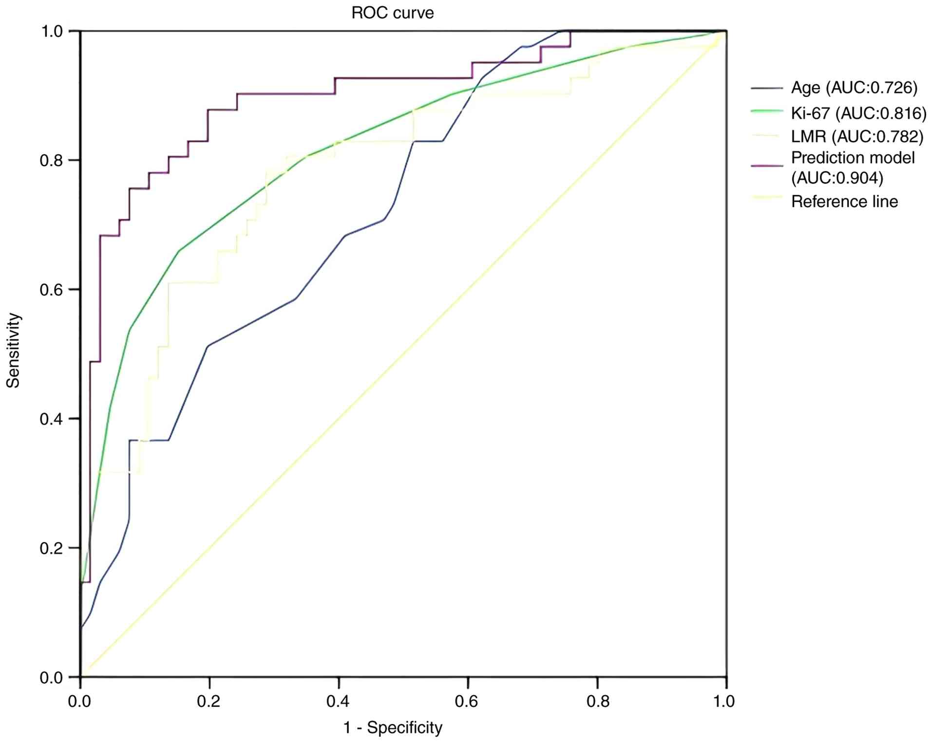
prediction model was performed. In the training set, the ROC curve
demonstrated that age [area under the curve (AUC)=0.726; 95% CI,
0.631–0.821], the Ki-67 index (AUC=0.816; 95% CI, 0.730–0.902) and
the LMR (AUC=0.782; 95% CI, 0.690–0.875) each demonstrated moderate
performance in individually predicting high-risk EC (Fig. 1). The combined prediction model
(based on age, the Ki-67 index and the LMR) had excellent
predictive value (AUC=0.904; 95% CI, 0.840–0.968; Fig. 1). The Youden's J index of the model
was 0.695, with a sensitivity of 75.6%, a specificity of 93.9% and
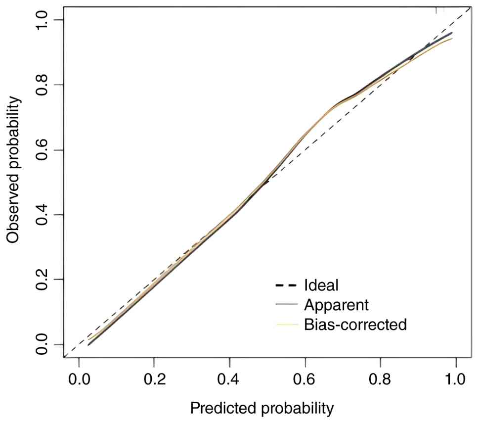
an optimal cut-off value of 0.45. The Hosmer-Lemeshow test
indicated an adequate goodness-of-fit for the combined prediction
model (X2=9.185; P=0.327). As shown in Fig. 2, the calibration curve of the model
primarily lies along the 45-degree line, indicating that the
predicted probability is close to the actual probability and that
the model is well calibrated.
External validation
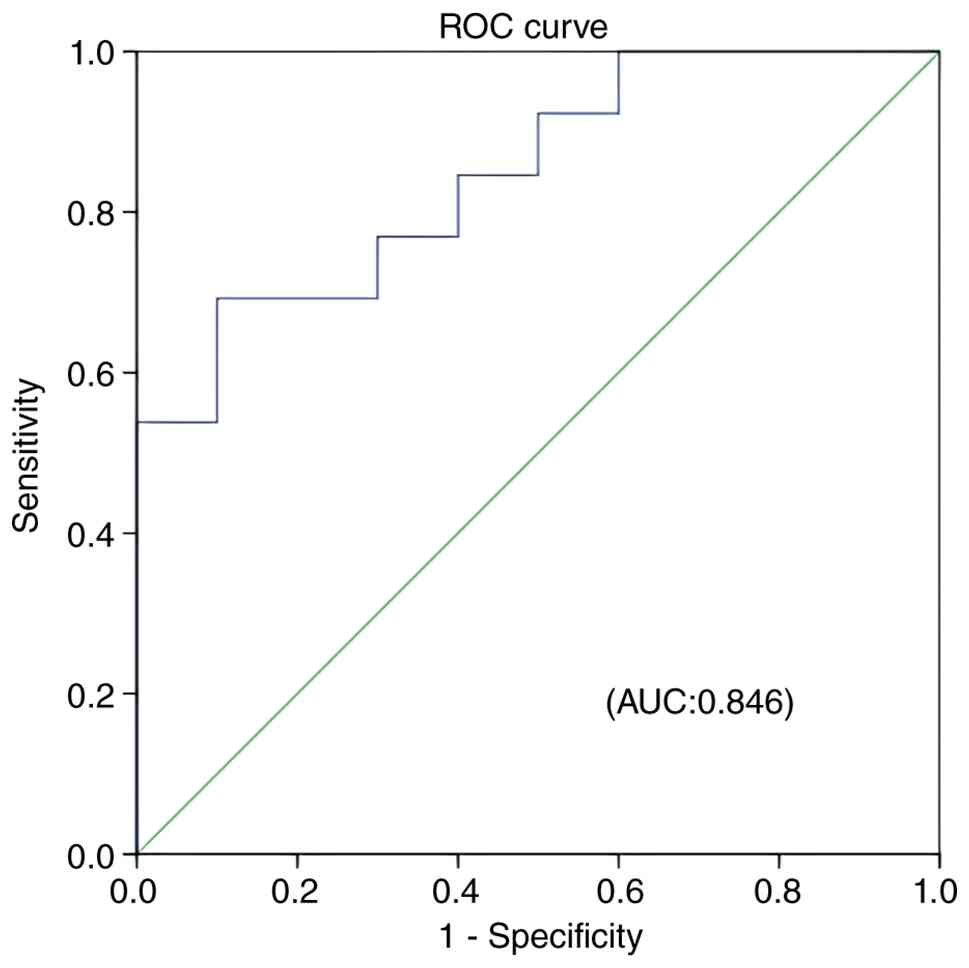
The predictive model was used to evaluate the
patients in the validation set. The ROC curve revealed that the
model had good predictive performance (AUC=0.846; 95% CI,
0.688–1.000; P<0.05; Fig. 3). On
the basis of the optimal cut-off value, patients were divided into
predicted highrisk and predicted lowrisk groups, and the results
were compared with the actual conditions. The results revealed that
among the 8 patients predicted to be highrisk, 6 were actually high
risk; among the 15 patients predicted to be lowrisk, 11 were
actually lowrisk. The calculated prediction accuracy was (11+6)/23
×100%=73.9%.
Development of a nomogram prediction
model
Firstly, the Harman single-factor method was used to
test the data bias correction, and the first common factor
accounted for only 39.04% of the total variance explained, which
was <40% of the evaluation criterion. Therefore, it could be
considered that there was no serious bias. Secondly, the VIF was
used to assess the multicollinearity of the data. The results
showed that the VIF values for age, Ki-67 and LMR were 1.009, 1.073
and 1.068, respectively, all of which were <5, indicating that
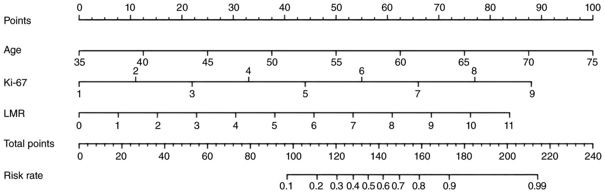
there was no strong collinearity among them. Then, a nomogram
prediction model was developed using R software (Fig. 4), and a higher total score in the
nomogram indicated a greater probability of developing high-risk
EC. As shown in Fig. 4, the
patient's age score was 100 points, the Ki-67 expression score was
87.5 points and the LMR score was 83.75 points.
Discussion
EC is the most common gynaecological malignancy in
postmenopausal women (13). The
incidence of EC is steadily increasing (14), and it ranks as the 14th leading
cause of cancer-related mortality among women worldwide (15). EC treatment is primarily surgical,
with radiotherapy and chemotherapy being common adjuvant treatment
modalities. Generally, for patients with suspected EC or
endometrial lesions, a hysteroscopic endometrial biopsy is first
performed. The obtained endometrial specimen is examined
pathologically to determine the pathological type and
differentiation grade of the tumour. Subsequently, radical surgical
treatment is carried out. The basic surgical procedures include
total hysterectomy plus bilateral salpingo-oophorectomy, with or
without pelvic and para-aortic lymphadenectomy. Intraoperative
peritoneal washing fluid is collected for cytological examination.
Sentinel lymph node biopsy combined with pathological ultra staging
may be chosen as an alternative to systematic lymphadenectomy. For
patients whose diagnostic curettage pathological examination
reveals endometrial serous carcinoma, carcinosarcoma or
undifferentiated carcinoma, omentectomy or omental biopsy is also
needed (16,17). Although the majority of patients can
be cured with surgery, 10–20% of patients experience distant
recurrence (18). Patients with
high-risk EC are more prone to lymphatic metastasis and distant
invasion, which complicates treatment and leads to a worse
prognosis (19). Owing to the lack
of effective early screening markers and diagnostic indicators
(20), early prevention and
management of these high-risk patients remain major challenges and
priorities in clinical practice (21).
Some researchers have classified patients with FIGO
stage I–II EC into a younger group (<70 years) and an older
group (≥70 years) and reported that patients (≥70 years) belong to
a higher-risk group (22).
Additionally, a previous study (23) revealed that patients >50 years
tended to have worse 5-year overall survival and disease-free
survival rates, with a greater proportion of high-risk patients in
the older age group. Additional studies have indicated that the
incidence of metabolic diseases increases progressively with age,
thereby increasing the risk of EC development and progression
(24,25). In addition, oestrogen is commonly
used to treat menopausal syndrome in elderly women to reduce the
incidence of osteoporosis and postmenopausal cardio-cerebrovascular
events. However, this hormone therapy can increase the risk of
developing EC (26). In the present
study, logistic multivariate regression and ROC curve analyses
revealed that patient age was an independent influencing factor for
high-risk EC. The age of patients in the high-risk group was
significantly greater than that in the low-risk group, which is
consistent with the findings of the aforementioned studies.
Moreover, age demonstrated diagnostic value in predicting high-risk
EC (AUC=0.726; 95% CI, 0.631–0.821).
Previous studies have suggested that pregnancy,
miscarriage and childbirth might influence the occurrence and
development of EC. Husby et al (27) reported that both childbirth and
miscarriage could affect the risk of EC to a certain extent, and
this effect was influenced by factors such as age, socioeconomic
status and pregnancy duration; in addition, there was a marginally
smaller risk reduction of EC in women who gave birth compared with
women who induced abortions, perhaps because the pregnancy duration
of the former was longer than that of the latter. Jordan et
al (28) reported that the risk
of EC decreases with increasing age during pregnancy. However,
owing to the lack of other pregnancy-related data in the present
study, a comprehensive understanding of the relationship between
pregnancy and EC risk remains limited.
However, relying solely on traditional clinical
parameters has limited effectiveness in predicting high-risk EC.
These findings suggest that new predictive markers should be
explored to improve prognostic accuracy. Numerous studies have
shown that IHC markers play a notable role in the diagnosis and
treatment of various tumours. The ER can activate tumour cell
proliferation in multiple types of cancer and is closely associated
with the recurrence and metastasis of EC (29,30).
In the present study, although both the positive expression rate
and the intensity of ER expression were greater in high-risk
patients than in low-risk patients, logistic multivariate analysis
revealed no correlation between ER expression and EC risk
stratification. This lack of association may be attributed to the
greater proportion of non-endometrioid carcinomas in the high-risk
group (11/66). Clarke et al (31) suggested that non-endometrioid
tumours are oestrogen independent and exhibit a weaker association
with ER expression, which is consistent with the findings of the
present study. Ki-67, a nuclear antigen associated with
proliferating cells, is currently the most widely used and highly
sensitive marker of cell proliferation and plays a notable role in
the development and progression of various malignant tumours
(21,32). Wei et al (30) demonstrated that Ki-67 is closely
associated with distant metastasis and clinical staging of
malignant tumours, suggesting its potential as a reliable molecular
marker for assessing disease stage and prognosis. Furthermore, high
expression of the Ki-67 protein promotes rapid tumour cell
proliferation, metastasis and deep myometrial invasion in EC
(33). In the present study, high
expression of Ki-67 was identified as an independent influencing
factor for high-risk EC, which is consistent with the
aforementioned findings mentioned. ROC curve analysis demonstrated
that Ki-67 expression is valuable for predicting high-risk EC
(AUC=0.816; 95% CI: 0.730–0.902). These results suggest that Ki-67
could serve as a valuable biomarker for predicting high-risk
EC.
As a biomarker, serum LMR has many advantages such
as simple calculation, low cost and easy availability, and can be
used for high-risk stratification assessment and prognosis
prediction of various tumors. Researchers have found that the LMR
values for low-, intermediate- and high-risk patients with thyroid
cancer were 4.7 (range, 1–10), 5 (range, 2.3–15) and 3.7 (range,
1.6–7.7), respectively, indicating that LMR is associated with
tumour risk stratification (34).
In a retrospective analysis of EC, the LMR was found to be
associated with worse prognosis in patients (35), and markedly elevated LMR levels were
observed in patients with advanced EC (36). The present study revealed that
compared with low-risk patients, those in the high-risk group had
markedly higher LMRs. The LMR was identified as an independent risk
factor for high-risk EC and demonstrated a certain predictive value
(AUC=0.782; 95% CI, 0.690–0.875), which aligns with the findings
mentioned above. Inflammatory cells in peripheral blood and related
haematological indicators reflect the level of systemic immune and
inflammatory responses and can promote tumour cell proliferation,
invasion and migration (37).
Lymphocytes play a key role in chronic inflammatory responses
(38), and changes in lymphocyte
status may directly affect tumour progression (39). Qiu et al (40) reported that individuals in the high
lymphocyte count group had a notably increased risk of thyroid
cancer, suggesting that the lymphocyte count may be closely
associated with the occurrence of thyroid malignancy. Monocytes can
exert their immune evasion effects by limiting the biological
function of CD8+ T cells in lymphocytes (41). In the present study, the high LMR in
patients with high-risk EC indicated an immune status characterized
by increased lymphocytes and decreased monocytes, reflecting an
association among the inflammatory status, immune level and disease
progression in these patients with tumours.
Serum tumour marker testing offers advantages such
as simple operation, minimal invasiveness and high reproducibility
(42), whereas IHC staining is
characterized by high sensitivity, rapid processing and low cost.
Both methodologies are widely adopted in clinical practice and
demonstrate favourable cost-effectiveness. Therefore, the
integration of appropriate serum tumour markers, IHC markers, and
clinicopathological parameters for predicting high-risk EC has
significant practical value (43).
In the present study, individual analyses of patient age, the Ki-67
index and LMR each demonstrated certain predictive value for
high-risk EC. However, compared with any single indicator alone, a
nomogram model incorporating these three indicators showed
significantly superior predictive performance, with good
sensitivity, specificity and fit. The results of the external
validation also demonstrated that the model has good predictive
value.
In summary, the nomogram model incorporating age,
Ki-67 expression and the LMR demonstrated predictive efficacy for
high-risk EC, indicating that this approach can effectively enhance
early diagnostic capability and holds notable value for formulating
treatment strategies. However, the overall sample size of the
present study was limited. Although external validation was
performed using cases from another time period, this remains a
singlecentre retrospective analysis, and some selection bias is
inevitable. To further improve the accuracy of the present study
and validate the predictive value of the model, future studies with
larger sample sizes and a multicentre design are necessary.
Acknowledgements
Not applicable.
Funding
The present study was supported by the Lianyungang City Health
Science and Technology Project, Jiangsu (grant no. 202330).
Availability of data and materials
The data generated in the present study may be
requested from the corresponding author.
Authors' contributions
YYC and JWL analysed the data and wrote the
manuscript. LY, PLL and FFM were involved in the collection and
processing of data. YJZ was involved in the present study design
and participated in the evaluation of the results. All authors read
and approved the final version of the manuscript. YJZ and YYC
confirm the authenticity of all the raw data.
Ethics approval and consent to
participate
The present study was approved by the Ethical Review
Committee of the Lianyungang Maternal and Child Health Hospital
(approval no. XM2023027). Informed consent was waived for the
present study by the Medical Ethics Committee of Lianyungang
Maternal and Child Health Hospital (approval no. XM2023027).
Patient consent for publication
Not applicable.
Competing interests
The authors declare that they have no competing
interests.
Glossary
Abbreviations
Abbreviations:
|
EC
|
endometrial cancer
|
|
ROC
|
receiver operating characteristic
|
|
LMR
|
lymphocyte-to-monocyte ratio
|
|
BMI
|
body mass index
|
|
ER
|
oestrogen receptor
|
|
PR
|
progesterone receptor
|
References
|
1
|
Sung H, Ferlay J, Siegel RL, Laversanne M,
Soerjomataram I, Jemal A and Bray F: Global cancer statistics 2020:
GLOBOCAN estimates of incidence and mortality worldwide for 36
cancers in 185 countries. CA Cancer J Clin. 71:209–249.
2021.PubMed/NCBI
|
|
2
|
Filho AM, Laversanne M, Ferlay J, Colombet
M, Piñeros M, Znaor A, Parkin DM, Soerjomataram I and Bray F: The
GLOBOCAN 2022 cancer estimates: Data sources, methods, and a
snapshot of the cancer burden worldwide. Int J Cancer.
156:1336–1346. 2025. View Article : Google Scholar : PubMed/NCBI
|
|
3
|
Han B, Zheng R, Zeng H, Wang S, Sun K,
Chen R, Li L, Wei W and He J: Cancer incidence and mortality in
China, 2022. J Natl Cancer Cent. 4:47–53. 2024.PubMed/NCBI
|
|
4
|
Siegel RL, Miller KD, Fuchs HE and Jemal
A: Cancer statistics, 2021. CA Cancer J Clin. 71:7–33.
2021.PubMed/NCBI
|
|
5
|
Koskas M, Amant F, Mirza MR and Creutzberg
CL: Cancer of the corpus uteri:2021 update. Int J Gynaecol Obstet.
155 (Suppl 1):S45–S60. 2021. View Article : Google Scholar
|
|
6
|
Colombo N, Creutzberg C, Amant F, Bosse T,
González-Martín A, Ledermann J, Marth C, Nout R, Querleu D, Mirza
MR, et al: ESMO-ESGO-ESTRO consensus conference on endometrial
cancer: Diagnosis, treatment and follow-up. Ann Oncol. 27:16–41.
2016. View Article : Google Scholar : PubMed/NCBI
|
|
7
|
Reijnen C, Visser NC, Kasius JC, Boll D,
Geomini PM, Ngo H, Van Hamont D, Pijlman BM, Vos MC, Bulten J, et
al: Improved preoperative risk stratification with CA-125 in
low-grade endometrial cancer: A multicenter prospective cohort
study. J Gynecol Oncol. 30:e702019. View Article : Google Scholar : PubMed/NCBI
|
|
8
|
Lin Q: Application value of Ki67 and serum
CA125 in the deep myometrial invasion of endometrial
adenocarcinoma. BMC Cancer. 23:2402023. View Article : Google Scholar : PubMed/NCBI
|
|
9
|
Jiang P and Yuan R: Analysis of factors
related to lymph node metastasis in early-stage type 1 endometrial
cancer: Verifying the clinical value of positive threshold of the
IHC parameter Ki67. Cancer Manag Res. 13:6319–6328. 2021.
View Article : Google Scholar : PubMed/NCBI
|
|
10
|
Zhong JH, Huang DH and Chen ZY: Prognostic
role of systemic immune-inflammation index in solid tumors: A
systematic review and meta-analysis. Oncotarget. 8:75381–75388.
2017. View Article : Google Scholar : PubMed/NCBI
|
|
11
|
Huo D and Yang L: Significance of the
neutrophil-lymphocyte ratio, lymphocyte-monocyte ratio, and
platelet-lymphocyte ratio in peripheral blood for endometrial
cancer. Am J Transl Res. 16:6689–6698. 2024. View Article : Google Scholar : PubMed/NCBI
|
|
12
|
Yoshida H, Takigawa W, Kobayashi-Kato M,
Nishikawa T, Shiraishi K and Ishikawa M: Mismatch repair protein
expression in endometrial cancer: Assessing concordance and
unveiling pitfalls in two different immunohistochemistry assays. J
Pers Med. 13:12602023. View Article : Google Scholar : PubMed/NCBI
|
|
13
|
Siegel RL, Miller KD, Fuchs HE and Jemal
A: Cancer statistics, 2022. CA Cancer J Clin. 72:7–33.
2022.PubMed/NCBI
|
|
14
|
Crosbie EJ, Kitson SJ, McAlpine JN,
Mukhopadhyay A, Powell ME and Singh N: Endometrial cancer. Lancet.
399:1412–1428. 2022. View Article : Google Scholar : PubMed/NCBI
|
|
15
|
Salinas-Vera YM, Gallardo-Rincón D,
Ruíz-García E, Silva-Cázares MB, De la Peña-Cruz SC and
López-Camarillo C: The role of hypoxia in endometrial cancer. Curr
Pharm Biotechnol. 23:221–234. 2022. View Article : Google Scholar : PubMed/NCBI
|
|
16
|
Lu KH and Broaddus RR: Endometrial Cancer.
N Engl J Med. 2020 Nov 19;383:2053–2064. 2020. View Article : Google Scholar : PubMed/NCBI
|
|
17
|
Tu H, Huang H, Xian B, Li J, Wang P, Zhao
W, Chen X, Xie X, Wang C, Kong B, et al: Sentinel lymph node biopsy
versus pelvic lymphadenectomy in early-stage cervical cancer: A
multi-center randomized trial (PHENIX/CSEM 010). Int J Gynecol
Cancer. 30:1829–1833. 2020. View Article : Google Scholar : PubMed/NCBI
|
|
18
|
Ørtoft G, Lausten-Thomsen L, Høgdall C,
Hansen ES and Dueholm M: Lymph-vascular space invasion (LVSI) as a
strong and independent predictor for non-locoregional recurrences
in endometrial cancer: A Danish Gynecological Cancer Group Study. J
Gynecol Oncol. 30:e842019. View Article : Google Scholar : PubMed/NCBI
|
|
19
|
Song J, Le T, Hopkins L, Fung-Kee-Fung M,
Jooya A, Lupe K, Gaudet M and Samant R: Treatment of early stage
highrisk endometrioid-type endometrial cancer and patterns of
disease relapse: A retrospective analysis. Adv Radiat Oncol.
5:910–919. 2020. View Article : Google Scholar : PubMed/NCBI
|
|
20
|
de Boer SM, Nout RA, Bosse T and
Creutzberg CL: Adjuvant therapy for high-risk endometrial cancer:
Recent evidence and future directions. Expert Rev Anticancer Ther.
19:51–60. 2019. View Article : Google Scholar : PubMed/NCBI
|
|
21
|
Cai Y, Wang B, Xu W, Liu K, Gao Y, Guo C,
Chen J, Kamal MA and Yuan C: Endometrial cancer: Genetic, metabolic
characteristics, therapeutic strategies and nanomedicine. Curr Med
Chem. 28:8755–8781. 2021. View Article : Google Scholar : PubMed/NCBI
|
|
22
|
Barben J, Kamga MA, Guilloteau A, Quipourt
V, Bengrine-Lefevre L and Dabakuyo-Yonli TS: Are older women with
early-stage endometrial cancer undertreated and more likely to die
from their cancer? Maturitas. 202:1087412025. View Article : Google Scholar : PubMed/NCBI
|
|
23
|
Vuppu PD, Rajanbabu A, Bhati P, Nair IR,
Nair AS and Pavithran K: Beyond the number-age as a prognostic
indicator in endometrial cancer. Eur J Obstet Gynecol Reprod Biol.
314:1146642025. View Article : Google Scholar : PubMed/NCBI
|
|
24
|
Zhai L, Cheng Y, Wu M, Wang T, Yin M, Yang
X, Sun B, Li C, He M, Sun Y, et al: Metabolic syndrome promotes
endometrial cancer by Oleic acid-mediated polyamine accumulation.
Nat Commun. 17:3882025. View Article : Google Scholar : PubMed/NCBI
|
|
25
|
Wu Z, Hu Z, Li Q, Liu G, Oaknin A, Grau
Bejar JF, Mills GB, Ma D, Sun C and Chen G: Molecular and clinical
insights into early-onset endometrial cancer. Trends Cancer.
11:503–519. 2025. View Article : Google Scholar : PubMed/NCBI
|
|
26
|
Yu K, Huang ZY, Xu XL, Li J, Fu XW and
Deng SL: Estrogen receptor function:Impact on the human
endometrium. Front Endocrinol (Lausanne). 13:8277242022. View Article : Google Scholar : PubMed/NCBI
|
|
27
|
Husby A, Wohlfahrt J and Melbye M:
Pregnancy duration and endometrial cancer risk: Nationwide cohort
study. BMJ. 366:l46932019. View Article : Google Scholar : PubMed/NCBI
|
|
28
|
Jordan SJ, Na R, Weiderpass E, Adami HO,
Anderson KE, van den Brandt PA, Brinton LA, Chen C, Cook LS,
Doherty JA, et al: Pregnancy outcomes and risk of endometrial
cancer: A pooled analysis of individual participant data in the
Epidemiology of Endometrial Cancer Consortium. Int J Cancer.
148:2068–2078. 2020. View Article : Google Scholar : PubMed/NCBI
|
|
29
|
Schüler-Toprak S, Skrzypczak M, Gründker
C, Ortmann O and Treeck O: Role of estrogen receptor β, G-Protein
coupled estrogen receptor and estrogen-related receptors in
endometrial and ovarian cancer. Cancers (Basel). 15:28452023.
View Article : Google Scholar : PubMed/NCBI
|
|
30
|
Wei L, Shi C and Zhang Y: Expression of
miR-34a and Ki67 in nasopharyngeal carcinoma and the relationship
with clinicopathological features and prognosis. Oncol Lett.
19:1273–1280. 2020.PubMed/NCBI
|
|
31
|
Clarke MA, Devesa SS, Harvey SV and
Wentzensen N: Hysterectomy-corrected uterine corpus cancer
incidence trends and differences in relative survival reveal racial
disparities and rising rates of nonendometrioid cancers. J Clin
Oncol. 37:1895–1908. 2019. View Article : Google Scholar : PubMed/NCBI
|
|
32
|
Andrés-Sánchez N, Fisher D and Krasinska
L: Physiological functions and roles in cancer of the proliferation
marker Ki-67. J Cell Sci. 135:jcs2589322022. View Article : Google Scholar : PubMed/NCBI
|
|
33
|
Ding SX, Sun YF, Meng H, Wang JN, Xue LY,
Gao BL and Yin XP: Radiomics model based on multi-sequence MRI for
preoperative prediction of ki-67 expression levels in early
endometrial cancer. Sci Rep. 13:220522023. View Article : Google Scholar : PubMed/NCBI
|
|
34
|
Refaat L, Eissa MS, Elnaggar GN, Mehesen
M, Eissa MS, Kamal A and Abdellateif MS: Systemic inflammatory
indices comprising monocytes provide a clinical significance for
thyroid cancer identification. Sci Rep. 15:399672025. View Article : Google Scholar : PubMed/NCBI
|
|
35
|
Eo WK, Kwon S, Koh SB, Kim MJ, Ji YI, Lee
JY, Suh DS, Kim KH and Kim HY: The lymphocyte-monocyte ratio
predicts patient survival and aggressiveness of endometrial cancer.
J Cancer. 7:538–545. 2016. View Article : Google Scholar : PubMed/NCBI
|
|
36
|
Lin H, Zhong W, Zhong L, Que C and Lin X:
The inflammatory markers combined with CA125 may predict
postoperative survival in endometrial cancer. J Obstet Gynaecol.
44:23739372024. View Article : Google Scholar : PubMed/NCBI
|
|
37
|
Jin W, Yin H, Li H, Yu XJ, Xu HX and Liu
L: Neutrophil extracellular DNA traps promote pancreatic cancer
cells migration and invasion by activating EGFR/ERK pathway. J Cell
Mol Med. 25:5443–5456. 2021. View Article : Google Scholar : PubMed/NCBI
|
|
38
|
Kennel KB, Bozlar M, De Valk AF and Greten
FR: Cancer-Associated fibroblasts in inflammation and antitumor
immunity. Clin Cancer Res. 29:1009–1016. 2023. View Article : Google Scholar : PubMed/NCBI
|
|
39
|
Paijens ST, Vledder A, de Bruyn M and
Nijman HW: Tumor-infiltrating lymphocytes in the immunotherapy era.
Cell Mol Immunol. 18:842–859. 2021. View Article : Google Scholar : PubMed/NCBI
|
|
40
|
Qiu XP, Xie RY, Chen Y, Tu M and Wang T:
Clinical value of lymphocyte count in risk prediction of papillary
thyroid carcinoma: A case-control study in a Chinese population. J
Inflamm Res. 18:12971–12988. 2025. View Article : Google Scholar : PubMed/NCBI
|
|
41
|
Lesokhin AM, Hohl TM, Kitano S, Cortez C,
Hirschhorn-Cymerman D, Avogadri F, Rizzuto GA, Lazarus JJ, Pamer
EG, Houghton AN, et al: Monocytic CCR2(+) myeloid-derived
suppressor cells promote immune escape by limiting activated CD8
T-cell infiltration into the tumor microenvironment. Cancer Res.
72:876–886. 2012. View Article : Google Scholar : PubMed/NCBI
|
|
42
|
Doshi C, Zahir M, Dadabhoy A, Escobar D,
Xia L and Daneshmand S: Serum tumor markers for muscle-invasive
bladder cancer in clinical practice: A narrative review. Cancers
(Basel). 17:7282025. View Article : Google Scholar : PubMed/NCBI
|
|
43
|
Wakefield C and Hornick JL: Update on
immunohistochemistry in bone and soft tissue tumors:
Cost-effectively replacing molecular testing with
immunohistochemistry. Hum Pathol. 147:58–71. 2024. View Article : Google Scholar : PubMed/NCBI
|