Introduction
Cancer is a complex disease accompanied by the
collapse of regulatory networks governing cellular proliferation,
apoptosis, differentiation, metabolism and immunity. Essentially,
aberrant gene expression drives dysregulated cell proliferation and
differentiation, endowing cells with the capacities for unlimited
proliferation, invasion and metastasis (1). The regulation of gene expression
relies on interactions among numerous factors, the synergistic
effects of which generate cell- and tissue-specific expression
patterns. Transcription factors are downstream mediators of cancer
regulatory pathways and are a diverse group of proteins that
function to either suppress or activate target genes. During tumor
progression, dysregulation of transcription factors with oncogenic
or tumor-suppressive functions triggers multiple biological
behaviors in tumor cells, including uncontrolled proliferation
(2).
Research has indicated that the ETS transcription
factor family participates in numerous molecular biological
processes, including regulation of the cell cycle, proliferation,
differentiation, migration, apoptosis and angiogenesis (3,4).
Abnormal expression of ETS transcription factors often leads to
disease onset, including tumors, immune dysfunction and
neurodegenerative disorders (3,5,6). The
ETS transcription factor family is one of the largest transcription
factor families, with c-ets-1serving as its prototype. Notably,
v-ets was first identified in 1983 by Leprince et al
(7) as a fusion protein expressed
by avian retrovirus E26. Subsequently, homologous genes were
identified based on their specific ETS domains, with 29 found in
humans, 28 in mice, 10 in nematodes and 9 in fruit flies (8). The highly conserved ETS domain
comprises 85 amino acids forming three α-helices and four β-sheets;
it adopts a wing-helix-turn-helix topology with the sequence
α1-β1-β2-α2-α3-β3-β4, and is typically located at the C-terminus of
ETS family transcription factor proteins. This structure enables
highly specific recognition and binding to DNA binding sites
containing the 5′-GGA(A/T)-3′ sequence, thereby regulating the
expression of downstream genes. A study on the human ETS
transcription factor family have identified 12 subfamilies through
multiple sequence alignment to determine ETS domain homology
(9). These include: ETS, ERG, PEA3,
ETV2, TCF, GABP (GABPA), ERF, ELF, TEL, ESE, SPI and PDEF (SPDEF).
Additionally, 12 ETS transcription factor proteins harbor a PNT
domain at their N-terminus; this highly conserved domain comprises
four α-helices and one short α-helix or 310-helix formed by 65–80
amino acids, creating a specialized structure that enables
interactions with other proteins. Research has indicated that ETS
family participates in numerous molecular biological processes,
including regulation of the cell cycle, proliferation,
differentiation, migration, apoptosis and angiogenesis (3,4).
Abnormal expression of ETS transcription factors often leads to
disease onset, including tumors, immune dysfunction and
neurodegenerative disorders (3,5,6).
The ETS transcription factor ELK3, also known as
serum response factor (SRF) accessory protein 2 (SAP2), is encoded
by a gene located on chromosome 12 (12q23.1). The encoded protein
comprises 407 amino acids with a total molecular weight of 44,240
Da. Research has indicated that ELK3 functions as a transcription
repressor, but it can be phosphorylated by Ras and MAPKs, thereby
acquiring transcriptional activation activity. Due to its
structural specificity, ELK3 undergoes alternative splicing to
produce three distinct protein isoforms, each performing different
biological functions. It has been indicated that ELK3 serves a
crucial role in normal physiological processes, such as growth and
development, tissue repair and inflammatory responses (10). In tumors, including pancreatic,
breast, prostate colorectal, and liver cancer, ELK3 notably
influences biological characteristics, such as tumor cell
proliferation, metastasis, angiogenesis, microenvironmental
regulation and chemotherapy resistance (11–20).
The present review summarizes advances in ELK3 research,
elucidating its specific mechanisms influencing tumor progression,
and explores its potential as a molecular target for cancer
therapy.
Function and structure of the ELK3
protein
ELK3 (also known as NET and SAP2) belongs to the
subfamily of ternary complex factors (TCFs), other members of which
include ELK1 and ELK4 (SAP-1). Sequence alignment has revealed that
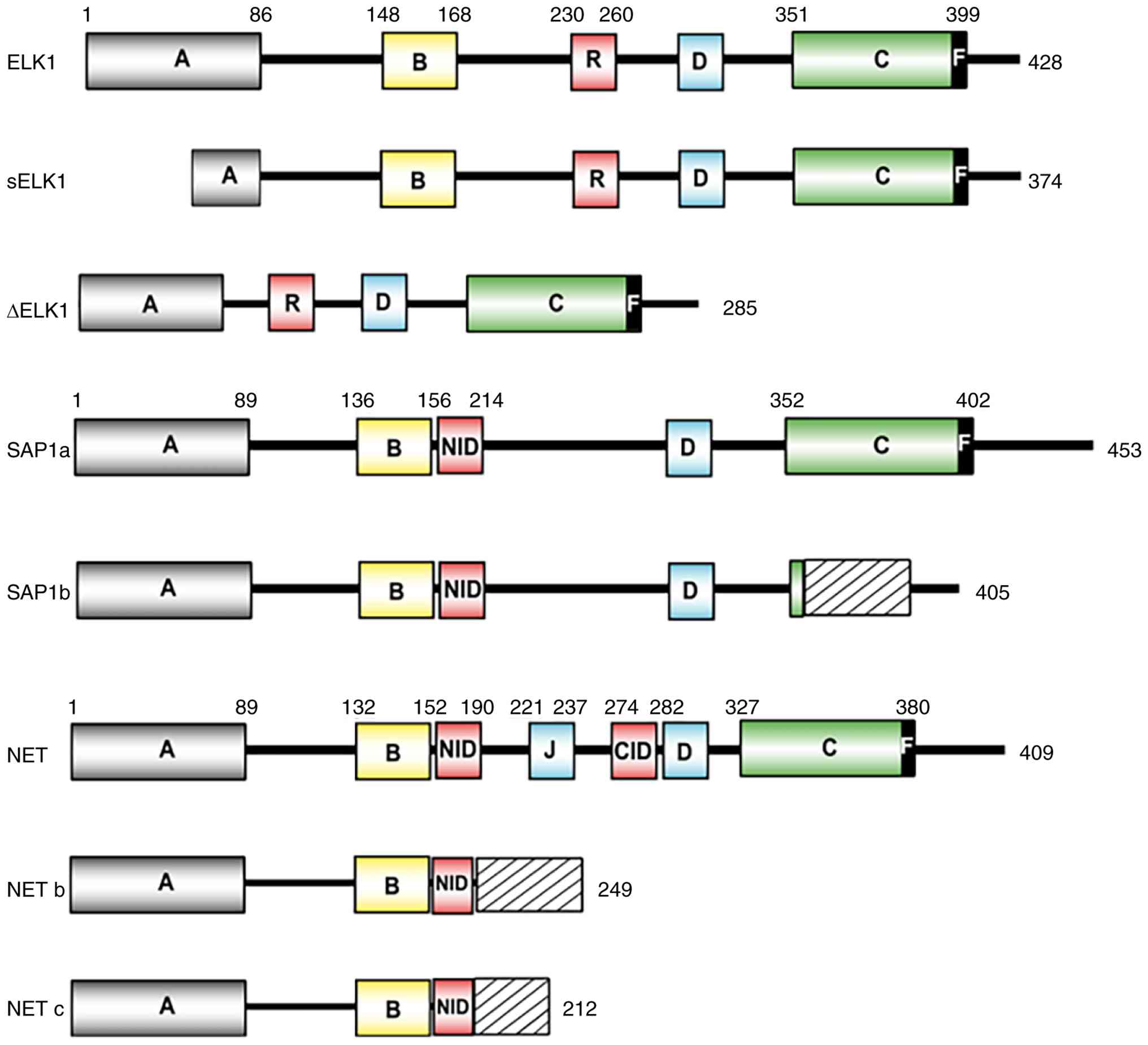
the TCF subfamily shares four similar A-D domains (Fig. 1). The N-terminal A domain
corresponds to the ETS DNA-binding domain, which enables this
subfamily to function as a transcriptional repressor by recruiting
transcriptional repressors and DNA-binding inhibitors. The B domain
can form a ternary nuclear protein complex with SRF, a member of
the MADS-box transcription factor family, on the serum response
element (SRE) of c-fos (3,18) (3,21). The
C-terminal C domain contains multiple S/TP MAPK phosphorylation
sites, enabling activation through MAPK phosphorylation. The D
domain serves as the MAPK and ELK3 nuclear localization signal
linker site. Additionally, TCF subfamily proteins possess an extra
MAPK linker site, the F domain, which exhibits distinct binding
characteristics from the D domain. Notably, beyond these shared
domains, each gene contains unique regions conferring distinct
biological functions. Specifically, the ELK1 protein contains an
R-motif between domains B and D that inhibits domain C activity.
The ELK3 protein possesses two inhibitory domains: The NET
inhibitory domain (NID) and the C-terminal binding protein
inhibitory domain (CID). The ELK4 protein (SAP-1a) also harbors an
NID domain between domains B and D (22).
Current research suggests that ELK3 functions as a
transcriptional repressor. Potential mechanisms include: NID
interacting with Ubc9 and PIAS1, leading to increased acetylation
at lysine 162; and CID interacting with the C-terminal binding
protein of the E1A corepressor, which inhibits transcription via
histone deacetylase activity (23–25).
Notably, reports have indicated that growth factors and Ras genes
can induce MAPK signaling, leading to phosphorylation and
activation of key residues in the C-domain of ELK3 by ERK and p38,
thereby triggering transcriptional activation (24–26).
Alternative splicing is a crucial post-transcriptional and
co-transcriptional mechanism for gene expression regulation,
enabling the production of multiple mature mRNA transcripts from a
single gene to generate substantial transcript and proteome
diversity (27). Three additional
ELK3 protein isoforms, ELK3b, ELK3c and ELK3d, have been
identified. ELK3b and ELK3c possess only the N-terminal A domain,
B-box and NID, lacking the C domain, D domain and CID (28,29).
The ELK3d isoform possesses a truncated ETS DNA-binding domain
(lacking the predicted β3 and β4 strands) while retaining most of
the transcription activation domain. It lacks the transcription
repression domains NID and CID, but can activate transcription via
ETS binding sites (28). ELK3d also
retains four of the five C-terminal Ser/Thr-Pro motifs (Thr 337,
Ser 359, Ser 365 and Ser 398) and the F domain from ELK3, which may
explain the transcriptional activation activity of ELK3d (26).
Alternative splicing events drive the translation of
ELK3 into functionally distinct protein isoforms, which may exhibit
markedly divergent biological activities and modes of action. To
the best of our knowledge, no studies to date have elucidated the
physiological and pathological functions of individual ELK3
isoforms. Direct experimental evidence in this research field
remains absent, indicating an urgent need for systematic in
vitro and in vivo investigations to identify their
biological roles.
ELK3 expression and prognostic impact in
tumors
Reports have indicated that ELK3 not only exhibits
abnormal expression in multiple tumors but that it is also
associated with tumor prognosis. The present review examined ELK3
expression across 34 tumor types using the Sangerbox online
analysis platform [Data source: The Cancer Genome Atlas (TCGA),
Therapeutically Applicable Research to Generate Effective
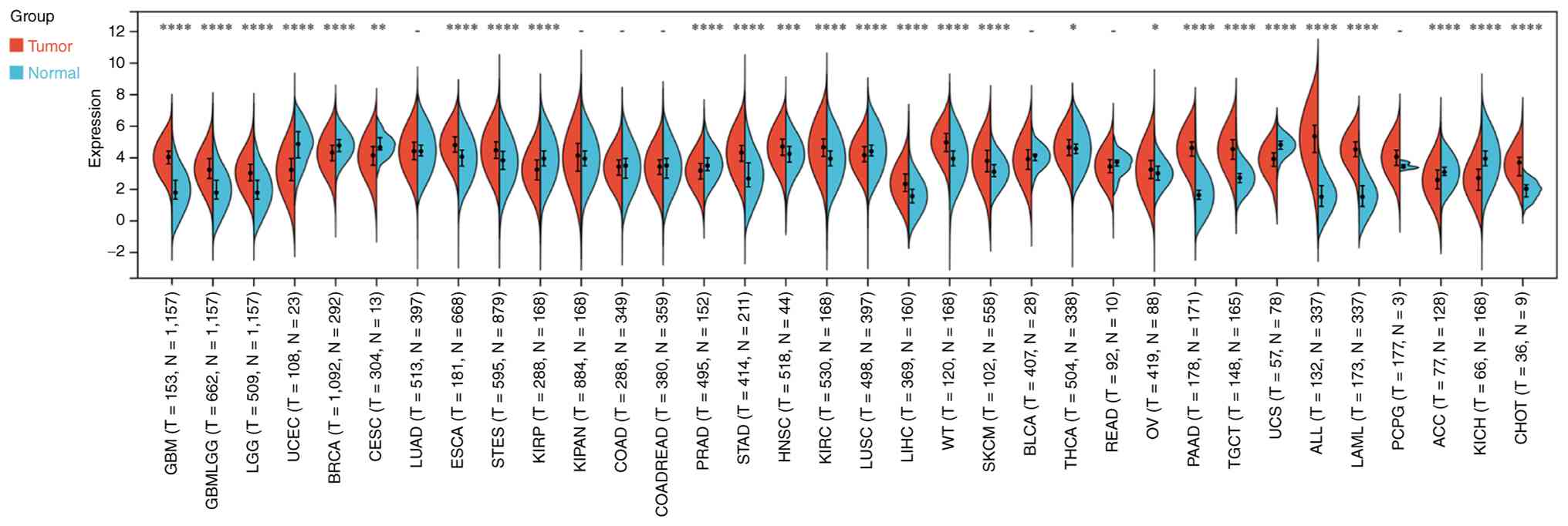
Treatments (TARGET) and GTEx]. Abnormal ELK3 expression was
observed in 27 tumor types, with high expression detected
predominantly in solid tumors, and low expression observed in
uterine corpus endometrial carcinoma, breast invasive carcinoma,
kidney renal papillary cell carcinoma, prostate adenocarcinoma,
lung squamous cell carcinoma, adrenocortical carcinoma and kidney
chromophobe (Fig. 2). Furthermore,
multiple experimental studies have suggested dysregulation of ELK3
expression in cancer. For example, Liu et al (13) reported that ELK3 expression was
markedly elevated in glioma and was increased in higher World
Health Organization grades, and that patients with high ELK3
expression exhibited a poor prognosis. Subsequent studies have
confirmed these findings (30,31).
Zhao et al (14) identified
that high ELK3 expression in 70 pancreatic cancer samples predicted
poorer overall prognosis. Cheng et al (32) similarly confirmed this association
in pancreatic cancer. An immunohistochemical study of 45
gallbladder cancer cases showed significantly higher ELK3 protein
expression in neoplastic tissue compared with in adjacent
non-neoplastic tissue, and patients with ELK3-positive expression
had a lower 3-year overall survival rate than the negative
expression group (33). In a study
on cervical cancer, ELK3 protein expression levels were revealed to
be markedly higher in tumor tissue than in adjacent non-neoplastic
tissue, and its expression was associated with poor overall
survival (34). Wang et al
(35) used bioinformatics analysis
to validate the upregulation of ELK3 as an independent prognostic
factor in ovarian cancer. In our previous studies, analysis of
multiple datasets revealed markedly higher ELK3 expression in
gastric cancer compared with in adjacent non-cancerous tissue,
predicting a poor prognosis in patients with gastric cancer
(36,37). A subsequent study by Wang et
al (38) in gastric cancer was
consistent with our prior findings. These studies collectively
indicate abnormal upregulation of ELK3 in tumors, potentially
promoting malignant progression. However, studies in pleural
mesothelioma have detected low ELK3 expression (18,39).
Moreover, contradictory conclusions exist within the same tumor
type. For example, Heo et al (12) demonstrated higher ELK3 expression in
breast cancer cell lines compared with in normal breast cell lines,
whereas He et al (40)
reported downregulation of both RNA and protein expression levels
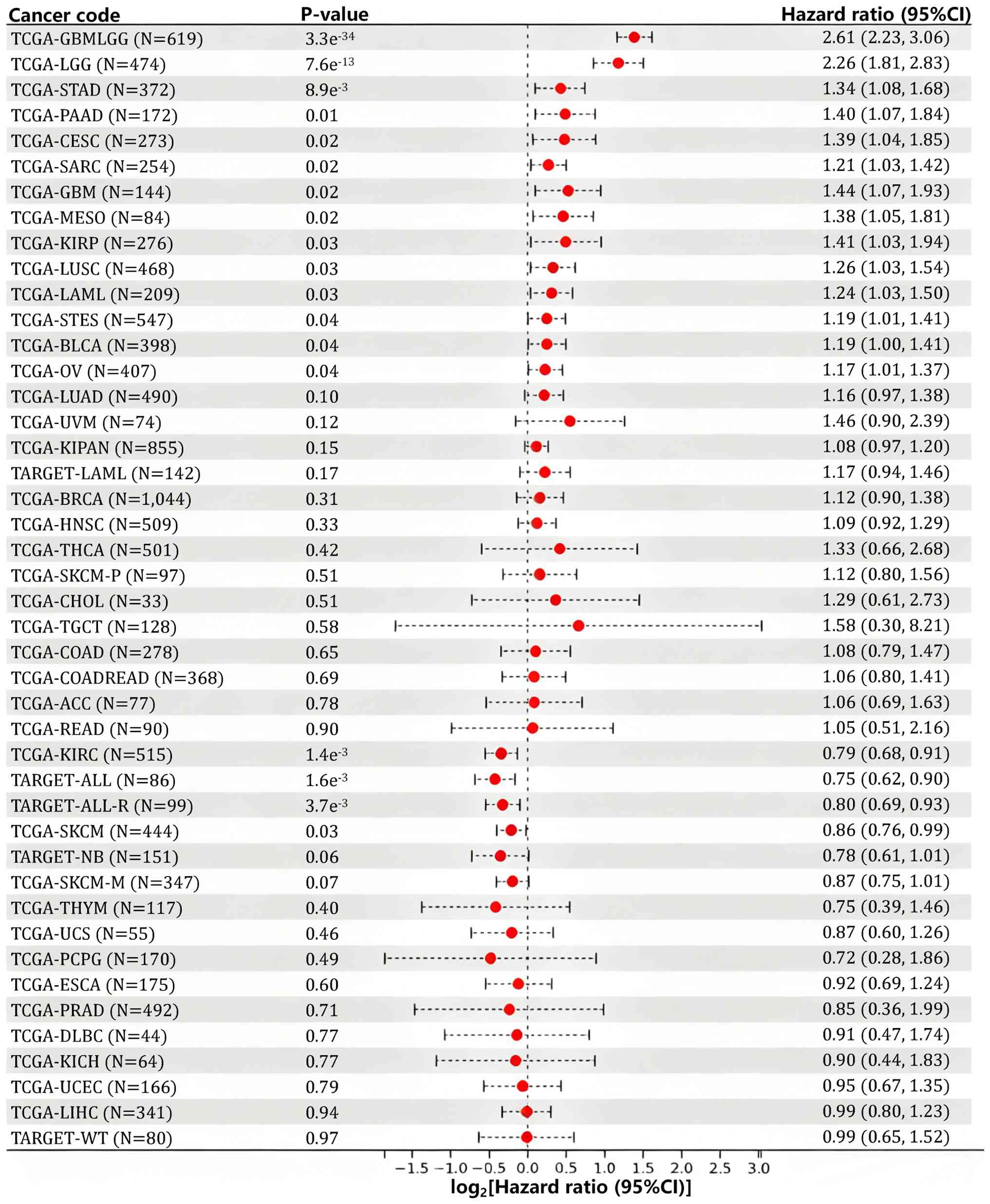
of ELK3 in primary breast cancer specimens. ELK3 prognostic data
from 44 tumors in the Sangerbox online analysis platform (Data
source: TCGA and TARGET) indicated that high ELK3 expression may be
associated with a poor prognosis in 14 tumors (glioblastoma and
lower grade glioma, brain lower grade glioma, stomach
adenocarcinoma, pancreatic adenocarcinoma, cervical squamous cell
carcinoma and endocervical adenocarcinoma, sarcoma, glioblastoma
multiforme, mesothelioma, kidney renal papillary cell carcinoma,
lung squamous cell carcinoma, acute myeloid leukemia, the combined
dataset of stomach and esophageal carcinoma, bladder urothelial
carcinoma and ovarian serous cystadenocarcinoma), whereas its high
expression predicted improved prognosis in four tumor types (kidney
renal clear cell carcinoma, acute lymphoblastic leukemia, relapsed
acute lymphoblastic leukemia and skin cutaneous melanoma) (Fig. 3).
ELK3 exhibits distinct or even opposing biological
functions across different tumor types, which may stem from the
combined effects of multiple factors: Heterogeneous isoforms
derived from alternative splicing, divergent backgrounds of tumor
driver mutations, lineage-specific transcriptomic profiles of
cells, regulation by the tumor microenvironment (TME),
dose-dependent expression effects (for example, ELK3 expression and
phosphorylation dictate the magnitude and polarity of its
regulation over angiogenesis, tumor invasion and endothelial
barrier function.) and tissue-specific post-translational
modifications.
ELK3 expression regulation
Regulation of gene and protein expression covers six
hierarchical layers: Chromatin remodeling, transcription,
post-transcriptional processing, mRNA homeostasis, translation and
post-translational modification. The expression regulation of ELK3
depends on the interaction among numerous factors, the synergistic
effects of which generate cell- and tissue-specific expression
patterns; these mechanisms are summarized in Table I.
 | Table I.Mechanisms of ELK3 expression
regulation. |
Table I.
Mechanisms of ELK3 expression
regulation.
| A, Transcription
initiation regulation |
|---|
|
|---|
| Regulatory
molecule/condition | Regulatory
mechanism | Expression
alteration | (Refs.) |
|---|
| SP1, SMAD3 | Binding to the ELK3
promoter | Upregulation | (2,14,41) |
|
| B,
Post-transcriptional regulation |
|
| Regulatory
molecule/condition | Regulatory
mechanism | Expression
alteration | (Refs.) |
|
| miR-200a,
miR-155-5p, miR-135a | Binds to ELK3 mRNA
3′UTR | Downregulation | (43,48,49) |
| Linc00662/miR-890,
Linc00525/miR-507, Linc01106/miR-3612 | Competing
endogenous RNA | Upregulation | (46,47,50) |
| METTL11A |
N6-methyladenosine | Upregulation | (32) |
|
| C, Protein
post-translational modification |
|
| Regulatory
molecule/condition | Regulatory
mechanism | Expression
alteration | (Refs.) |
|
| Ras/MAPK, RSK2 |
Phosphorylation | Upregulation | (23,53) |
| Ubc9, PIAS1,
E1A | Acetylation | Downregulation | (23–25) |
| Hypoxia | Ubiquitination | Downregulation | (51) |
Transcription initiation
regulation
Transcriptional regulation modulates gene expression
through tightly coordinated temporal and spatial interactions
between transcription machinery (including RNA polymerase II,
trans-acting factors and associated cofactors) and regulatory
elements on chromosomal DNA (1,41).
During the initiation phase of gene transcription, general
transcription factors associate with the promoter region of the
gene and bind to RNA polymerase II; however, efficient
transcription is difficult at this stage (1,2).
Transcription factors, also known as trans-acting factors, are
proteins that directly or indirectly recognize and bind to core
sequences of specific cis-acting elements. With the assistance of
gene-specific transcription factors, they can induce RNA polymerase
II to form an effective transcription initiation complex, thereby
exerting efficient transcriptional regulation (1). Thus, the variation in gene expression
leading to different types of cellular differentiation (such as
morphological features, biological functions and proliferative
capacity) is largely regulated by transcription factors. For
example, Tsoyi et al (42)
demonstrated that the transcription factor specificity protein 1
binds to the ELK3 promoter to promote its expression. Zinc finger
E-box binding homeobox 1 (ZEB1), a zinc finger transcription
factor, not only recruits co-repressors or co-activators, but also
binds to specific DNA sequences containing E-box elements to
regulate target gene expression. In studies of breast and
pancreatic cancer, ZEB1 has been shown to directly bind to ELK3
target genes to enhance their expression (14,43).
Research by Park and Park (44)
revealed that the ELK3 promoter region contains a transcription
factor SMAD3 binding site, which can directly activate
transcription and increase expression.
Post-transcriptional regulation
Post-transcriptional regulation of gene expression
serves a crucial role in various cellular processes, including cell
development, metabolism and cancer progression. Immediately after
transcription initiates the formation of mRNA precursors, these
precursors are enveloped by numerous proteins forming
ribonucleoprotein complexes. mRNA-binding proteins associate with
nascent mRNA precursors and mediate various RNA processing
reactions, ultimately yielding mature mRNA (45). N6-Methyladenine is the most common
post-transcriptional modification in eukaryotic mRNA. Methylation
modifications in the 5′UTR region of mRNA serve crucial roles in
mRNA splicing, editing, stability, degradation and polyadenylation,
while methylation in the 3′UTR region facilitates nuclear export,
translation initiation and maintains mRNA structural stability in
conjunction with polyA-binding proteins (46). Methyltransferase-like protein 11A
(METTL11A) binds to the 5′UTR region for methylation. Zhang et
al (34) demonstrated that
METTL11A can promote tumor progression by upregulating ELK3 in
cervical cancer. MicroRNAs (miRNAs/miRs) are a class of regulatory,
endogenously expressed non-coding small RNA molecules that
primarily control mRNA expression by binding to the 3′UTR (47). Studies have indicated that miR-507,
miR-200a, miR-3612, miR-155-5p, miR-890 and miR-135a suppress ELK3
expression (45,48–52).
Based on the mechanism of miRNA-mediated gene suppression, long
non-coding RNAs can exert competing endogenous RNA (ceRNA)
regulatory functions by competitively binding to shared miRNA
sequences and affecting the expression levels of their downstream
target mRNA (53). Xia et al
(52) demonstrated that
Linc00662/miR-890 regulates ELK3 expression in melanoma via a ceRNA
mechanism. Similarly, Linc00525/miR-507 and Linc01106/miR-3612
regulate ELK3 in colorectal cancer and bladder cancer,
respectively.
Post-translational modification (PTM)
of proteins
PTM of proteins refers to covalent and generally
enzyme-catalyzed modifications occurring after protein
biosynthesis. After mRNA is translated into polypeptide chains,
PTMs occur to form mature protein products; this process forms the
molecular basis for protein dynamic equilibrium and interactions,
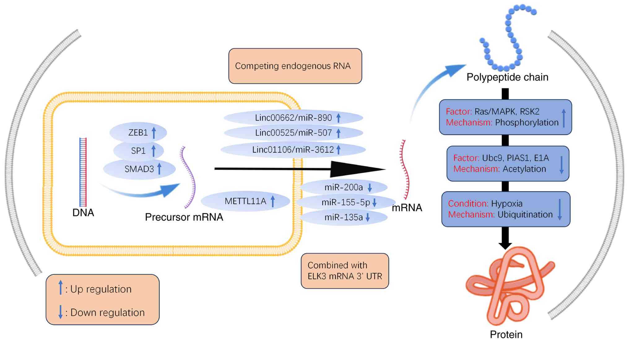
participating in nearly all biological processes (54). Among PTMs, we have previously
demonstrated that acetylation modifies ELK3 to inhibit its
transcriptional activity, whereas phosphorylation by Ras promotes
its transcriptional activity. Furthermore, Yoo et al
(55) investigated a novel
mechanism of ELK3 phosphorylation in breast cancer, revealing that
ELK3 serves as a substrate for ribosomal S6 kinase 2, which
phosphorylates ELK3 to mediate its activation. Gross et al
(56) also demonstrated that ELK3
ubiquitination levels are increased under hypoxic conditions,
leading to reduced ELK3 expression (Fig. 4).
Biological functions of ELK3 in tumors
Appropriate regulation of gene expression is crucial
for normal cellular development. Given that the aforementioned
multi-layered regulatory mechanisms exhibit pronounced
lineage-specific, tumor microenvironmental and tissue-specific
characteristics, they drive aberrant ELK3 expression, thereby
facilitating the initiation and progression of diseases such as
cancer. Notably, these divergent regulatory patterns result in
distinct ELK3 functional phenotypes across various tumor types
(Fig. 5).
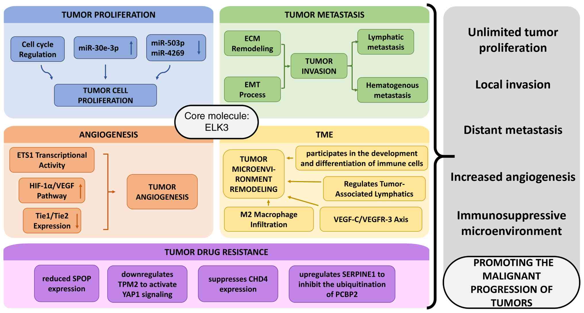 | Figure 5.Mechanisms underlying ELK3-mediated
regulation of tumor proliferation, metastasis, angiogenesis and the
tumor microenvironment. ECM, extracellular matrix; EMT,
epithelial-mesenchymal transition; HIF-1α, hypoxia-inducible
factor-1α; miR, microRNA; TPM2, tropomyosin-2; VEGF, vascular
endothelial growth factor; VEGFR, VEGF receptor; TME, tumor
microenvironment. |
ELK3 regulates tumor
proliferation
Tumor cells exhibit 10 hallmark characteristics,
among which the maintenance of chronic proliferation is the most
fundamental feature (57). Normal
cells are regulated by multiple signals that promote or inhibit
proliferation, guiding cells into normal division and growth
cycles. This ensures proper cell numbers and morphology, thereby
maintaining normal tissue structure and function. By contrast,
tumor cells can operate independently of these regulatory signals,
acquiring the ability for unlimited proliferation. Research has
indicated that ELK3 promotes cell proliferation in multiple tumor
types, including glioma, pancreatic cancer, breast cancer and
melanoma (12–15,58).
Park et al (59)
demonstrated that ELK3 can promote the tumor-associated lymphatic
secretion of exosomes. Exosomes enriched with ELK3 increase the
expression of oncogenic miR-30e-3p in tumor cells, while
downregulating the expression of tumor-suppressing miR-503-3p and
miR-4269, thereby driving breast cancer proliferation (15). ELK3 also regulates the cell cycle,
where checkpoints serve as critical control points for
proliferation. The G1-S checkpoint governs cell entry
into S phase, the S checkpoint prevents DNA-damaged cells from
synthesizing DNA, and the G2 checkpoint blocks damaged
cells from entering mitosis (60).
Mao et al (16) demonstrated
that ELK3 downregulation in prostate cancer cells increased S-phase
and G2-M phase durations by 58 and 62%, respectively,
thus promoting tumor proliferation. Activated ELK3 can also bind to
SRF and associate with the SRE of the c-fos proto-oncogene,
promoting its oncogenic activity (3,21,55). A
study on pancreatic cancer demonstrated that ELK3 overexpression
suppresses c-fos transcription via its dual inhibitory domains NID
and CID, thereby downregulating c-fos expression and restraining
the proliferation of pancreatic cancer cells (61). Whether ELK3 facilitates cell
proliferation in pancreatic cancer remains controversial; however,
most existing studies generally regard ELK3 as an oncogene that
accelerates the proliferation of solid tumors, marking it as a
promising biomarker.
ELK3 regulates tumor metastasis
Metastasis represents another critical tumor
characteristic, and a primary cause of treatment resistance and
mortality. Tumor cell dissemination primarily occurs via two
pathways: Lymphatic invasion, where cells infiltrate lymph nodes
and drain to distant organs; and hematogenous spread, where cells
circulate through the bloodstream to distant sites such as the
liver, brain, bone or lungs. Abnormal expression of genes
associated with tumor metastasis often enhances metastatic
potential. Studies have confirmed that ELK3 influences cell
metastasis in multiple tumor types (12,14,16–20,62,63).
For example, Sloan et al (17) demonstrated that ELK3 may affect
caveolin-1 transcription in lung cancer, thereby altering cancer
cell metastatic capacity. Choi et al (64) demonstrated that ELK3 regulates ID4
promoter activity, and the ELK3-ID4 axis modulates metastatic
properties in triple-negative breast cancer cells. Additionally, it
has been indicated that ELK3 promotes ovarian cancer cell migration
and invasion by targeting AEG1 (65).
Moreover, inhibiting tumor metastasis is notably
more challenging than suppressing tumor cell proliferation, as
metastasis involves a number of complex interactions between tumors
and the extracellular matrix (ECM). The ECM comprises diverse
proteins, including collagens, glycoproteins and secreted proteins,
which interact with cells and transmit extracellular signals,
thereby altering cellular phenotypes (66). ECM remodeling alters the TME,
leading to metastasis (67). In
gastric cancer, Lee et al (18) reported that ELK3 promotes cancer
cell spread by regulating the expression of ECM remodeling-related
genes, such as bone morphogenetic protein 1 and lysyl oxidase-like
2. Lee et al (18) also
demonstrated that inhibiting ELK3 can accelerate tumor cell
metastasis by modulating the expression of genes associated with
ECM remodeling, such as bone morphogenetic protein 1 and lysyl
oxidase-like 2. Furthermore, the adhesion of tumor cells to the
matrix serves a crucial role not only in cell proliferation, but
also markedly contributes to the process of tumor metastasis. In
breast cancer, ELK3 may act as a transcriptional activator for
MT1-MMP gene expression, degrading multiple ECM components
(12). Conversely, Mao et al
(16) demonstrated that decreased
ELK3 expression upregulates serpin family E member 1, which binds
to MMPs to mediate ECM degradation; this reduces prostate cancer
cell adhesion to the matrix, thereby promoting metastasis.
Further studies have indicated that ELK3 mediates
epithelial-mesenchymal transition (EMT) (68,69).
Li et al (70,71) demonstrated that interfering with
ELK3 expression inhibits EMT in hepatocellular carcinoma cells. EMT
is a critical cellular process that induces polarized epithelial
cells to adopt a mesenchymal phenotype, enhancing cell motility.
Cancer cells undergo EMT to detach from the primary tumor and
disseminate into the bloodstream, leading to metastasis. The loss
of E-cadherin, which disrupts intercellular junctions and promotes
cell separation, serves as a key biomarker for EMT (72). In breast cancer research, ELK3 has
been shown to suppress GATA3 expression through epigenetic
mechanisms, and GATA3 promotes E-cadherin transcription (19,20).
Cho et al (43) demonstrated
that ELK3 directly regulates E-cadherin, and revealed that ZEB1
binds to the C-domain of ELK3, forming a protein complex that
inhibits ELK3 phosphorylation activity, thereby suppressing
E-cadherin expression at the transcriptional level.
EMT is driven by multiple transcription factors,
which regulate the expression of classic EMT markers. These
transcription factors are initiated and controlled by pathways
including TGF-β signaling, Wnt/β-catenin signaling and
hypoxia-inducible factor (HIF)-1α (73). Studies in pancreatic and breast
cancer have demonstrated that inhibiting ELK3 affects Wnt/β-catenin
and TGF-β signaling-induced EMT in tumor cells; however, the
precise mechanisms remain unclear (14,19,20,74).
Additionally, research has confirmed that ELK3 maintains the
invasive and metastatic capabilities of liver cancer stem cells by
regulating HIF-1α (75). Research
by Gross et al (76)
indicated that ELK3 may partially participate in prolyl hydroxylase
domain (PHD) protein degradation via the E3 ligase SIAH2. PHD
proteins serve as primary regulators of HIF-1α, hydroxylating
HIF-1α based on intracellular oxygen levels and enabling its
recognition by the E3 ligase VHL for degradation (76). In summary, ELK3 facilitates the
metastasis of solid tumors by modulating ECM degradation, directly
or indirectly regulating EMT, and sustaining the stemness of tumor
cells.
ELK3 regulates angiogenesis
Angiogenesis is critical for biological processes
such as proliferation and development, with new blood vessels
emerging from existing capillaries. In adult mammals, dysregulated
angiogenesis is frequently associated with the progression of
various diseases, including tumors (77). Using in situ hybridization,
Ayadi et al (78) detected
ELK3 expression in sites of angiogenesis and vasogenesis within
mouse bladder vessels, cardiac endothelium and the dorsal aorta,
providing evidence of its involvement in vascular development.
Angiogenesis comprises three steps: Endothelial cell activation,
endothelial cell proliferation and migration, and maturation of
newly formed vessels. The transition of endothelial cells to an
angiogenic phenotype is induced by soluble factors such as vascular
endothelial growth factor (VEGF) and fibroblast growth factor
(FGF). One of the most potent activators is VEGF, an endothelial
cell-specific mitogen that serves a central role in
neovascularization (79). Molecular
studies by Mou et al (80)
revealed that ELK3 enhances VEGF-A expression and secretion by
promoting HIF-1α ubiquitination and degradation, ultimately
promoting glioma angiogenesis. In vivo and in vitro
studies by Zheng et al (81)
demonstrated that Ras can phosphorylate ELK3, leading to increased
activity in the VEGF promoter region (−80 to −53) and elevated
expression, indicating its decisive role in angiogenesis.
Conversely, Heo et al (82)
observed that VEGF may influence ELK3 expression at the protein
level, potentially through modulating MAPK signaling activity.
FGF-2 can also induce ELK3 phosphorylation via an ERK-1/2-dependent
pathway, converting ELK3 into an activator of angiogenesis
(81). An additional report has
indicated that VEGF expression exhibits no marked changes in
retinal vessels in ELK3-knockout mouse models, whereas the RNA
expression levels of Tie receptors for angiopoietin, Tie1 and Tie2,
are markedly reduced. It was subsequently revealed that Tie2
expression gradually recovers to normal levels with mouse growth,
whereas Tie1 expression remains downregulated; this may reflect the
involvement of ELK3 in the transcriptional regulation of Tie1
(83). The aforementioned studies
collectively demonstrate that ELK3 promotes angiogenesis. However,
an in vitro study has also suggested that ELK3 recruits the
co-repressor Sin3A to inhibit ETS1 activity, leading to
dysregulation of MT1-MMP transcription and reduced angiogenic
capacity in human umbilical vein endothelial cells (84). Although this contradicts the other
findings, based on the mechanism of ELK3 activation, this
discrepancy may arise from variations in specific experimental
conditions and cell types affecting ELK3 function.
ELK3 regulates the TME
The cellular environment surrounding tumor cells is
termed the TME, which is composed of cellular and non-cellular
components. These primarily include diverse types of stromal cells
(tumor-associated fibroblasts, lymphocytes, macrophages and
endothelial cells), immune cells (for example, T and B lymphocytes)
and extracellular components (including cytokines, growth factors,
hormones and ECM components). These elements envelop tumor cells
and are nourished by the surrounding tumor vasculature. Matrix
cells within the TME promote tumor growth by directly activating
growth signals in tumor cells or remodeling the surrounding area
through the release of various molecules (85). Concurrently, they enhance critical
biological processes such as tumor heterogeneity, adaptability and
evasion of immune surveillance (86–89).
Consequently, cells within the TME have emerged as targets for
effectively suppressing cancer. Tumor-associated lymphatic vessels
(TALVs), composed of lymphatic endothelial cells, constitute a
major component of the TME; due to their higher permeability
compared with blood vessels, TALVs serve a particularly crucial
role in tumor dissemination and metastasis (90). Research by Kim et al
(15) demonstrated that ELK3
promotes the viability of tumor-associated lymphatic endothelial
cells and enhances their capacity to stimulate breast cancer
proliferation. Furthermore, Wang et al (38) revealed through bioinformatics
analysis that the abnormally high expression of ELK3 in tumor cells
is closely associated with the VEGF-C/VEGF receptor (VEGFR)-3 axis.
Conversely, Park et al (59)
reported that suppressing ELK3 expression in TALVs can lead to
decreased VEGFR-3 expression. The protein encoded by VEGF-C
promotes angiogenesis and endothelial cell proliferation by binding
to and activating VEGFR-2 and VEGFR-3. Oh et al (91) explored the potential mechanism by
which ELK3 regulates the VEGF-C/VEGFR-3 axis. ELK3 was shown to
induce elevated levels of phosphorylated and acetylated NF-κB in
lymphatic endothelial cells, which may subsequently promote
upregulation of VEGFC expression. Furthermore, ELK3 expression in
gastric cancer has been reported to promote the infiltration of
tumor-associated M2 macrophages (38). Macrophages are the most abundant
infiltrating immune cells in the TME, participating in tumor cell
aerobic glycolysis, proliferation and other biological processes
that drive malignant progression. However, the mechanism by which
ELK3 regulates macrophages remains unclear.
The majority of current investigations into
ELK3-mediated regulation of the TME center primarily on the
mechanisms underlying ECM remodeling, lymphangiogenesis and
angiogenesis. However, Yang et al (92) reported that ELK3 participates in the
development and differentiation of immune cells, and a previous
study indicated that ELK3 is a key regulator of natural killer (NK)
cell therapy response in triple-negative breast cancer (93). Highly expressed in triple-negative
breast cancer, ELK3 acts as a core regulator by modulating
mitochondrial dynamics to influence NK cell cytotoxicity; knocking
down ELK3 has been shown to enhance NK cell antitumor activity
(94). Jung et al (95) further elucidated the molecular
mechanism of ELK3-based immunotherapy. Depletion of ELK3 was shown
to increase CXCL16 expression, which in turn can mediate chemotaxis
to recruit NK cells, thereby amplifying NK cell cytotoxicity. This
process ultimately enhances the antitumor immune response, further
solidifying ELK3 as a potential therapeutic target for TNBC
(95).
ELK3 regulates tumor drug
resistance
Chemotherapy resistance in tumor cells leads to
cancer recurrence and metastasis, constituting a notable factor
affecting the quality of life and clinical prognosis of patients
with cancer, and a major obstacle in cancer treatment. Current
molecular mechanisms of tumor chemotherapy resistance include
abnormal activation of ABC transporters, mitochondrial alterations,
DNA repair, autophagy, EMT, cancer stemness and exosomes (96). Wang et al (38) demonstrated that high ELK3 expression
counteracts the anticancer effects of axitinib. In a clinical
study, Lee et al (97)
revealed that elevated ELK3 levels in prostate cancer tissues leads
to decreased SPOP expression. Docetaxel induces cell death through
SPOP-mediated degradation of ELK3, whereas SPOP deletion or
mutation confers docetaxel resistance in prostate cancer cells.
This indicates that SPOP mutations may promote tumor cell
resistance by affecting ELK3 stability, suggesting a novel
therapeutic pathway for SPOP-positive prostate cancer. Furthermore,
studies have indicated that ELK3 promotes tumor stemness and
resistance to oxaliplatin (48,98).
When chemotherapy drugs stimulate tumor cells, autophagy-related
signaling pathways are activated and autophagy levels are elevated;
this enables tumor cells to degrade chemotherapeutic agents,
enhancing their resistance to chemotherapy (99). Park et al (100) elucidated the potential mechanism
by which ELK3 promotes resistance: Inhibiting ELK3 expression was
shown to disrupt autophagy mediated by the PI3K/Akt/mTOR pathway,
thereby restoring sensitivity to doxorubicin chemotherapy in breast
cancer cells. Another study revealed that ELK3 determines
chemotherapy sensitivity to cisplatin in triple-negative breast
cancer cells by regulating mitochondrial dynamics. Therefore,
inhibiting ELK3 expression holds promise as a potential therapeutic
strategy to overcome chemotherapy resistance or induce sensitivity
in triple-negative breast cancer (10). Wang et al (101) discovered that ELK3
transcriptionally upregulates SERPINE1, which inhibits the
ubiquitination of PCBP2, which restricts ferroptosis and
consequently induces gefitinib resistance in lung cancer. In
ovarian cancer, ELK3 has been verified to function as a
transcriptional repressor; it downregulates tropomyosin-2 protein
expression to activate YAP1 signaling, thereby resulting in
cisplatin resistance (102).
Furthermore, experiments by Peng et al (103) confirmed that ELK3 suppresses CHD4
expression and mediates the cisplatin-resistant phenotype.
Collectively, available evidence indicates that ELK3 drives tumor
chemoresistance through three key mechanisms: Promoting cancer
stemness, triggering autophagy and modulating mitochondrial
dynamics.
ELK3-targeting drugs
As for potent ELK3-targeted small-molecule
therapeutics, Wasylyk et al (104) identified a novel pyrazole
compound, XRP44X, through screening for small-molecule inhibitors
targeting Ras-activated ELK3 transcriptional activity in cell-based
assays. XRP44X markedly suppresses tubulin polymerization by
interacting with the colchicine-binding site. XRP44X consists of
four rings (Rings A-D), along with a carbonyl linkage between Rings
B and C. Notably, XRP44X is a multifunctional molecule that not
only interacts with tubulin but also exhibits diverse biological
activities. This compound inhibits ELK3 activation by targeting the
Ras-Erk signaling pathway upstream of Ras. Subsequent experiments
have demonstrated that XRP44X can suppress tumor growth and reduce
metastasis in three distinct mouse models (subcutaneous xenografts,
intra-cardiac injection-bone metastasis and TRAMP transgenic mouse
model of prostate cancer progression) of tumor progression and
metastasis, although minor side effects were observed (105). Kim and Park (106) discovered that XRP44X stimulates NK
cells to potentiate their cytotoxicity against breast cancer cells,
and while XRP44X exerts no influence on NK cell apoptosis or cell
cycle progression, it upregulates interferon-γ production and
activates the c-JNK signaling pathway, thereby amplifying NK
cell-mediated cytotoxicity. Chen et al (107) further demonstrated that XRP44X
triggers early activation of the JNK pathway, which induces tubulin
depolymerization and G2/M phase arrest in tumor
cells.
Conclusion
ELK3 is a key member of the ETS transcription factor
family, participating in multiple molecular regulatory pathways and
serving roles in various biological processes. The present review
provides an initial overview of the structure and function of the
ELK3 protein and its impact on biological functions, such as
tumorigenesis and progression. Due to alternative splicing of the
ELK3 gene, four structurally distinct ELK3 protein isoforms are
generated. Nevertheless, existing studies have not yet
characterized the biological functions of these isoforms. Regarding
full-length ELK3, most research confirms that it binds to ETS
consensus motifs within the promoters of target genes to regulate
transcription; however, there remains controversy over whether it
exerts transcriptional activation or repression. This dual
functional discrepancy arises as a dynamic outcome shaped by
multiple factors, including the activation status of upstream
kinases, the abundance of intracellular co-regulatory molecules,
cis-regulatory chromatin elements at target loci, the subcellular
localization of the ELK3 protein, crosstalk among multiple
signaling cascades, as well as tumor differentiation and
pathological stage (108).
Accumulating studies have verified that ELK3 is
highly expressed in tumor tissues, and its upregulation is
associated with poor clinical prognosis. These findings highlight
ELK3 as a central oncogene across multiple solid malignancies. ELK3
acts as an oncogene in tumors, promoting tumor cell proliferation
and metastasis, inducing tissue angiogenesis and TME formation, and
resulting in chemotherapy resistance. In tumor cells, ELK3 mediates
the aforementioned biological functions by acting as a molecular
bridge to modulate target gene expression and activate diverse
signaling pathways. ELK3 drives cell proliferation through
regulating cell cycle progression and c-fos transcription,
upregulating oncogenic miRNAs while downregulating
tumor-suppressive miRNAs. Accumulating reports have demonstrated
that ELK3 regulates its transcriptional activity and isoform
expression via RSK2-mediated phosphorylation, thereby affecting the
expression of genes related to extracellular matrix degradation
such as MMPs, and directly or indirectly governs EMT-related
factors and cascades including TGF-β, Wnt/β-catenin and HIF-1α
signaling to facilitate tumor metastasis (97). ELK3 indirectly and profoundly
affects angiopoietin-mediated vascular remodeling by regulating the
HIF-1α/VEGF-A signaling axis, maintaining endothelial barrier
stability, and responding to inflammatory signals (80). Studies focusing on ELK3-mediated
chemoresistance are relatively comprehensive. ELK3 confers drug
resistance via multiple mechanisms: Activating
PI3K/Akt/mTOR-dependent autophagy, maintaining cancer stem cell
properties, rewiring mitochondrial dynamics, suppressing
ferroptosis and modulating YAP1 signaling activity. To date,
research exploring the roles of ELK3 in remodeling the tumor immune
microenvironment remains limited. Available evidence has suggested
that ELK3 promotes the infiltration of M2-type macrophages and
suppresses NK cell-mediated tumor cytotoxicity by suppressing
CXCL16 expression. Collectively, the specific molecular mechanisms
by which ELK3 promotes tumorigenesis and progression remain
incompletely elucidated. However, a small number of studies suggest
that ELK3 can inhibit tumor progression, potentially due to
specific activation mechanisms that alter research conditions (such
as when the RSK2 signaling pathway is blocked) and thereby modify
ELK3 function. Furthermore, cancer cells often harbor multiple
concurrently abnormal intracellular signaling pathways. Future
research is therefore needed to explore whether ELK3 possesses
broadly targetable common mechanisms.
Given the pivotal role of ELK3 in the progression of
multiple types of cancer, ELK3 represents a promising therapeutic
target and therapeutic strategies targeting ELK3 are currently
under investigation. Specific small-molecule compounds can be
screened and candidate drugs rationally designed based on the
structural characteristics and regulatory mechanisms of ELK3.
Focusing on ELK3 regulatory networks, the present review
systematically summarizes upstream transcription factors governing
ELK3 expression, critical enzymes mediating ELK3 post-translational
modifications, ELK3-targeted miRNAs, as well as ceRNAs that
modulate ELK3 activity. Wasylyk et al (104) developed XRP44X according to the
molecular cascade whereby Ras signaling activates ELK3, and
multiple subsequent studies have validated its specific inhibitory
effect on ELK3 as well as its potent antitumor efficacy. With the
advancement of personalized precision medicine, improving the
understanding of the oncogenic molecular mechanisms of ELK3 and
developing ELK3-targeted drugs holds promise for the treatment of
malignant tumors. Research on ELK3-targeting anticancer drugs
remains limited, with XRP44X representing an early example. Future
efforts should focus on screening and optimizing more effective
small-molecule inhibitors for the ELK3 target. Additionally,
combination therapeutic strategies, integrating ELK3 inhibitors
with existing chemotherapeutic agents or immune checkpoint
inhibitors, should be considered to overcome drug resistance and
enhance therapeutic efficacy. The role and mechanisms of ELK3 in
tumor cells discussed in the present review represent only one of
numerous factors regulating tumor cell behavior. Ongoing
advancements in single-cell technologies, protein structural
studies and artificial intelligence, coupled with the initiation of
clinical trials for ELK3-targeted drugs, may propel rapid progress
in this research field.
Acknowledgements
Not applicable.
Funding
The present study was funded by the National Natural Science
Foundation of China (grant no. 82460485).
Availability of data and materials
Not applicable.
Authors' contributions
ZZL wrote the manuscript as the first author. ZW
performed acquisition, analysis and interpretation of data.. ZSL
reviewed and revised the manuscript. LQZ guided and supervised the
review, reviewed the manuscript and provided funding support. Data
authentication is not applicable. All authors have read and
approved the final manuscript.
Ethics approval and consent to
participate
Not applicable.
Patient consent for publication
Not applicable.
Competing interests
The authors declare that they have no competing
interests.
References
|
1
|
Lambert SA, Jolma A, Campitelli LF, Das
PK, Yin Y, Albu M, Chen X, Taipale J, Hughes TR and Weirauch MT:
The human transcription factors. Cell. 172:650–665. 2018.
View Article : Google Scholar : PubMed/NCBI
|
|
2
|
Ell B and Kang Y: Transcriptional control
of cancer metastasis. Trends Cell Biol. 23:603–611. 2013.
View Article : Google Scholar : PubMed/NCBI
|
|
3
|
Sizemore GM, Pitarresi JR, Balakrishnan S
and Ostrowski MC: The ETS family of oncogenic transcription factors
in solid tumours. Nat Rev Cancer. 17:337–351. 2017. View Article : Google Scholar : PubMed/NCBI
|
|
4
|
Randi AM, Sperone A, Dryden NH and Birdsey
GM: Regulation of angiogenesis by ETS transcription factors.
Biochem Soc Trans. 37((Pt 6)): 1248–1253. 2009. View Article : Google Scholar : PubMed/NCBI
|
|
5
|
Goldman D and Sapru MK: Molecular
mechanisms mediating synapse-specific gene expression during
development of the neuromuscular junction. Can J Appl Physiol.
23:390–395. 1998. View Article : Google Scholar : PubMed/NCBI
|
|
6
|
Nowling TK and Gilkeson GS: Regulation of
Fli1 gene expression and lupus. Autoimmun Rev. 5:377–382. 2006.
View Article : Google Scholar : PubMed/NCBI
|
|
7
|
Leprince D, Gegonne A, Coll J, de Taisne
C, Schneeberger A, Lagrou C and Stehelin D: A putative second
cell-derived oncogene of the avian leukaemia retrovirus E26.
Nature. 306:395–397. 1983. View Article : Google Scholar : PubMed/NCBI
|
|
8
|
Poon GMK and Kim HM: Signatures of DNA
target selectivity by ETS transcription factors. Transcription.
8:193–203. 2017. View Article : Google Scholar : PubMed/NCBI
|
|
9
|
Kodandapani R, Pio F, Ni CZ, Piccialli G,
Klemsz M, McKercher S, Maki RA and Ely KR: A new pattern for
helix-turn-helix recognition revealed by the PU.l ETS-domain-DNA
complex. Nature. 380:456–460. 1996. View Article : Google Scholar : PubMed/NCBI
|
|
10
|
Park JD, Jang HJ, Choi SH, Jo GH, Choi JH,
Hwang S, Park W and Park KS: The ELK3-DRP1 axis determines the
chemosensitivity of triple-negative breast cancer cells to CDDP by
regulating mitochondrial dynamics. Cell Death Discov. 9:2372023.
View Article : Google Scholar : PubMed/NCBI
|
|
11
|
Ghysdael J: Transcriptional regulators of
the Ets family and their implication in normal physiological
processes and carcinogenesis. Pathol Biol (Paris). 43:202–207.
1995.(In French). PubMed/NCBI
|
|
12
|
Heo SH, Lee JY, Yang KM and Park KS: ELK3
expression correlates with cell migration, invasion, and membrane
Type 1-Matrix metalloproteinase expression in MDA-MB-231 breast
cancer cells. Gene Expr. 16:197–203. 2015. View Article : Google Scholar : PubMed/NCBI
|
|
13
|
Liu Z, Ren Z, Zhang C, Qian R, Wang H,
Wang J, Zhang W, Liu B, Lian X, Wang Y, et al: ELK3: A new
molecular marker for the diagnosis and prognosis of glioma. Front
Oncol. 11:6087482021. View Article : Google Scholar : PubMed/NCBI
|
|
14
|
Zhao Q, Ren Y, Xie H, Yu L, Lu J, Jiang W,
Xiao W, Zhu Z, Wan R and Li B: ELK3 Mediated by ZEB1 facilitates
the growth and metastasis of pancreatic carcinoma by activating the
Wnt/β-catenin pathway. Front Cell Dev Biol. 9:7001922021.
View Article : Google Scholar : PubMed/NCBI
|
|
15
|
Kim KS, Park JI, Oh N, Cho HJ, Park JH and
Park KS: ELK3 expressed in lymphatic endothelial cells promotes
breast cancer progression and metastasis through exosomal miRNAs.
Sci Rep. 9:84182019. View Article : Google Scholar : PubMed/NCBI
|
|
16
|
Mao Y, Li W, Hua B, Gu X, Pan W, Chen Q,
Xu B, Wang Z and Lu C: Silencing of ELK3 Induces S-M phase arrest
and apoptosis and upregulates SERPINE1 expression reducing
migration in prostate cancer cells. Biomed Res Int.
2020:24061592020. View Article : Google Scholar : PubMed/NCBI
|
|
17
|
Sloan KA, Marquez HA, Li J, Cao Y, Hinds
A, O'Hara CJ, Kathuria S, Ramirez MI, Williams MC and Kathuria H:
Increased PEA3/E1AF and decreased Net/Elk-3, both ETS proteins,
characterize human NSCLC progression and regulate caveolin-1
transcription in Calu-1 and NCI-H23 NSCLC cell lines.
Carcinogenesis. 30:1433–1442. 2009. View Article : Google Scholar : PubMed/NCBI
|
|
18
|
Lee M, Cho HJ, Park KS and Jung HY: ELK3
controls gastric cancer cell migration and invasion by regulating
ECM remodeling-related genes. Int J Mol Sci. 23:37092022.
View Article : Google Scholar : PubMed/NCBI
|
|
19
|
Kim KS, Kim J, Oh N, Kim MY and Park KS:
ELK3-GATA3 axis modulates MDA-MB-231 metastasis by regulating
cell-cell adhesion-related genes. Biochem Biophys Res Commun.
498:509–515. 2018. View Article : Google Scholar : PubMed/NCBI
|
|
20
|
Kong SY, Kim KS, Kim J, Kim MK, Lee KH,
Lee JY, Oh N, Park JI, Park JH, Heo SH, et al: The ELK3-GATA3 axis
orchestrates invasion and metastasis of breast cancer cells in
vitro and in vivo. Oncotarget. 7:65137–65146. 2016. View Article : Google Scholar : PubMed/NCBI
|
|
21
|
Buchwalter G, Gross C and Wasylyk B: Ets
ternary complex transcription factors. Gene. 324:1–14. 2004.
View Article : Google Scholar : PubMed/NCBI
|
|
22
|
Shaw PE and Saxton J: Ternary complex
factors: Prime nuclear targets for mitogen-activated protein
kinases. Int J Biochem Cell Biol. 35:1210–1226. 2003. View Article : Google Scholar : PubMed/NCBI
|
|
23
|
Giovane A, Pintzas A, Maira SM,
Sobieszczuk P and Wasylyk B: Net, a new ets transcription factor
that is activated by Ras. Genes Dev. 8:1502–1513. 1994. View Article : Google Scholar : PubMed/NCBI
|
|
24
|
Criqui-Filipe P, Ducret C, Maira SM and
Wasylyk B: Net, a negative Ras-switchable TCF, contains a second
inhibition domain, the CID, that mediates repression through
interactions with CtBP and de-acetylation. EMBO J. 18:3392–3403.
1999. View Article : Google Scholar : PubMed/NCBI
|
|
25
|
Maira SM, Wurtz JM and Wasylyk B: Net
(ERP/SAP2) one of the Ras-inducible TCFs, has a novel inhibitory
domain with resemblance to the helix-loop-helix motif. EMBO J.
15:5849–5865. 1996. View Article : Google Scholar : PubMed/NCBI
|
|
26
|
Treisman R: Ternary complex factors:
Growth factor regulated transcriptional activators. Curr Opin Genet
Dev. 4:96–101. 1994. View Article : Google Scholar : PubMed/NCBI
|
|
27
|
Bates DO, Morris JC, Oltean S and
Donaldson LF: Pharmacology of modulators of alternative splicing.
Pharmacol Rev. 69:63–79. 2017. View Article : Google Scholar : PubMed/NCBI
|
|
28
|
Kerr N, Pintzas A, Holmes F, Hobson SA,
Pope R, Wallace M, Wasylyk C, Wasylyk B and Wynick D: The
expression of ELK transcription factors in adult DRG: Novel
isoforms, antisense transcripts and upregulation by nerve damage.
Mol Cell Neurosci. 44:165–177. 2010. View Article : Google Scholar : PubMed/NCBI
|
|
29
|
Giovane A, Sobieszczuk P, Ayadi A, Maira
SM and Wasylyk B: Net-b, a Ras-insensitive factor that forms
ternary complexes with serum response factor on the serum response
element of the fos promoter. Mol Cell Biol. 17:5667–5678. 1997.
View Article : Google Scholar : PubMed/NCBI
|
|
30
|
Jing S, Wang L, Liu W and Li C: ELK3
expression as a prognostic marker in patients with glioma. Neurol
Res. 47:550–556. 2025. View Article : Google Scholar : PubMed/NCBI
|
|
31
|
Xin Z, Guo Y, Zhou Q, Wang L, Liu W, Jiang
X and Ren C: High ELK3 expression is associated with the wild type
IDH1 in glioma and enhances infiltration of M2 macrophages. Int
Immunopharmacol. 161:1150642025. View Article : Google Scholar : PubMed/NCBI
|
|
32
|
Cheng T, Li H and Wang L: Expression and
clinical significance of miR-155-5p and ELK3 in pancreatic cancer.
Lab Med Clin. 16:2816–2820. 2019.(In Chinese).
|
|
33
|
Wang S: The expressions and clinical
significances of miR-155-5p and ELK3 in gallbladder carcinoma
tissues. Mod Oncol. 28:1707–1711. 2020.(In Chinese).
|
|
34
|
Zhang J, Song H, Chen C, Chen L, Dai Y,
Sun PH, Zou C and Wang X: Methyltransferase-like protein 11A
promotes migration of cervical cancer cells via up-regulating ELK3.
Pharmacol Res. 172:1058142021. View Article : Google Scholar : PubMed/NCBI
|
|
35
|
Wang Y, Li BX and Li X: Identification and
validation of angiogenesis-related gene expression for predicting
prognosis in patients with ovarian cancer. Front Oncol.
11:7836662022. View Article : Google Scholar : PubMed/NCBI
|
|
36
|
Zhou L, Wu Y and Xin L: Expression
characteristics and functional analysis of ELK3 in gastric cancer.
Nan Fang Yi Ke Da Xue Xue Bao. 41:1287–1295. 2021.(In Chinese).
PubMed/NCBI
|
|
37
|
Zhou L, Chen Z, Wu Y, Lu H and Xin L:
Prognostic signature composed of transcription factors accurately
predicts the prognosis of gastric cancer patients. Cancer Cell Int.
21:3572021. View Article : Google Scholar : PubMed/NCBI
|
|
38
|
Wang D, Jiao Z and Ren C: High ELK3
expression is associated with the VEGF-C/VEGFR-3 axis and gastric
tumorigenesis and enhances infiltration of M2 macrophages. Future
Med Chem. 12:2209–2224. 2020. View Article : Google Scholar : PubMed/NCBI
|
|
39
|
Mohr S, Bottin MC, Lannes B, Neuville A,
Bellocq JP, Keith G and Rihn BH: Microdissection, mRNA
amplification and microarray: A study of pleural mesothelial and
malignant mesothelioma cells. Biochimie. 86:13–19. 2004. View Article : Google Scholar : PubMed/NCBI
|
|
40
|
He J, Pan Y, Hu J, Albarracin C, Wu Y and
Dai JL: Profile of Ets gene expression in human breast carcinoma.
Cancer Biol Ther. 6:76–82. 2007. View Article : Google Scholar : PubMed/NCBI
|
|
41
|
Bradner JE, Hnisz D and Young RA:
Transcriptional addiction in cancer. Cell. 168:629–643. 2017.
View Article : Google Scholar : PubMed/NCBI
|
|
42
|
Tsoyi K, Geldart AM, Christou H, Liu X,
Chung SW and Perrella MA: Elk-3 is a KLF4-regulated gene that
modulates the phagocytosis of bacteria by macrophages. J Leukoc
Biol. 97:171–180. 2015. View Article : Google Scholar : PubMed/NCBI
|
|
43
|
Cho HJ, Oh N, Park JH, Kim KS, Kim HK, Lee
E, Hwang S, Kim SJ and Park KS: ZEB1 Collaborates with ELK3 to
Repress E-Cadherin expression in triple-negative breast cancer
cells. Mol Cancer Res. 17:2257–2266. 2019. View Article : Google Scholar : PubMed/NCBI
|
|
44
|
Park JH and Park KS: SMAD3 promotes ELK3
expression following transforming growth factor β-mediated
stimulation of MDA-MB231 cells. Oncol Lett. 19:2749–2754.
2020.PubMed/NCBI
|
|
45
|
Kim HK, Park JD, Choi SH, Shin DJ, Hwang
S, Jung HY and Park KS: Functional link between miR-200a and ELK3
regulates the metastatic nature of breast cancer. Cancers (Basel).
12:12252020. View Article : Google Scholar : PubMed/NCBI
|
|
46
|
Wang T, Kong S, Tao M and Ju S: The
potential role of RNA N6-methyladenosine in cancer progression. Mol
Cancer. 19:882020. View Article : Google Scholar : PubMed/NCBI
|
|
47
|
Shukla GC, Singh J and Barik S: MicroRNAs:
Processing, maturation, target recognition and regulatory
functions. Mol Cell Pharmacol. 3:83–92. 2011.PubMed/NCBI
|
|
48
|
Wang S, Li J and Yang X: Long Non-Coding
RNA LINC00525 promotes the stemness and chemoresistance of
colorectal cancer by targeting miR-507/ELK3 axis. Int J Stem Cells.
12:347–359. 2019. View Article : Google Scholar : PubMed/NCBI
|
|
49
|
Meng L, Xing Z, Guo Z and Liu Z: LINC01106
post-transcriptionally regulates ELK3 and HOXD8 to promote bladder
cancer progression. Cell Death Dis. 11:10632020. View Article : Google Scholar : PubMed/NCBI
|
|
50
|
Robertson ED, Wasylyk C, Ye T, Jung AC and
Wasylyk B: The oncogenic MicroRNA Hsa-miR-155-5p targets the
transcription factor ELK3 and links it to the hypoxia response.
PLoS One. 9:e1130502014. View Article : Google Scholar : PubMed/NCBI
|
|
51
|
Ahmad A, Zhang W, Wu M, Tan S and Zhu T:
Tumor-suppressive miRNA-135a inhibits breast cancer cell
proliferation by targeting ELK1 and ELK3 oncogenes. Genes Genomics.
40:243–251. 2018. View Article : Google Scholar : PubMed/NCBI
|
|
52
|
Xia XQ, Lu WL, Ye YY and Chen J: LINC00662
promotes cell proliferation, migration and invasion of melanoma by
sponging miR-890 to upregulate ELK3. Eur Rev Med Pharmacol Sci.
24:8429–8438. 2020.PubMed/NCBI
|
|
53
|
Zhao M, Feng J and Tang L: Competing
endogenous RNAs in lung cancer. Cancer Biol Med. 18:1–20. 2021.
View Article : Google Scholar : PubMed/NCBI
|
|
54
|
Osamor VC, Chinedu SN, Azuh DE, Iweala EJ
and Ogunlana OO: The interplay of post-translational modification
and gene therapy. Drug Des Devel Ther. 10:861–871. 2016. View Article : Google Scholar : PubMed/NCBI
|
|
55
|
Yoo SM, Lee CJ, An HJ, Lee JY, Lee HS,
Kang HC, Cho SJ, Kim SM, Park J, Kim DJ and Cho YY: RSK2-Mediated
ELK3 activation enhances cell transformation and breast cancer cell
growth by regulation of c-fos promoter activity. Int J Mol Sci.
20:19942019. View Article : Google Scholar : PubMed/NCBI
|
|
56
|
Gross C, Buchwalter G, Dubois-Pot H, Cler
E, Zheng H and Wasylyk B: The ternary complex factor net is
downregulated by hypoxia and regulates hypoxia-responsive genes.
Mol Cell Biol. 27:4133–4141. 2007. View Article : Google Scholar : PubMed/NCBI
|
|
57
|
Hanahan D: Hallmarks of cancer: New
dimensions. Cancer Discov. 12:31–46. 2022. View Article : Google Scholar : PubMed/NCBI
|
|
58
|
Wu D, Liu J, Yu L, Wu S and Qiu X:
Circular RNA hsa_circ_0000144 aggravates ovarian Cancer progression
by regulating ELK3 via sponging miR-610. J Ovarian Res. 15:1132022.
View Article : Google Scholar : PubMed/NCBI
|
|
59
|
Park JI, Kim KS, Kong SY and Park KS:
Novel function of E26 transformation-specific domain-containing
protein ELK3 in lymphatic endothelial cells. Oncol Lett. 15:55–60.
2018.PubMed/NCBI
|
|
60
|
Matthews HK, Bertoli C and de Bruin RAM:
Cell cycle control in cancer. Nat Rev Mol Cell Bio. 23:74–88. 2022.
View Article : Google Scholar : PubMed/NCBI
|
|
61
|
Li B, Ni P, Zhu Q, Cao H, Xu H, Zhang S,
Au C and Zhang Y: Growth inhibitory effect of the ternary complex
factor Net on human pancreatic carcinoma cell lines. Tohoku J Exp
Med. 216:139–147. 2008. View Article : Google Scholar : PubMed/NCBI
|
|
62
|
Gao ZX, Li CL, Zhang H, Zhang GH, Zhang Y,
Guo XY, Tang ZY, Gao P and Liu HT: LINC00882, transcriptionally
activated by CEBP-β and post-transcriptionally stabilized by
METTL14-mediated m(6)A modification, exerts tumorigenesis by
promoting PABPC1-mediated stabilization of ELK3 mRNA. Oncogene.
44:363–377. 2025. View Article : Google Scholar : PubMed/NCBI
|
|
63
|
Liu C, Zhou L and Chen Z: Construction of
gastric cancer prognostic signature based on the E26 transcription
factor and the identification of novel oncogene ELK3. Am J Cancer
Res. 14:1831–1849. 2024. View Article : Google Scholar : PubMed/NCBI
|
|
64
|
Choi JH, Park JD, Choi SH, Ko ES, Jang HJ
and Park KS: ELK3-ID4 axis governs the metastatic features of
triple negative breast cancer. Oncol Res. 32:127–138. 2023.
View Article : Google Scholar : PubMed/NCBI
|
|
65
|
Yu X, Du C, Cui Y, Jiang Y and Feng D:
ELK3 targeting AEG1 promotes migration and invasion of ovarian
cancer cells under hypoxia. Biol Pharm Bull. 46:883–892. 2023.
View Article : Google Scholar : PubMed/NCBI
|
|
66
|
Humphrey JD, Dufresne ER and Schwartz MA:
Mechanotransduction and extracellular matrix homeostasis. Nat Rev
Mol Cell Biol. 15:802–812. 2014. View Article : Google Scholar : PubMed/NCBI
|
|
67
|
Winkler J, Abisoye-Ogunniyan A, Metcalf KJ
and Werb Z: Concepts of extracellular matrix remodelling in tumour
progression and metastasis. Nat Commun. 11:51202020. View Article : Google Scholar : PubMed/NCBI
|
|
68
|
Li TZ, Kim SM, Hur W, Choi JE, Kim JH,
Hong SW, Lee EB, Lee JH and Yoon SK: Elk-3 contributes to the
progression of liver fibrosis by regulating the
epithelial-mesenchymal transition. Gut Liver. 11:102–111. 2017.
View Article : Google Scholar : PubMed/NCBI
|
|
69
|
Choi SH, Jang HJ, Park JD, Ryu KS, Maeng
E, Cho S, Park H, Jung HY and Park KS: Correction: ELK3-CYFIP2
axis-mediated actin remodeling modulates metastasis and natural
killer cell responses in triple-negative breast cancer. J Exp Clin
Cancer Res. 44:1232025. View Article : Google Scholar : PubMed/NCBI
|
|
70
|
Li T, Shi T, Zhou J, Jin G, Zhang J, Hao D
and Bai C: ELK-3 interference inhibits the epithelial-mesenchymal
transition of human hepatocellular carcinoma cells. Basic and
Clinical Medicine. 37:211–216. 2017.(In Chinese).
|
|
71
|
Li T, Cui F, Wang W, Bai Y, Shi T and Rui
Y: Sorafenib inhibits epithelial-mesenchymal transition of human
hepatocellular carcinoma cells by regulating ELK-3. Journal of
Chifeng University (Natural Science Edition). 33:135–137. 2017.(In
Chinese).
|
|
72
|
Lu W and Kang Y: Epithelial-Mesenchymal
plasticity in cancer progression and metastasis. Dev Cell.
49:361–374. 2019. View Article : Google Scholar : PubMed/NCBI
|
|
73
|
Bao B, Azmi AS, Ali S, Ahmad A, Li Y,
Banerjee S, Kong D and Sarkar FH: The biological kinship of hypoxia
with CSC and EMT and their relationship with deregulated expression
of miRNAs and tumor aggressiveness. Biochim Biophys Acta.
1826:272–296. 2012.PubMed/NCBI
|
|
74
|
Xu B, Chen H, Sun X, Cheng H, Chipusu K,
Hua W and Chen T: Regulation of RHBDD1 in the invasion of
esophageal cancer cells via ELK3/Wnt/β-catenin signaling pathway.
Front Bioeng Biotechnol. 13:16048592025. View Article : Google Scholar : PubMed/NCBI
|
|
75
|
Lee JH, Hur W, Hong SW, Kim JH, Kim SM,
Lee EB and Yoon SK: ELK3 promotes the migration and invasion of
liver cancer stem cells by targeting HIF-1α. Oncol Rep. 37:813–822.
2017. View Article : Google Scholar : PubMed/NCBI
|
|
76
|
Gross C, Dubois-Pot H and Wasylyk B: The
ternary complex factor Net/Elk-3 participates in the
transcriptional response to hypoxia and regulates HIF-1 alpha.
Oncogene. 27:1333–1341. 2008. View Article : Google Scholar : PubMed/NCBI
|
|
77
|
Carmeliet P and Jain RK: Angiogenesis in
cancer and other diseases. Nature. 407:249–257. 2000. View Article : Google Scholar : PubMed/NCBI
|
|
78
|
Ayadi A, Suelves M, Dollé P and Wasylyk B:
Net, an Ets ternary complex transcription factor, is expressed in
sites of vasculogenesis, angiogenesis, and chondrogenesis during
mouse development. Mech Dev. 102:205–208. 2001. View Article : Google Scholar : PubMed/NCBI
|
|
79
|
Leung DW, Cachianes G, Kuang WJ, Goeddel
DV and Ferrara N: Vascular endothelial growth factor is a secreted
angiogenic mitogen. Science. 246:1306–1309. 1989. View Article : Google Scholar : PubMed/NCBI
|
|
80
|
Yueyang M, Yaqin H, Guolian X, Wenjian Z,
Yang J, Chen L, Haiyan C, Min C, Jianping D, Penggao D, et al:
Glioma angiogenesis is boosted by ELK3 activating the HIF-1
α/VEGF-A signaling axis. BMC Cancer. 23:6622023. View Article : Google Scholar : PubMed/NCBI
|
|
81
|
Zheng H, Wasylyk C, Ayadi A, Abecassis J,
Schalken JA, Rogatsch H, Wernert N, Maira SM, Multon MC and Wasylyk
B: The transcription factor Net regulates the angiogenic switch.
Genes Dev. 17:2283–2297. 2003. View Article : Google Scholar : PubMed/NCBI
|
|
82
|
Heo SH, Choi YJ, Ryoo HM and Cho JY:
Expression profiling of ETS and MMP factors in VEGF-activated
endothelial cells: role of MMP-10 in VEGF-induced angiogenesis. J
Cell Physiol. 224:734–742. 2010. View Article : Google Scholar : PubMed/NCBI
|
|
83
|
Weinl C, Wasylyk C, Garcia Garrido M,
Sothilingam V, Beck SC, Riehle H, Stritt C, Roux MJ, Seeliger MW,
Wasylyk B and Nordheim A: Elk3 deficiency causes transient
impairment in post-natal retinal vascular development and formation
of tortuous arteries in adult murine retinae. PLoS One.
9:e1070482014. View Article : Google Scholar : PubMed/NCBI
|
|
84
|
Heo SH and Cho JY: ELK3 suppresses
angiogenesis by inhibiting the transcriptional activity of ETS-1 on
MT1-MMP. Int J Biol Sci. 10:438–447. 2014. View Article : Google Scholar : PubMed/NCBI
|
|
85
|
Ferrara N, Hillan KJ, Gerber HP and
Novotny W: Discovery and development of bevacizumab, an anti-VEGF
antibody for treating cancer. Nat Rev Drug Discov. 3:391–400. 2004.
View Article : Google Scholar : PubMed/NCBI
|
|
86
|
Mashukov A, Shapochka D, Seleznov O,
Kobyliak N, Falalyeyeva T, Kirkilevsky S, Yarema R and Sulaieva O:
Histological differentiation impacts the tumor immune
microenvironment in gastric carcinoma: Relation to the immune
cycle. World J Gastroenterol. 27:5259–5271. 2021. View Article : Google Scholar : PubMed/NCBI
|
|
87
|
Wang M, Zhao J, Zhang L, Wei F, Lian Y, Wu
Y, Gong Z, Zhang S, Zhou J, Cao K, et al: Role of tumor
microenvironment in tumorigenesis. J Cancer. 8:761–773. 2017.
View Article : Google Scholar : PubMed/NCBI
|
|
88
|
Choi H and Na KJ: Integrative analysis of
imaging and transcriptomic data of the immune landscape associated
with tumor metabolism in lung adenocarcinoma: Clinical and
prognostic implications. Theranostics. 8:1956–1965. 2018.
View Article : Google Scholar : PubMed/NCBI
|
|
89
|
Cai WY, Dong ZN, Fu XT, Lin LY, Wang L, Ye
GD, Luo QC and Chen YC: Identification of a tumor
microenvironment-relevant gene set-based prognostic signature and
related therapy targets in gastric cancer. Theranostics.
10:8633–8647. 2020. View Article : Google Scholar : PubMed/NCBI
|
|
90
|
Yong LC and Jones BE: A comparative study
of cultured vascular and lymphatic endothelium. Exp Pathol.
42:11–25. 1991. View Article : Google Scholar : PubMed/NCBI
|
|
91
|
Oh N, Park JI, Park JH, Kim KS, Lee DR and
Park KS: The role of ELK3 to regulate peritumoral lymphangiogenesis
and VEGF-C production in triple negative breast cancer cells.
Biochem Biophys Res Commun. 484:896–902. 2017. View Article : Google Scholar : PubMed/NCBI
|
|
92
|
Yang Y, Han X, Sun L, Shao F, Yin Y and
Zhang W: ETS transcription factors in immune cells and
immune-related diseases. Int J Mol Sci. 25:100042024. View Article : Google Scholar : PubMed/NCBI
|
|
93
|
Jung HY and Lee Y: Abstract 2692: ELK3 is
a key regulator as the response of natural killer cell therapy in
triple-negative breast cancer. Cancer Res. 84 (6_Suppl):S26922024.
View Article : Google Scholar
|
|
94
|
Park JD, Kim KS, Choi SH, Jo GH, Choi JH,
Park SW, Ko ES, Lee M, Lee DK, Jang HJ, et al: ELK3 modulates the
antitumor efficacy of natural killer cells against triple negative
breast cancer by regulating mitochondrial dynamics. J Immunother
Cancer. 10:e0048252022. View Article : Google Scholar : PubMed/NCBI
|
|
95
|
Jung HY, Lee DK, Lee M, Choi SH, Park JD,
Ko ES, Lee J, Park KS and Jung HY: ELK3-CXCL16 axis determines
natural killer cell cytotoxicity via the chemotactic activity of
CXCL16 in triple negative breast cancer. Oncoimmunology.
12:21906712023. View Article : Google Scholar : PubMed/NCBI
|
|
96
|
Wang Z, Li Y, Ahmad A, Banerjee S, Azmi
AS, Kong D and Sarkar FH: Pancreatic cancer: Understanding and
overcoming chemoresistance. Nat Rev Gastroenterol Hepatol. 8:27–33.
2011. View Article : Google Scholar : PubMed/NCBI
|
|
97
|
Lee CJ, Lee H, Kim SR, Nam SB, Lee GE,
Yang KE, Lee GJ, Chun SH, Kang HC, Lee JY, et al: ELK3
destabilization by speckle-type POZ protein suppresses prostate
cancer progression and docetaxel resistance. Cell Death Dis.
15:2742024. View Article : Google Scholar : PubMed/NCBI
|
|
98
|
Mei Y, Chen D, He S, Ye J, Luo M, Wu Q and
Huang Y: Transcription factor ELK3 promotes stemness and
oxaliplatin resistance of glioma cells by regulating RNASEH2A. Horm
Metab Res. 55:149–155. 2023. View Article : Google Scholar : PubMed/NCBI
|
|
99
|
Hou X, Jiang J, Tian Z and Wei L:
Autophagy and tumour chemotherapy. Adv Exp Med Biol. 1207:351–374.
2020. View Article : Google Scholar : PubMed/NCBI
|
|
100
|
Park JH, Kim KP, Ko JJ and Park KS:
PI3K/Akt/mTOR activation by suppression of ELK3 mediates
chemosensitivity of MDA-MB-231 cells to doxorubicin by inhibiting
autophagy. Biochem Biophys Res Commun. 477:277–282. 2016.
View Article : Google Scholar : PubMed/NCBI
|
|
101
|
Wang Y, Xu Z, He C, Qu H, Yang Y, Lu Y and
Dong X: E26 transformation-specific transcription factor 3 tips the
balance: Repressing tropomyosin 2 to fuel Yes-associated protein
1-driven cisplatin resistance in ovarian cancer. Anticancer Drugs.
36:788–796. 2025. View Article : Google Scholar : PubMed/NCBI
|
|
102
|
Zhu X, Wei J, Chen X, Zhang X, Li Z, Yang
F, Jiang S, Yang Z, Ge H and Shen X: ELK3-SERPINE1-PCBP2 axis
promotes gefitinib resistance in lung cancer by inhibiting
ferroptosis. Int Immunopharmacol. 169:1159632026. View Article : Google Scholar : PubMed/NCBI
|
|
103
|
Peng Q, Leung K, Sun Y, Zhang S, Teng Y
and Dong X: ELK3 promotes cisplatin resistance in ovarian cancer
via regulating CHD4 gene expression. Discov Oncol. May
19–2026.(Epub ahead of print). View Article : Google Scholar
|
|
104
|
Wasylyk C, Zheng H, Castell C, Debussche
L, Multon MC and Wasylyk B: Inhibition of the Ras-Net (Elk-3)
pathway by a novel pyrazole that affects microtubules. Cancer Res.
68:1275–1283. 2008. View Article : Google Scholar : PubMed/NCBI
|
|
105
|
Semenchenko K, Wasylyk C, Cheung H,
Tourrette Y, Maas P, Schalken JA, van der Pluijm G and Wasylyk B:
XRP44X, an inhibitor of ras/erk activation of the transcription
factor Elk3, inhibits tumour growth and metastasis in mice. PLoS
One. 11:e01595312016. View Article : Google Scholar : PubMed/NCBI
|
|
106
|
Kim KS and Park KS: XRP44X enhances the
cytotoxic activity of natural killer cells by activating the c-JUN
N-terminal kinase signaling pathway. Dev Reprod. 24:53–62. 2020.
View Article : Google Scholar : PubMed/NCBI
|
|
107
|
Chen J, Sun WL, Wasylyk B, Wang YP and
Zheng H: c-Jun N-terminal kinase mediates
microtubule-depolymerizing agent-induced microtubule
depolymerization and G2/M arrest in MCF-7 breast cancer cells.
Anticancer Drugs. 23:98–107. 2012. View Article : Google Scholar : PubMed/NCBI
|
|
108
|
Wasylyk C, Criqui-Filipe P and Wasylyk B:
Sumoylation of the net inhibitory domain (NID) is stimulated by
PIAS1 and has a negative effect on the transcriptional activity of
Net. Oncogene. 24:820–828. 2005. View Article : Google Scholar : PubMed/NCBI
|