Introduction
Gastric cancer is one of the most common
malignancies worldwide and remains a leading cause of
cancer-related mortality. Gastric cancer is one of the most common
malignancies worldwide, with ~1.09 million new cases and ~769,000
deaths reported globally in 2020 (1). The pathogenesis of gastric cancer is
complex and involves interactions among numerous genetic,
environmental and lifestyle factors. Established risk factors
include a high-salt diet, smoking, Helicobacter pylori
infection, alcohol consumption, poor nutrition and gastric polyps.
Current treatment strategies for gastric cancer include surgery,
chemotherapy, radiotherapy, targeted therapy and immunotherapy,
administered either alone or in combination. Despite recent
therapeutic advances (1), patients
with advanced-stage, metastatic, recurrent or treatment-resistant
gastric cancer experience poor clinical outcomes. Therefore,
identifying potential prognostic markers and therapeutic targets
remains key.
In recent years, increasing attention has been
directed toward cancer-associated fibroblasts (CAFs) in gastric
cancer. CAFs are key components of the tumor microenvironment and
serve key roles in tumor growth, dissemination, metastasis and drug
resistance (2). A number of
advances have been reported in this field. CAFs in gastric cancer
may originate from resident fibroblasts in adjacent tissues, bone
marrow-derived mesenchymal cells, adipocytes or other cell types
(3). CAFs secrete extracellular
matrix (ECM) components, growth factors, cytokines and other
soluble mediators, thereby contributing to the regulation of tumor
growth, invasion and metastasis. Second, research has revealed a
complex interaction network between CAFs and other cell types (such
as tumor and immune cells) within the gastric cancer
microenvironment (4). These
reciprocal interactions shape both the phenotype and function of
CAFs and contribute to tumor progression. Third, CAFs have
increasingly been recognized as potential therapeutic targets and
strategies aimed at inhibiting or reprogramming CAF function may
remodel the tumor microenvironment and restrict tumor growth and
dissemination (5–7).
Research regarding lipid metabolism in gastric
cancer progression has also begun to attract increasing attention
(8,9) Lipid metabolism is a central biological
process required for cell proliferation, differentiation and
survival, serving an important role in the initiation and
progression of gastric cancer. Abnormalities in lipid metabolism
may promote the proliferation of gastric cancer cells and influence
their invasive and metastatic potential (10); second, the role of lipid metabolism
in regulating the tumor microenvironment has been increasingly
recognized as lipid metabolites such as free fatty acids,
lysophosphatidic acid and prostaglandin E2 influence CAF
activation, extracellular matrix remodeling and tumor-cell
interactions with the surrounding matrix (11,12);
and third, abnormalities in lipid metabolism are associated with
resistance to chemotherapy and targeted therapy in gastric cancer
(13). Therefore, understanding the
role of lipid metabolism in gastric cancer progression may
facilitate the development of improved treatment strategies.
Given the importance of both CAF biology and lipid
metabolic remodeling in gastric cancer, a lipid-associated CAF
state may represent a clinically relevant but insufficiently
characterized stromal component (6,12).
Therefore, the present study aimed to characterize
lipo-CAF-associated genes in gastric cancer and evaluate their
association with prognosis, metabolic features and immune features
of the tumor microenvironment. Public gastric cancer datasets were
used to construct and validate a lipo-CAF-associated prognostic
model, and selected candidate genes were further examined in
fibroblast-based co-culture experiments to assess their potential
influence on gastric cancer cell behavior. The present study was
designed to clarify the clinical and functional relevance of
lipo-CAF-associated genes and to provide a basis for future studies
of stromal lipid metabolism in gastric cancer.
Methods and materials
Patient data acquisition
RNA sequencing data and corresponding clinical
information for patients with gastric cancer were obtained from The
Cancer Genome Atlas Stomach adenocarcinoma (TCGA-STAD, portal.gdc.cancer.gov/projects/TCGA-STAD) cohort.
A total of 443 patients were initially identified. Subsequently, 5
patients were excluded due to incomplete follow-up information
required for survival analysis, including missing survival time or
survival status. Therefore, 438 patients were retained for
follow-up and prognostic analyses. No newly collected patient data
were included in the present study for the first time. All
patient-level data were obtained from TCGA-GDC
(portal.gdc.cancer.gov/), GEO (https://www.ncbi.nlm.nih.gov/geo/), HPA (proteinatlas.org/), TISIDB (cis.hku.hk/TISIDB/), TIDE
(tide.dfci.harvard.edu/) and STRING (https://string-db.org/). GO (http://geneontology.org/) and KEGG (https://www.genome.jp/kegg/) databases were used for
enrichment analyses (14–19). The external validation cohort
GSE84437 (20) was downloaded from
the Gene Expression Omnibus database (https://www.ncbi.nlm.nih.gov/geo/) and included
transcriptomic and clinical information from 483 patients with
gastric cancer. The GSE84437 SuperSeries contains 483 gastric
cancer samples and was generated using the GPL6947 Illumina
HumanHT-12 V3.0 expression beadchip platform. Gene expression
matrices were annotated using the GPL6947 Illumina HumanHT-12 V3.0
expression beadchip platform annotation file.. When multiple probes
corresponded to the same gene symbol, the mean expression value was
used for downstream analysis with gastric cancer.
Lipo-CAF-associated gene co-expression
network
A total of 28 lipo-CAF-associated genes were
selected through a structured literature review. Genes were
included if previous studies reported their association with lipid
metabolism, lipid transport, fatty acid handling, adipocyte-like
stromal features, lipid-rich fibroblast phenotypes, CF-associated
stromal remodeling or cancer-associated lipid metabolic programs.
The selected genes and information regarding their supporting
studies are listed in Table I
(21–48). This literature-derived gene set was
used as an initial biologically informed signature to evaluate
lipid-associated CAF-associated features in gastric cancer. To
identify gastric cancer phenotype-associated biological processes,
differential expression analysis was performed between the
lipo-CAF-high and -low groups using the limma package (version
3.52.0) in R software version 4.2.1 (49). Genes with adjusted P-value <0.05
and |log2 fold change| >1 were considered significantly
differentially expressed. followed by Gene Ontology (50) and Kyoto Encyclopedia of Genes and
Genomes (51) enrichment analyses.
A protein-protein interaction (PPI) network of lipo-CAF-associated
genes was constructed and visualized using the STRING database
(19).
 | Table I.Lipid rich cancer associated
fibroblast-associated genes. |
Table I.
Lipid rich cancer associated
fibroblast-associated genes.
| First author,
year | Gene | Function | (Refs.) |
|---|
| Yoshida et
al, 2024 | CFD | Hydrolase, protease
and serine protease | (21) |
| Dai et al,
2008 | CA3 | Lyase | (22) |
| Luis et al,
2021 | FABP4 | Lipid transport
protein in adipocytes | (23) |
| Wang et al,
2019 | APOC3 | Lipid degradation,
lipid metabolism and transport | (24) |
| Gu et al,
2023 | APOC1 | Lipid
transport | (25) |
| Dwivedi et
al, 2022 | APOA2 | Host-virus
interaction, lipid transport | (26) |
| Wang et al,
2021 | APOC2 | Lipid degradation,
lipid metabolism, lipid transport | (27) |
| Yamanaka et
al, 2022 | ROBO4 | Developmental
protein and receptor | (28) |
| Hagemann et
al, 2022 | SERPINF2 | Protease inhibitor
and serine protease inhibitor | (29) |
| Qiu et al,
2023 | FABP5 | Lipid transport and
transport | (30) |
| Duan et al,
2016 | FASN | Fatty acid
biosynthesis, fatty acid metabolism, lipid biosynthesis and lipid
metabolism | (31) |
| Nishimoto et
al, 2018 | CBR1 | Oxidoreductase | (32) |
| Djurec et
al, 2018 | SAA3P | Major acute phase
reactant | (33) |
| Zhu et al,
2023 | CD36 | Cell adhesion,
lipid transport | (34) |
| Wiltshire et
al, 2002 | SH3BP5 | Guanine-nucleotide
releasing factor | (35) |
| Yagi et al,
2011 | ARAP3 | GTPase
activation | (36) |
| Yang et al,
2021 | ABCA8 | Lipid transport and
transport | (37) |
| Nieminen et
al, 2011 | BMPR1A | Kinase, receptor,
serine/threonine-protein kinase and transferase | (38) |
| Saetrom et
al, 2009 | BMPR1B | Kinase, receptor,
serine/threonine-protein kinase and transferase | (39) |
| Owens et al,
2012 | BMPR2 | Kinase, receptor,
serine/threonine-protein kinase and transferase | (40) |
| Deng et al,
2020 | BMP4 | Cytokine,
developmental protein and growth factor | (41) |
| Li et al,
2023 | TFCP2 | Transcription and
transcription regulation | (42) |
| Zhang et al,
2020 | WNT5B | Developmental
protein | (43) |
| Mizokami et
al, 2008 | HIF1A | Host-virus
interaction, transcription and transcription regulation | (44) |
| Lin et al,
2019 | PTGS2 | Fatty acid
biosynthesis, fatty acid metabolism, lipid biosynthesis, lipid
metabolism, prostaglandin biosynthesis and prostaglandin
metabolism | (45) |
| Wang et al,
2023 | HILPDA | Increases
intracellular lipid accumulation | (46) |
| Zhang et al,
2022 | PNPLA2 | Lipid degradation
and lipid metabolism | (47) |
| Zhang et al,
2022 | IL1R1 | Interleukin-1
receptor involved in inflammatory cytokine signaling | (48) |
Construction of a lipo-CAF gene
prognostic signature in gastric cancer
Any association between lipo-CAF gene expression and
overall survival (OS) was evaluated using univariate Cox regression
analysis. To identify a prognosis-associated signature, Least
Absolute Shrinkage and Selection Operator (LASSO) Cox regression
analysis was performed. Lipo-CAF genes were selected based on the
Akaike information criterion (52),
which was used for model selection by balancing model fit and
complexity. The risk score for each patient was calculated as the
sum of the expression value of each selected gene multiplied by its
corresponding regression coefficient. Univariate and multivariate
Cox regression analyses were performed to evaluate independent
prognostic factors, including clinical features and risk score, in
gastric cancer. Kaplan-Meier survival analysis was performed using
mRNA expression and overall survival data from TCGA-STAD and
GSE84437 gastric cancer cohorts. Patients were stratified into
high- and low-risk groups according to the median risk score cutoff
of 2.20 and survival differences were compared using the log-rank
test.
Tumor microenvironment analysis
Stromal, immune and ESTIMATE score and tumor purity
were calculated using the ESTIMATE algorithm (53). ESTIMATE is an expression-based
method that infers the levels of stromal and immune cell
infiltration in tumor tissue based on gene expression signatures.
The ESTIMATE algorithm was implemented using the estimate package
in R software, and normalized gene expression data from TCGA-STAD
cohort were used as input. The stromal score, immune score,
ESTIMATE score and tumor purity were compared between the
lipo-CAF-high and lipo-CAF-low groups.
Immune escape analysis
Tumor Immune Dysfunction and Exclusion (https://tide.dfci.harvard.edu) was used to evaluate
immune escape in patients classified into high- and low-risk groups
according to the prognostic signature. The expression levels of
human leukocyte antigen (HLA) family genes and immune checkpoint
molecules (TIGIT, PDCD1, SIGLEC15, CTLA4, CD274, PDCD1LG2, LAG3 and
HAVCR2) were compared between the high- and low-risk groups.
Spearman correlation analysis was conducted to assess the
association between immune cell infiltration and risk score.
Cell lines and culture conditions
NIH-3T3 mouse fibroblasts were obtained from the
American Type Culture Collection (cat. no. CRL-1658) and mouse
forestomach carcinoma (MFC) gastric cancer cells were obtained from
Procell Life Science & Technology Co., Ltd. (cat. no. CL-0156).
NIH-3T3 cells were cultured in high-glucose DMEM supplemented with
10% FBS and 1% penicillin-streptomycin. MFC cells were cultured in
RPMI-1640 medium supplemented with 10% FBS and 1%
penicillin-streptomycin. Cells were maintained at 37°C in a
humidified incubator containing 5% CO2. Cells were used
within 10 passages after thawing and were determined to be free of
Mycoplasma contamination. NIH-3T3 cells were used as a
technically tractable fibroblast-based model to evaluate whether
knockdown of candidate lipo-CAF-associated genes in fibroblasts
could affect gastric cancer cell behavior in co-culture. Given that
the gastric cancer cells used in the co-culture system were mouse
MFC cells, NIH-3T3 cells were selected to maintain species
compatibility.
Cell Counting Kit-8 (CCK-8) assay
3×xTransduced NIH-3T3 mouse fibroblasts were
co-cultured with MFC mouse gastric cancer cells in 96-well plates.
Briefly, NIH-3T3 cells were seeded into 96-well plates and allowed
to adhere overnight at 37°C. MFC cells were added, and co-cultured
in complete medium at 37°C. CCK-8 assays were performed at 0, 24,
48 and 72 h after co-culture. CCK-8 (Beyotime Biotechnology, cat.
no. C0038) was added to each well and incubated for 2 h at 37°C.
Absorbance was measured at 450 nm using a microplate reader.
TUNEL assay
xxTransduced NIH-3T3 mouse fibroblasts were
co-cultured with MFC mouse gastric cancer cells for 48 h at 37°C.
Cells were fixed with 4% paraformaldehyde for 30 min at room
temperature. TUNEL assay was performed using a One Step TUNEL
Apoptosis Assay kit (Beyotime Biotechnology, cat. no. C1088)
according to the manufacturer's instructions. Briefly, cells were
incubated with the TUNEL reaction mixture for 60 min at 37°C in the
dark. After washing with PBS, nuclei were counterstained with DAPI
(1 µg/ml) for 5 min at room temperature. Samples were mounted using
anti-fade fluorescence mounting medium and observed under a
fluorescence microscope. In total, ≥5 randomly selected fields of
view were captured for each sample. Fluorescence signals were
analyzed using ImageJ software version 1.53t (National Institutes
of Health). The percentage of apoptotic cells was calculated as the
number of TUNEL-positive cells divided by the total number of
DAPI-positive cells.
Human Protein Atlas (HPA) and
Tumor-Immune System Interaction Database (TISIDB) analyses
HPA (proteinatlas.org) was used to evaluate the protein
expression of candidate genes in gastric cancer tissues and normal
gastric tissues. For HPA immunohistochemistry images, the
corresponding HPA antibody accession numbers were recorded. The
antibody accession numbers used for normal and tumor tissues were
as follows: Apolipoprotein (APO)-C3 (accession no. HPA073918 for
both normal and tumor tissues); APOA2 (accession no. HPA072575 for
normal tissue and CAB025885 for tumor tissue); APOC2 (accession no.
HPA055877 for both normal and tumor tissue);
prostaglandin-endoperoxide synthase 2 (PTGS2; accession no.
HPA001335 for both normal and tumor tissues); FABP4 (accession no.
CAB024961 for normal tissue and HPA002188 for tumor tissue); CD36
(accession no. CAB025866 for both normal and tumor tissues); and
ABCA8 (accession no. HPA044914 for both normal and tumor tissues).
The TISIDB database (cis.hku.hk/TISIDB/) was used to assess the
associations between candidate gene expression and clinical
features in stomach adenocarcinoma. The TISIDB analyses were based
on the cohort from TCGA-STAD. The source cohort was recorded as
TCGA-STAD, with Genomic Data Commons project ID: TCGA-STAD
(Database Of Genotypes And Phenotypes accession no. phs000178).
Western blotting analysis
Transduced NIH-3T3 mouse fibroblasts were lysed
using RIPA lysis buffer (Thermo Fisher Scientific, Inc.; cat. no.
89900) supplemented with protease inhibitor cocktail. Total protein
concentration was determined using a bicinchoninic acid protein
assay kit. Equal amounts of protein (30 µg/lane) were loaded onto
10% SDS-PAGE gels and transferred onto PVDF membranes. The
membranes were blocked with 5% skimmed milk in Tris-buffered saline
containing 0.1% Tween-20 (TBST) for 1 h at room temperature. The
membranes were incubated with primary antibodies against FABP4
(1:1,000; cat. no. #2120S; Cell Signaling Technology, Inc.), CD36
(cat. no. #74002S; Cell Signaling Technology, Inc.), ABCA8 (all
1:1,000; cat. no. ab230896; Abcam) and GAPDH (1:5,000; cat. no.
ab8245; Abcam) overnight at 4°C. After washing three times with
TBST, the membranes were incubated with HRP-conjugated anti-rabbit
(1:5,000; cat. no. #7074S; Cell Signaling Technology, Inc.) and
anti-mouse IgG (1:5,000; cat. no. #7076S; Cell Signaling
Technology, Inc.), for 1 h at room temperature. Protein bands were
detected using Pierce ECL Western Blotting Substrate (Thermo Fisher
Scientific, Inc.; cat. no. 32106) and visualized using a
chemiluminescence imaging system. Band intensities were quantified
using ImageJ software version 1.53t (National Institutes of Health)
and normalized to GAPDH.
Lentiviral transduction and co-culture
assay
Lentiviral shRNA plasmids targeting mouse Fabp4,
Cd36 or Abca8a were constructed using the pLKO.1-puro lentiviral
vector. The pLKO.1-puro vector and psPAX2 packaging and pMD2.G
envelope plasmid were obtained from Addgene, Inc. Lentiviral
particles were generated using a second-generation lentiviral
packaging system. Briefly, 293T cells (American Type Culture
Collection; cat. no. CRL-3216) were maintained in high-glucose DMEM
supplemented with 10% FBS and 1% penicillin-streptomycin at 37°C.
For lentivirus production, 293T cells were co-transfected with 10
µg pLKO.1-shRNA plasmid, 7.5 µg psPAX2 packaging plasmid and 2.5 µg
pMD2.G envelope plasmid at a mass ratio of 4:3:1 using
Lipofectamine 3000 transfection reagent. Transfection was performed
at 37°C for 6 h, after which the medium was replaced with fresh
complete medium. Lentiviral supernatant was collected at 48 and 72
h after transfection. NIH-3T3 mouse fibroblasts were infected with
the collected lentiviral particles at a multiplicity of infection
of 10 in the presence of 8 µg/ml polybrene. After 24 h of
transduction at 37°C in a humidified incubator containing 5%
CO2, the medium was replaced with fresh complete medium.
Cells were selected with 2 µg/ml puromycin for 72 h and maintained
in medium containing 1 µg/ml puromycin. Knockdown efficiency was
assessed by western blotting 72 h after transduction, and the
sequence with the highest knockdown efficiency for each target gene
was used for subsequent functional assays. The shRNA target
sequences were as follows: shFABP4-1, 5′-CACCGAGATTTCCTTCAAA-3′;
shFABP4-2, 5′-CTGGATGGAAATTTGCATCAA-3′; shFABP4-3,
5′-TGTGTGATGCCTTTGTGGG-3′; shCD36-1, 5′-GGACCATTGGTGATGAGAAGG-3′;
shCD36-2, 5′-GGCTGTGTTTGGAGGTATTCT-3′; shCD36-3,
5′-GCTGTGTTTGGAGGTATTCTG-3′; shABCA8A-1,
5′-GCTGCTATGTTCTTCCTGAAA-3′; shABCA8A-2,
5′-GCAGATGATGCTGCTGATGAA-3′; shABCA8A-3,
5′-GCTGATGACCTTCTTCATCAA-3′; and sh-negative control (NC),
5′-TTCTCCGAACGTGTCACGT-3′. For co-culture assays, transduced
NIH-3T3 cells were seeded into culture plates and allowed to adhere
overnight at 37°C. MFC mouse gastric cancer cells were added and
co-cultured with NIH-3T3 cells in complete medium at 37°C.
Co-culture was performed for 0, 24, 48 and 72 h for CCK-8 assays
and 48 h for TUNEL assays.
Statistical analysis
Statistical analyses were performed using R software
(version 4.2.1; Posit Software, PBC) and GraphPad Prism (version
9.5.1; Dotmatics). Data are presented as the mean ± SD unless
otherwise stated. Data normality was assessed using the
Shapiro-Wilk test. For two-group comparisons, unpaired two-tailed
Student's t-tests or Wilcoxon rank-sum tests were used as
appropriate. For comparisons among >2 groups, one-way ANOVAs
followed by Tukey's multiple-comparison tests or Kruskal-Wallis
tests followed by Dunn's multiple-comparison tests were used as
appropriate. For time-course experiments, two-way ANOVA followed by
Sidak's multiple-comparison tests was used. Kaplan-Meier survival
curves were compared using the log-rank test. Cox regression
analysis was used to evaluate prognostic factors and LASSO Cox
regression was used to construct the prognostic model. Spearman
correlation analysis was used to assess correlations between
variables. All tests were two-sided and P<0.05 was considered to
indicate a statistically significant difference.
Results
Lipid-rich CAFs are upregulated in
gastric cancer
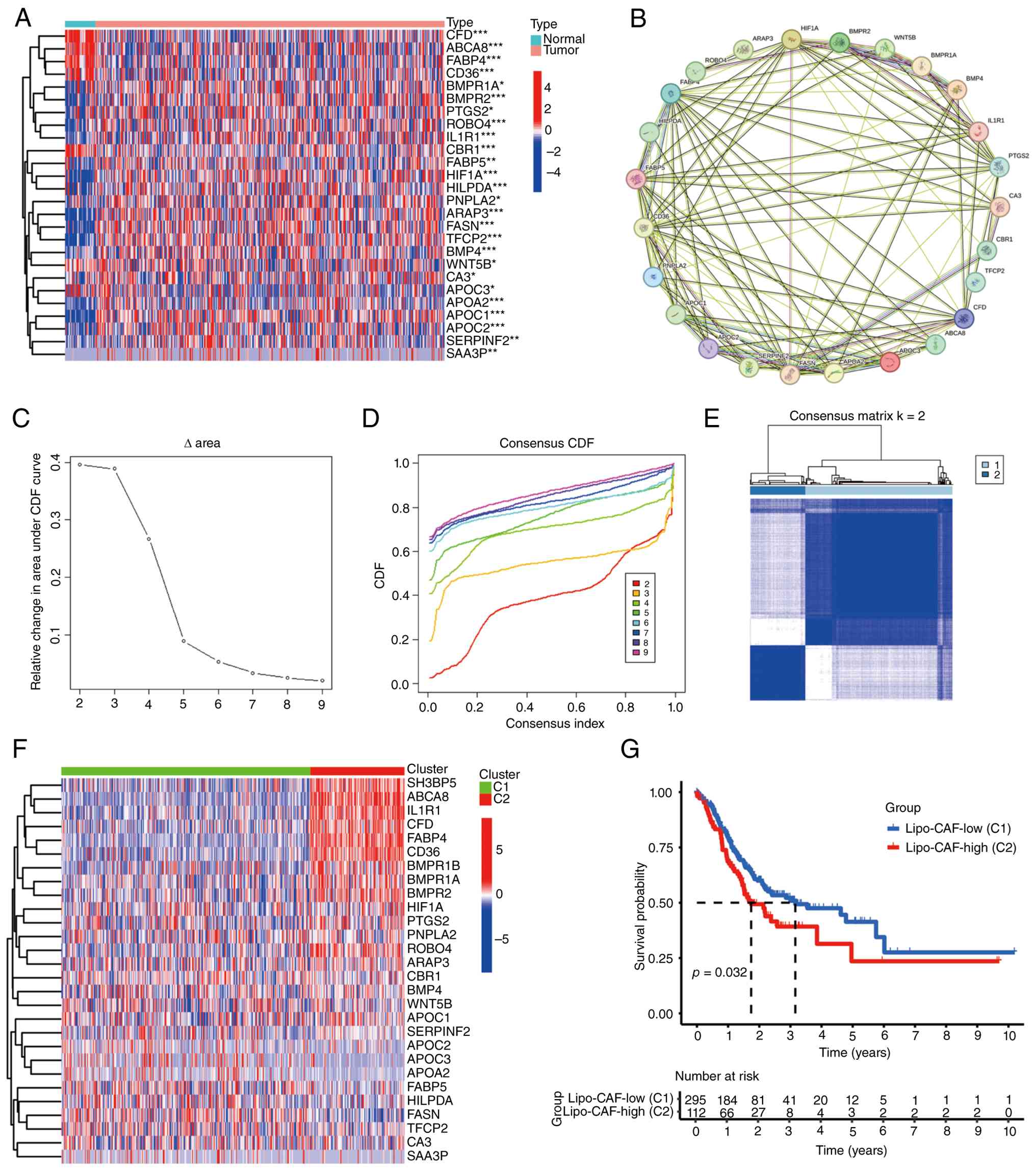
To investigate the role of lipo-CAFs in gastric
cancer progression, 28 lipo-CAF-associated genes were collected
(Table I). Differential expression
analysis revealed that the majority of lipo-CAF-associated genes
were expressed at higher levels in gastric cancer compared with in
normal tissues (Fig. 1A).
protein-protein interaction (PPI) analysis demonstrated that these
genes were closely interconnected (Fig.
1B). Based on the expression patterns of these
lipo-CAF-associated genes, patients with gastric cancer from
TCGA-STAD cohort were classified into two subtypes (Fig. 1C-E). Significant differences in
lipo-CAF gene expression were observed between the two subtypes
(Fig. 1F). In addition, patients
with the C2 subtype (lipo-CAF-high) exhibited significantly shorter
OS compared with those with the C1 subtype (Fig. 1G).
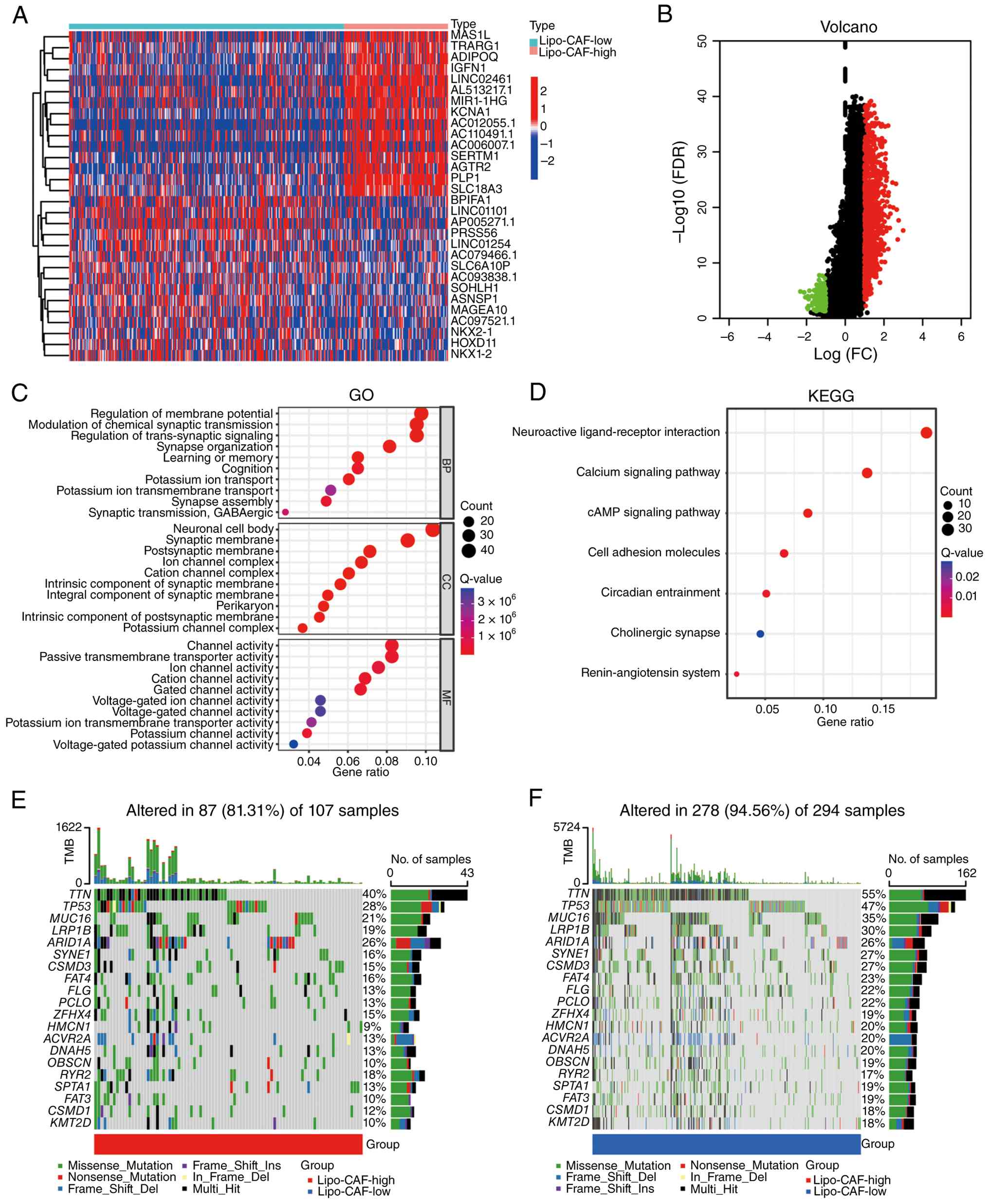
Differential gene expression and gene enrichment
analyses were performed to compare the two subtypes (Fig. 2A and B). GO enrichment analysis
showed that differentially expressed genes between the
lipo-CAF-high and -low groups were enriched in terms associated
with synaptic signaling, membrane potential regulation and
ion-channel activity, including ‘regulation of membrane potential’,
‘modulation of chemical synaptic transmission’, ‘synapse
organization’, ‘potassium ion transport’, ‘ion channel complex’ and
‘channel activity’ (Fig. 2C).
Pathway enrichment analysis further demonstrated that these genes
were enriched in energy metabolism-associated pathways, including
the ‘calcium signaling pathway’, ‘cell adhesion molecules’ and the
‘cAMP signaling pathway’ (Fig. 2D).
In addition, mutation analysis revealed that patients with gastric
cancer in the lipo-CAF-high group exhibited a lower overall
mutation rate (Fig. 2E and F).
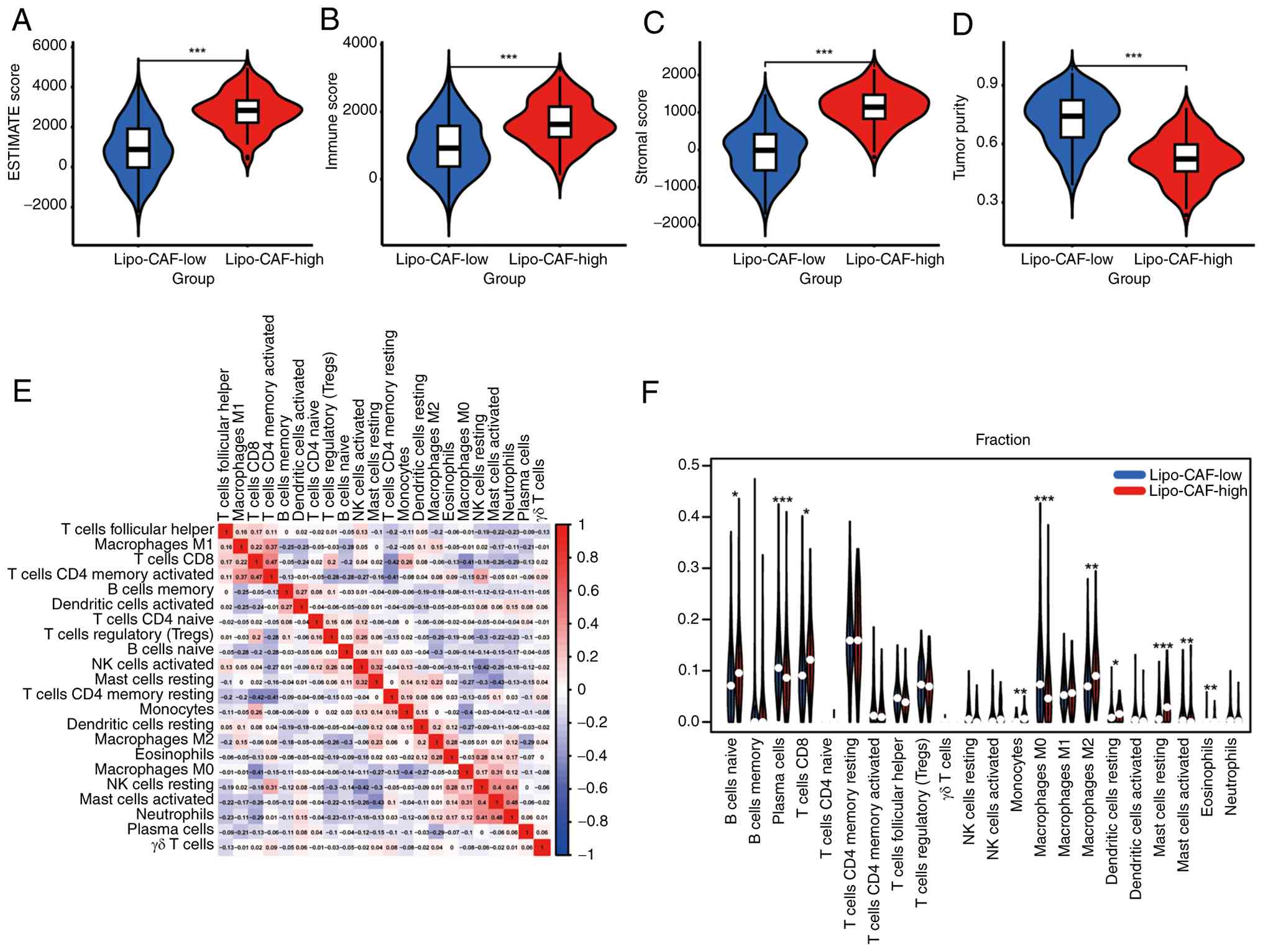
Estimation of Stromal and Immune cells in Malignant Tumours using
Expression data-based analyses showed that patients in the
lipo-CAF-high group exhibited significantly higher immune and
stromal scores, whereas tumor purity was significantly lower,
compared with the lipo-CAF-low group (Fig. 3A-D). Immune infiltration analysis
(Fig. 3E) further indicated that
the lipo-CAF-high group exhibited increased infiltration of naïve B
cells, CD8+ T cells, monocytes, M2 macrophages,
dendritic cells and mast cells (Fig.
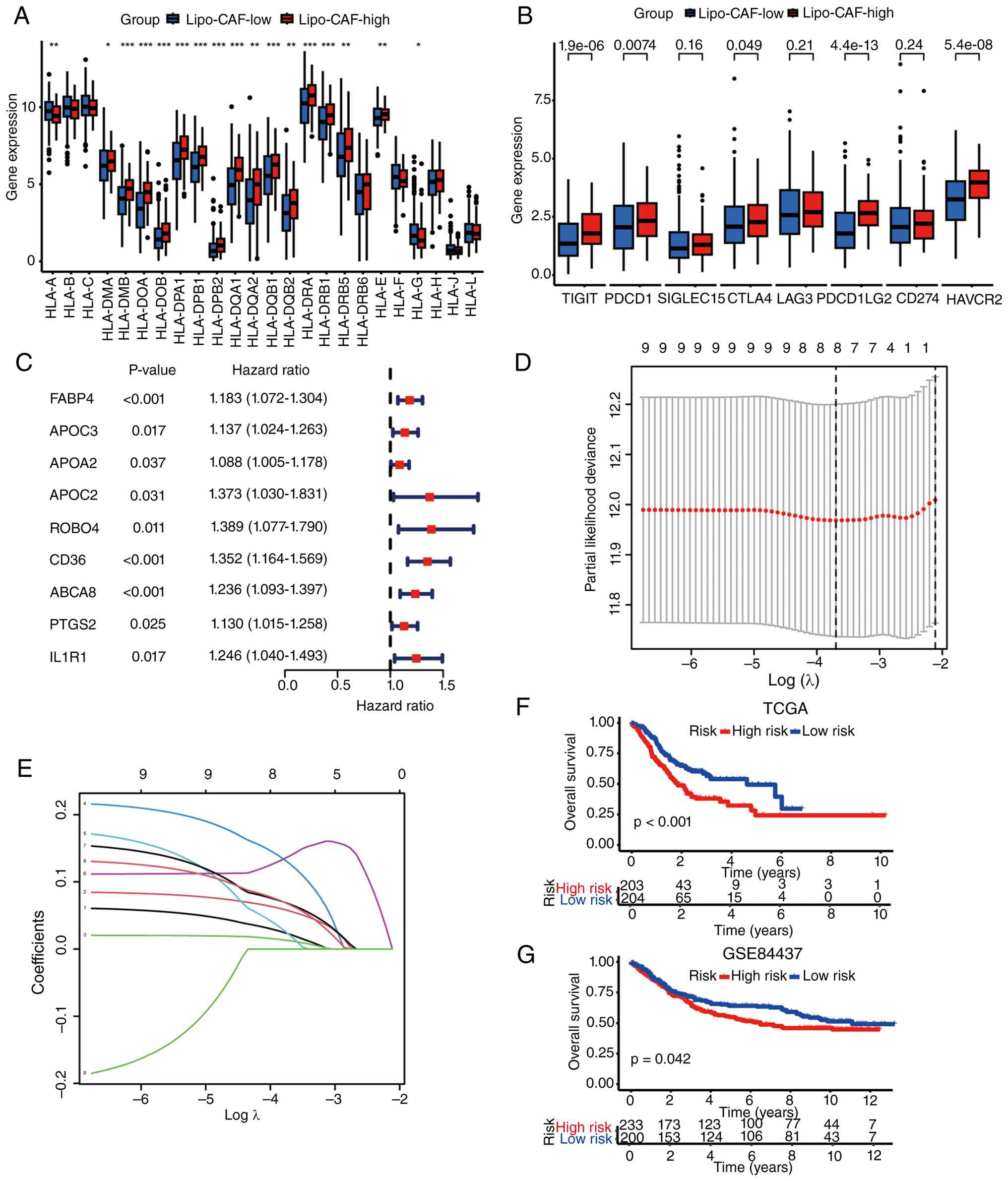
3F). Finally, the lipo-CAF-high group demonstrated higher
expression levels of HLA family genes (Fig. 4A) and immune checkpoint molecules,
including T cell immunoreceptor with Ig and ITIM domains,
programmed cell death 1 and cytotoxic T lymphocyte-associated
protein 4 (Fig. 4B).
Lipo-CAF-associated genes to predict
the prognosis of patients with gastric cancer
Based on the role of lipo-CAF-associated genes in
gastric cancer as aforementioned, a prognostic model was
constructed using these genes. Univariate Cox regression analysis
identified nine genes with prognostic value (Fig. 4C). Subsequently, a prognostic
signature was established using LASSO Cox regression analysis
(Fig. 4D and E). The model was
internally validated in the TCGA cohort and externally validated in
the GSE84437 cohort and was significantly associated with OS in
patients with gastric cancer (Fig. 4F
and G). Visualization using a heatmap demonstrated that these
genes were highly expressed in patients at high-risk and were
associated with poor survival outcomes (Fig. 5A-C). Furthermore, univariate and
multivariate Cox regression analyses showed that the prognostic
model-derived risk score was an independent prognostic factor for
patients with gastric cancer (Fig. 5D
and E).
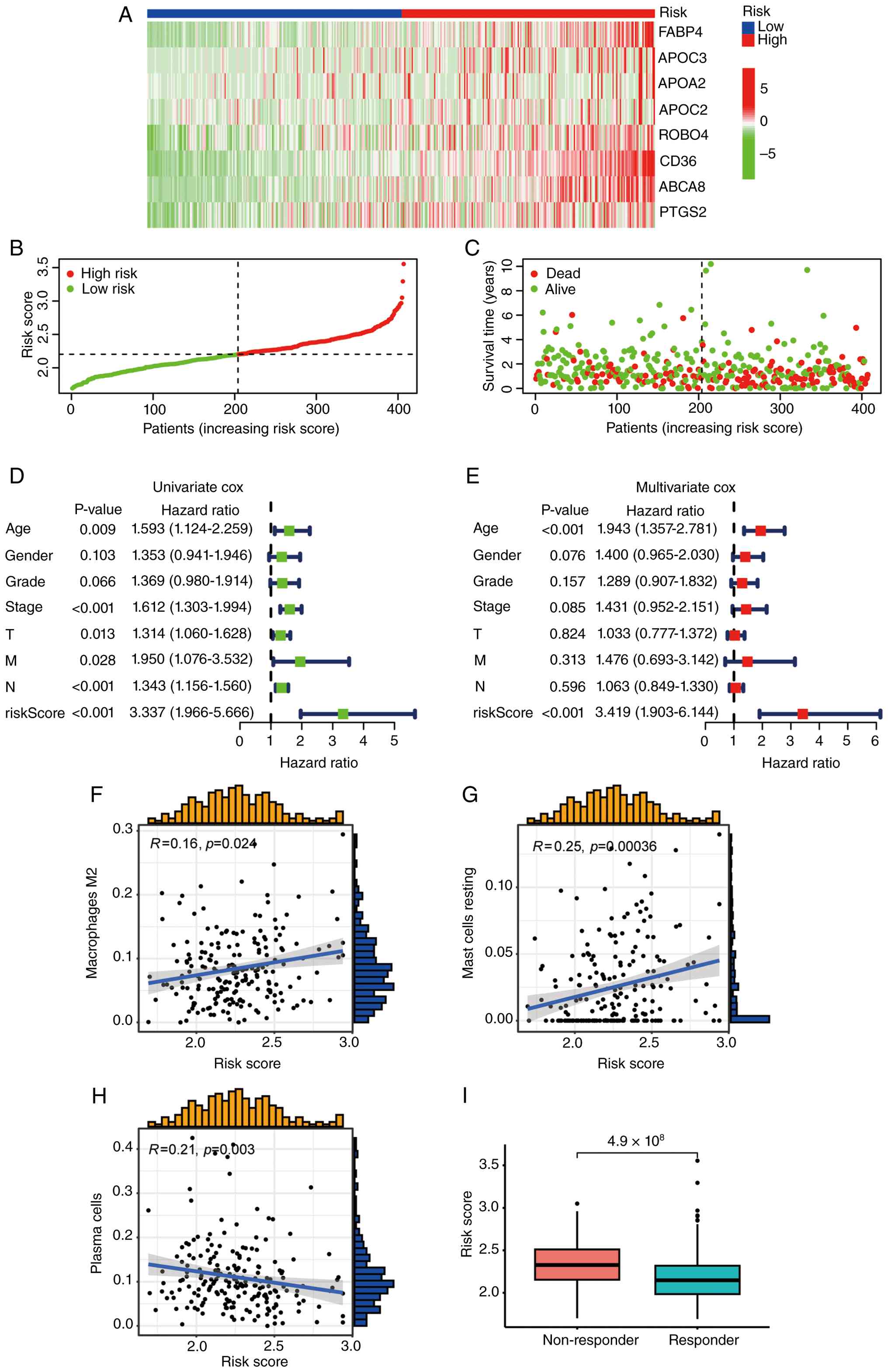 | Figure 5.Lipo-CAF risk score associated with
prognosis and immune features in gastric cancer. (A) Heatmap
showing the expression of lipo-CAF signature genes in the high-risk
and low-risk groups. (B) Survival status distribution in high-risk
and low-risk patients. (C) Risk score distribution in high-risk and
low-risk patients. (D) Univariate Cox regression analysis of age,
sex, grade, stage, T, N stage, M stage and risk score in patients
with gastric cancer. (E) Multivariate Cox regression analysis of
age, sex, grade, stage, T, N stage, M stage and risk score in
patients with gastric cancer. (F) Correlation between the lipo-CAF
risk score and M2 macrophage infiltration. (G) Correlation between
the lipo-CAF risk score and resting mast cell infiltration. (H)
Correlation between the lipo-CAF risk score and plasma cell
infiltration. (I) Association between the lipo-CAF risk score and
predicted immunotherapy response. Lipo-CAF, lipid-rich
cancer-associated fibroblast; T stage, tumor stage; N stage, lymph
node stage; M stage, metastasis stage; FABP4, fatty acid-binding
protein 4; APO, apolipoprotein; ROBO4, roundabout homolog 4; ABCA8,
ATP-binding cassette subfamily A member 8; PTGS2,
prostaglandin-endoperoxide synthase 2. |
Lipo-CAF signature is associated with
immune function
To determine whether the prognostic model was
associated with immune cell infiltration, correlation analysis was
performed between the risk score and immune cell infiltration
levels. Results showed that the risk score was positively
correlated with M2 macrophages and mast cells and negatively
correlated with plasma cells (Fig.
5F-H). Immune therapy response scoring was conducted in
TCGA-STAD patients with gastric cancer, with results suggesting
that high-risk patients, as defined by the prognostic model, were
more likely to exhibit no response to immunotherapy (Fig. 5I).
Lipo-CAF genes (FABP4, CD36 and ABCA8)
promote gastric cancer progression
To determine whether the lipo-CAF genes included in
the prognostic model were expressed in CAFs in gastric cancer,
single-cell transcriptomic datasets GSE134520 and GSE167297 were
analyzed. GSE134520 was originally generated to construct a
single-cell transcriptomic atlas of gastric premalignant and
early-malignant lesions, including non-atrophic gastritis, chronic
atrophic gastritis, intestinal metaplasia and early gastric cancer.
GSE167297 was originally generated from diffuse-type gastric cancer
samples to characterize spatially distinct tumor microenvironment
features The results showed that FABP4, CD36, ABCA8 and PTGS2 were
expressed in CAFs (Figs. S1 and
S2). Subsequently, clinical
validation analyses were performed using publicly available online
databases. FABP4, CD36 and ABCA8 were associated with poor
prognosis in gastric cancer and showed clinicopathological
associations with tumor stage and grade (Fig. S3A, F and G), whereas the remaining
genes did not show significant differences (Fig. S3B-E and H). Furthermore, protein
expression of lipo-CAF-associated genes was evaluated using the HPA
database, which showed that FABP4, CD36 and ABCA8 were highly
expressed in CAFs within gastric cancer tissue (Fig. 6A-G). To further investigate their
functional roles, these three genes were individually knocked down
in mouse fibroblasts using lentiviral vectors (Fig. 6H-J) and subsequently co-cultured
with mouse gastric cancer MFC cells (Fig. 6K). Functional assays demonstrated
that knockdown of these genes significantly reduced MFC cell
proliferation, as measured by CCK-8 assays and was accompanied by
increased TUNEL-positive staining in the co-culture system
(Fig. 6L-Q). These findings
indicate that lipo-CAF-associated genes, particularly FABP4, CD36
and ABCA8, may exert tumor-promoting effects in fibroblast-based
co-culture systems.
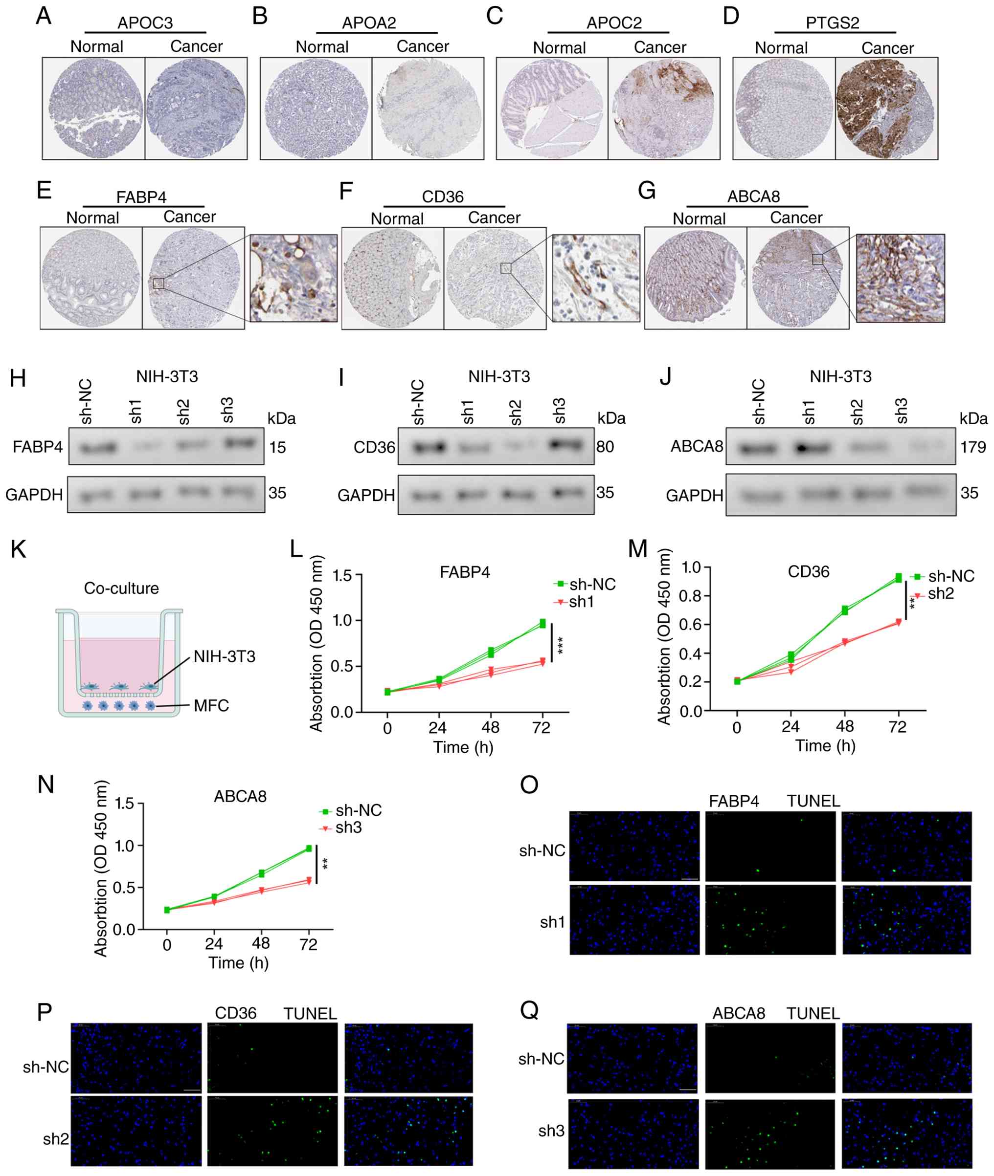 | Figure 6.FABP4, CD36 and ABCA8 promote gastric
cancer cell growth in a fibroblast-based co-culture system. (A)
Representative HPA immunohistochemistry showing APOC3 protein
expression in normal gastric tissue and gastric cancer tissue. (B)
Representative HPA immunohistochemistry image showing APOA2 protein
expression in normal gastric tissue and gastric cancer tissue. (C)
Representative HPA immunohistochemistry showing APOC2 protein
expression in normal gastric tissue and gastric cancer tissue. (D)
Representative HPA immunohistochemistry image showing PTGS2 protein
expression in normal gastric tissue and gastric cancer tissue. (E)
Representative HPA immunohistochemistry image showing FABP4 protein
expression in normal gastric tissue and gastric cancer tissue. (F)
Representative HPA immunohistochemistry image showing CD36 protein
expression in normal gastric tissue and gastric cancer tissue. (G)
Representative HPA immunohistochemistry image showing ABCA8 protein
expression in normal gastric tissue and gastric cancer tissue. (H)
Western blotting analysis showing FABP4 knockdown in NIH-3T3 cells.
(I) Western blotting analysis showing CD36 knockdown in NIH-3T3
cells. (J) Western blotting analysis showing ABCA8 knockdown in
NIH-3T3 cells. (K) Schematic diagram of the co-culture system using
NIH-3T3 mouse fibroblasts and MFC mouse gastric cancer cells.(L)
Cell Counting Kit-8 assay showing the effect of FABP4 knockdown in
NIH-3T3 cells on cell viability in the co-culture system. (M) Cell
Counting Kit-8 assay showing the effect of CD36 knockdown in
NIH-3T3 cells on cell viability in the co-culture system. (N) Cell
Counting Kit-8 assay showing the effect of ABCA8 knockdown in
NIH-3T3 cells on cell viability in the co-culture system. (O) TUNEL
assay showing the effect of FABP4 knockdown in NIH-3T3 cells on
apoptosis in the co-culture system. (P) TUNEL assay showing the
effect of CD36 knockdown in NIH-3T3 cells on apoptosis in the
co-culture system. (Q) TUNEL assay showing the effect of ABCA8
knockdown in NIH-3T3 cells on apoptosis in the co-culture system.
**P<0.01 and ***P<0.001. Scale bar, 50 µm. FABP4, fatty
acid-binding protein 4; ABCA8, ATP-binding cassette subfamily A
member 8; HPA, Human Protein Atlas; lipo-CAF, lipid-rich
cancer-associated fibroblast; sh, short hairpin; NC, negative
control; OD, optical density; MFC, mouse forestomach carcinoma;
PTGS2, prostaglandin-endoperoxide synthase 2. |
Discussion
Marked advances have been made in understanding the
role of lipid metabolism in CAFs and tumors. Fatty acids are the
primary building blocks of lipids and enter metabolic pathways such
as triglyceride synthesis, phospholipid and sphingolipid synthesis
and cholesterol ester formation to generate more complex lipid
species (54). The structural
diversity of fatty acids serves a key role in regulating the
cellular lipid pool and associated biochemical processes in normal
cells (55). Aberrant fatty acid
metabolism as a key feature of cancer, as fatty acids not only
contribute to cell membrane structure but also serve central roles
in cell signaling and energy metabolism (56), Pancreatic stellate cells (PSCs),
which represent fibroblast-like stromal cells associated with the
PDAC tumor microenvironment, undergo metabolic remodeling during
activation, including loss of vitamin A-containing lipid droplets,
altered lipid-droplet homeostasis and secretion of
lysophosphatidylcholine (LPC) species that can be converted into
lysophosphatidic acid to support pancreatic tumor progression
(57). Activated PSCs lose neutral
lipids, resulting in a marked increase in intracellular
lysophospholipid levels (58). In
addition, LPCs are abundantly secreted into the tumor
microenvironment. A proportion of these LPCs is taken up by PDAC
cells for membrane lipid synthesis, whereas the remaining LPCs are
converted into lysophosphatidic acid (LPA) by autotaxin (ATX)
secreted by PDAC cells, thereby activating LPA receptor signaling
in tumor cells (59). Similarly,
tumor-derived LPA promotes metabolic reprogramming in both cancer
cells and stromal fibroblasts. In fibroblasts, ovarian cancer
cell-derived LPA induces a glycolytic shift and CAF-like phenotype
through LPA receptors, with involvement of hypoxia-inducible factor
1α signaling, highlighting metabolic crosstalk between fibroblasts
and cancer cells (60). The
ATX/LPA/LPA receptor signaling pathway exerts notable effects on
tumor growth, cell proliferation and metabolism and has therefore
been proposed as a potential therapeutic target, particularly in
liver cancer, and as a signaling pathway involved in
cancer-associated pain (61,62).
CAFs undergo lipid metabolic reprogramming and can promote CRC cell
migration and invasion through lipid-mediated metabolic crosstalk
(63). Collectively, these findings
advance understanding of lipid metabolic mechanisms in CAFs and
tumor cells and provide insights for future research and
therapeutic development. Collectively, these observations provide a
biological rationale for interpreting lipo-CAF-tumor interactions
in gastric cancer from the perspective of metabolic symbiosis, in
which lipid-associated stromal cells may facilitate tumor growth by
modulating lipid availability, transport and utilization (13).
In the present study, it was demonstrated that
lipo-CAF-associated genes were highly expressed in gastric cancer.
Based on their expression profiles, patients with gastric cancer
were classified into two subtypes with distinct prognostic
outcomes. Further analyses revealed that the subtype associated
with poor prognosis exhibited increased tumor immune infiltration.
In addition, the lipo-CAF-based prognostic model indicated that
patients with high expression of lipo-CAF-associated genes were
more likely to exhibit resistance to immunotherapy. Lipid-mediated
cellular interactions within the tumor microenvironment may confer
survival advantages to cancer cells and promote metastasis. Fatty
acids secreted by adipocytes and other tumor-associated stromal
cells, such as CAFs, can directly enhance tumor cell proliferation,
invasion and migration, thereby exerting tumor-promoting effects.
In addition, lipid metabolic remodeling may modulate tumor
progression by influencing immune cells in the tumor
microenvironment, including natural killer and cytotoxic
CD8+ T cells, tumor-associated macrophages, dendritic
cells, regulatory T cells and myeloid-derived suppressor cells
(64). For example, lipid transfer
from lipid-laden stromal cells has been reported to impair natural
killer cell function, while aberrant lipid metabolism can also
affect CD8+ T cell effector activity, including
cytotoxic function and the production of effector cytokines such as
IFN-γ and TNF-α (10,65–67).
Lipid accumulation can also reduce dendritic cell activity, promote
infiltration of myeloid-derived suppressor cells, and enhance
regulatory T cell activity, thereby suppressing cytotoxic T cell
function (68). Furthermore, lipids
can induce tumor-promoting polarization of tumor-associated
macrophages. Finally, lipids may facilitate communication with
neural cells, such as Schwann cells, leading to increased secretion
of ECM components that promote metastasis (69). In this context, the present findings
suggest that lipo-CAF-associated genes may define a metabolically
active stromal state that is associated not only with
tumor-supportive features but also with an immunosuppressive
microenvironment. The elevated expression of immune checkpoint
molecules and the poorer predicted response to immunotherapy in the
lipo-CAF-high group further support this interpretation (9,67).
However, these observations are just associations in the present
study and should not be overinterpreted as direct mechanistic
evidence that lipo-CAFs drive immune evasion. Rather, the present
results indicate that lipo-CAF-associated programs may be
associated with gastric cancer progression partly through
stromal-tumor metabolic crosstalk and partly through their
association with an immunosuppressive niche.
In total, three lipo-CAF-associated genes (FABP4,
CD36 and ABCA8) associated with tumor-promoting effects in gastric
cancer were identified. FABP4 is a lipid-binding protein that
serves a key role in lipid metabolism and intracellular fatty acid
transport (23). It is upregulated
in a number of cancer types, including breast, prostate and
colorectal cancers (70–72). FABP4 promotes cancer cell
proliferation, migration, invasion and therapy resistance by
regulating fatty acid transport, lipid uptake and metabolic
adaptation (73–76). In addition, FABP4 is reported to
enhance tumor angiogenesis by interacting with endothelial cells
and promoting vascularization within the tumor microenvironment
(77,78). Furthermore, FABP4 may modulate
antitumor immune responses by influencing immune cell function and
facilitating immunosuppressive mechanisms in the tumor
microenvironment (79,80). These findings have important
implications for the efficacy of cancer immunotherapy and the
overall antitumor immune response. In the context of lipo-CAFs,
FABP4 may facilitate intracellular lipid trafficking and handling
in stromal cells, thereby supporting the storage, buffering or
delivery of lipid metabolites that can be utilized by adjacent
gastric cancer cells (81).
CD36 is associated with lipid metabolism and tumor
biology (34). As a lipid scavenger
receptor, CD36 serves a central role in regulating lipid metabolism
under physiological conditions by mediating lipid uptake,
metabolism and storage. However, in tumor settings, CD36 function
may be dysregulated, contributing to tumor initiation and
progression. CD36 is upregulated in a number of cancer types,
including breast and prostate cancer and melanoma. In addition,
CD36 expression has been associated with resistance to chemotherapy
and targeted therapies in certain cancer types. Previous studies
(34,82,83)
have examined the role of CD36 in angiogenesis and tumor vascular
biology, but these effects appear to be context- and
ligand-dependent. CD36 mediates thrombospondin-induced
anti-angiogenic signaling, whereas CD36-mediated lipid uptake has
been linked to tumor growth, metastasis, immune regulation and
therapy resistance (83,84). Furthermore, CD36 may contribute to
tumor immune evasion by modulating immune cell function and
attenuating antitumor immune responses. From the perspective of
metabolic symbiosis, CD36 may enhance the uptake and exchange of
extracellular lipids within the tumor microenvironment, thereby
strengthening metabolic coupling between lipo-CAFs and tumor
cells.
ABCA8 is an ATP-binding cassette transporter
involved in the transmembrane transport of cellular substrates
under physiological conditions (37). ABCA8, an ATP-binding cassette
transporter, may influence tumor-associated biological processes
(37,85) by regulating substrate transport and
lipid-associated signaling. For example, ABCA8-mediated efflux of
taurocholic acid contributes to gemcitabine insensitivity in
pancreatic cancer through the S1PR2/ERK pathway, and ABCA8-positive
lipid-metabolic CAFs have been associated with immunotherapy
resistance in triple-negative breast cancer (37,85).
In addition, ABCA8 may be involved in the remodeling of tumor lipid
metabolism, therapeutic resistance and lipid-mediated signaling
within the tumor microenvironment. By regulating lipid transport
and associated metabolic pathways, ABCA8 may influence tumor cell
survival, proliferation and metastasis. It may also modulate
intercellular signaling and treatment responsiveness within the
tumor microenvironment. Compared with FABP4 and CD36, the role of
ABCA8 in cancer remains less well characterized. This limitation
makes ABCA8 one of the more distinctive findings within the present
(85). As a transmembrane
transporter, ABCA8 may contribute to gastric cancer malignancy by
regulating the transport of lipid-associated molecules, altering
membrane composition or facilitating stromal-tumor lipid exchange
within the microenvironment. Although these mechanisms remain
speculative, the present findings suggest that ABCA8 is not merely
a passive marker but may represent a distinct component of the
lipo-CAF program that warrants further mechanistic
investigation.
A key strength of the present study was the
identification of lipo-CAF-associated genes and the successful
development of a prognostic signature in gastric cancer. Despite
this, a number of limitations should be acknowledged. Only in
vitro experiments were performed for functional validation and
no in vivo studies were conducted. In addition, further
experimental validation of the effects of lipo-CAF-associated genes
on tumor immunity was not undertaken. In addition, the in
vitro functional assays were conducted using an immortalized
mouse fibroblast cell line rather than primary gastric CAFs or
directly isolated lipo-CAFs. Given the marked heterogeneity of
fibroblasts, this model may not fully recapitulate the biological
characteristics of lipo-CAFs within the gastric tumor
microenvironment. Also, migration or invasion assays were not
performed and the effects of these genes on tumor growth and
stromal-immune interactions were not validated in vivo.
Therefore, the present findings should be interpreted as
bioinformatics-supported and in vitro functional evidence
rather than definitive proof of lipo-CAF-specific biological
activity in gastric cancer. Despite these limitations, the
lipo-CAF-associated signature may provide a useful framework for
prognostic stratification and for identifying tumors characterized
by a metabolically remodeled stromal microenvironment. From a
translational perspective, FABP4, CD36 and ABCA8 may represent
candidate stromal targets for future investigation, particularly in
studies aimed at disrupting tumor-supportive lipid crosstalk or
enhancing immunotherapy responsiveness.
In conclusion, a lipo-CAF-associated gene prognostic
model was established to predict the prognosis of gastric cancer.
Patients classified as high risk by this model exhibited poorer
survival and an altered immune infiltration profile. Notably, the
lipo-CAF-associated genes with marked prognostic value (FABP4, CD36
and ABCA8), were all associated with gastric cancer progression.
The present co-culture experiments further demonstrated that the
expression of these genes in fibroblasts was associated with
tumor-promoting effects on gastric cancer cells in vitro.
Collectively, these findings suggested that lipo-CAF-associated
genes exhibit notable potential for prognostic stratification and
therapeutic targeting in gastric cancer. However, their precise
mechanistic roles, particularly in immune regulation and
stromal-tumor metabolic coupling, remain to be elucidated in future
studies using primary CAF-based systems and in vivo
models.
Supplementary Material
Supporting Data
Acknowledgements
Not applicable.
Funding
Funding: No funding was received.
Availability of data and materials
The data generated in the present study may be
requested from the corresponding author.
Authors' contributions
LW and JL conceived the present study and wrote,
reviewed and edited the manuscript. LZ and QL analyzed and
interpreted the data. PZ performed the statistical and
computational analyses, and supervised the study. LW and JL confirm
the authenticity of all the raw data. All authors have read and
approved the final manuscript.
Ethics approval and consent to
participate
Not applicable.
Patient consent for publication
Not applicable.
Competing interests
The authors declare that they have no competing
interests.
References
|
1
|
Guan WL, He Y and Xu RH: Gastric cancer
treatment: Recent progress and future perspectives. J Hematol
Oncol. 16:572023. View Article : Google Scholar : PubMed/NCBI
|
|
2
|
Caligiuri G and Tuveson DA: Activated
fibroblasts in cancer: Perspectives and challenges. Cancer Cell.
41:434–449. 2023. View Article : Google Scholar : PubMed/NCBI
|
|
3
|
Lavie D, Ben-Shmuel A, Erez N and
Scherz-Shouval R: Cancer-associated fibroblasts in the single-cell
era. Nat Cancer. 3:793–807. 2022. View Article : Google Scholar : PubMed/NCBI
|
|
4
|
Kennel KB, Bozlar M, De Valk AF and Greten
FR: Cancer-associated fibroblasts in inflammation and antitumor
immunity. Clin Cancer Res. 29:1009–1016. 2023. View Article : Google Scholar : PubMed/NCBI
|
|
5
|
Guo D, Ji X, Xie H, Ma J, Xu C, Zhou Y,
Chen N, Wang H and Fan Cand Song H: Targeted reprogramming of
vitamin B(3) metabolism as a nanotherapeutic strategy towards
chemoresistant cancers. Adv Mater. 35:e23012572023. View Article : Google Scholar : PubMed/NCBI
|
|
6
|
Jia H, Chen X, Zhang L and Chen M: Cancer
associated fibroblasts in cancer development and therapy. J Hematol
Oncol. 18:362025. View Article : Google Scholar : PubMed/NCBI
|
|
7
|
Yamazaki M and Ishimoto T: Targeting
cancer-associated fibroblasts: Eliminate or reprogram? Cancer Sci.
116:613–621. 2025. View Article : Google Scholar : PubMed/NCBI
|
|
8
|
Li C, Zhang L, Qiu Z, Deng W and Wang W:
Key molecules of fatty acid metabolism in gastric cancer.
Biomolecules. 12:7062022. View Article : Google Scholar : PubMed/NCBI
|
|
9
|
Zhan Q, Ni H, Zhou M, Mao X, Ouyang Y, Shi
T and Li R: Recent advances in understanding the relationship
between lipid metabolism and immune escape in the tumor
microenvironment of gastric cancer. Med Rev (2021). 5:378–399.
2025. View Article : Google Scholar : PubMed/NCBI
|
|
10
|
Gong Z, Li Q, Shi J, Liu ET, Shultz LD and
Ren G: Lipid-laden lung mesenchymal cells foster breast cancer
metastasis via metabolic reprogramming of tumor cells and natural
killer cells. Cell Metabolism. 34:1960–1976.e9. 2022. View Article : Google Scholar : PubMed/NCBI
|
|
11
|
Goswami S, Zhang Q, Celik CE, Reich EM and
Yilmaz OH: Dietary fat and lipid metabolism in the tumor
microenvironment. Biochim Biophys Acta Rev Cancer. 1878:1889842023.
View Article : Google Scholar : PubMed/NCBI
|
|
12
|
Li R and Li Y: Role of metabolic
reprogramming of cancer-associated fibroblasts in tumor development
and progression (Review). Int J Oncol. 67:902025. View Article : Google Scholar : PubMed/NCBI
|
|
13
|
Wong TL, Loh JJ, Lu S, Yan HHN, Siu HC, Xi
R, Chan D, Kam MJF, Zhou L, Tong M, et al: ADAR1-mediated RNA
editing of SCD1 drives drug resistance and self-renewal in gastric
cancer. Nat Commun. 14:28612023. View Article : Google Scholar : PubMed/NCBI
|
|
14
|
Cancer Genome Atlas Research Network, .
Weinstein JN, Collisson EA, Mills GB, Shaw KR, Ozenberger BA,
Ellrott K, Shmulevich I, Sander C and Stuart JM: The cancer genome
atlas pan-cancer analysis project. Nat Genet. 45:1113–1120. 2013.
View Article : Google Scholar : PubMed/NCBI
|
|
15
|
Edgar R, Domrachev M and Lash AE: Gene
expression omnibus: NCBI gene expression and hybridization array
data repository. Nucleic Acids Res. 30:207–210. 2002. View Article : Google Scholar : PubMed/NCBI
|
|
16
|
Uhlen M, Fagerberg L, Hallstrom BM,
Lindskog C, Oksvold P, Mardinoglu A, Sivertsson Å, Kampf C,
Sjöstedt E, Asplund A, et al: Proteomics. Tissue-based map of the
human proteome. Science. 347:12604192015. View Article : Google Scholar : PubMed/NCBI
|
|
17
|
Ru B, Wong CN, Tong Y, Zhong JY, Zhong
SSW, Wu WC, Chu KC, Wong CY, Lau CY, Chen I, et al: TISIDB: an
integrated repository portal for tumor-immune system interactions.
Bioinformatics. 35:4200–4202. 2019. View Article : Google Scholar : PubMed/NCBI
|
|
18
|
Jiang P, Gu S, Pan D, Fu J, Sahu A, Hu X,
Li Z, Traugh N, Bu X, Li B, et al: Signatures of T cell dysfunction
and exclusion predict cancer immunotherapy response. Nat Med.
24:1550–1558. 2018. View Article : Google Scholar : PubMed/NCBI
|
|
19
|
Szklarczyk D, Kirsch R, Koutrouli M,
Nastou K, Mehryary F, Hachilif R, Gable AL, Fang T, Doncheva NT,
Pyysalo S, et al: The STRING database in 2023: protein-protein
association networks and functional enrichment analyses for any
sequenced genome of interest. Nucleic Acids Res. 51((D1)):
D638–D646. 2023. View Article : Google Scholar : PubMed/NCBI
|
|
20
|
Cheong JH, Yang HK, Kim H, Kim WH, Kim YW,
Kook MC, Park YK, Kim HH, Lee HS, Lee KH, et al: Predictive test
for chemotherapy response in resectable gastric cancer: A
multi-cohort, retrospective analysis. Lancet Oncol. 19:629–638.
2018. View Article : Google Scholar : PubMed/NCBI
|
|
21
|
Yoshida J, Hayashi T, Munetsuna E,
Khaledian B, Sueishi F, Mizuno M, Maeda M, Watanabe T, Ushida K,
Sugihara E, et al: Adipsin-dependent adipocyte maturation induces
cancer cell invasion in breast cancer. Sci Rep. 14:184942024.
View Article : Google Scholar : PubMed/NCBI
|
|
22
|
Dai HY, Hong CC, Liang SC, Yan MD, Lai GM,
Cheng AL and Chuang SE: Carbonic anhydrase III promotes
transformation and invasion capability in hepatoma cells through
FAK signaling pathway. Mol Carcinog. 47:956–963. 2008. View Article : Google Scholar : PubMed/NCBI
|
|
23
|
Luis G, Godfroid A, Nishiumi S, Cimino J,
Blacher S, Maquoi E, Wery C, Collignon A, Longuespée R,
Montero-Ruiz L, et al: Tumor resistance to ferroptosis driven by
Stearoyl-CoA Desaturase-1 (SCD1) in cancer cells and Fatty Acid
Biding Protein-4 (FABP4) in tumor microenvironment promote tumor
recurrence. Redox Biol. 43:1020062021. View Article : Google Scholar : PubMed/NCBI
|
|
24
|
Wang M, Wang J and Jiang H: Diagnostic
value of apolipoprotein C-I, transthyretin and apolipoprotein C-III
in gastric cancer. Oncol Lett. 17:3227–3232. 2019.PubMed/NCBI
|
|
25
|
Gu Q, Zhan T, Guan X, Lai C, Lu NA, Wang
G, Xu L, Gao X and Zhang J: Apolipoprotein C1 promotes tumor
progression in gastric cancer. Oncol Res. 31:287–297. 2023.
View Article : Google Scholar : PubMed/NCBI
|
|
26
|
Dwivedi S, Hernandez-Montes G, Montano LF
and Rendon-Huerta EP: Chromosomally unstable gastric cancers
overexpressing claudin-6 disclose cross-talk between HNF1A and
HNF4A, and upregulated cholesterol metabolism. Int J Mol Sci.
23:139772022. View Article : Google Scholar : PubMed/NCBI
|
|
27
|
Wang C, Yang Z, Xu E, Shen X, Wang X, Li
Z, Yu H, Chen K, Hu Q, Xia X, et al: Apolipoprotein C-II induces
EMT to promote gastric cancer peritoneal metastasis via
PI3K/AKT/mTOR pathway. Clin Transl Med. 11:e5222021. View Article : Google Scholar : PubMed/NCBI
|
|
28
|
Yamanaka M, Hayashi M, Sonohara F, Yamada
S, Tanaka H, Sakai A, Mii S, Kobayashi D, Kurimoto K, Tanaka N, et
al: Downregulation of ROBO4 in pancreatic cancer serves as a
biomarker of poor prognosis and indicates increased cell motility
and proliferation through activation of MMP-9. Ann Surg Oncol.
29:7180–7189. 2022. View Article : Google Scholar : PubMed/NCBI
|
|
29
|
Hagemann CA, Legart C, Mollerhoj MB,
Madsen MR, Hansen HH, Konig MJ, Helgstrand F, Hjørne FP, Toxværd A,
Langhoff JL, et al: A liver secretome gene signature-based approach
for determining circulating biomarkers of NAFLD severity. PLoS One.
17:e02759012022. View Article : Google Scholar : PubMed/NCBI
|
|
30
|
Qiu MQ, Wang HJ, Ju YF, Sun L, Liu Z, Wang
T, Kan SF, Yang Z, Cui YY, Ke YQ, et al: Fatty acid binding protein
5 (FABP5) promotes aggressiveness of gastric cancer through
modulation of tumor immunity. J Gastric Cancer. 23:340–354. 2023.
View Article : Google Scholar : PubMed/NCBI
|
|
31
|
Duan J, Sun L, Huang H, Wu Z, Wang L and
Liao W: Overexpression of fatty acid synthase predicts a poor
prognosis for human gastric cancer. Mol Med Rep. 13:3027–3035.
2016. View Article : Google Scholar : PubMed/NCBI
|
|
32
|
Nishimoto Y, Murakami A, Sato S, Kajimura
T, Nakashima K, Yakabe K, Sueoka K and Sugino N: Decreased carbonyl
reductase 1 expression promotes tumor growth via epithelial
mesenchymal transition in uterine cervical squamous cell
carcinomas. Reprod Med Biol. 17:173–181. 2018. View Article : Google Scholar : PubMed/NCBI
|
|
33
|
Djurec M, Grana O, Lee A, Troule K,
Espinet E, Cabras L, Navas C, Blasco MT, Martín-Díaz L, Burdiel M,
et al: Saa3 is a key mediator of the protumorigenic properties of
cancer-associated fibroblasts in pancreatic tumors. Proc Natl Acad
Sci USA. 115:E1147–E1156. 2018. View Article : Google Scholar : PubMed/NCBI
|
|
34
|
Zhu GQ, Tang Z, Huang R, Qu WF, Fang Y,
Yang R, Tao CY, Gao J, Wu XL, Sun HX, et al: CD36(+)
cancer-associated fibroblasts provide immunosuppressive
microenvironment for hepatocellular carcinoma via secretion of
macrophage migration inhibitory factor. Cell Discov. 9:252023.
View Article : Google Scholar : PubMed/NCBI
|
|
35
|
Wiltshire C, Matsushita M, Tsukada S,
Gillespie DA and May GH: A new c-Jun N-terminal kinase
(JNK)-interacting protein, Sab (SH3BP5), associates with
mitochondria. Biochem J. 367((Pt 3)): 577–585. 2002. View Article : Google Scholar : PubMed/NCBI
|
|
36
|
Yagi R, Tanaka M, Sasaki K, Kamata R,
Nakanishi Y, Kanai Y and Sakai R: ARAP3 inhibits peritoneal
dissemination of scirrhous gastric carcinoma cells by regulating
cell adhesion and invasion. Oncogene. 30:1413–1421. 2011.
View Article : Google Scholar : PubMed/NCBI
|
|
37
|
Yang C, Yuan H, Gu J, Xu D, Wang M, Qiao
J, Yang X, Zhang J, Yao M, Gu J, et al: ABCA8-mediated efflux of
taurocholic acid contributes to gemcitabine insensitivity in human
pancreatic cancer via the S1PR2-ERK pathway. Cell Death Discov.
7:62021. View Article : Google Scholar : PubMed/NCBI
|
|
38
|
Nieminen TT, Abdel-Rahman WM, Ristimaki A,
Lappalainen M, Lahermo P, Mecklin JP, Järvinen HJ and Peltomäki P:
BMPR1A mutations in hereditary nonpolyposis colorectal cancer
without mismatch repair deficiency. Gastroenterology. 141:e23–e26.
2011. View Article : Google Scholar : PubMed/NCBI
|
|
39
|
Saetrom P, Biesinger J, Li SM, Smith D,
Thomas LF, Majzoub K, Rivas GE, Alluin J, Rossi JJ, Krontiris TG,
et al: A risk variant in an miR-125b binding site in BMPR1B is
associated with breast cancer pathogenesis. Cancer Res.
69:7459–7465. 2009. View Article : Google Scholar : PubMed/NCBI
|
|
40
|
Owens P, Pickup MW, Novitskiy SV, Chytil
A, Gorska AE, Aakre ME, West J and Moses HL: Disruption of bone
morphogenetic protein receptor 2 (BMPR2) in mammary tumors promotes
metastases through cell autonomous and paracrine mediators. Proc
Natl Acad Sci USA. 109:2814–2819. 2012. View Article : Google Scholar : PubMed/NCBI
|
|
41
|
Deng G, Chen Y, Guo C, Yin L, Han Y, Li Y,
Fu Y, Cai C, Shen H and Zeng S: BMP4 promotes the metastasis of
gastric cancer by inducing epithelial-mesenchymal transition via
ID1. J Cell Sci. 133:jcs2372222020. View Article : Google Scholar : PubMed/NCBI
|
|
42
|
Li Y, Mo N, Yang D, Lin Q, Huang W and
Wang R: Predictive value of DNA methylation in the efficacy of
chemotherapy for gastric cancer. Front Oncol. 13:12383102023.
View Article : Google Scholar : PubMed/NCBI
|
|
43
|
Zhang Q, Fan H, Liu H, Jin J, Zhu S, Zhou
L, Liu H, Zhang F, Zhan P, Lv T and Song Y: WNT5B exerts oncogenic
effects and is negatively regulated by miR-5587-3p in lung
adenocarcinoma progression. Oncogene. 39:1484–1497. 2020.
View Article : Google Scholar : PubMed/NCBI
|
|
44
|
Mizokami K, Kakeji Y, Oda S, Irie K,
Yonemura T, Konishi F and Maehara Y: Clinicopathologic significance
of hypoxia-inducible factor 1alpha overexpression in gastric
carcinomas. J Surg Oncol. 94:149–154. 2006. View Article : Google Scholar : PubMed/NCBI
|
|
45
|
Lin XM, Li S, Zhou C, Li RZ, Wang H, Luo
W, Huang YS, Chen LK, Cai JL, Wang TX, et al: Cisplatin induces
chemoresistance through the PTGS2-mediated anti-apoptosis in
gastric cancer. Int J Biochem Cell Biol. 116:1056102019. View Article : Google Scholar : PubMed/NCBI
|
|
46
|
Wang X, Zou A, Zhang J, Gao G, Shan W, Li
J and Liu X: High expression of HILPDA is an adverse prognostic
prognostic factor in hepatocellular carcinoma. Medicine
(Baltimore). 102:e331452023. View Article : Google Scholar : PubMed/NCBI
|
|
47
|
Zhang R, Meng J, Yang S, Liu W, Shi L,
Zeng J, Chang J, Liang B, Liu N and Xing D: Recent advances on the
role of ATGL in cancer. Front Oncol. 12:9440252022. View Article : Google Scholar : PubMed/NCBI
|
|
48
|
Zhang P, Gu Y, Fang H, Cao Y, Wang J, Liu
H, Zhang H, Li H, He H, Li R, et al: Intratumoral IL-1R1 expression
delineates a distinctive molecular subset with therapeutic
resistance in patients with gastric cancer. J Immunother Cancer.
10:e0040472022. View Article : Google Scholar : PubMed/NCBI
|
|
49
|
Ritchie ME, Phipson B, Wu D, Hu Y, Law CW,
Shi W and Smyth GK: limma powers differential expression analyses
for RNA-sequencing and microarray studies. Nucleic Acids Res.
43:e472015. View Article : Google Scholar : PubMed/NCBI
|
|
50
|
Gene Ontology Consortium, . Aleksander SA,
Balhoff J, Carbon S, Cherry JM, Drabkin HJ, Ebert D, Feuermann M,
Gaudet P, Harris NL, et al: The gene ontology knowledgebase in
2023. Genetics. 224:iyad0312023. View Article : Google Scholar : PubMed/NCBI
|
|
51
|
Kanehisa M, Goto S, Kawashima S, Okuno Y
and Hattori M: The KEGG resource for deciphering the genome.
Nucleic Acids Res. 32((Database Issue)): D277–D280. 2004.
View Article : Google Scholar : PubMed/NCBI
|
|
52
|
Wagenmakers EJ and Farrell S: AIC model
selection using Akaike weights. Psychon Bull Rev. 11:192–196. 2004.
View Article : Google Scholar : PubMed/NCBI
|
|
53
|
Yoshihara K, Shahmoradgoli M, Martinez E,
Vegesna R, Kim H, Torres-Garcia W, Treviño V, Shen H, Laird PW,
Levine DA, et al: Inferring tumour purity and stromal and immune
cell admixture from expression data. Nat Commun. 4:26122013.
View Article : Google Scholar : PubMed/NCBI
|
|
54
|
Heath BR, Gong W, Taner HF, Broses L,
Okuyama K, Cheng W, Jin M, Fitzsimonds ZR, Manousidaki A, Wu Y, et
al: Saturated fatty acids dampen the immunogenicity of cancer by
suppressing STING. Cell Rep. 42:1123032023. View Article : Google Scholar : PubMed/NCBI
|
|
55
|
Dyall SC, Balas L, Bazan NG, Brenna JT,
Chiang N, da Costa Souza F, Dalli J, Durand T, Galano JM, Lein PJ,
et al: Polyunsaturated fatty acids and fatty acid-derived lipid
mediators: Recent advances in the understanding of their
biosynthesis, structures, and functions. Prog Lipid Res.
86:1011652022. View Article : Google Scholar : PubMed/NCBI
|
|
56
|
Lee H, Woo SM, Jang H, Kang M and Kim SY:
Cancer depends on fatty acids for ATP production: A possible link
between cancer and obesity. Semin Cancer Biol. 86((Pt 2)): 347–357.
2022. View Article : Google Scholar : PubMed/NCBI
|
|
57
|
Al-Zoubi M, Chipitsyna G, Saxena S,
Sarosiek K, Gandhi A, Kang CY, Relles D, Andrelsendecki J, Hyslop
T, Yeo CJ and Arafat HA: Overexpressing TNF-alpha in pancreatic
ductal adenocarcinoma cells and fibroblasts modifies cell survival
and reduces fatty acid synthesis via downregulation of sterol
regulatory element binding protein-1 and activation of acetyl CoA
carboxylase. J Gastrointest Surg. 18:257–268; discussion 268. 2014.
View Article : Google Scholar : PubMed/NCBI
|
|
58
|
Auciello FR, Bulusu V, Oon C, Tait-Mulder
J, Berry M, Bhattacharyya S, Tumanov S, Allen-Petersen BL, Link J,
Kendsersky ND, et al: A stromal lysolipid-autotaxin signaling axis
promotes pancreatic tumor progression. Cancer Discov. 9:617–627.
2019. View Article : Google Scholar : PubMed/NCBI
|
|
59
|
Ando R, Shiraki Y, Miyai Y, Shimizu H,
Furuhashi K, Minatoguchi S, Kato K, Kato A, Iida T, Mizutani Y, et
al: Meflin is a marker of pancreatic stellate cells involved in
fibrosis and epithelial regeneration in the pancreas. J Pathol.
262:61–75. 2024. View Article : Google Scholar : PubMed/NCBI
|
|
60
|
Radhakrishnan R, Ha JH, Jayaraman M, Liu
J, Moxley KM, Isidoro C, Sood AK, Song YS and Dhanasekaran DN:
Ovarian cancer cell-derived lysophosphatidic acid induces
glycolytic shift and cancer-associated fibroblast-phenotype in
normal and peritumoral fibroblasts. Cancer Lett. 442:464–474. 2019.
View Article : Google Scholar : PubMed/NCBI
|
|
61
|
Khasabova IA, Khasabov SG, Johns M,
Juliette J, Zheng A, Morgan H, Flippen A, Allen K, Golovko MY,
Golovko SA, et al: Exosome-associated lysophosphatidic acid
signaling contributes to cancer pain. Pain. 164:2684–2695. 2023.
View Article : Google Scholar : PubMed/NCBI
|
|
62
|
Kaffe E, Magkrioti C and Aidinis V:
Deregulated lysophosphatidic acid metabolism and signaling in liver
cancer. Cancers (Basel). 11:16262019. View Article : Google Scholar : PubMed/NCBI
|
|
63
|
Liu L, Mo M, Chen X, Chao D, Zhang Y, Chen
X, Wang Y, Zhang N, He N, Yuan X, et al: Targeting inhibition of
prognosis-related lipid metabolism genes including CYP19A1 enhances
immunotherapeutic response in colon cancer. J Exp Clin Cancer Res.
42:852023. View Article : Google Scholar : PubMed/NCBI
|
|
64
|
Chen E, Wang C, Lv H and Yu J: The role of
fatty acid desaturase 2 in multiple tumor types revealed by bulk
and single-cell transcriptomes. Lipids Health Dis. 22:252023.
View Article : Google Scholar : PubMed/NCBI
|
|
65
|
Nava Lauson CB, Tiberti S, Corsetto PA,
Conte F, Tyagi P, Machwirth M, Ebert S, Loffreda A, Scheller L,
Sheta D, et al: Linoleic acid potentiates CD8(+) T cell metabolic
fitness and antitumor immunity. Cell Metab. 35:633–650. e92023.
View Article : Google Scholar : PubMed/NCBI
|
|
66
|
Tang Y, Chen Z, Zuo Q and Kang Y:
Regulation of CD8+ T cells by lipid metabolism in cancer
progression. Cell Mol Immunol. 21:1215–1230. 2024. View Article : Google Scholar : PubMed/NCBI
|
|
67
|
Chen S, Chen W, Xu T, Li J, Yu J, He Y and
Qiu S: The impact of aberrant lipid metabolism on the immune
microenvironment of gastric cancer: A mini review. Front Immunol.
16:16398232025. View Article : Google Scholar : PubMed/NCBI
|
|
68
|
Lim SA, Su W, Chapman NM and Chi H: Lipid
metabolism in T cell signaling and function. Nat Chem Biol.
18:470–481. 2022. View Article : Google Scholar : PubMed/NCBI
|
|
69
|
Cai J, Chen T, Jiang Z, Yan J, Ye Z, Ruan
Y, Tao L, Shen Z, Liang X, Wang Y, et al: Bulk and single-cell
transcriptome profiling reveal extracellular matrix mechanical
regulation of lipid metabolism reprograming through YAP/TEAD4/ACADL
axis in hepatocellular carcinoma. Int J Biol Sci. 19:2114–2131.
2023. View Article : Google Scholar : PubMed/NCBI
|
|
70
|
Guaita-Esteruelas S, Saavedra-Garcia P,
Bosquet A, Borras J, Girona J, Amiliano K, Rodríguez-Balada M,
Heras M, Masana L and Gumà J: Adipose-derived fatty acid-binding
proteins plasma concentrations are increased in breast cancer
patients. Oncologist. 22:1309–1315. 2017. View Article : Google Scholar : PubMed/NCBI
|
|
71
|
Herroon MK, Rajagurubandara E, Hardaway
AL, Powell K, Turchick A, Feldmann D and Podgorski I: Bone marrow
adipocytes promote tumor growth in bone via FABP4-dependent
mechanisms. Oncotarget. 4:2108–2123. 2013. View Article : Google Scholar : PubMed/NCBI
|
|
72
|
Tian W, Zhang W, Zhang Y, Zhu T, Hua Y, Li
H, Zhang Q and Xia M: FABP4 promotes invasion and metastasis of
colon cancer by regulating fatty acid transport. Cancer Cell Int.
20:5122020. View Article : Google Scholar : PubMed/NCBI
|
|
73
|
Guaita-Esteruelas S, Bosquet A, Saavedra
P, Guma J, Girona J, Lam EW, Amillano K, Borràs J and Masana L:
Exogenous FABP4 increases breast cancer cell proliferation and
activates the expression of fatty acid transport proteins. Mol
Carcinog. 56:208–217. 2017. View Article : Google Scholar : PubMed/NCBI
|
|
74
|
Gyamfi J, Yeo JH, Kwon D, Min BS, Cha YJ,
Koo JS, Jeong J, Lee J and Choi J: Interaction between CD36 and
FABP4 modulates adipocyte-induced fatty acid import and metabolism
in breast cancer. NPJ Breast Cancer. 7:1292021. View Article : Google Scholar : PubMed/NCBI
|
|
75
|
Mukherjee A, Chiang CY, Daifotis HA,
Nieman KM, Fahrmann JF, Lastra RR, Romero IL, Fiehn O and Lengyel
E: Adipocyte-induced FABP4 expression in ovarian cancer cells
promotes metastasis and mediates carboplatin resistance. Cancer
Res. 80:1748–1761. 2020. View Article : Google Scholar : PubMed/NCBI
|
|
76
|
Cheng YC, Chen MY, Yadav VK, Pikatan NW,
Fong IH, Kuo KT, Yeh CT and Tsai JT: Targeting FABP4/UCP2 axis to
overcome cetuximab resistance in obesity-driven CRC with
drug-tolerant persister cells. Transl Oncol. 53:1022742025.
View Article : Google Scholar : PubMed/NCBI
|
|
77
|
Elmasri H, Ghelfi E, Yu CW, Traphagen S,
Cernadas M, Cao H, Shi GP, Plutzky J, Sahin M, Hotamisligil G and
Cataltepe S: Endothelial cell-fatty acid binding protein 4 promotes
angiogenesis: Role of stem cell factor/c-kit pathway. Angiogenesis.
15:457–468. 2012. View Article : Google Scholar : PubMed/NCBI
|
|
78
|
Harjes U, Bridges E, Gharpure KM, Roxanis
I, Sheldon H, Miranda F, Mangala LS, Pradeep S, Lopez-Berestein G,
Ahmed A, et al: Antiangiogenic and tumour inhibitory effects of
downregulating tumour endothelial FABP4. Oncogene. 36:912–921.
2017. View Article : Google Scholar : PubMed/NCBI
|
|
79
|
Hao J, Yan F, Zhang Y, Triplett A, Zhang
Y, Schultz DA, Sun Y, Zeng J, Silverstein KAT, Zheng Q, et al:
Expression of adipocyte/macrophage fatty acid-binding protein in
tumor-associated macrophages promotes breast cancer progression.
Cancer Res. 78:2343–2355. 2018. View Article : Google Scholar : PubMed/NCBI
|
|
80
|
Liu Z, Gao Z, Li B, Li J, Ou Y, Yu X,
Zhang Z, Liu S, Fu X, Jin H, et al: Lipid-associated macrophages in
the tumor-adipose microenvironment facilitate breast cancer
progression. Oncoimmunology. 11:20854322022. View Article : Google Scholar : PubMed/NCBI
|
|
81
|
Wang M, Hawanga M, Wan S, Wu C, Liang C,
Zhang X, Zhang D, Hu F, Liu Y, Li Z, et al: FABP4 in lipid
metabolism and the tumor microenvironment: Mechanisms and
therapeutic potential. Lipids Health Dis. 25:292025. View Article : Google Scholar : PubMed/NCBI
|
|
82
|
Pascual G, Avgustinova A, Mejetta S,
Martin M, Castellanos A, Attolini CS, Berenguer A, Prats N, Toll A,
Hueto JA, et al: Targeting metastasis-initiating cells through the
fatty acid receptor CD36. Nature. 541:41–45. 2017. View Article : Google Scholar : PubMed/NCBI
|
|
83
|
Dawson DW, Pearce SF, Zhong R, Silverstein
RL, Frazier WA and Bouck NP: CD36 mediates the In vitro inhibitory
effects of thrombospondin-1 on endothelial cells. J Cell Biol.
138:707–717. 1997. View Article : Google Scholar : PubMed/NCBI
|
|
84
|
Klenotic PA, Page RC, Li W, Amick J, Misra
S and Silverstein RL: Molecular basis of antiangiogenic
thrombospondin-1 type 1 repeat domain interactions with CD36.
Arterioscler Thromb Vasc Biol. 33:1655–1662. 2013. View Article : Google Scholar : PubMed/NCBI
|
|
85
|
Qin W, Li D and Zhang J, Wang S, Hou L,
Zhang C, Wang D and Zhang J: ABCA8-positive lipid-metabolic CAFs
mediate immunotherapy resistance in TNBC. Front Oncol.
15:17292752026. View Article : Google Scholar : PubMed/NCBI
|