Introduction
Papillary thyroid carcinoma (PTC) is the most common
type of thyroid malignancy worldwide, accounting for 70–80% of all
thyroid cancer cases (1). Over the
past decades, the incidence of PTC has risen markedly, increasing
from 4.8 to 14.9 per 100,000 individuals from 1975 to 2012, largely
due to increased detection rather than carcinogenic effects of
diagnostic procedures. In particular, the widespread use of
high-resolution thyroid ultrasonography, ultrasound-guided
fine-needle aspiration biopsy and more sensitive cytopathological
assessment has improved the detection of small, asymptomatic or
subclinical papillary thyroid lesions that may not have been
identified by conventional diagnostic approaches, such as
symptom-based clinical assessment and palpation-based neck
examination (2). PTC primarily
affects middle-aged women, 40–60 years of age, with a median age at
presentation of ~50 years (female-to-male ratio, ~3:1) (3) and typically follows an indolent
course. Metastasis occurs mainly through the lymphatic system, with
cervical lymph node involvement serving as an important predictor
of recurrence and poor outcomes (4,5). The
coexistence of PTC with hematological malignancies is exceedingly
rare. A focused literature search identified five directly
comparable case reports of PTC coexisting with chronic lymphocytic
leukemia (CLL)/small lymphocytic lymphoma (SLL). Reid-Nicholson
et al (6) reported a case of
PTC with concurrent CLL/SLL involving the thyroid gland, while
Ahmed and Salih (7) described a
case of PTC and SLL within the thyroid gland diagnosed by
cytohistological correlation. Sezer et al (8) reported a case of metastatic
well-differentiated thyroid carcinoma and CLL in the same lymph
node, accompanied by internal jugular vein thrombus. Furthermore,
Rejeb et al (9) described a
case of synchronous cervical lymph node involvement by metastatic
PTC and SLL, and Farnin et al (10) reported a case of synchronous
papillary thyroid microcarcinoma and CLL in a patient presenting
with cervical lymphadenopathy. Collectively, these reports show
that the main challenge in diagnosing and managing concurrent PTC
and CLL/SLL is the diagnostic and therapeutic ambiguity caused by
overlapping lymphadenopathy. Cervical lymph nodes may represent
metastatic PTC, CLL/SLL involvement or coexistence of both lesions,
whereas imaging and laboratory findings are often non-specific and
fine-needle aspiration may sample only one component. This
uncertainty can affect tumor staging, the extent of lymph node
dissection and subsequent hematological management. Therefore,
integrated assessment using imaging, cytology or histopathology,
immunohistochemistry and flow cytometry is essential for an
accurate diagnosis. The present case attempts to contribute to the
literature by describing a rare case of the synchronous occurrence
of PTC and CLL with the leukemic manifestation of SLL, emphasizing
the diagnostic challenges and multidisciplinary management
required.
Case report
Presentation
In October 2023, a 64-year-old female patient was
admitted to the Second Hospital of Lanzhou University (Lanzhou,
China) with complaints of a thyroid nodule. Specifically, the
nodule had gradually enlarged over the past 4 years, accompanied by
multiple enlarged lymph nodes in the bilateral axillae, inguinal
regions and abdominal cavity. The patient had experienced no neck
pain, hoarseness, dysphagia or dyspnea. Systemic symptoms, such as
fever, palpitations, diarrhea and facial flushing, were also
absent. On admission, the physical examination revealed multiple
enlarged lymph nodes palpable in the bilateral cervical, axillary
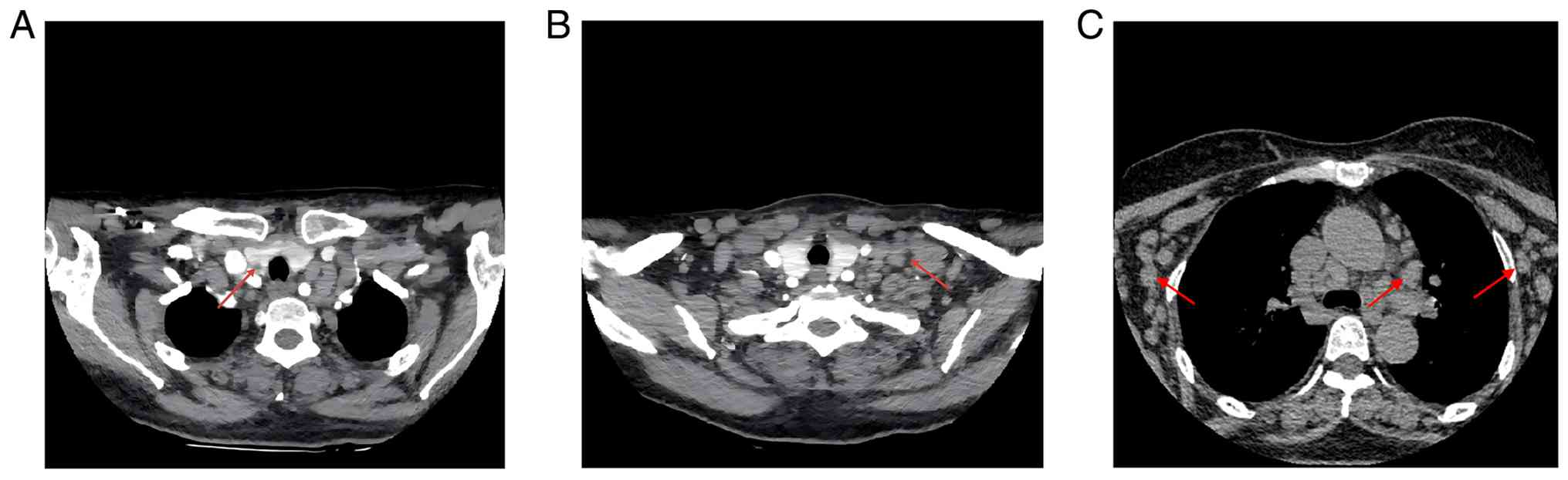
and inguinal regions. Baseline contrast-enhanced neck CT showed
that the largest node in the right cervical region measured
~2.0×1.0 cm, whilst the largest on the left side measured ~1.0×1.0
cm (Fig. 1A and B). No hepatomegaly
or splenomegaly was detected on abdominal palpation.
Past medical history
The patient had a 30-year history of hypertension
and was receiving irbesartan/hydrochlorothiazide tablets (150/12.5
mg), one tablet orally once daily. Blood pressure measured at
admission was 162/78 mmHg. The patient had undergone a
cholecystectomy 15 years prior for gallstones with acute
cholecystitis. In addition, 7 years before the present admission,
the patient had been diagnosed with CLL and had since been followed
up regularly, with no clear evidence of clinical progression. The
patient was resident in an urban district of Lanzhou, China, and no
specific environmental or occupational carcinogenic exposure was
identified. The patient also had no prior exposure to ionizing
radiation, radioactive materials or hazardous chemicals, with no
history of smoking, alcohol consumption or known chronic infectious
disease.
Laboratory tests
A complete blood count revealed a white blood cell
count of 8.1×10⁹/l (reference range,
3.5–9.5×10⁹/l), a lymphocyte count of
3.14×10⁹/l (reference range, 1.1–3.2×10⁹/l),
a hemoglobin level of 139 g/l (reference range, 115–150 g/l) and a
platelet count of 185×109/l (reference range,
125–350×109/l). Biochemistry showed elevated levels of
β2-microglobulin and uric acid at 6.44 mg/l (reference
range, 0.8–2.2 mg/l) and 558 µmol/l (reference range, 155–357
µmol/l), respectively. Immunological analysis demonstrated the
following results: Immunoglobulin (Ig)A, 0.29 g/l (reference range,
0.7–4.0 g/l); IgG, 3.32 g/l (reference range, 7.0–16.0 g/l); IgM,
0.26 g/l (reference range, 0.4–2.3 g/l); complement (C)3, 0.88 g/l
(reference range, 0.9–1.8 g/l); and C4, 0.03 g/l (reference range,
0.1–0.4 g/l), with a κ/λ light chain ratio of 1.81 (reference
range, 1.35–2.65). Thyroid function tests showed the following
results: Triiodothyronine (T3), 1.5 nmol/l (reference range,
1.3–3.1 nmol/l); thyroxine (T4), 161.10 nmol/l (reference range,
66–181 nmol/l); free (F)T3, 4.57 pmol/l (reference range, 3.1–6.8
pmol/l); FT4, 16.62 pmol/l (reference range, 12.0–22.0 pmol/l);
thyroid-stimulating hormone (TSH), 4.954 µIU/ml (reference range,
0.27–4.20 µIU/ml); calcitonin, 4.47 pg/ml (reference range, 0–8.4
pg/ml); vitamin D, 7.0 ng/ml (reference range, 30–100 ng/ml); and
parathyroid hormone, 42.90 pg/ml (reference range, 15–65
pg/ml).
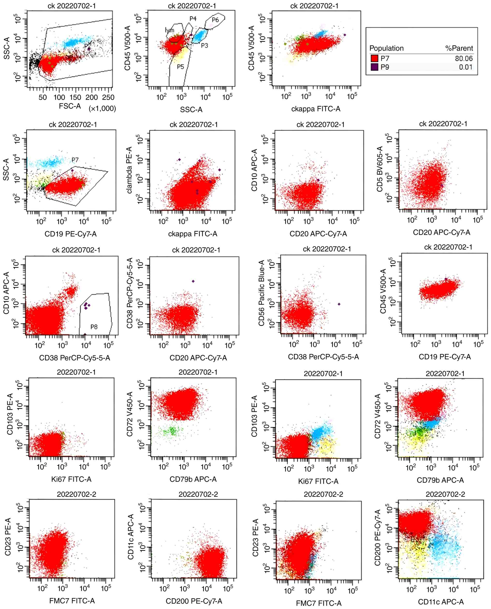
Peripheral blood flow cytometry was performed using
ethylenediaminetetraacetic acid-anticoagulated peripheral blood as
part of the routine clinical hematopathological work-up. After
erythrocyte lysis, nucleated cells were stained with
fluorochrome-conjugated antibody panels. Detailed antibody
combinations and brief method details are provided in Data S1. Lymphocytes were gated according
to CD45/side-scatter characteristics, followed by analysis of the
CD19-positive B-cell population, light-chain restriction and
CLL/SLL-associated markers. Representative dot plots/histograms
with population frequencies are shown in Fig. 2. Flow cytometry identified an
abnormal mature B-cell population accounting for 80.06% of the
parent population, with κ light-chain restriction. The
immunophenotype was characterized as CD5 (+), CD19 (+), CD20
(partial+), CD22 (partial+), CD23 (partial+), CD200 (weak+), CD10
(−) and FMC7 (−). These findings support a clonal mature B-cell
lymphoproliferative disorder compatible with CLL/SLL.
Bone marrow cytomorphological examination showed
active hematopoiesis with an increased proportion of lymphocytes,
including certain immature forms. Fine-needle aspiration (FNA) of
the left supraclavicular lymph node indicated an indolent small
B-cell lymphoma (Fig. 3).
Laryngoscopy showed no abnormalities.
Pathology examination
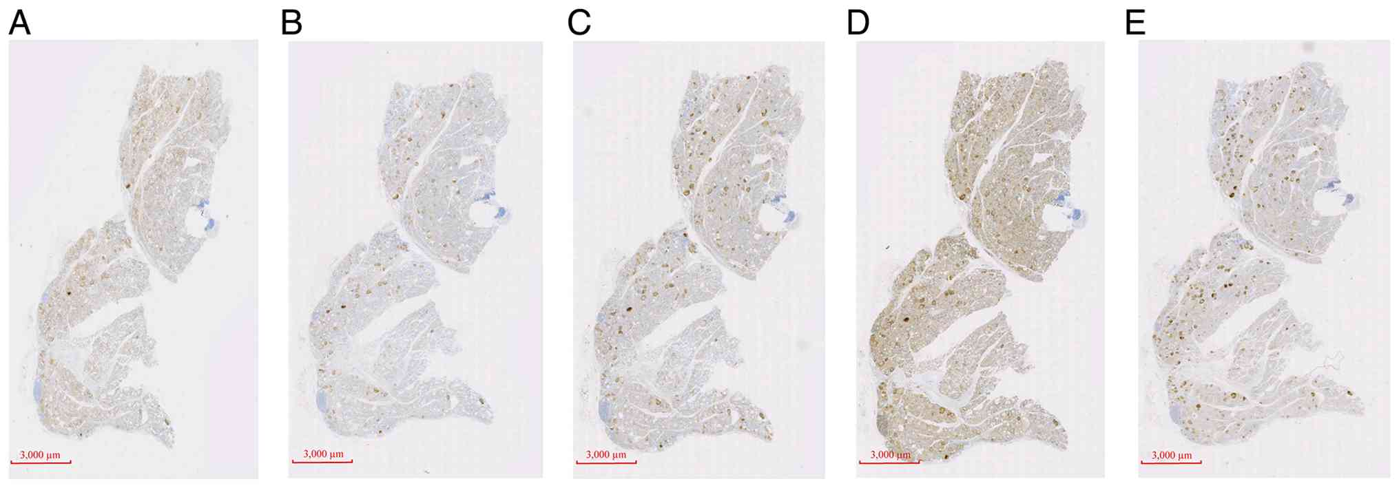
In November 2023, FNA of the right thyroid nodule
revealed PTC. Cytological and immunohistochemical evaluation was
performed according to routine clinical diagnostic pathology
procedures. Briefly, the available cytopathological material was
processed for immunohistochemical staining, followed by incubation
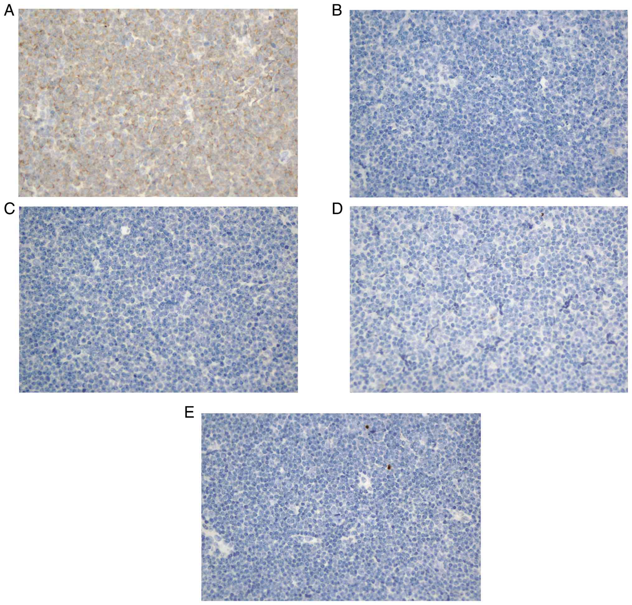
with antibodies against cytokeratin (CK)19, galectin-3, Hector
Battifora mesothelial-1 (HBME-1), CD56 and Ki-67. Appropriate
positive and negative controls were used. Immunohistochemical
staining demonstrated strong positivity for CK19, galectin-3 and
HMBE1, absence of CD56 expression and a Ki-67 proliferation index
of ~3% (Fig. 4). Other thyroid
lesions were excluded based on the combined cytological,
immunohistochemical and laboratory findings. Strong CK19,
galectin-3 and HBME-1 positivity with loss of CD56 supported PTC
rather than benign nodular disease. Medullary thyroid carcinoma was
unlikely as the serum calcitonin level was within the reference
range and no medullary carcinoma morphology was identified.
Lymphoid involvement of the thyroid nodule was also not supported
as the tumor cells showed an epithelial rather than lymphoid
immunophenotype. The detailed pathological and immunohistochemical
staining protocol is provided in Data
S1.
FNA biopsy of the right supraclavicular lymph node
suggested an indolent lymphoma of small B-cell origin.
Immunohistochemistry (Data S1)
showed positivity for CD20, CD79a, BCL-2, lymphoid enhancer-binding
factor 1 (LEF-1; focal), and multiple myeloma oncogene-1 (MUM-1;
focal). CD5 expression was partially positive, CD23 was weakly
positive, and Cyclin D1, SOX11, CD2, CD3, CD10, CD21, CD30, c-Myc,
BCL-6, myeloperoxidase, terminal deoxynucleotidyl transferase,
anaplastic lymphoma kinase and Epstein-Barr virus (EBV)-encoded
small RNA results were negative. The Ki-67 proliferation index was
~20% (Figs. 5 and 6).
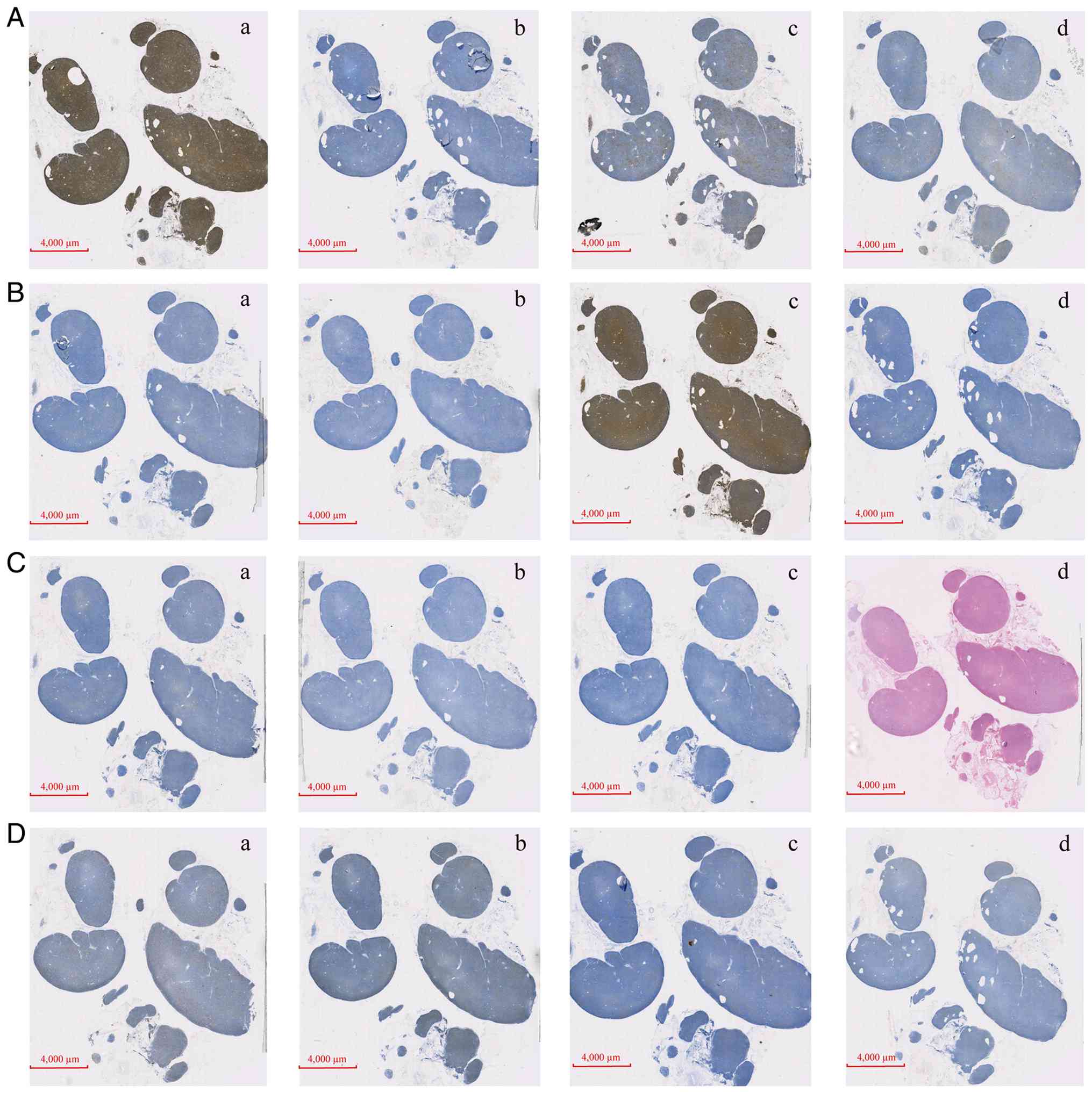 | Figure 5.Fine-needle aspiration biopsy of the
right supraclavicular lymph node suggesting an indolent lymphoma of
small B-cell origin. Immunohistochemical staining demonstrated the
following results: (A-a) CD79a (positive), (A-b) BCL-6 (negative),
(A-c) CD2 (negative) and (A-d) CD3 (negative). (B-a) CD5 (partial
positive), (B-b) CD10 (negative), (B-c) CD20 (positive) and (B-d)
CD21 (negative). (C-a) CD23 (weak positive), (C-b) CD30 (negative),
(C-c) Cyclin D1 (negative) and (C-d) Epstein-Barr Virus-encoded
small RNA (negative). (D-a) Ki-67 (~20% positive), (D-b) lymphoid
enhancer-binding factor 1 (focal positive), (D-c) multiple myeloma
oncogene-1 (focal positive) and (D-d) c-Myc (negative). All panels
are whole-section scanned images; conventional microscopic
magnification is not applicable. Scale bars, 4,000 µm. |
Taken together, the immunophenotypic profile of the
lymph node tissue, namely positivity for CD20, CD79a, LEF-1
(focal), BCL-2 and MUM-1 (focal), with partially positive
expression of CD5 and CD23, was consistent with CLL. Other
hematological malignancies were excluded based on the
immunohistochemical profile. Negative cyclin D1 and SOX11 results
argued against mantle cell lymphoma, the absence of CD10 and BCL-6
excluded germinal center-derived B-cell lymphomas, and negative
CD3, terminal deoxynucleotidyl transferase and myeloperoxidase
results did not support T-cell, lymphoblastic or myeloid neoplasms.
Negativity for CD30, anaplastic lymphoma kinase and EBV-encoded
small RNA further excluded CD30-positive, ALK-positive or
EBV-associated lymphoid neoplasms.
Imaging examination
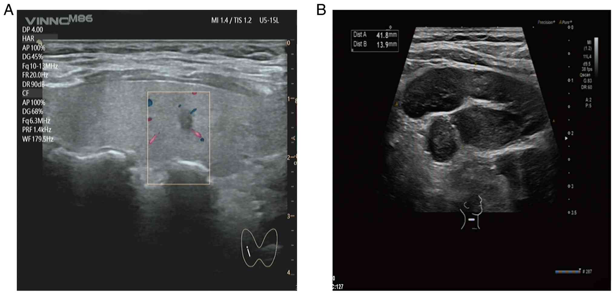
In November 2023, neck ultrasonography revealed a
0.38×0.31-cm hypoechoic nodule in the mid-portion of the right
thyroid lobe [Thyroid Imaging Reporting and Data System (TI-RADS)
4a], a 1.0×0.8-cm hypoechoic nodule in the lower pole of the right
lobe (TI-RADS 3) and a 0.5×0.3-cm mixed-echoic nodule in the left
lobe (TI-RADS 3), according to the Chinese TI-RADS classification
(11). Multiple enlarged lymph
nodes were detected in cervical regions I–V bilaterally, the
largest measuring ~3.1×1.0 cm on the right and 1.5×1.0 cm on the
left (Fig. 7).
During the same initial diagnostic work-up at the
index admission, contrast-enhanced CT of the neck demonstrated a
0.9-cm hypodense nodule in the right thyroid lobe, along with
multiple enlarged lymph nodes in the bilateral cervical and
supraclavicular regions (Fig. 1A and
B). Chest CT revealed multiple enlarged lymph nodes in the
bilateral axillae and mediastinum, suggestive of lymphoma (Fig. 1C).
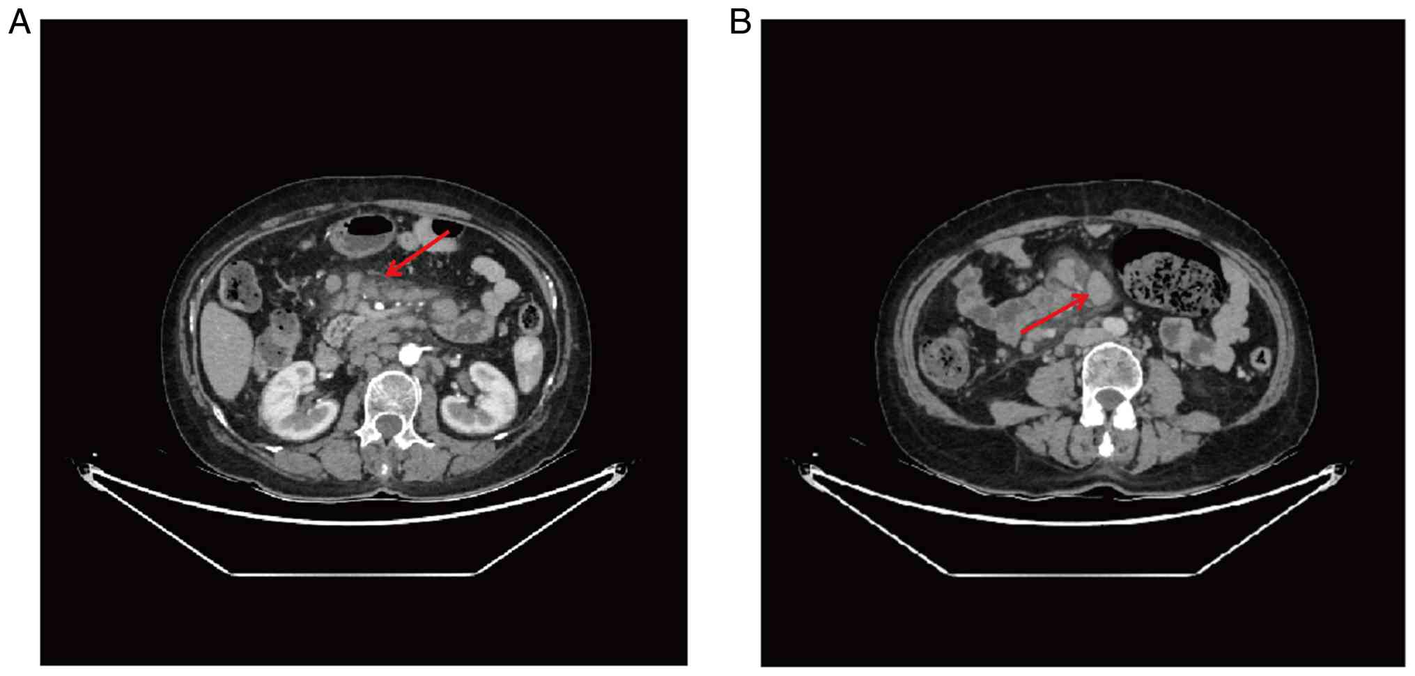
Contrast-enhanced abdominal CT showed multiple
enlarged lymph nodes in the abdominal and pelvic cavities, the
largest measuring ~4.7×2.4 cm, with mild enhancement (Fig. 8). The chronological sequence of
patient clinical presentation, diagnostic evaluations,
interventions and outcomes is summarized in Table I.
 | Table I.Timeline of patient clinical
course. |
Table I.
Timeline of patient clinical
course.
| Date/interval | Event/clinical
findings | Diagnostic
assessment |
Intervention/management |
Outcome/follow-up |
|---|
| ~7 years prior to
admission | History of CLL
treated with traditional Chinese medicine | - | Symptomatic
management | Stable disease |
| Several months
prior to admission | Cervical
lymphadenopathy, thyroid nodule detected | Physical exam,
ultrasound | - | Suspicion of
synchronous malignancy |
| Work-up phase | Enlarged
cervical/axillary/inguinal lymph nodes | Flow cytometry,
immuno-histochemistry, radiology | - | Diagnosis of PTC +
CLL |
| Surgery (index
admission) | Thyroidectomy
performed | Histopathology
confirmed PTC (pT1aN0M0) | Surgery +
initiation of levothyroxine (50 µg daily) | Successful
recovery |
| 2-year
follow-up | Regular imaging and
labs | Ultrasound,
thyroglobulin tests | - | No recurrence,
disease-free |
The initial suspected diagnosis was a solitary
thyroid malignancy. However, the presence of persistent and
generalized lymphadenopathy raised suspicion for a concurrent
hematological disorder, prompting further hematological evaluation,
including flow cytometry and bone marrow cytomorphological
examination, which confirmed CLL.
Diagnosis and treatment
In November 2023, the patient underwent a total
thyroidectomy with right central compartment lymph node dissection
under general anesthesia at the Second Hospital of Lanzhou
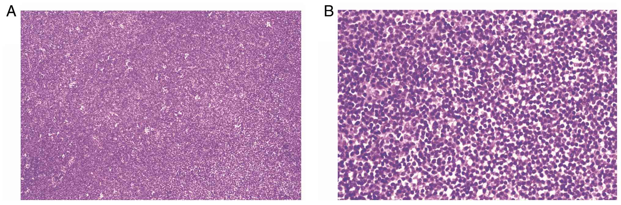
University. The surgical thyroid specimens were fixed in 10%
neutral buffered formalin at room temperature for 12–36 h, embedded
in paraffin, sectioned at 4 µm, stained with hematoxylin and eosin
at room temperature 20–25°C for 45–50 min, and examined and imaged
using an Olympus CX43 light microscope at ×100 or ×200
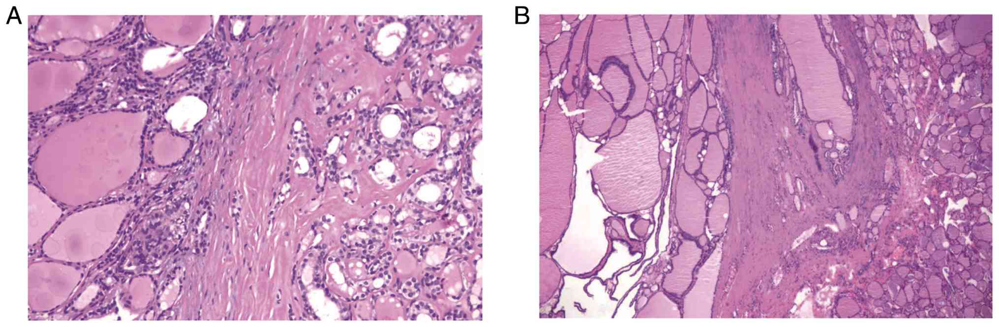
magnification. Postoperative histopathological examination of
hematoxylin and eosin-stained sections revealed a micropapillary
carcinoma of the right lobe and isthmus, with the largest focus
measuring 0.3 cm in diameter, accompanied by nodular goiter
(Fig. 9A). The left lobe showed a
nodular goiter with adenomatoid hyperplasia (Fig. 9B). No metastatic carcinoma was
identified in the dissected right central compartment and level 6B
lymph nodes.
According to the American Joint Committee on Cancer
8th edition staging system (12),
the diagnosis was papillary thyroid microcarcinoma, stage pT1aN0M0.
The staging was supported by the largest tumor focus measuring 0.3
cm in diameter, consistent with T1a disease, no metastatic
carcinoma being identified in the dissected lymph nodes, supporting
N0 status, and no distant metastasis being suggested by the
available examinations, supporting M0 status. The patient had an
uneventful postoperative recovery and was discharged on
postoperative day 7 without complications.
The subsequent treatment plan included lifelong oral
endocrine suppressive therapy with levothyroxine (50 µg daily,
taken in the morning on an empty stomach), along with dynamic
follow-up of thyroid function and cervical ultrasonography.
Given the indolent course of CLL and the absence of
treatment indications, a watch-and-wait strategy was adopted in
consultation with the hematology team.
At a 2-year postoperative follow-up in November
2025, the patient remained asymptomatic. The thyroid function test
results were as follows: T3, 1.09 nmol/l; T4, 207.50 nmol/l; FT3,
4.06 pmol/l; FT4, 23.95 pmol/l; and TSH, 6.107 µIU/ml. Compared
with the baseline values, T3 was lower, T4 and FT4 were higher, FT3
remained within the reference range and TSH remained mildly
elevated. Follow-up cervical CT showed no thyroid-bed lesion after
the thyroidectomy. Multiple soft-tissue nodules in the bilateral
cervical, supraclavicular/infraclavicular and mediastinal regions
were considered to be lymphoma-related lymphadenopathy in the
context of known CLL/SLL. The patient was followed every 3 months
during the first postoperative year and every 6 months during the
second postoperative year, and remains under regular hematology
follow-up.
Discussion
PTC originates from the thyroid follicular
epithelium and is the most common and well-differentiated thyroid
malignancy, accounting for ~85% all thyroid cancer cases (4,13).
Over the past decade, the incidence of PTC has risen markedly with
advances in diagnostic techniques. PTC predominantly affects
middle-aged women, and the disease typically follows an indolent
course (4). The typical
histopathological features include complex papillary structures and
the characteristic ground-glass (‘Orphan Annie-eye’) nuclear
appearance (3). FNA cytology under
ultrasound guidance remains the cornerstone of preoperative
diagnosis, providing high accuracy in identifying PTC (14). Immunohistochemistry serves as an
adjunct in difficult cases, with PTC tumor cells frequently
expressing thyroid-specific markers [such as thyroid transcription
factor-1 and thyroglobulin (Tg)] and CKs (CK19, HBME-1 and
galectin-3) (3,15). CLL, by contrast, is an indolent
hematological malignancy that is characterized by the clonal
proliferation of mature B lymphocytes, which represents the most
common form of leukemia in adults (16). CLL primarily occurs in middle-aged
and elderly individuals and is typically identified through
peripheral blood lymphocytosis and generalized painless
lymphadenopathy. Bone marrow biopsy and immunophenotyping are
critical for achieving a definitive diagnosis (17,18).
The typical immunophenotypic profile of CLL includes positivity for
CD5, CD19 and CD23 (19,20). Overall, PTC is confined to the
thyroid gland, progresses relatively slowly and carries a favorable
prognosis. By contrast, CLL has a protracted clinical course, with
long-term remission possible but a cure remaining elusive.
The simultaneous occurrence of PTC and CLL in the
same patient is exceedingly rare, with only sporadic case reports
documented in the literature (7,8). Such
‘dual primary’ malignancies have been described almost exclusively
in isolated cases and small case series, with an estimated
incidence of ~0.04%. In a large cohort study of 17,196 patients
with PTC, only 0.04% were found to have a concomitant indolent
B-cell lymphoma, underscoring its rarity (21). The coexistence of PTC and CLL may
not be entirely coincidental. CLL-associated immune dysregulation
provides a biologically plausible, although unproven, explanation
for the development of a second primary solid tumor. CLL is
accompanied by abnormalities across both humoral and cellular
immune compartments, where patients with CLL have an increased risk
of ‘secondary’ primary malignancies compared with the general
healthy population (22). The most
frequently reported secondary cancers in this context include
cutaneous malignancies, other lymphoid neoplasms and various solid
tumors, such as lung and bronchus cancer, colorectal cancer, kidney
cancer, thyroid cancer and soft-tissue sarcoma. Notably, the
relative risk of thyroid cancer among patients with CLL has been
reported to be nearly 3 times greater compared with that in the
general population (23). In a
previous cohort study of 2,028 untreated patients with CLL, 11.2%
developed a subsequent malignancy during follow-up, with the
overall risk of secondary cancer being 2.2 times higher compared
with that observed in the general population (24). These epidemiological observations
support the concept that CLL-related immune failure may create a
permissive host environment for the emergence such ‘secondary’
primary solid tumors, including PTC. Genetic predisposition may
also serve a contributory role. Certain molecular alterations have
been implicated in both diseases. The deleted in lymphocytic
leukemia 1 gene, frequently deleted or downregulated in CLL, has
been found to be upregulated in PTC, where it promotes thyroid
cancer cell proliferation and invasion by sponging specific
microRNAs (25). This raises the
possibility that shared genetic or epigenetic alterations may
contribute to the pathogenesis of coexisting PTC and CLL.
Nevertheless, no definitive common pathogenic mutations have been
established to date. Therefore, the concurrence of PTC and CLL is
considered to be an opportunistic comorbidity arising from multiple
contributing factors rather than a direct causal relationship
(22,23).
In the present case, no recognized exogenous
carcinogenic exposure was identified from the residential,
occupational or lifestyle history of the patient, including
ionizing radiation, radioactive materials, hazardous chemicals,
smoking, alcohol consumption or known chronic infectious disease.
Therefore, the coexistence of PTC and long-standing CLL is unlikely
to be explained by a single environmental exposure. A more
plausible interpretation is that the dual malignancies arose
through a multifactorial process in which CLL-associated immune
dysregulation may have lowered antitumor immune surveillance and
facilitated the emergence of a second primary solid tumor. Beyond
its identity as a clonal mature B-cell neoplasm, CLL is
increasingly recognized as a systemic immune-disruptive disease,
characterized by impaired humoral immunity, dysfunctional T-cell
and natural killer-cell responses, altered antigen presentation,
upregulated immune-checkpoint signaling and a tolerogenic tumor
microenvironment (23,26). Recent reviews have further
emphasized that immune dysfunction in CLL is persistent and
clinically relevant, affecting not only infection susceptibility
but also immune surveillance, immune-cell cross-talk and the
broader risk of secondary malignancies (26,27).
Together, these immune defects may impair antitumor surveillance
and reduce host capacity to eliminate transformed epithelial cells,
thereby creating a permissive milieu for secondary malignancies,
including solid tumors, such as PTC.
Nevertheless, a direct causal relationship between
CLL and PTC cannot be proven from a single case. The development of
dual primary malignancies in the present patient is best
interpreted as the result of interacting factors, including
age-related accumulation of oncogenic alterations, sex-related
susceptibility to PTC, possible genetic or epigenetic
predisposition and CLL-related impairment of immune surveillance.
Therefore, rather than representing metastatic spread or a single
unifying pathogenic pathway, the present case likely reflects the
biologically plausible coexistence of two independent primary
malignancies in the setting of long-standing immune dysregulation.
This interpretation also underscores the need for the careful
evaluation of new solid lesions in patients with CLL, even when the
hematological disease appears clinically indolent. A recent 2026
case of synchronous PTC and CLL presenting with cervical
lymphadenopathy similarly emphasized that lymph node enlargement in
patients with thyroid nodules may reflect hematological disease
rather than metastatic thyroid carcinoma, whereby definitive
histopathological and immunohistochemical assessment is essential
for appropriate management (10).
The coexistence of thyroid carcinoma and
hematological malignancy presents substantial diagnostic
challenges. In the present case, the thyroid nodule was accompanied
by widespread lymphadenopathy, a presentation that could easily
suggest thyroid carcinoma with regional lymph node metastasis.
However, further investigations revealed that the enlarged lymph
nodes were not metastatic PTC but rather the result of CLL
involvement. Lymphocytic infiltration is frequently observed in PTC
tissue, particularly in association with Hashimoto's thyroiditis
(28,29). By contrast, infiltration of the
thyroid gland by CLL may closely mimic autoimmune lymphocytic
infiltration under microscopy, making histological differentiation
difficult (30). Therefore, in
cases of thyroid carcinoma with dense monomorphic lymphocytic
infiltration, clinicians should maintain a high index of suspicion
for concomitant lymphoproliferative disorders, such as CLL.
Immunohistochemistry and flow cytometry are indispensable adjuncts
for establishing the correct lineage and avoiding diagnostic
misclassification. Detection of monoclonal B-cell populations (such
as clones co-expressing CD5, CD19 and CD23) by flow cytometry
strongly supports the diagnosis of CLL (7,16).
Histopathologically, immunohistochemistry enables separation of the
two tumor components. In particular, PTC cells express epithelial
and thyroid-related markers, whereas infiltrating lymphocytes
express B-cell and CLL-associated markers (such as CD20, CD5 and
CD23) but lack T-cell and germinal center markers. A recent report
described the case of a patient with preexisting indolent SLL who
developed suspicious cervical lymphadenopathy after thyroidectomy
for PTC. FNA yielded only CLL-like lymphocytes, yet the aspirate
demonstrated markedly elevated thyroglobulin levels, raising
suspicion for occult PTC metastasis. Subsequent lymph node
dissection confirmed the presence of metastatic PTC deposits within
CLL-involved lymph nodes (9). This
case, reported by Rejeb et al (9), highlights the diagnostic complexity of
PTC coexisting with CLL, underscoring the need for the integration
of tumor markers and when necessary, a repeat biopsy, to avoid
misdiagnosis.
In the present case, the combined
immunohistochemical and flow-cytometric findings established the
thyroid lesion as PTC and the nodal disease as CLL. The distinct
histomorphological and immunophenotypic profiles of the two lesions
prevented misclassification of CLL-related lymphadenopathy as
metastatic PTC and ensured appropriate staging and management.
Although active surveillance is recognized in several international
guidelines as a reasonable option for carefully selected patients
with low-risk papillary thyroid microcarcinoma, management in the
present case was individualized and was not dictated by tumor size
alone (31–33). Recent reviews continue to support
active surveillance as a safe and increasingly accepted option for
carefully selected low-risk PTC, but also emphasize that
eligibility depends on reliable imaging, the absence of suspicious
nodal disease, patient preference and the feasibility of structured
follow-up (34–36). According to Recommendation 42 of the
Chinese Guidelines for the Diagnosis and Treatment of Thyroid
Nodules and Differentiated Thyroid Cancer (Second Edition, 2023),
thyroid lobectomy with isthmusectomy is strongly recommended, with
moderate-quality evidence, for differentiated thyroid cancer
lesions measuring <1 cm in the absence of high-risk features.
Therefore, surgery represented a guideline-concordant management
option rather than a departure from accepted care (37).
Crucially, the present case was not a
straightforward case of an isolated, incidentally detected PTC. The
patient had a 7-year history of CLL and presented with persistent,
generalized lymphadenopathy involving the cervical, axillary,
mediastinal, abdominal, pelvic and inguinal regions. In such a
setting, preoperative risk stratification was particularly
challenging, as enlarged lymph nodes could represent CLL
involvement, occult PTC metastasis or coexistence of both processes
within the same nodal basin. Although preoperative cytology and
flow cytometry favored CLL-related nodal involvement, they could
not fully exclude occult metastatic PTC, particularly in the
setting of extensive cervical lymphadenopathy. Surgical resection
with a central compartment lymph node dissection therefore provided
definitive histopathological staging, excluded occult nodal
metastasis in the dissected nodal basin and informed subsequent
surveillance intensity.
In addition, patient preference was an important
ethical and clinical consideration. The diagnosis of concurrent
malignancies imposed a substantial psychological burden and the
patient consistently expressed a strong preference for definitive
surgical treatment rather than prolonged observation. After shared
decision-making and multidisciplinary discussion involving the
thyroid surgery and hematology teams, this patient-centered
approach also enabled definitive pathological staging and remained
consistent with the patient's informed values and psychological
needs.
The prognosis of patients with concomitant PTC and
CLL depends on the stage and biological behavior of each
malignancy, in addition to the effectiveness of treatment (4). Overall, early-stage PTC, with
papillary microcarcinoma as a representative subtype, is associated
with a highly favorable prognosis. Papillary thyroid microcarcinoma
(≤1 cm) has an exceptionally low mortality rate and >90% 10-year
survival rate (38). CLL is an
indolent lymphoma that can also have a prolonged natural history.
Low-risk patients may survive for years with observation alone
already sufficient (16). In the
present case, the 64-year-old patient had a 0.3-cm papillary
carcinoma with no lymph node metastases (stage pT1aN0M0) and was
considered clinically cured following surgery. This specific CLL,
which was diagnosed 7 years earlier, remained in a stable early
stage, where no disease progression was observed during the 2-year
follow-up. The patient has since maintained a good quality of life
without additional therapy for CLL. However, the prognosis in
similar cases may vary depending on tumor burden and pathological
subtype of PTC. Sezer et al (8) reported the case of a patient with PTC
who had local invasion and distant metastases alongside CLL and
succumbed within 1 year postoperatively. In addition, in CLL,
late-stage complications, such as Richter's transformation, which
refers to the progression of CLL into aggressive diffuse large
B-cell lymphoma, can markedly shorten survival time (39). These considerations highlight the
necessity of comprehensive surveillance.
As a result of the present case, a
multidisciplinary, long-term follow-up strategy is recommended. For
the monitoring of thyroid carcinoma recurrence and metastasis,
postoperative surveillance should include the periodic measurement
of serum Tg and anti-Tg antibodies, in addition to high-resolution
neck ultrasonography at least every 6–12 months. In patients who
have undergone a total thyroidectomy, any unexplained rise in Tg or
suspicious imaging findings should prompt further imaging (such as
whole-body positron emission tomography-CT or iodine scans) to
localize potential recurrent or metastatic disease (3,40). For
the monitoring of hematological disease progression, in stable CLL,
hematological evaluation with blood counts and relevant chemistries
is recommended every 3 months. Key parameters include lymphocyte
doubling time, cytopenias and constitutional symptoms, such as
night sweats or weight loss. Early-stage asymptomatic disease is
generally managed with observation, whilst rapid lymphocyte
escalation, progressive lymphadenopathy or splenomegaly, or bone
marrow failure warrant treatment initiation.
In summary, patients with concomitant PTC and CLL
require sustained, multidisciplinary follow-up. Surveillance must
focus not only on detecting the recurrence of thyroid carcinoma but
also on monitoring the evolution of leukemia. With individualized
follow-up plans and proactive management strategies, the majority
of patients with this rare dual malignancy can achieve a good
quality of life and prolonged survival time.
Supplementary Material
Supporting Data
Acknowledgements
Not applicable.
Funding
The present study was supported by the Cuiying Technology
Innovation Project of Lanzhou University Second Hospital (grant no.
CY2023-MS-B16).
Availability of data and materials
The data generated in the present study are included
in the figures and/or tables of this article.
Authors' contributions
JM contributed to image acquisition and drafting of
the manuscript. TG analyzed patient data and contributed to the
revision of the manuscript. SX performed histopathological image
acquisition and assisted in manuscript drafting. HT contributed to
data interpretation and critical revision of the manuscript. AS
contributed to the conception and design of the study,
interpretation of the clinical data, and critical revision of the
manuscript. LZ contributed to the conception and design of the
study, interpretation of the clinical and pathological data, and
critical revision of the manuscript. JM, TG and SX confirm the
authenticity of all the raw data. All authors have read and
approved the final version of the manuscript.
Ethics approval and consent to
participate
The study was reviewed and approved by the
Institutional Medical Ethics Committee of the Second Hospital of
Lanzhou University (approval no. 2025A-951). All procedures were
performed in accordance with the Declaration of Helsinki and
relevant guidelines and regulations.
Patient consent for publication
Written informed consent was obtained from the
patient for publication of the present study and associated
images.
Competing interests
The authors declare that they have no competing
interests.
References
|
1
|
Han Y, Hou L, Zhao B, Gao L and Li S: Risk
factors for neck nodal metastasis in papillary thyroid cancer with
BRAF V600E mutation. Front Endocrinol (Lausanne). 13:8844282022.
View Article : Google Scholar : PubMed/NCBI
|
|
2
|
Kitahara CM and Schneider AB: Epidemiology
of thyroid cancer. Cancer Epidemiol Biomarkers Prev. 31:1284–1297.
2022. View Article : Google Scholar : PubMed/NCBI
|
|
3
|
Limaiem F, Rehman A and Mazzoni T:
Papillary Thyroid Carcinoma. StatPearls (Internet). StatPearls
Publishing; Treasure Island, FL: 2025
|
|
4
|
Boucai L, Zafereo M and Cabanillas ME:
Thyroid cancer: A review. JAMA. 331:425–435. 2024. View Article : Google Scholar : PubMed/NCBI
|
|
5
|
Yu K, Wu X, Dai L, Le Q, Xie Y, Wang Y and
Zhang W: Risk factors associated with lymph node metastasis in
papillary thyroid cancer: A retrospective analysis based on 2,428
cases. Front Oncol. 14:14738582024. View Article : Google Scholar : PubMed/NCBI
|
|
6
|
Reid-Nicholson M, Moreira A and Ramalingam
P: Cytologic features of mixed papillary carcinoma and chronic
lymphocytic leukemia/small lymphocytic lymphoma of the thyroid
gland. Diagn Cytopathol. 36:813–817. 2008. View Article : Google Scholar : PubMed/NCBI
|
|
7
|
Ahmed OI and Salih ZT: Dual malignancy in
a thyroid; papillary thyroid carcinoma and small lymphocytic
lymphoma; a report of a case with a cyto-histologic correlation.
Diagn Cytopathol. 45:851–856. 2017. View
Article : Google Scholar : PubMed/NCBI
|
|
8
|
Sezer H, Yazici D, Tezelman S, Demirkol
MO, Ünal OF, Dilege S, Taşkın OC, Peker O, Kapran Y, Çolakoğlu B,
et al: Concomittant occurence of well-differentiated thyroid
carcinoma metastasis and chronic lymphocytic leukemia in the same
lymph node along with internal jugular vein thrombus: A case
report. Indian J Pathol Microbiol. 64:149–151. 2021. View Article : Google Scholar : PubMed/NCBI
|
|
9
|
Rejeb S, Sakly S, Hachicha A, Dridi K and
Turki S: Synchronous cervical lymph node involvement with a
papillary thyroid carcinoma and small lymphocytic lymphoma: A case
report. J Surg Case Rep. 2025:rjaf0382025. View Article : Google Scholar : PubMed/NCBI
|
|
10
|
Farnin R, Altindag H, Lienenluke R, Hammer
A, Gebrehiwot L, Engler J, Mittermeier J and Vorländer C:
Synchronous papillary thyroid microcarcinoma and chronic
lymphocytic leukemia: A rare interdisciplinary case. Cureus.
18:e1071942026.PubMed/NCBI
|
|
11
|
Zhou J, Yin L, Wei X, Zhang S, Song Y, Luo
B, Li J, Qian L, Cui LG, Chen W, et al: 2020 Chinese guidelines for
ultrasound malignancy risk stratification of thyroid nodules: The
C-TIRADS. Endocrine. 70:256–279. 2020. View Article : Google Scholar : PubMed/NCBI
|
|
12
|
Tuttle RM, Haugen B and Perrier ND:
Updated American joint committee on cancer/tumor-node-metastasis
staging system for differentiated and anaplastic thyroid cancer
(Eighth Edition): What changed and why? Thyroid. 27:751–756. 2017.
View Article : Google Scholar : PubMed/NCBI
|
|
13
|
Hao Z, Cui H, Li Y, Wu W, Wang Y, Dan H,
Lou L, Wang H and Zhao P: Simultaneous occurrence of papillary
thyroid carcinoma, medullary thyroid carcinoma, and lymphoma: A
case report. Medicine (Baltimore). 103:e393632024. View Article : Google Scholar : PubMed/NCBI
|
|
14
|
Haugen BR, Alexander EK, Bible KC, Doherty
GM, Mandel SJ, Nikiforov YE, Pacini F, Randolph GW, Sawka AM,
Schlumberger M, et al: 2015 American thyroid association management
guidelines for adult patients with thyroid nodules and
differentiated thyroid cancer: The American thyroid association
guidelines task force on thyroid nodules and differentiated thyroid
cancer. Thyroid. 26:1–133. 2016. View Article : Google Scholar : PubMed/NCBI
|
|
15
|
Laishram H, Jagadale K, Shelke P and
Bharadwaj R: Cross-sectional study to determine the diagnostic
accuracy of cytokeratin-19 immunomarker in the diagnosis of thyroid
malignancy with histopathology as gold standard. Niger Postgrad Med
J. 32:61–67. 2025. View Article : Google Scholar : PubMed/NCBI
|
|
16
|
Mukkamalla SKR, Taneja A, Malipeddi D and
Master SR: Chronic lymphocytic leukemia. StatPearls (Internet).
StatPearls Publishing; Treasure Island, FL: 2025
|
|
17
|
Rawstron AC, Fazi C, Agathangelidis A,
Villamor N, Letestu R, Nomdedeu J, Palacio C, Stehlikova O, Kreuzer
KA, Liptrot S, et al: A complementary role of multiparameter flow
cytometry and high-throughput sequencing for minimal residual
disease detection in chronic lymphocytic leukemia: an European
Research Initiative on CLL study. Leukemia. 30:929–936. 2016.
View Article : Google Scholar : PubMed/NCBI
|
|
18
|
Rawstron AC, Kreuzer KA, Soosapilla A,
Spacek M, Stehlikova O, Gambell P, McIver-Brown N, Villamor N,
Psarra K, Arroz M, et al: Reproducible diagnosis of chronic
lymphocytic leukemia by flow cytometry: An European research
initiative on CLL (ERIC) & European society for clinical cell
analysis (ESCCA) Harmonisation project. Cytometry B Clin Cytom.
94:121–128. 2018. View Article : Google Scholar : PubMed/NCBI
|
|
19
|
Hallek M, Shanafelt TD and Eichhorst B:
Chronic lymphocytic leukaemia. Lancet. 391:1524–1537. 2018.
View Article : Google Scholar : PubMed/NCBI
|
|
20
|
Rasul KH, Wsoo MA, Hassan DH, HamadaminS K
and Qadr ZJ: Immunophenotypic properties association of CLL and ALL
patient cells by flow cytometry analysis. J Taibah Univ Med Sci.
19:981–989. 2024.PubMed/NCBI
|
|
21
|
Zhang Y, Song Y, Cheng R and Zhai Q:
Concurrent papillary thyroid carcinoma and incidental cervical
lymph node indolent B cell non-Hodgkin lymphoma:
Clinicopathological features, outcomes, and potential
relationships. World J Surg Oncol. 23:1452025. View Article : Google Scholar : PubMed/NCBI
|
|
22
|
van der Straten L, Levin MD, Dinnessen
MAW, Visser O, Posthuma EFM, Doorduijn JK, Langerak AW, Kater AP
and Dinmohamed AG: Risk of second primary malignancies in patients
with chronic lymphocytic leukemia: A population-based study in the
Netherlands, 1989–2019. Blood Cancer J. 13:152023. View Article : Google Scholar : PubMed/NCBI
|
|
23
|
Sayin S, Kiliacslan E, Yildirim M, Ozturk
HBA, Yilmaz ES, Albayrak M, Kaptan MK and Ayli M: Evaluation of
second primary cancer risk among chronic lymphocytic leukemia
patients: Multicenter study. Asian Pac J Cancer Prev. 24:1971–1977.
2023. View Article : Google Scholar : PubMed/NCBI
|
|
24
|
Xiong YY, Liu ZJ, Chen L, Yuan FF, Yin QS,
Mi RH, Zhang B, Du JW, Zhang QL, Lin QD, et al: Synchronous
lymphoma and carcinoma-clinical analyses of 17 patients. Zhonghua
Xue Ye Xue Za Zhi. 39:277–280. 2018.(In Chinese). PubMed/NCBI
|
|
25
|
Li R, Wan T, Qu J, Yu Y and Zheng R: Long
non-coding RNA DLEUI promotes papillary thyroid carcinoma
progression by sponging miR-421 and increasing ROCK1 expression.
Aging. 12:20127–20138. 2020.PubMed/NCBI
|
|
26
|
Narkhede M and Ujjani CS: Immune
dysfunction and consequences in chronic lymphocytic leukemia. J
Natl Compr Canc Netw. 23:e2470902025. View Article : Google Scholar : PubMed/NCBI
|
|
27
|
Juarez-Salcedo LM and Loscertales J:
Clinical implications of immune dysfunction in chronic lymphocytic
leukemia. Cancers (Basel). 18:13232026. View Article : Google Scholar : PubMed/NCBI
|
|
28
|
Yao S and Zhang H: Papillary thyroid
carcinoma with Hashimoto's thyroiditis: Impact and correlation.
Front Endocrinol (Lausanne). 16:15124172025. View Article : Google Scholar : PubMed/NCBI
|
|
29
|
Monabbati S, Fu P, Asa SL, Pathak T,
Willis JE, Shi Q and Madabhushi A: Machine vision-detected
peritumoral lymphocytic aggregates are associated with disease-free
survival in patients with papillary thyroid carcinoma. Lab Invest.
104:1021682024. View Article : Google Scholar : PubMed/NCBI
|
|
30
|
Vurgun NA, Yalcin N, Turk NS, Fenkci SM
and Çağlıyan GA: Thyroid infiltration by chronic lymphocytic
leukemia: A rare case report with cytological and flow cytometry
findings. Diagn Cytopathol. 53:E133–E137. 2025. View Article : Google Scholar : PubMed/NCBI
|
|
31
|
Sawka AM, Ghai S, Rotstein L, Irish JC,
Pasternak J, Monteiro E, Chung J, Su J, Xu W, Esemezie AO, et al:
Long-term durability of active surveillance of small, low-risk
papillary thyroid cancer. JAMA Surg. 160:1117–1124. 2025.
View Article : Google Scholar : PubMed/NCBI
|
|
32
|
Kim MJ, Moon JH, Lee EK, Song YS, Jung KY,
Ji Ye Lee JY, Kim JH, Kim K, Park SK and Park YJ: Active
surveillance for low-risk thyroid cancers: A review of current
practice guidelines. Endocrinol Metab (Seoul). 39:47–60. 2024.
View Article : Google Scholar : PubMed/NCBI
|
|
33
|
Lee EK, Kim MJ, Kang SH, Koo BS, Kim K,
Kim M, Kim BH, Kim JH, Moon S, Back K, et al: 2025 Korean thyroid
association clinical management guideline on active surveillance
for low-risk papillary thyroid carcinoma. Endocrinol Metab (Seoul).
40:307–341. 2025. View Article : Google Scholar : PubMed/NCBI
|
|
34
|
Wu Z, Zhang K, Zhou H, Wan L, Zhong J, Li
C, Gong L and Peng Y: Active surveillance for low-risk papillary
thyroid carcinoma: Integrating guidelines, emerging evidence, and
directions. iScience. 29:1158322026. View Article : Google Scholar : PubMed/NCBI
|
|
35
|
Lee JY, Lee MK, Lim HK, Lee CY, Sung JY,
Yoon JH, Hahn SY, Shin JH, Kim JH, Jung SL, et al: Standardized
ultrasound evaluation for active surveillance of low-risk thyroid
microcarcinoma in adults: 2024 Korean society of thyroid radiology
consensus statement. Korean J Radiol. 25:942–958. 2024. View Article : Google Scholar : PubMed/NCBI
|
|
36
|
Nguyen VC, Song CM, Ji YB, Moon S, Park
JH, Kim DS and Tae K: Outcomes and effectiveness of active
surveillance for low-risk papillary thyroid carcinoma: A systematic
review and meta-analysis. Eur Arch Otorhinolaryngol. 282:2239–2252.
2025. View Article : Google Scholar : PubMed/NCBI
|
|
37
|
Chinese Medical Association Endocrinology
Society, Subspecialty Group of Thyroid and Metabolic Surgery of
Chinese Medical Association Surgery Society, Head and Neck Cancer
Specialized Committee of China Anti-Cancer Association, .
Guidelines for the Diagnosis and Treatment of Thyroid Nodules and
Differentiated Thyroid Cancer (Second Edition). Chin J Endocrinol
Metab. 39:181–226. 2023.(In Chinese).
|
|
38
|
Lan Y, Luo Y, Zhang M, Jin Z, Xiao J, Yan
L and Zhu Y: Quality of life in papillary thyroid microcarcinoma
patients undergoing radiofrequency ablation or surgery: A
comparative study. Front Endocrinol (Lausanne). 11:2492020.
View Article : Google Scholar : PubMed/NCBI
|
|
39
|
Shen Y, Coyle L, Kerridge I, Stevenson W,
Arthur C, McKinlay N, Fay K, Ward C, Greenwood M, Best O, et al:
Second primary malignancies in chronic lymphocytic leukaemia: Skin,
solid organ, haematological and Richter's syndrome. EJHaem.
3:129–138. 2022. View Article : Google Scholar : PubMed/NCBI
|
|
40
|
Filetti S, Durante C, Hartl D, Leboulleux
S, Locati LD, Newbold K, Papotti MG and Berruti A; ESMO Guidelines
Committee, : Thyroid cancer: ESMO Clinical Practice Guidelines for
diagnosis, treatment and follow-up†. Ann Oncol. 30:1856–1883. 2019.
View Article : Google Scholar : PubMed/NCBI
|