Introduction
Breast cancer, the most common malignant tumor among
women worldwide, accounts for >2.3 million new cases annually
(1). Breast cancer usually arises
from the combined effects of multiple factors, including genetic
factors, hormone levels, lifestyle and environmental factors, which
typically lead to the malignant transformation of breast epithelial
cells (2). Among all types of
breast cancer, the luminal subtype contributes to ~70% of cases,
including luminal A (ERα+, PR+, HER2- and low Ki-67) and luminal B
(ERα+, PR+/-, HER2-/+ and high Ki-67) (3). The luminal subtype is a
hormone-dependent molecular subtype of breast cancer, and patients
at the early stage typically achieve favorable therapeutic outcomes
with endocrine therapy. Of all patients, ~20% of patients with the
luminal subtype still experience recurrence and distant metastasis
because of endocrine therapy resistance, which progresses to
advanced-stage disease and results in poor disease prognosis
(4,5). Therefore, close attention to the
dynamic progression of luminal breast cancer is highly
important.
The application of CDK4/6 inhibitors has reshaped
the therapeutic landscape for patients with endocrine therapy
failure. As a first-line clinical CDK4/6 inhibitor, palbociclib has
been shown to achieve favorable clinical efficacy and considerably
prolong the survival of patients with advanced luminal breast
cancer (6). However, the issue of
palbociclib resistance has become increasingly prominent, further
hindering its widespread clinical application (7). Currently, the clinical determination
of therapeutic resistance relies mainly on disease
progression-driven tumor protein markers and imaging examinations.
However, these approaches exhibit poor specificity and sensitivity
in minimal residual disease detection, along with drawbacks such as
an inherent time lag (8). Thus,
developing innovative efficacy assessment methods and resistance
prediction strategies is key for improving patient outcomes.
Tumor initiation and progression are accompanied by
metabolic reprogramming, and metabolomics is favorable for
identifying biological phenotypes. The methionine cycle, a key
component of one-carbon metabolism, is a universal cellular
metabolic process that primarily supplies methyl groups for
intracellular biological reactions (9). A total of four metabolites,
methionine, S-adenosylmethionine (SAM), S-adenosylhomocysteine
(SAH) and homocysteine, are collectively involved in the methionine
cycle. Dysregulation of the methionine cycle usually leads to hypo-
or hypermethylation, thereby causing physiological abnormalities
and even tumorigenesis (10).
Previous studies have shown that the methionine cycle drives
tumorigenesis through multiple mechanisms, such as epigenetic
regulation (methylation), immune regulation and redox balance
(11–13). Previous studies have reported
aberrations in the abundance of the methionine cycle-related
metabolites in breast cancer tissue compared with normal tissue
(14,15). Moreover, SAM can inhibit breast
cancer cell proliferation by regulating microRNAs (miRNAs),
apoptosis and autophagy (16,17).
These findings indicate an intricate and association between the
methionine cycle and breast cancer. Nevertheless, the association
of the methionine cycle with luminal breast cancer, along with its
function and value in tumor progression and palbociclib resistance,
still requires further exploration.
In the present study, targeted metabolomics based on
liquid chromatography-tandem mass spectrometry (LC-MS/MS) was
applied to measure methionine cycle levels in plasma from luminal
breast cancer patients and healthy controls (HCs). Intergroup
differences in metabolite levels were also analyzed and variations
in metabolite abundance across early to advanced disease stages was
investigated. In addition, the potential value of the methionine
cycle in luminal breast cancer and developed a palbociclib
resistance prediction model is clarified. Consequently, the present
study sheds light on the metabolic features of luminal breast
cancer and provides a new approach for accurate clinical efficacy
evaluation and optimization of drug resistance treatment
strategies.
Materials and methods
Reagents and chemicals
Standards of the methionine cycle-related
metabolites (methionine, SAM, SAH and homocysteine) were obtained
from Shanghai Macklin Biochemical Co., Ltd. The isotopic internal
standard including Methionine-d3 (Met-d3), S-Adenosylmethionine-d3
(SAM-d3), S-Adenosylhomocysteine-d4 (SAH-d4), and Homocysteine-d4
(Hcy-d4) were purchased from MedChemExpress. LC-MS grade methyl
alcohol and acetonitrile were obtained from MilliporeSigma.
Ammonium formate and LC-MS grade formic acid were both obtained
from Thermo Fisher Scientific, Inc.
Study cohort and blood sample
collection
A total of 146 patients with luminal breast cancer
and 36 HCs were recruited for plasma sample collection at Jiangsu
Cancer Hospital (Nanjing, China) between August 2023 and March
2024, including 94 (64.4%) patients with luminal A subtype and 52
(35.6%) patients with luminal B subtype. The inclusion criteria
were as follows: i) Age >18 years; ii) histopathologically
confirmed luminal A or luminal B breast cancer based on surgically
resected or biopsy tissues; iii) no history of other concurrent
malignant tumors or previous malignancies; and iv) patients with
locally advanced or advanced luminal breast cancer who subsequently
received first-line palbociclib-based therapy, as these patients
were further included in the predictive model analysis for
palbociclib treatment response. Breast cancer staging was performed
according to established clinical staging criteria (18) and molecular subtypes were determined
by professional pathologists based on molecular marker expression.
Individuals that underwent physical examinations during the same
period that had no clinical symptoms or abnormal findings upon
examination were recruited as the HCs group. The present study was
approved by the Ethics Committee of Jiangsu Cancer Hospital
(Nanjing, China) and written informed consent was obtained from all
participants.
For patients included in the palbociclib response
prediction cohort, peripheral blood samples were collected prior to
initiation of palbociclib-based therapy, at the time when patients
were confirmed to receive treatment. Patients were subsequently
followed longitudinally and classified according to clinical
outcomes. Treatment response was evaluated based on the Response
Evaluation Criteria in Solid Tumors (RECIST) version 1.1.
Progressive disease was defined as ≥20% increase in the sum of the
diameters of target lesions relative to the nadir, with an absolute
increase of at least 5 mm or the appearance of new lesions.
Palbociclib resistance was operationally defined as RECIST
v1.1-confirmed disease progression occurring ≤6 months after the
initiation of palbociclib treatment, whereas patients who achieved
complete response, partial response (PR) or durable stable disease
with progression-free survival >6 months were considered
treatment-responsive. Thus, the present study was designed as a
predictive biomarker study based on pretreatment plasma samples
rather than a cross-sectional analysis of established
resistance.
Blood samples were collected into K2EDTA
anticoagulant tubes, and plasma separation was performed <2 h
after collection. Briefly, 5 ml of whole blood was gently inverted
several times immediately after collection and centrifuged at 1,200
× g for 10 min at 4°C. The supernatant was collected as plasma and
further centrifuged at 14,000 × g for 10 min at 4°C to remove
residual cellular debris. The resulting plasma supernatant was
aliquoted and stored at −80°C until further analysis.
Plasma metabolite extraction
In total, 200 µl of plasma was pipetted into a 1.5
ml microcentrifuge tube (EP tube), followed by the addition of 800
µl of precooled (−80°C) methanol containing an isotopically labeled
internal standard (5.00 µM Met-d3, 0.10 µM
SAM-d3, 0.05 µM SAH-d4 and 5.00 µM
HCY-d4). The mixture was vigorously vortexed to ensure
thorough homogenization and then incubated in a −80°C freezer for 4
h. After incubation, the samples were centrifuged at 14,000 × g for
10 min at 4°C. The resulting supernatant was transferred to a new
EP tube and subjected to centrifugal concentration under 4°C. The
concentrated sample was reconstituted with 50% methanol-water
(methanol-water=1:1, v/v), thoroughly vortexed and centrifuged
again at 14,000 × g for 10 min at 4°C. Finally, the
metabolite-containing supernatant was transferred to a mass
spectrometry vial for subsequent analysis.
LC-MS/MS detection
Metabolite detection was performed using an LC-MS/MS
system equipped with an Agilent 1260 Infinity LC system and an
Agilent G6460 triple quadrupole mass spectrometer (Agilent
Technologies, Inc.). An electrospray ionization (ESI) source was
used for metabolite ionization and detection was conducted in
positive ion mode. The mass spectrometry parameters were set as
follows: Capillary voltage, 4,000 V; nebulizer pressure, 20 psi;
drying gas flow rate, 11 l/min; and nitrogen gas temperature,
300°C. The multiple reaction monitoring (MRM) transitions were as
follows: Methionine: 150.1>104.1/133.0; SAM:
399.1>136.2/250.2; SAH: 385.0>134.1/136.1; homocysteine:
136.1>56.2/90.1; methionine-d3: 153.1>107.2/136.1;
SAM-d3: 402.0>136.2/250.1; SAH: 389.1>136.0/138.1;
homocysteine: 140.0>60.1/94.0. A InfinityLab Poroshell 120
HILIC-Z column (2.1×100 mm; 2.7 µm) was used for metabolite
separation with an injection volume of 5 µl. The column temperature
was maintained at 40°C, and the flow rate was set to 0.4 ml/min.
The aqueous phase (Phase A) consisted of a 5 mM ammonium formate
aqueous solution containing 0.1% formic acid, while the organic
phase was 5 mM ammonium formate in acetonitrile with 0.1% formic
acid. The gradient elution program used was as follows: Phase A:
10% (0 min)>10% (1 min)>30% (4 min)>10% (4.1 min)>10%
(5 min).
Calibration curve and preparation of
quality control (QC)
Stock solutions of metabolite standards were
prepared using ultrapure water and stored for subsequent use. A
series of standard solutions with gradient concentrations were
prepared by serially diluting the metabolite stock solutions in a
5% bovine serum albumin (BSA) solution (used as a surrogate
matrix). The concentration points of the calibration curve were set
as follows: methionine: 1.00, 5.00, 10.0, 25.0 and 50.0 µM; SAM:
0.05, 0.10, 0.20, 0.50 and 1.00 µM; SAH: 0.01, 0.05, 0.10, 0.25 and
0.50 µM; homocysteine: 1.00, 5.00, 10.0, 25.0 and 50.0 µM. QC
samples were prepared for the methionine cycle-related metabolites,
including samples at the lower limit of quantification (LLOQ)
concentration, low QC (3XLLOQ), middle QC (at the mid-concentration
point of the calibration curve), and high QC (at 80% of the highest
concentration point of the calibration curve).
Isotopically labeled analogs of the target
metabolites were used as internal standards for quantitative
analysis in the present study. A precise mass of each isotopic
internal standard was accurately weighed, and then dissolved in
ultrapure water to prepare 100 mM stock solutions. The internal
standard stock solutions were serially diluted with water
containing 0.1% formic acid to obtain working internal standard
solutions, which were stored for subsequent use.
Statistical analysis
Acquisition and analysis of the raw mass
spectrometry data were performed using the Agilent MassHunter
Workstation Qualitative Analysis Software (version B.06.00; Agilent
Technologies, Inc.). The mass spectrometry data was uploaded to
Metabolomics Workbench (https://www.metabolomicsworkbench.org/; study_id:
ST004922) (19).
Receiver operating characteristic (ROC) analysis of
the combined metabolites was performed using multivariable logistic
regression analysis. A total of five metabolites (methionine, SAM,
SAH, homocysteine and SAM/SAH) were included as independent
variables in the logistic regression model, with treatment response
status as the dependent variable. The predicted probability
generated from this multivariable logistic regression model was
subsequently used as the combined metabolites score for ROC
analysis. ROC analysis and the Delong test were performed with IBM
SPSS Statistics version 27 (IBM Corp.).
ANCOVA was performed using SPSS (version 27.0; IBM
Corp.). For each metabolite, concentration was entered as the
dependent variable, disease status was entered as the fixed factor
and age was entered as a covariate. The adjusted means and P-values
for group differences after controlling for age were calculated.
Statistical significance was set at P<0.05 (two-sided).
The Mann-Whitney U test was applied to compare
metabolite level differences among three pairs of groups: Patients
with luminal breast cancer vs. HCs, patients in the early-stage vs.
patients in the late-stage and patients that are
palbociclib-sensitive vs. patients that are palbociclib-resistant.
Similarly, ROC analysis was used to evaluate the diagnostic
efficacy of the independent/combined metabolites in these three
comparisons. Least absolute shrinkage and selection operator
(LASSO) regression was employed to screen for metabolites and
clinical variables associated with palbociclib resistance. A
logistic regression model was constructed for predicting
palbociclib resistance, which was further visualized via a
nomogram. The performance of the prediction model was assessed
using ROC curves, calibration curves and decision curve analysis
(DCA). Internal validation with bootstrap resampling (500
iterations) was performed to reduce the risk of overfitting. LASSO,
logistic regression and bootstrap resampling were performed using
RStudio (https://posit.co/products/open-source/rstudio).
Results
Experimental workflow and clinical
characteristics
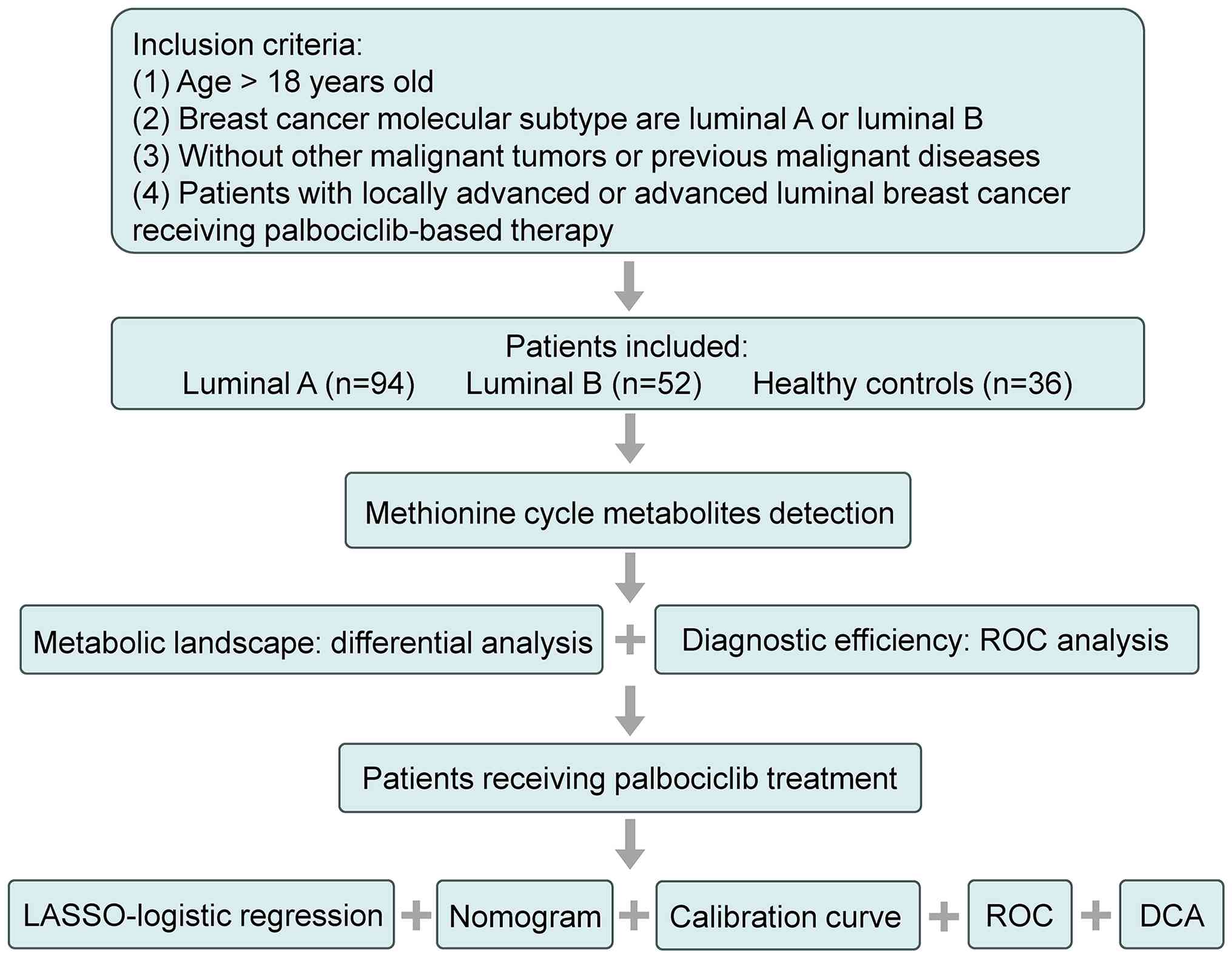
The overall experimental workflow is illustrated in
Fig. 1. First, patients with breast
cancer and HCs were enrolled according to the inclusion criteria,
with 94 patients with the luminal A subtype, 52 patients with the
luminal B subtype and 36 HCs being recruited. An MS detection
method for methionine cycle-related metabolites was optimized and
established in advance. Subsequently, the metabolites were
extracted from the plasma after collection and targeted MS
detection was performed. Differential analysis of the metabolic
landscape among groups was conducted, and the diagnostic efficacy
of individual/integrated metabolites was analyzed. Subsequently,
the clinical data of the patients was collected and integrated the
metabolic landscape. LASSO was used to screen variables related to
palbociclib resistance, and a logistic regression model was
constructed and visualized with a nomogram. The performance of the
prediction model was evaluated using calibration curves, ROC curves
and DCA.
The clinical characteristics of the enrolled
subjects are listed in Table I. The
average ages of patients with luminal A/B breast cancer were 58
(31–83) years and 59 (37–82) years, respectively, and the
average age of HCs was 51 (24–72)
years. The differential analysis of the methionine cycle-related
metabolites is presented in the subsequent sections.
 | Table I.Clinical characteristics of the
patients with breast cancer and HCs. |
Table I.
Clinical characteristics of the
patients with breast cancer and HCs.
|
| Patients with
breast cancer |
|
|
|---|
|
|
|
|
|
|---|
| Clinical
characteristics | Luminal A | Luminal B | HCs | Luminal vs.
HCs |
|---|
| Sample size | 94 | 52 | 36 |
|
| Age (years) | 58 (31–83) | 59 (37–82) | 51 (24–72) |
P=2.2×10⁻² |
| TNM stage |
|
|
|
|
| I | 33 | 14 |
|
|
| II | 34 | 26 |
|
|
|
III | 21 | 10 |
|
|
| IV | 6 | 2 |
|
|
| Grade |
|
|
|
|
| 1 | 29 | 10 |
|
|
| 2 | 39 | 24 |
|
|
| 3 | 26 | 18 |
|
|
| CEA, ng/ml |
|
|
|
|
| ≤5 | 63 | 36 |
|
|
|
>5 | 31 | 16 |
|
|
| CA125, U/ml |
|
|
|
|
|
≤25 | 67 | 33 |
|
|
|
>25 | 27 | 19 |
|
|
| CA15-3, U/ml |
|
|
|
|
|
≤24 | 58 | 27 |
|
|
|
>24 | 36 | 25 |
|
|
| Ki67 |
|
|
|
|
|
<40% | 52 | 37 |
|
|
|
≥40% | 42 | 15 |
|
|
| cfDNA, ng/ml |
|
|
|
|
|
<12 | 73 | 37 |
|
|
|
≥12 | 211 | 15 |
|
|
| Local/distant
metastasis |
|
|
|
|
| No | 67 | 40 |
|
|
|
Yes | 27 | 12 |
|
|
| Palbociclib
treatment |
|
|
|
|
| No | 67 | 40 |
|
|
|
Yes | 27 | 12 |
|
|
| Methionine, µM | 17.70±6.78 | 17.75±6.46 | 21.08±6.75 |
P=1.2×10⁻² |
| SAM, µM | 0.24±0.15 | 0.27±0.15 | 0.39±0.14 |
P=2.0×10⁻⁶ |
| SAH, µM | 0.095±0.048 | 0.088±0.06 | 0.112±0.055 |
P=2.5×10⁻² |
| Homocysteine,
µM | 8.42±4.49 | 8.65±4.42 | 10.49±4.97 |
P=2.0×10⁻² |
| SAM/SAH | 3.14±2.49 | 5.13±4.25 | 5.25±4.84 |
P=7.7×10⁻² |
Quantification of methionine
cycle-related metabolites
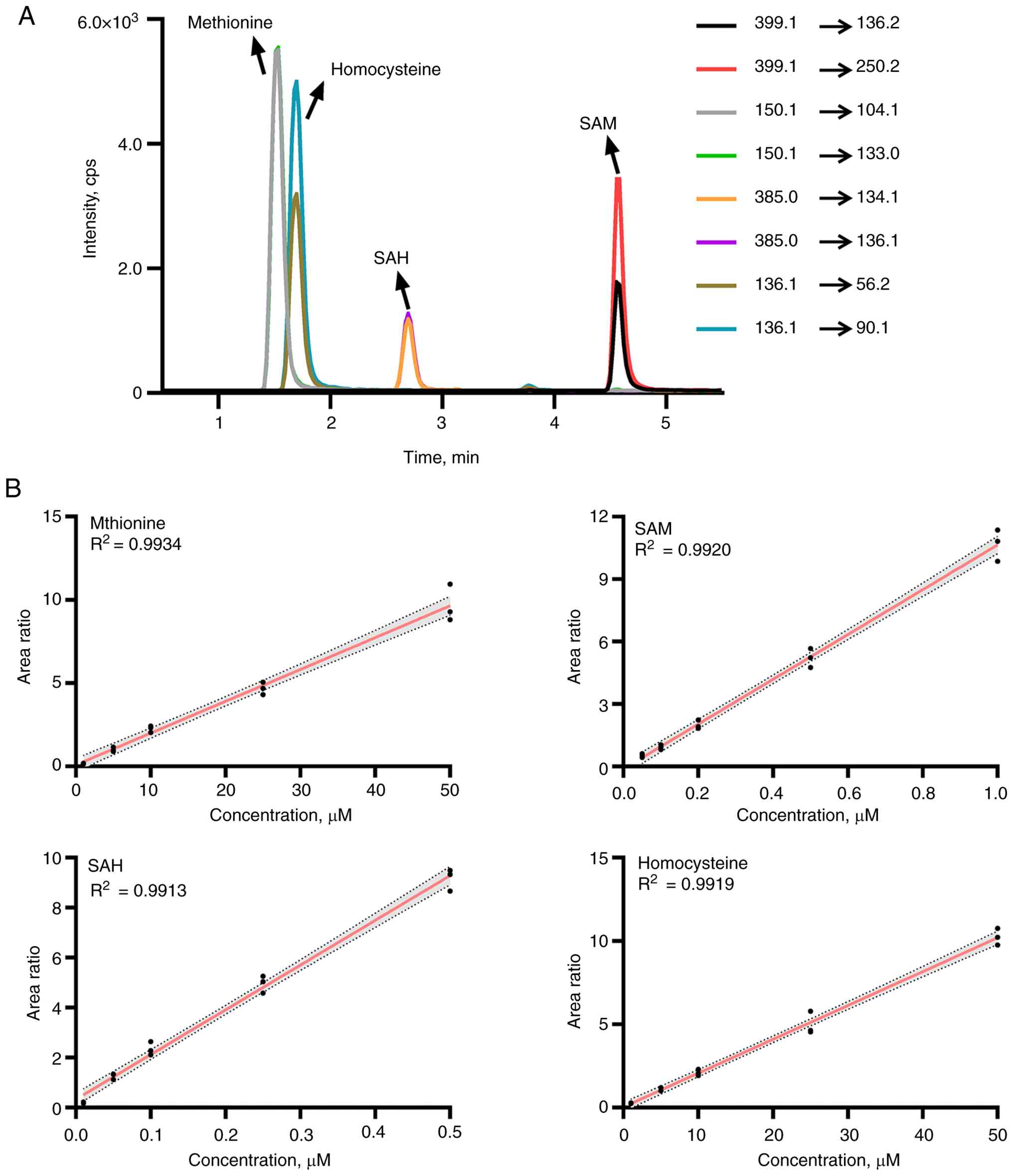
Targeted metabolomics was employed for metabolite
quantification (20,21). First, an MS detection method was
established using metabolite standards. By optimizing parameters
such as collision energy and fragment voltage, two MRM transitions
were ultimately selected for each metabolite and its isotopic
internal standard. The optimized mass spectrometry parameters are
listed in Table SI. After
validation, the methionine cycle-related metabolites exhibited
excellent separation efficiency and mass spectrometric response
within the 5-min gradient (Fig.
2A).
To achieve absolute quantification, calibration
curves were constructed for the target metabolites. Specifically,
calibration curves were constructed by plotting the ratio of the
total MRM peak areas of target metabolites to those of their
isotopic internal standards against metabolite concentrations. As
shown in Fig. 2B, using a weighting
factor of 1/x2, all the calibration curves exhibited
excellent linearity within the established concentration range
(R2 >0.99). Additionally, to verify the precision and
accuracy of the quantification, a quality control (QC) assessment
was performed for the method. The precision of the method,
including intraday and interday precision, was quantified using the
percent coefficient of variation (% CV). Method accuracy was
evaluated by calculating the percentage bias (% bias), which
reflects the discrepancy between the measured concentration of
samples and their respective standard concentrations. For all QC
samples, the resulting precision and accuracy values met the
acceptance criteria of ≤15% (LLOQ ≤20%). The detailed results of
the QC analysis are presented in Table
SII.
The clinical value of the methionine
cycle in luminal breast cancer
To evaluate the potential clinical value of the
methionine cycle, the present study first assessed the diagnostic
efficacy of metabolites for luminal breast cancer. Given the
significant difference in age between the luminal group and the HCs
group (P=2.4×10⁻²), an analysis of covariance (ANCOVA)
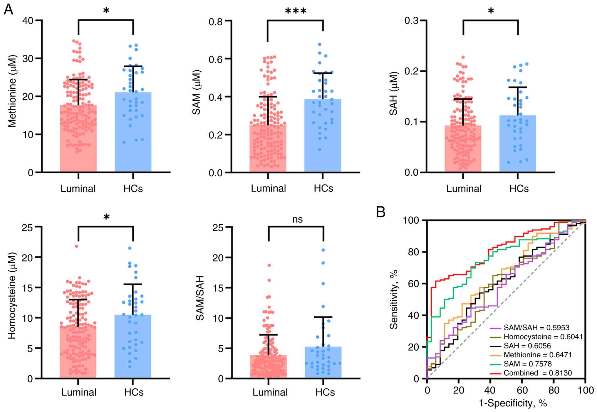
was performed to correct the impact of age. Compared with HCs,
patients with luminal breast cancer had lower plasma concentrations
of methionine (P=1.2×10⁻²), SAM (P=2.0×10⁻⁶),
SAH (P=2.5×10⁻²) and homocysteine
(P=2.0×10⁻²) (Fig. 3A).
The SAM/SAH ratio, a common indicator of methylation level
(22,23), did not differ
(P=7.7×10⁻²; Table
SIII). ROC analysis of individual metabolites revealed that SAM
exhibited the best diagnostic efficacy (AUC=0.7578). Notably, the
combined metabolites yielded improved diagnostic results when
compared with any single metabolite alone (AUC=0.8130; Fig. 3B). To elucidate this difference, a
DeLong test was conducted to compare the ROC curves of the combined
metabolites and individual metabolites. The results revealed that
the discriminatory performance of the combined metabolites was
significantly improved when compared with that of the individual
metabolites (Table SIV). Heatmap
analysis also revealed a marked difference in metabolic landscape
between patients with luminal breast cancer and HCs (Fig. S1).
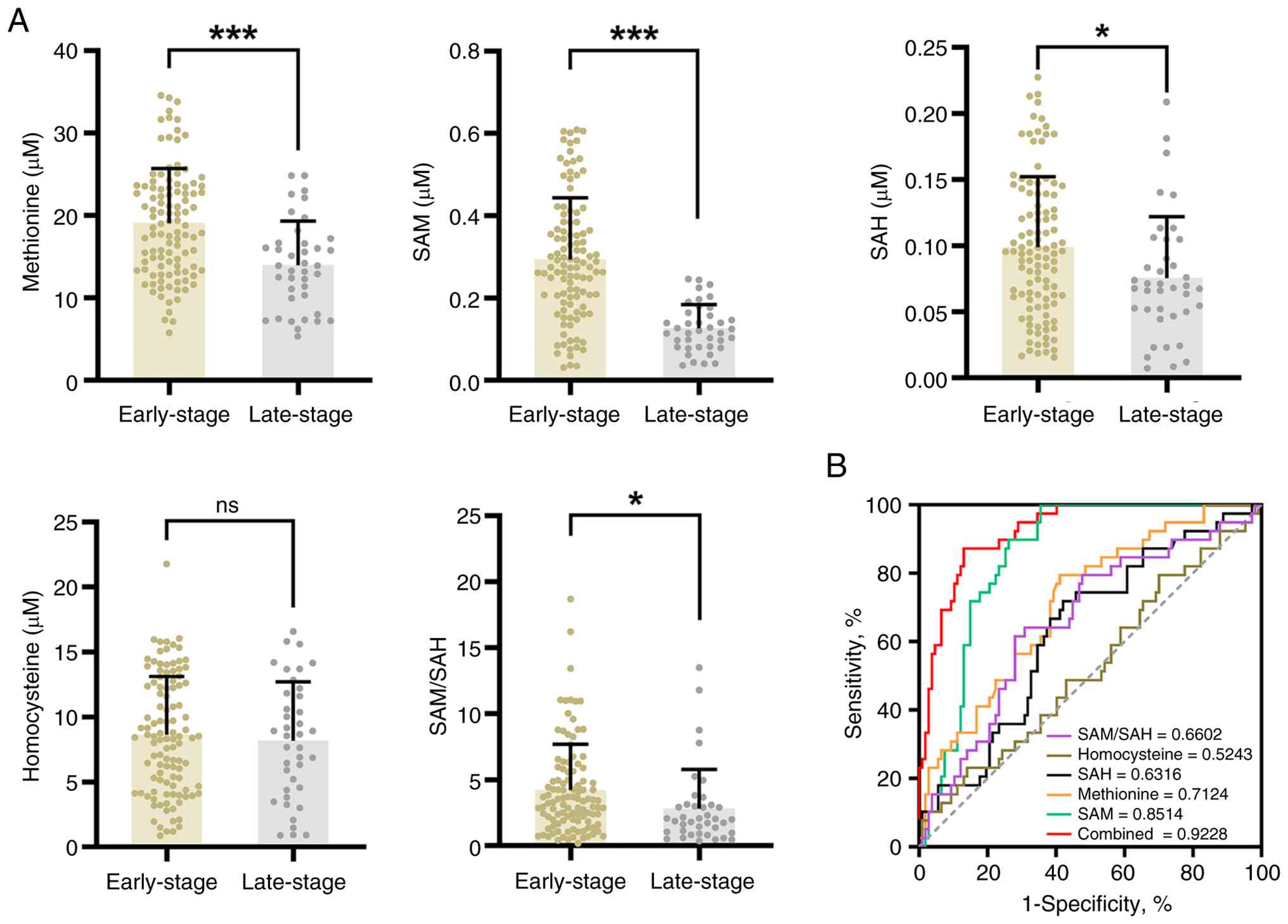
To further investigate changes in the methionine
cycle during the progression of luminal breast cancer, metabolite
levels were compared between patients at the early-stage and
late-stage. As shown in Fig. 4A,
methionine (P<0.001), SAM (P<0.001), SAH (P<0.05) and the
SAM/SAH (P<0.05) ratio were downregulated in patients at the
late-stage, whereas homocysteine abundance did not vary with
disease progression. ROC analysis revealed that SAM had the optimal
performance in distinguishing between early-stage and late-stage
patients with luminal breast cancer (AUC=0.8514), and the combined
use of multiple metabolites resulted in an even greater diagnostic
AUC of 0.9228 (Fig. 4B, Table SIV). Finally, to assess the value
of the methionine cycle in palbociclib resistance, the present
study compared the abundance of metabolites between patients who
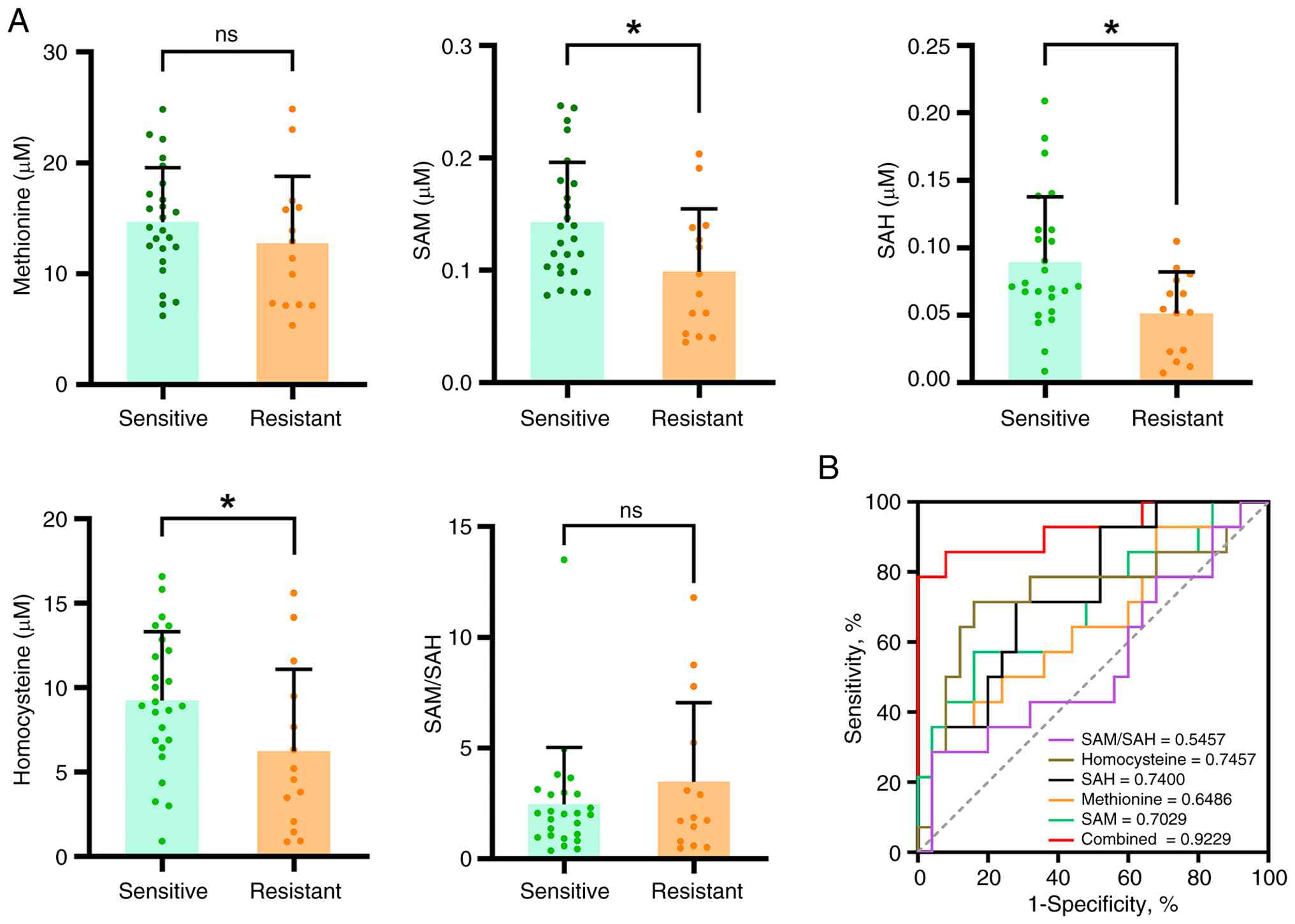
are palbociclib-sensitive and palbociclib-resistant. The abundances
of SAM, SAH and homocysteine were lower in resistant patients,
whereas methionine levels and the SAM/SAH ratio did not
significantly differ between sensitive and resistant patients
(Fig. 5A). ROC analysis
demonstrated that the diagnostic efficacy of SAM (AUC=0.7029), SAH
(AUC=0.7400) and homocysteine (AUC=0.7457) was favorable, and the
combination of all metabolite indicators achieved an AUC of 0.9229
for distinguishing palbociclib-sensitive/resistant patients
(Fig. 5B, Table SIV).
Construction of a prediction model for
palbociclib resistance
Given the limitations of current clinical methods
for determining therapeutic resistance, the present study aimed to
construct a palbociclib-resistance prediction model by combining
the methionine cycle with clinical information. A total of 39
palbociclib-treated patients (25 sensitive vs. 14 resistant) were
included in the model construction. After collecting clinical data,
the correlations between the levels of methionine cycle-related
metabolites and clinical characteristics were first analyzed. As
shown in Fig. S2, except for a
weak correlation (r=0.15, P<0.05) between methionine and age, no
significant correlations were observed between the other
metabolites and clinical indicators. ANCOVA further confirmed that
there was no significant linear relationship between age and
metabolites. These findings indirectly indicate that these
metabolic indicators differ from traditional clinical
indicators.
Subsequently, patients were divided into an internal
training set and an internal validation set at a 7:3 ratio based on
their resistance status. The training cohort included 18 sensitive
and 11 resistant patients, while the validation cohort included 7
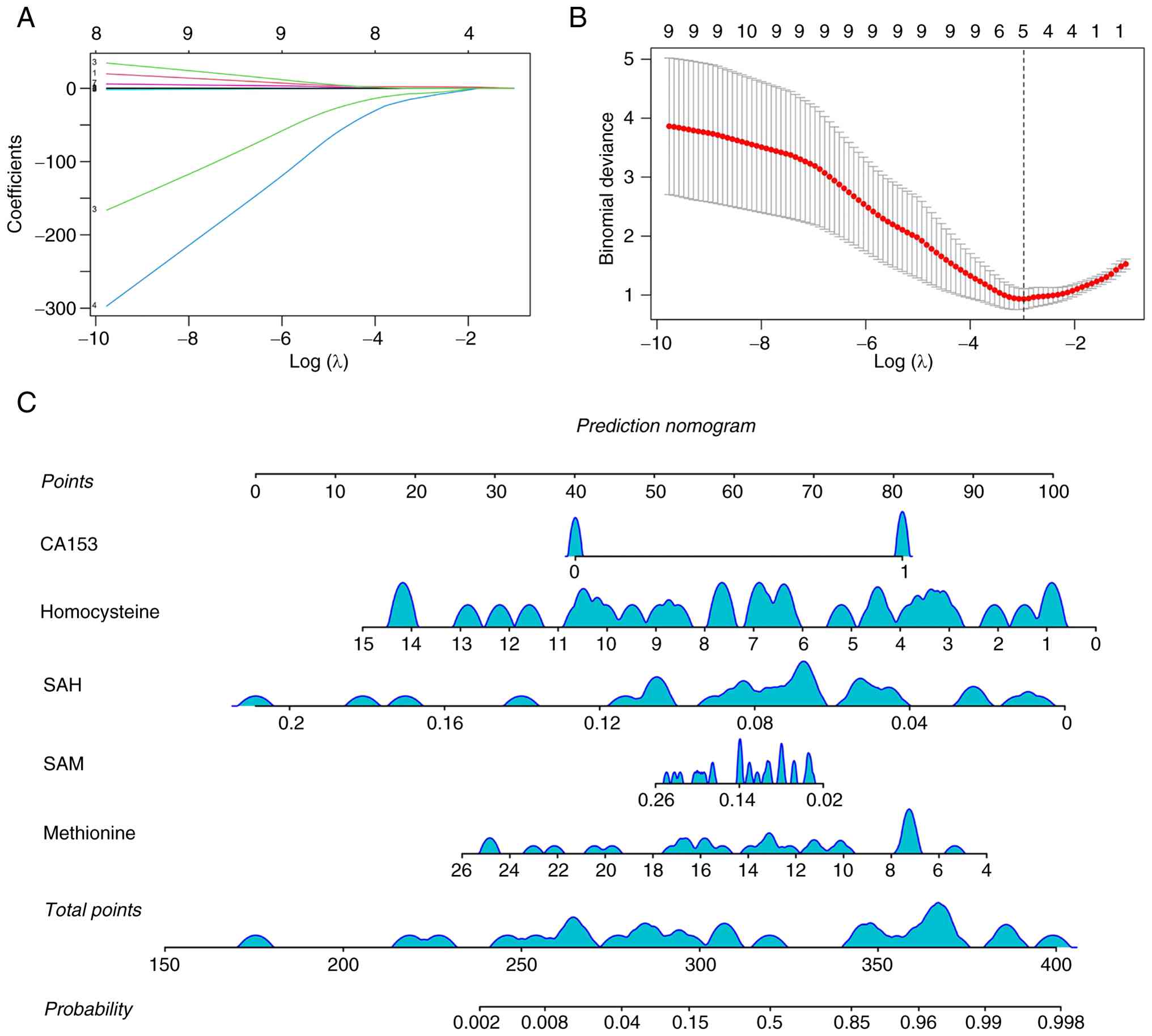
sensitive and 3 resistant patients. Here, LASSO regression was used
to screen variables associated with resistance (Fig. 6A), and 10-fold cross-validation was
employed for iterative analysis (Fig.
6B). The optimal model performance was achieved when the
minimum criterion was applied (lambda.min=0.0424), with a total of
5 variables selected, including methionine, SAM, SAH, homocysteine
and carbohydrate antigen 153 (CA153). On the basis of these
variables, the present study developed a multivariate logistic
regression model and further established a prediction model for the
probability of palbociclib resistance. This prediction model was
visualized as a nomogram (Fig. 6C),
which calculates the probability of palbociclib resistance based on
the total score corresponding to each variable. To reduce the risk
of overfitting, the present study further performed internal
validation using bootstrap resampling (500 iterations) for
correction (Fig. S3).
Multiple metrics were employed to validate the model
for accuracy and reliability. ROC analysis revealed an apparent AUC
of 0.9795 (95% CI, 0.9409–1.0000) in the training set (Fig. S4). The mean optimism was 0.0724,
yielding an optimism-corrected AUC of 0.9089, indicating that the
model maintained good discrimination after correction. The internal
validation set yielded an AUC of 0.8000, but its confidence
interval was unstable because of the small sample size. The
Hosmer-Lemeshow test (training set: P=7.64×10⁻¹;
validation set: P=3.57×10⁻¹) confirmed that the
correction was acceptable. Calibration curve analysis further
confirmed the favorable agreement between observed and predicted
results (Fig. S5). DCA
demonstrated the model's clinical benefits (Fig. S6). In summary, all these results
support the potential clinical utility of the prediction model.
Discussion
As a source of cellular nutrients, amino acid
metabolism provides the material basis for tumor growth and
proliferation. Amino acids, their metabolites and metabolic enzymes
directly or indirectly affect tumor cells, influence the tumor
microenvironment and regulate the host immune system, collectively
controlling tumor progression. Methionine metabolism has recently
become an emerging focus and potential therapeutic target in tumor
metabolism research (24). As the
core component of methionine metabolism, the methionine cycle has
been found to be associated with metabolic dysregulation in various
diseases (25,26). As the most prevalent subtype of
breast cancer, luminal breast cancer is defined by core clinical
features of hormone dependence and endocrine therapy sensitivity.
However, the emergence of endocrine resistance and subsequent
resistance to CDK4/6 inhibitors (for example, palbociclib) remains
a key bottleneck limiting improved disease prognosis. While
previous studies have mostly focused on genetic variations and
aberrant signaling pathways to elucidate luminal breast cancer
pathogenesis (27,28), metabolic reprogramming, especially
methionine cycle dysregulation, has received less attention in this
subtype.
The present study, for the first time, explored the
potential association between the methionine cycle and luminal
breast cancer, and clarified its utility in diagnosis, disease
progression and subsequent therapeutic resistance. On the basis of
the variables screened via LASSO, the present study constructed a
multivariate logistic regression model and a probability-prediction
nomogram for palbociclib resistance, with further reliability
confirmation of this strategy.
As a central branch of one-carbon metabolism, the
methionine cycle regulates methylation reactions, epigenetic
remodeling and redox homeostasis through the sequential conversion
of methionine, SAM, SAH and homocysteine. Previous studies have
reported reduced methionine-cycle activity in breast cancer cells
compared with normal breast epithelial cells (29,30).
Consistent with these findings, the present study observed
significantly decreased plasma methionine, SAM, SAH, homocysteine
levels, as well as SAM/SAH ratios in patients with luminal breast
cancer, suggesting systemic suppression of methionine cycle
metabolism during tumor development.
Tumor cells are known to exhibit ‘methionine
dependence’, characterized by enhanced methionine uptake and
utilization to support rapid proliferation, nucleotide synthesis
and transmethylation reactions. Accordingly, methionine restriction
suppresses tumor growth, whereas EMSY-driven methionine metabolism
promotes breast cancer progression (31). Intermittent methionine deprivation
can also enhance CD8+ T-cell-mediated antitumor immunity (32). In the present study, serum
methionine levels further decreased in advanced-stage and
palbociclib-resistant patients, suggesting that aggressive and
resistant tumors may have greater methionine demand and metabolic
consumption. As the major methyl donor in vivo, SAM
regulates DNA, RNA and histone methylation and thereby influences
gene expression and tumor progression. SAM-dependent methylation
has been implicated in m6A modification and histone trimethylation
during cancer metastasis (33).
Increased SAM enhances SETD1A-mediated trimethylation of histones,
ultimately facilitating the metastasis of colorectal cancer
(34). Enhanced methyltransferase
activity in tumors may accelerate SAM consumption and methyl-group
turnover. Consistently, the present study found significantly
decreased SAM levels in advanced-stage and resistant patients,
indicating increased methylation demand and epigenetic
reprogramming during tumor progression and palbociclib
resistance.
SAH, the downstream product of methylation
reactions, is a potent inhibitor of methyltransferases, while the
SAM/SAH ratio is considered a better indicator of methylation
potential than either metabolite alone. Reduced SAM/SAH ratios have
been reported in patients with liver cancer and cancer cachexia
(25). Recent studies have
associated serum SAH levels with cancer prognosis (35,36),
while cellular investigations have revealed the potential role of
SAH hydrolase in tumor invasion and metastasis (37,38).
In the present cohort, both SAH and the SAM/SAH ratio progressively
decreased from healthy controls to advanced-stage and resistant
patients, suggesting impaired methylation capacity and disrupted
one-carbon metabolic homeostasis in aggressive luminal breast
cancer.
Homocysteine associates methionine metabolism with
folate metabolism, transsulfuration pathways and glutathione
synthesis. Although the findings of previous studies on
homocysteine alterations in breast cancer remain controversial
(39–41), the present study observed
significantly lower homocysteine levels in luminal breast cancer
patients compared with HCs, particularly in advanced-stage and
resistant patients. Since estrogen can suppress homocysteine levels
(42,43). this may partly explain the findings
in the endocrine-responsive cohort. In addition, resistant tumor
cells may consume more homocysteine for glutathione synthesis and
antioxidant defense under endocrine therapy and CDK4/6
inhibitor-induced oxidative stress.
Collectively, the present results demonstrated a
consistent downward trend in methionine, SAM, SAH, homocysteine
levels, as well as the SAM/SAH ratio from HCs to advanced-stage and
palbociclib-resistant patients with luminal breast cancer. These
findings suggest that progressive exhaustion of methionine cycle
metabolism may accompany tumor progression and therapeutic
resistance, potentially reflecting increased metabolic demand,
accelerated transmethylation activity, epigenetic remodeling and
oxidative stress adaptation in luminal breast cancer.
The present study has several limitations. First,
this was a single-center study with a limited sample size, and
external validation in an independent cohort was not performed.
Therefore, the generalizability of the nomogram remains to be
confirmed in future studies. This is a major limitation of the
present study. Second, in terms of the breadth and depth of the
metabolomic analysis, the present study focused only on four key
metabolites of the methionine cycle. The methionine cycle is not
isolated but interacts with other pathways, and uncollected
confounders (for example, renal function, folate and B12) could not
be adjusted, precluding a full understanding of global metabolic
reprogramming in luminal breast cancer. Finally, in terms of
mechanistic research and clinical translation, the present study
primarily relied on correlation analysis of clinical samples. The
specific molecular mechanisms by which methionine cycle metabolites
affect luminal breast cancer progression and palbociclib resistance
were not validated in cell or animal models. Thus, these methionine
cycle metabolites are best viewed as potential predictive
biomarkers that require validation in larger multicenter cohorts.
Future work should improve research on the metabolic mechanisms of
luminal breast cancer and its clinical translation through
multicenter prospective cohorts, multiomics techniques and
basic/clinical experiments.
Using LC-MS/MS-based targeted metabolomics, the
present study systematically analyzed plasma differences in key
methionine cycle metabolites (methionine, SAM, SAH and
homocysteine) between patients with luminal breast cancer and
healthy controls, revealing significant alterations in the majority
of metabolites after adjustment for confounders. The present study
further explored the clinical value of the cycle in disease
progression and palbociclib resistance, identifying distinct
metabolic signatures associated with tumor aggressiveness and drug
resistance. Finally, the present study developed and validated a
resistance prediction model that integrates these metabolic
indicators with routine clinical parameters, offering a practical
tool for risk stratification in patients with luminal breast
cancer. Limitations include a single-center study with a small
sample size without external validation, unadjusted confounders
(for example, renal function, B12) and lack of mechanistic
validation beyond correlation analysis. Future studies with
independent cohorts, expanded metabolomic coverage and experimental
validation are needed to confirm these observations.
Supplementary Material
Supporting Data
Supporting Data
Acknowledgements
Not applicable.
Funding
The present study was supported by the Research Project of
Jiangsu Cancer Hospital (grant no. XHMS202504).
Availability of data and materials
The data generated in the present study may be
requested from the corresponding author. The mass spectrometry data
generated in the present study may be found in the Metabolomics
Workbench database under accession number ST004922 or at the
following URL: https://www.metabolomicsworkbench.org/data/DRCCMetadata.php?Mode=Study&StudyID=ST004922.
Authors' contributions
FX and ZW conceived and designed the project. WW
carried out the mass spectrometry experiment. CM and FX
participated in acquisition of clinical data. YT, HY and YM
analyzed and interpreted the data. WW, YT and ZW drafted the
manuscript. FX and ZW confirm the authenticity of all the raw data.
All authors read and approved the final manuscript.
Ethics approval and consent to
participate
Blood sample collection was approved by the Ethics
Committee of Jiangsu Cancer Hospital (Nanjing, China) (approval no.
2023021), and all patients and HCs provided written informed
consent.
Patient consent for publication
All patients and HCs signed written informed consent
forms and agreed to the publication.
Competing interests
The authors declare that they have no competing
interests
References
|
1
|
Bray F, Laversanne M, Sung H, Ferlay J,
Siegel RL, Soerjomataram I and Jemal A: Global cancer statistics
2022: GLOBOCAN estimates of incidence and mortality worldwide for
36 cancers in 185 countries. CA Cancer J Clin. 74:229–263.
2024.PubMed/NCBI
|
|
2
|
Abdulkareem IH: Aetio-pathogenesis of
breast cancer. Niger Med J. 54:371–375. 2013. View Article : Google Scholar : PubMed/NCBI
|
|
3
|
Hany D, Vafeiadou V and Picard D:
CRISPR-Cas9 screen reveals a role of purine synthesis for estrogen
receptor α activity and tamoxifen resistance of breast cancer
cells. Sci Adv. 9:eadd36852023. View Article : Google Scholar : PubMed/NCBI
|
|
4
|
Giaquinto AN, Sung H, Newman LA, Freedman
RA, Smith RA, Star J, Jemal A and Siegel RL: Breast cancer
statistics 2024. CA Cancer J Clin. 74:477–495. 2024.PubMed/NCBI
|
|
5
|
Makhlouf S, Wahab N, Toss M, Ibrahim A,
Lashen AG, Atallah NM, Ghannam S, Jahanifar M, Lu W, Graham S, et
al: Evaluation of tumour infiltrating lymphocytes in luminal breast
cancer using artificial intelligence. Br J Cancer. 129:1747–1758.
2023. View Article : Google Scholar : PubMed/NCBI
|
|
6
|
Turner NC, Slamon DJ, Ro J, Bondarenko I,
Im SA, Masuda N, Colleoni M, DeMichele A, Loi S, Verma S, et al:
Overall survival with palbociclib and fulvestrant in advanced
breast cancer. N Engl J Med. 379:1926–1936. 2018. View Article : Google Scholar : PubMed/NCBI
|
|
7
|
Park YH, Im SA, Park K, Wen J, Lee KH,
Choi YL, Lee WC, Min A, Bonato V, Park S, et al: Longitudinal
multi-omics study of palbociclib resistance in
HR-positive/HER2-negative metastatic breast cancer. Genome Med.
15:552023. View Article : Google Scholar : PubMed/NCBI
|
|
8
|
Seale KN and Tkaczuk KHR: Circulating
biomarkers in breast cancer. Clin Breast Cancer. 22:e319–e331.
2022. View Article : Google Scholar : PubMed/NCBI
|
|
9
|
Mentch SJ and Locasale JW: One-carbon
metabolism and epigenetics: Understanding the specificity. Ann N Y
Acad Sci. 1363:91–98. 2016. View Article : Google Scholar : PubMed/NCBI
|
|
10
|
Ducker GS and Rabinowitz JD: One-carbon
metabolism in health and disease. Cell Metab. 25:27–42. 2017.
View Article : Google Scholar : PubMed/NCBI
|
|
11
|
Jin X, Liu L, Liu D, Wu J, Wang C, Wang S,
Wang F, Yu G, Jin X, Xue YW, et al: Unveiling the methionine cycle:
A key metabolic signature and NR4A2 as a methionine-responsive
oncogene in esophageal squamous cell carcinoma. Cell Death Differ.
31:558–573. 2024. View Article : Google Scholar : PubMed/NCBI
|
|
12
|
Zhao T and Lum JJ: Methionine
cycle-dependent regulation of T cells in cancer immunity. Front
Oncol. 12:9695632022. View Article : Google Scholar : PubMed/NCBI
|
|
13
|
Martínez Y, Li X, Liu G, Bin P, Yan W, Más
D, Valdivié M, Hu CA, Ren W and Yin Y: The role of methionine on
metabolism, oxidative stress, and diseases. Amino Acids.
49:2091–2098. 2017. View Article : Google Scholar : PubMed/NCBI
|
|
14
|
Liu F, Zhou H, Peng Y, Qiao Y, Wang P, Si
C, Wang X, Gong J, Chen K and Song F: Plasma one-carbon
metabolism-related micronutrients and the risk of breast cancer:
Involvement of DNA methylation. Nutrients. 15:36212023. View Article : Google Scholar : PubMed/NCBI
|
|
15
|
Santos JR, Waitzberg DL, da Silva IDCG,
Junior TCT, Barros LRC, Canuto GAB, Faccio AT, Yamaguchi LF, Kato
MJ, Tavares MFM, et al: Distinct pattern of one-carbon metabolism,
a nutrient-sensitive pathway, in invasive breast cancer: A
metabolomic study. Oncotarget. 11:16372020. View Article : Google Scholar : PubMed/NCBI
|
|
16
|
Ilisso CP, Delle Cave D, Mosca L, Pagano
M, Coppola A, Mele L, Caraglia M, Cacciapuoti G and Porcelli M:
S-Adenosylmethionine regulates apoptosis and autophagy in MCF-7
breast cancer cells through the modulation of specific microRNAs.
Cancer Cell Int. 18:1972018. View Article : Google Scholar : PubMed/NCBI
|
|
17
|
Ilisso CP, Castellano M, Zappavigna S,
Lombardi A, Vitale G, Dicitore A, Cacciapuoti G, Caraglia M and
Porcelli M: The methyl donor S-adenosylmethionine potentiates
doxorubicin effects on apoptosis of hormone-dependent breast cancer
cell lines. Endocrine. 50:212–222. 2015. View Article : Google Scholar : PubMed/NCBI
|
|
18
|
Amin MB, Greene FL, Edge SB, Compton CC,
Gershenwald JE, Brookland RK, Meyer L, Gress DM, Byrd DR and
Winchester DP: The eighth edition AJCC cancer staging manual:
Continuing to build a bridge from a population-based to a more
‘personalized’ approach to cancer staging. CA Cancer J Clin.
67:93–99. 2017.PubMed/NCBI
|
|
19
|
Sud M, Fahy E, Cotter D, Azam K, Vadivelu
I, Burant C, Edison A, Fiehn O, Higashi R, Nair KS, et al:
Metabolomics workbench: An international repository for
metabolomics data and metadata, metabolite standards, protocols,
tutorials and training, and analysis tools. Nucleic Acids Res.
44:D463–D470. 2016. View Article : Google Scholar : PubMed/NCBI
|
|
20
|
Wang Z, Li L, Kuang Y, Yao J, Xu F and
Chen Y: Simultaneous quantification of multiple single nucleotide
variants in PIK3CA ctDNA using mass-tagged LCR probe sets. Talanta.
258:1244262023. View Article : Google Scholar : PubMed/NCBI
|
|
21
|
Xu Q, Zhu M, Yang T, Xu F, Liu Y and Chen
Y: Quantitative assessment of human serum transferrin receptor in
breast cancer patients pre-and post-chemotherapy using peptide
immunoaffinity enrichment coupled with targeted proteomics. Clin
Chim Acta. 448:118–123. 2015. View Article : Google Scholar : PubMed/NCBI
|
|
22
|
Liu Z, Li X, Wang T, Zhang H, Li X, Xu J,
Zhang Y, Zhao Z, Yang P, Zhou C, et al: SAH and SAM/SAH ratio
associate with acute kidney injury in critically ill patients: A
case-control study. Clin Chim Acta. 553:1177262024. View Article : Google Scholar : PubMed/NCBI
|
|
23
|
Xiao J, You Y, Chen X, Tang Y, Chen Y, Liu
Q, Liu Z and Ling W: Higher S-adenosylhomocysteine and lower ratio
of S-adenosylmethionine to S-adenosylhomocysteine were more closely
associated with increased risk of subclinical atherosclerosis than
homocysteine. Front Nutr. 9:9186982022. View Article : Google Scholar : PubMed/NCBI
|
|
24
|
Ma C, Xu A, Zuo L, Li Q, Fan F, Hu Y and
Sun C: Methionine dependency and restriction in cancer: Exploring
the pathogenic function and therapeutic potential. Pharmaceuticals
(Basel). 18:6402025. View Article : Google Scholar : PubMed/NCBI
|
|
25
|
Lin K, Wei L, Wang R, Li L, Song S, Wang
F, He M, Pu W, Wang J, Wazir J, et al: Disrupted methionine cycle
triggers muscle atrophy in cancer cachexia through epigenetic
regulation of REDD1. Cell Metab. 37:460–476.e8. 2025. View Article : Google Scholar : PubMed/NCBI
|
|
26
|
Li F, Liu P, Mi W, Li L, Anderson NM,
Lesner NP, Burrows M, Plesset J, Majer A, Wang G, et al: Blocking
methionine catabolism induces senescence and confers vulnerability
to GSK3 inhibition in liver cancer. Nat Cancer. 5:131–146. 2024.
View Article : Google Scholar : PubMed/NCBI
|
|
27
|
Santarpia L, Bottai G, Kelly CM, Győrffy
B, Székely B and Pusztai L: Deciphering and targeting oncogenic
mutations and pathways in breast cancer. Oncologist. 21:1063–1078.
2016. View Article : Google Scholar : PubMed/NCBI
|
|
28
|
Langille E, Al-Zahrani KN, Ma Z, Liang M,
Uuskula-Reimand L, Espin R, Teng K, Malik A, Bergholtz H, Ghamrasni
SE, et al: Loss of epigenetic regulation disrupts lineage
integrity, induces aberrant alveogenesis, and promotes breast
cancer. Cancer Discov. 12:2930–2953. 2022. View Article : Google Scholar : PubMed/NCBI
|
|
29
|
Pankevičiūtė-Bukauskienė M, Mikalayeva V,
Žvikas V, Skeberdis VA and Bordel S: Multi-omics analysis revealed
increased de novo synthesis of serine and lower activity of the
methionine cycle in breast cancer cell lines. Molecules.
28:45352023. View Article : Google Scholar : PubMed/NCBI
|
|
30
|
Lien EC, Ghisolfi L, Geck RC, Asara JM and
Toker A: Oncogenic PI3K promotes methionine dependency in breast
cancer cells through the cystine-glutamate antiporter xCT. Sci
Signal. 10:eaao66042017. View Article : Google Scholar : PubMed/NCBI
|
|
31
|
Liu CC, Chen L, Cai YW, Chen YF, Liu YM,
Zhou YJ, Shao ZM and Yu KD: Targeting EMSY-mediated methionine
metabolism is a potential therapeutic strategy for triple-negative
breast cancer. Cell Rep Med. 5:1013962024. View Article : Google Scholar : PubMed/NCBI
|
|
32
|
Xue Y, Lu F, Chang Z, Li J, Gao Y, Zhou J,
Luo Y, Lai Y, Cao S, Li X, et al: Intermittent dietary methionine
deprivation facilitates tumoral ferroptosis and synergizes with
checkpoint blockade. Nat Commun. 14:47582023. View Article : Google Scholar : PubMed/NCBI
|
|
33
|
Li T, Tan YT, Chen YX, Zheng XJ, Wang W,
Liao K, Mo HY, Lin J, Yang W, Piao HL, et al: Methionine deficiency
facilitates antitumour immunity by altering m6A methylation of
immune checkpoint transcripts. Gut. 72:501–511. 2023. View Article : Google Scholar : PubMed/NCBI
|
|
34
|
Zhang Y, Yu H, Zhang J, Gao H, Wang S, Li
S, Wei P, Liang J, Yu G, Wang X, et al: Cul4A-DDB1-mediated
monoubiquitination of phosphoglycerate dehydrogenase promotes
colorectal cancer metastasis via increased S-adenosylmethionine. J
Clin Invest. 131:e1461872021. View Article : Google Scholar : PubMed/NCBI
|
|
35
|
Wusiman M, Huang SY, Liu ZY, He TT, Fang
AP, Li MC, Yang MT, Wang C, Zhang YJ and Zhu HL: Serum
S-adenosylhomocysteine, rather than homocysteine, is associated
with hepatocellular carcinoma survival: A prospective cohort study.
Am J Clin Nutr. 120:481–490. 2024. View Article : Google Scholar : PubMed/NCBI
|
|
36
|
Yalley NN, Armasu SM, Fan WZ, Yan IK,
Ahmed FY, Stål P, Roberts LR, Patel T and Antwi SO: Associations of
S-adenosylmethionine and S-adenosylhomocysteine with hepatocellular
carcinoma. Metabolites. 15:7402025. View Article : Google Scholar : PubMed/NCBI
|
|
37
|
Park SJ, Kong HK, Kim YS, Lee YS and Park
JH: Inhibition of S-adenosylhomocysteine hydrolase decreases cell
mobility and cell proliferation through cell cycle arrest. Am J
Cancer Res. 5:21272015.PubMed/NCBI
|
|
38
|
Pavičić I, Rokić F and Vugrek O: Effects
of S-adenosylhomocysteine hydrolase downregulation on Wnt signaling
pathway in SW480 cells. Int J Mol Sci. 24:161022023. View Article : Google Scholar : PubMed/NCBI
|
|
39
|
Hasan T, Arora R, Bansal AK, Bhattacharya
R, Sharma GS and Singh LR: Disturbed homocysteine metabolism is
associated with cancer. Exp Mol Med. 51:1–13. 2019. View Article : Google Scholar : PubMed/NCBI
|
|
40
|
He Q, Yang Z, Sun Y, Qu Z, Jia X, Li J,
Lin Y and Luo Y: The impact of homocysteine on the risk of
hormone-related cancers: A Mendelian randomization study. Front
Nutr. 8:6453712021. View Article : Google Scholar : PubMed/NCBI
|
|
41
|
Naushad SM, Reddy CA, Kumaraswami K,
Divyya S, Kotamraju S, Gottumukkala SR, Digumarti RR and Kutala VK:
Impact of hyperhomocysteinemia on breast cancer initiation and
progression: Epigenetic perspective. Cell Biochem Biophys.
68:397–406. 2014. View Article : Google Scholar : PubMed/NCBI
|
|
42
|
Gaikwad NW: Mass spectrometry evidence for
formation of estrogen-homocysteine conjugates: Estrogens can
regulate homocysteine levels. Free Radic Biol Med. 65:1447–1454.
2013. View Article : Google Scholar : PubMed/NCBI
|
|
43
|
Dimitrova KR, DeGroot K, Myers AK and Kim
YD: Estrogen and homocysteine. Cardiovasc Res. 53:577–588. 2002.
View Article : Google Scholar : PubMed/NCBI
|