Introduction
Breast cancer remains the most common malignancy
among women worldwide (1) and the
leading cause of cancer-related deaths in women, with an estimated
2.3 million new cases and 670,000 deaths globally in 2022 (2,3).
Although notable progress has been made in early diagnosis and
comprehensive treatment strategies, tumor migration continues to be
the primary cause of treatment failure and patient mortality
(4,5). The invasion and migration of tumor
cells constitute a multi-step complex process, involving metabolic
reprogramming, epithelial-mesenchymal transition and remodeling of
the tumor microenvironment (6).
Among these, metabolic dysregulation, an emerging hallmark of
cancer, serves a key role in providing precursors for
macromolecular synthesis and maintaining redox homeostasis. In
particular, dysregulated amino acid metabolism has been closely
associated with malignant tumor progression (7).
Branched-chain amino acids (BCAAs), including
leucine, isoleucine and valine, serve not only as key substrates
for protein synthesis but also as notable signaling molecules and
energy sources (8). BCAA metabolism
is frequently dysregulated in cancer and the key enzyme BCAA
transaminase 1 (BCAT1) has been implicated in tumor progression
(9). BCAT1 catalyzes the initial
step of BCAA catabolism, sustaining intracellular BCAA reserves to
fuel anabolic processes such as mTOR signaling and nucleotide
synthesis (8,9). In breast cancer, BCAT1 is upregulated
in aggressive subtypes including triple-negative breast cancer
(TNBC) and this upregulation is associated with poor prognosis
(10). Specifically,
immunohistochemical analysis of breast cancer tissue microarrays in
a previous study confirmed that high BCAT1 expression is associated
with higher histological grade (Nottingham grading system), lymph
node migration and worse prognosis (11). Functional studies have demonstrated
that BCAT1 promotes breast cancer cell proliferation, migration and
invasion, partly by activating mTOR signaling and sustaining
intracellular BCAA reserves (11,12),
with emerging evidence implicating downstream pathways such as
SHOC2-Ras-ERK in TNBC (13). BCAT1
has also been associated with endocrine therapy resistance;
specifically, its upregulation sustains the proliferation of
antiestrogen-resistant and estrogen receptor (ER)α−
breast cancer cells (12).
However, despite these advances, key knowledge gaps
remain regarding the oncogenic functions and underlying mechanisms
of BCAT1 in TNBC, particularly its contribution to migration
through metabolic reprogramming (10,12,13).
To the best of our knowledge, the majority of mechanistic studies
have focused on ER+ models, and the specific role of
BCAT1 in TNBC migration, particularly its contribution through
metabolic reprogramming, has not been fully elucidated. Notably,
whether pharmacological targeting of BCAT1-mediated BCAA metabolism
can suppress TNBC progression has not been explored, to the best of
our knowledge. Due to the potent pro-metastatic functions of BCAT1
in TNBC, identifying agents that modulate BCAT1 activity and BCAA
metabolism may offer novel therapeutic opportunities in the
future.
Astragalus membranaceus, a traditional
Chinese medicine that is believed to reinforce vital energy and
enhance immune function, contains the active ingredient
astragaloside IV (AS-IV), which has been demonstrated to possess
diverse pharmacological activities including immunomodulatory,
anti-inflammatory, antioxidant and antitumor effects (14–16).
Recent studies have suggested that AS-IV can inhibit proliferation,
invasion and induce apoptosis in multiple types of cancer cell,
including hepatocellular carcinoma, nasopharyngeal carcinoma, oral
cancer, ovarian cancer, gastric cancer and triple-negative breast
cancer (17–23). Nevertheless, whether AS-IV exerts
antitumor effects through the modulation of tumor metabolism,
particularly amino acid metabolic pathways, remains to be
elucidated.
The concept of network pharmacology was first
proposed by the British scholar Andrew L. Hopkins in 2007 (24). This discipline integrates biology
systems, traditional pharmacology computer network science, systems
biology, bioinformatics and network science. Based on a
systems-level perspective and a holistic view of biological
networks, network pharmacology elucidates the molecular connections
between drugs and diseases, clarifies drug therapeutic mechanisms
and provides guidance for novel drug development and clinical
practice (25). The analytical
methods of network pharmacology for target genes align well with
the multi-component, -target and -mechanism characteristics of
traditional Chinese medicine, making it a current research hotspot
in the field (26–30). Molecular docking technology, by
contrast, is a computer-aided drug design method. The core
mechanism lies in embedding small molecule compounds into
macromolecular structures, aiming to predict interactions between
‘target-ligand’ and ‘structure-activity’ at the molecular level.
Molecular docking technology is extensively applied in predicting
the efficacy of novel compounds, exploring polypharmacology and
drug repurposing (31–33).
Among breast cancer subtypes, TNBC is particularly
aggressive, lacks targeted therapies and is associated with poor
prognosis. Increasing evidence suggests that BCAA metabolism may be
particularly key in TNBC, as these tumors exhibit metabolic
vulnerabilities distinct from hormone receptor or HER2-amplified
subtypes (12,34,35).
Therefore, as an initial exploration of whether AS-IV exerts
anti-metastatic effects through BCAT1-mediated BCAA metabolism, the
present study focused on the MDA-MB-231 TNBC cell line as a
representative and clinically relevant model.
Building upon this background, we hypothesized that
AS-IV suppresses the invasive and migratory capabilities of
MDA-MB-231 TNBC cells and that this effect may involve
BCAT1-mediated reprogramming of BCAA metabolism. This hypothesis
was derived from network pharmacology predictions, which identified
multiple potential targets of AS-IV associated with breast cancer.
BCAT1 was prioritized for experimental validation based on three
criteria: i) BCAT1 ranked among the top core targets in the
protein-protein interaction (PPI) network based on degree
centrality; ii) pathway enrichment analysis demonstrated notable
involvement of the BCAA degradation pathway; and iii) emerging
evidence suggests BCAA metabolism serves a key role in TNBC
progression (10,12,13);
however, whether AS-IV modulates this pathway has not been
explored. Notably, although previous studies have reported the
antitumor effects of AS-IV in various cancer types, its direct
targeting of a metabolic enzyme, particularly BCAT1, has not yet
been demonstrated. Thus, the present study, to the best of our
knowledge, represents the first attempt to associate AS-IV with
BCAT1-mediated BCAA metabolic reprogramming in breast cancer cells.
Specifically, the present study aimed to investigate this
hypothesis and further explore the underlying molecular mechanisms,
with a focus on examining whether AS-IV interacts with BCAT1 to
modulate BCAA metabolism in TNBC cells. The findings may provide a
theoretical basis for further investigation of AS-IV in metabolic
intervention strategies against breast cancer migration in the
future.
Materials and methods
Experimental materials
Cell source
The MDA-MB-231 TNBC cell line [American Type Culture
Collection (ATCC)® HTB-26™] was obtained from the ATCC.
All cells were cultured in high-glucose DMEM medium (cat. no.
11965084; Gibco; Thermo Fisher Scientific, Inc.) supplemented with
10% fetal bovine serum (cat. no. 30-2020; ATCC) and 1%
penicillin/streptomycin at 37°C under 5% CO2.
Reagents
AS-IV (purity, ≥98%; cat. no. S1101-01019) was
purchased from Nanjing Jingzhu Biotechnology Co., Ltd. The rabbit
polyclonal antibody against BCAT1 (cat. no. ab197941) was acquired
from Abcam. Lipofectamine® 3000 transfection reagent was
obtained from Thermo Fisher Scientific, Inc. BCAT1-specific
small-interfering RNA (siRNA/si) and non-targeting negative control
(NC) siRNA, BCAT1 overexpression (OE) plasmid (pcDNA3.1-BCAT1) and
empty vector were synthesized by Shanghai GenePharma Co., Ltd.
Matrigel matrix was procured from Corning, Inc. The Seahorse XF
Cell Energy Metabolism Assay Kit (cat. no. 103325-100) was sourced
from Agilent Technologies, Inc. BCAA standards (leucine, isoleucine
and valine) and isotopic internal standards were purchased from
MilliporeSigma.
Network pharmacological analysis
Screening of drug and disease targets
Potential targets of AS-IV were identified using
databases including SwissTargetPrediction (based on 2D and 3D
similarity measures; http://www.swisstargetprediction.ch/), Similarity
Ensemble Approach Search Server (based on set-wise chemical
similarity among ligands; http://sea.bkslab.org/), PharmMapper (based on reverse
pharmacophore mapping; http://www.lilab-ecust.cn/pharmmapper/), SuperPred
(based on machine learning and 2D similarity; http://prediction.charite.de/subpages/target_prediction.php)
and TargetNet (based on Naïve Bayes QSAR models; http://targetnet.scbdd.com/calcnet/index/). Breast
cancer-associated targets were retrieved from disease databases
such as GeneCards (http://www.genecards.org/), Online Mendelian
Inheritance in Man (OMIM; http://omim.org/)
and Disease Gene Network (DisGeNET; http://disgenet.com/). The intersections of the drug
and disease targets were selected as the candidate targets.
Screening of core targets for AS-IV in
the treatment of breast cancer
The intersections between the obtained targets of
AS-IV and the disease targets of breast cancer were identified
using the Venny online platform (version 2.1.0; http://bioinfogp.cnb.csic.es/tools/venny/). This
process yielded the potential therapeutic targets of AS-IV for
breast cancer and a Venn diagram was generated accordingly.
PPI network construction and
topological analysis
The potential therapeutic targets of AS-IV for
breast cancer were imported into the Search Tool for Retrieval of
Interacting Genes/Proteins database (https://string-db.org/). Under ‘Multiple proteins’ the
‘Organisms’ parameter was set to ‘Homo sapiens’ to conduct
PPI analysis and obtain PPI information, which was then saved in
tab-separated values format. Subsequently, the data was imported
into the Cytoscape software (version 3.10.0; http://cytoscape.org/) for visualization. Topological
analysis was performed using the CytoHubba (version 1.6) and MCODE
(version 2.0.3)plugins.
Gene Ontology (GO) and Kyoto
Encyclopedia of Genes and Genomes (KEGG) enrichment analysis
To further characterize the roles of the potential
targets of AS-IV in treating breast cancer in terms of gene
function and signaling pathways, KEGG signaling pathway and GO
functional enrichment analyses were performed on the potential
therapeutic targets using the Metascape database (https://metascape.org; version 3.5.20260201). The
enrichment analysis results were visualized via the bioinformatics
online plotting platform (http://www.bioinformatics.com.cn/).
Construction of the ‘AS-IV-core
targets-key pathways-breast cancer’ network
The top five entries from both the KEGG pathway and
the GO functional enrichment analyses results were selected. The
corresponding target data for these entries were collected. These
targets, along with the files containing the intersecting targets
between the drug and disease, were imported into the Cytoscape
software (version 3.10.0; http://cytoscape.org/)) to construct the ‘AS-IV-core
targets-key pathways-breast cancer’ network diagram.
Cell culture and treatment
MDA-MB-231 TNBC cells were routinely cultured in
DMEM medium (cat. no. 11965084; Gibco; Thermo Fisher Scientific,
Inc.) supplemented with 10% FBS (cat. no. 30-2020; ATCC) and 1%
penicillin/streptomycin (cat. no. 15140122; Gibco; Thermo Fisher
Scientific, Inc.). AS-IV was dissolved in DMSO to prepare a 20 mM
stock solution, which was stored at −20°C. For experiments, the
stock was diluted to desired concentrations with serum-free medium,
ensuring that the final DMSO concentration was not >0.1%.
Establishment of BCAT1 knockdown (KD)
cell model
BCAT1 expression was knocked down using siRNA
technology. In total, three specific siRNA sequences targeting
BCAT1 and a non-targeting NC siRNA were designed and synthesized by
Shanghai GenePharma Co., Ltd. The target sequences are presented in
Table SI. MDA-MB-231 TNBC cells
were seeded into 6-well plates (5×10⁵ cells/well) and
transfected at 60–70% confluence with 50 nM siRNA using
Lipofectamine® 3000 (Thermo Fisher Scientific, Inc.)
according to the manufacturer's instructions. Transfection was
performed at 37°C for 6 h, after which the medium was replaced with
fresh complete medium. Cells were harvested 48 h post-transfection
and KD efficiency was validated by western blotting and
quantitative PCR (qPCR) as later described.
After screening, only si-BCAT1-1 exhibited a stable
and notably efficient KD effect (the comparison of the three
siBCAT1 KD sequences is presented in Fig. S1). Subsequent experiments were all
conducted using this sequence (siBCAT1-1).
Establishment of BCAT1-overexpression
cell model
The full-length BCAT1 complementary DNA (cDNA) was
cloned into the pcDNA3.1 vector to construct the OE plasmid. The
target sequence is presented in Table
SI. The PCR product was digested and inserted into the
pcDNA3.1(+) vector. The construct was verified by Sanger
sequencing. Cell seeding and transfection procedures were performed
as described in the previous section. Briefly, cells were
transfected with 2.5 µg BCAT1 overexpression plasmid or empty
vector per well using Lipofectamine® 3000 (Thermo Fisher
Scientific, Inc.) at 37°C for 6 h, after which the medium was
replaced with fresh complete medium. OE efficiency was confirmed
via western blotting and qPCR 48 h after transfection.
Overexpression efficiency was confirmed via western blotting and
quantitative PCR (qPCR) 48 h after transfection, as later
described.
Validation of the binding interaction
between AS-IV and BCAT1
Molecular docking
Molecular docking simulations were performed using
the AutoDock Vina software (version 1.2.0; The Scripps Research
Institute; http://vina.scripps.edu/). The
crystal structure of BCAT1 (Protein Data Bank database identifier,
9BFA) was retrieved from the Protein Data Bank database (PDB;
http://www.rcsb.org/). Water molecules and
original ligands were removed using the PyMOL software (version
2.5.0; Schrödinger, LLC; http://pymol.org/); hydrogen atoms were added and
charges were assigned. The 3D structure of AS-IV (compound
identifier, 13943299) was obtained from the PubChem database
(https://pubchem.ncbi.nlm.nih.gov/)
and energy-minimized. The docking grid was centered on the original
ligand coordinates with a predefined box size. Default parameters
were used for docking and the conformation with the lowest binding
free energy was selected as the optimal binding mode.
Protein-ligand interactions (for example, hydrogen bonds) were
visualized using the PyMOL software.
Cellular thermal shift assay
(CETSA)
MDA-MB-231 TNBC cells were divided into control and
AS-IV-treated groups (10 µM AS-IV for 24 h). Cells were collected
and lysed in RIPA lysis buffer [50 mM Tris-HCl (pH 7.4), 150 mM
NaCl, 1% NP-40, 0.5% sodium deoxycholate and 0.1% SDS; cat. no.
P0013B; Beyotime Biotechnology] supplemented with protease and
phosphatase inhibitors. The lysates were aliquoted and heated at
temperatures ranging from 37°C to 67°C for 3 min. After
centrifugation at 20,000 × g for 20 min at 4°C, the supernatants
were subjected to western blotting to examine changes in BCAT1
thermal stability, as later described.
Functional cell assays
Wound healing assay
Cells were seeded into 6-well plates at a density of
3×10⁵ cells/well and cultured to 90% confluence. A
sterile 200 µl pipette tip was used to create a linear scratch
across the cell monolayer. Detached cells were removed by gently
washing twice with PBS. The medium was replaced with serum-reduced
medium (DMEM containing 1% FBS) supplemented with various
concentrations of AS-IV (0, 10, 20 and 40 µM). For each well, three
pre-marked positions along the scratch were captured under an
inverted microscope (Olympus CKX53; ×10 objective; Olympus
Corporation) at 0 and 24 h after scratching. The scratch area was
quantified using ImageJ software (version 1.53; National Institutes
of Health, Bethesda, MD, USA) by tracing the cell-free region. For
each image, the wound area was measured in three independent
regions and averaged.
Transwell invasion assay
Transwell inserts (8-µm pore size; 24-well format;
Corning, Inc.) were coated with 50 µl of Matrigel matrix (diluted
1:8 in serum-free DMEM; Corning, Inc.) and allowed to solidify at
37°C for 2 h. Cells from different treatment groups were harvested,
washed twice with PBS and resuspended in serum-free DMEM. A total
of 5×10⁴ cells in 200 µl serum-free medium were seeded
into the upper chamber. The lower chamber was filled with 600 µl
DMEM containing 10% FBS as a chemoattractant. After 24 h of
incubation at 37°C in 5% CO2, non-invading cells on the
upper surface of the membrane were carefully removed with a cotton
swab. The inserts were then fixed with 4% paraformaldehyde for 20
min at room temperature, washed with PBS and stained with 0.1%
crystal violet for 15 min at room temperature. After washing with
distilled water and air-drying, invaded cells on the lower surface
were imaged under an inverted light microscope (Olympus CKX53; ×20
objective; Olympus Corporation). For each insert, five randomly
selected fields were captured and the number of invaded cells was
counted manually using the cell counter plugin in ImageJ software
(version 1.53; National Institutes of Health). The average cell
count per field was calculated for each insert. All experiments
were performed with three independent biological replicates (n=3),
each with three technical replicates (three inserts per
condition).
Detection of BCAT1 and downstream
protein expression
Western blotting
Total protein was extracted from three independent
biological replicates per treatment group using RIPA lysis buffer
[50 mM Tris-HCl (pH 7.4), 150 mM NaCl, 1% NP-40, 0.5% sodium
deoxycholate and 0.1% SDS; cat. no. P0013B; Beyotime Biotechnology]
supplemented with protease and phosphatase inhibitors. Protein
concentration was quantified using the BCA method. Equal amounts of
protein (30 µg per lane) from each biological replicate were
separated on 10% gels using SDS-PAGE and transferred to PVDF
membranes. After blocking with 5% bovine serum albumin (BSA; cat.
no. A7030; MilliporeSigma) in TBST [20 mM Tris-HCl (pH 7.6), 137 mM
NaCl and 0.1% Tween-20] for 1 h at room temperature, the membranes
were incubated with specific primary antibodies (anti-BCAT1 rabbit
polyclonal antibody: cat. no. ab197941; dilution 1:1,000; Abcam;
and anti-β-actin mouse monoclonal antibody: cat. no. A5441;
dilution 1:5,000; Merck KGaA) overnight at 4°C. Subsequently, the
membranes were incubated with HRP-conjugated secondary antibodies
(goat anti-rabbit IgG-HRP: cat. no. 7074; dilution 1:2,000; Cell
Signaling Technology, Inc.; and goat anti-mouse IgG-HRP: cat. no.
7076; dilution 1:2,000; Cell Signaling Technology, Inc.) for 1 h at
room temperature. After thorough washing with TBST, protein bands
were visualized using an ECL chemiluminescence substrate (Clarity™
Western ECL Substrate; cat. no. 1705061; Bio-Rad Laboratories,
Inc.,) and imaged. β-actin was used as the internal control and
band intensities were semi-quantified using ImageJ software
(version 1.53; National Institutes of Health).
Reverse transcription-qPCR
Total RNA was extracted from three independent
biological replicates per treatment group using TRIzol®
reagent (cat. no. 15596018; Thermo Fisher Scientific, Inc.). RNA
concentration and purity were measured using a NanoDrop™ 2000
spectrophotometer (Thermo Fisher Scientific, Inc.). cDNA was
synthesized from 1 µg RNA per sample using the
SuperScript® IV First-Strand Synthesis System (cat. no.
18091050; Thermo Fisher Scientific, Inc.) according to the
manufacturer's protocol (25°C for 5 min, 50°C for 10 min and 80°C
for 10 min). qPCR was performed in triplicate technical replicates
for each biological sample using specific primers and
SYBR® Green Mix (iTaq™ Universal SYBR Green Supermix;
cat. no. 1725121; Bio-Rad Laboratories, Inc.) on a Bio-Rad CFX96
real-time PCR system (under the following conditions: 95°C for 30
sec; 40 cycles of 95°C for 5 sec and 60°C for 30 sec). The target
sequences are presented in Table
SI. β-actin was used as the reference gene and relative mRNA
expression levels were calculated using the 2−ΔΔCq
method (36).
Analysis of BCAA levels
Sample preparation
Cells (1×10⁶) from three independent
biological replicates per treatment group were collected and washed
twice with ice-cold PBS. Then, 1 ml of pre-chilled
methanol/acetonitrile (8:2; v/v) solution was added. Each
biological sample was prepared in duplicate as technical replicates
for liquid chromatography-mass spectrometry (LC-MS) analysis. The
cells were disrupted by sonication on ice (300 W; 5 min) and
incubated at −20°C for 30 min. After centrifugation at ~15,000 × g
for 15 min at 4°C, the supernatant was collected, dried under
nitrogen stream at a flow rate of 10–15 l/min and reconstituted in
100 µl acetonitrile: water (1:1; v/v). The sample was filtered
through a 0.22-µm membrane prior to LC-MS analysis.
LC-MS conditions
i) Chromatographic conditions. Column: ACQUITY ultra
performance liquid chromatography HSS T3 (2.1×100 mm; 1.8 µm;
Waters Corporation); mobile phase: i) 0.1% formic acid in water;
and ii) 0.1% formic acid in acetonitrile; flow rate: 0.3 ml/min;
column temperature: 40°C; injection volume: 2 µl. The gradient
elution program was as follows: i) 0–2 min, 5–30% B; ii) 2–8 min,
30–100% B; iii) 8–10 min, 100–5% B; and iv) 10.1–12 min, 5% B.
ii) Mass spectrometric conditions. Ion source,
electrospray ionization; scanning mode, positive ion mode;
capillary voltage, 3.0 kV; nebulizer pressure, 40 psi; source
temperature, 150°C; desolvation temperature, 500°C; desolvation gas
flow, 1,000 l/h; cone gas flow, 50 l/h; scan range, 50–1,000 m/z;
collision energy, 20 eV.
Statistical analysis
All experiments were performed with at least three
independent biological replicates (n=3), each with three technical
replicates. Data are presented as mean ± SD. Statistical analysis
was performed using GraphPad Prism software (version 8.0;
Dotmatics). Comparisons between two groups were conducted using
two-tailed unpaired Student's t-test. For multiple-group
comparisons, one-way ANOVA followed by Tukey's post hoc test was
used to adjust for multiple comparisons.
For functional assays (wound healing and Transwell),
data were analyzed using two-way ANOVA followed by Bonferroni's
post hoc test to evaluate the main effects of AS-IV concentration
and BCAT1 modification, as well as their interaction. To compare
the efficacy of AS-IV across different genetic backgrounds, data
were also normalized to the respective 0 µM control within each
cell line group and expressed as percentage inhibition compared
with the baseline. P<0.05 was considered to indicate a
statistically significant difference. Specific statistical tests
and exact P-values are indicated in the figure legends.
Results
Network pharmacology prediction of
potential targets and pathways of AS-IV for breast cancer
treatment
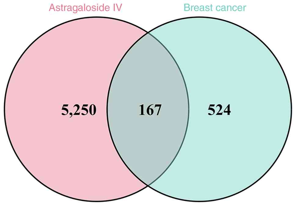
Using network pharmacology methods, a total of 524
potential targets of AS-IV were screened from databases including
SwissTargetPrediction, while 5,250 breast cancer-associated targets
were obtained from databases such as GeneCards, OMIM and DisGeNET.
Of note, the 5,250 breast cancer-associated genes encompass a
comprehensive collection of genes associated with breast cancer,
including driver, tumor suppressor and other cancer-associated
genes that may serve as biomarkers or therapeutic targets. After
taking the intersection, 167 common candidate targets were
identified (Fig. 1).
PPI network analysis and topological screening
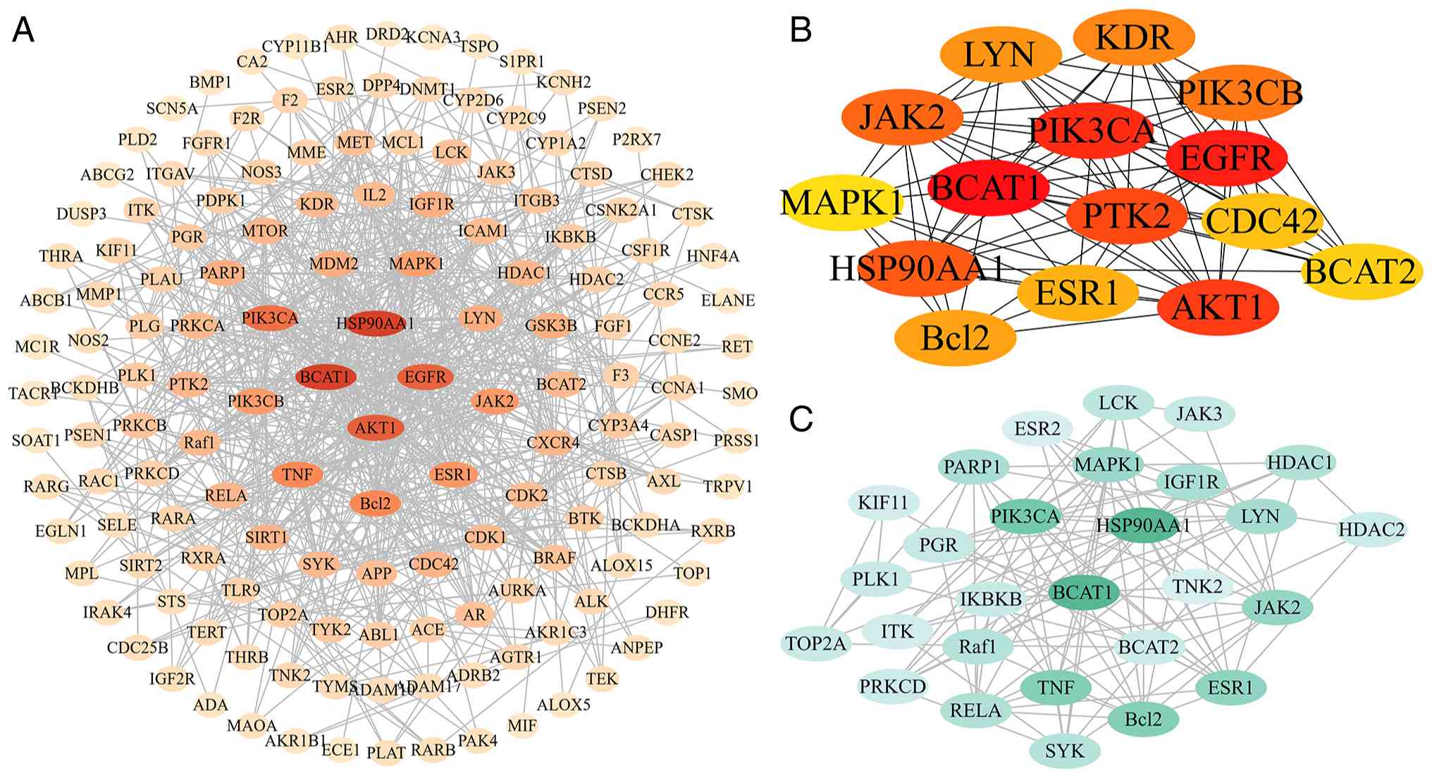
(using ‘CytoHubba’ and ‘MCODE’ plugins) revealed that targets such
as BCAT1, AKT1, PIK3CA, EGFR and HSP90AA1 are located at the core
of the network (Fig. 2A-C).
Quantitative ranking based on degree centrality identified the top
10 core targets, as presented in Table
SII. Among these, HSP90AA1 and BCAT1 exhibited the highest
degree values (both 46), followed by AKT1 with 40, EGFR with 39 and
PIK3CA with 36, indicating their potential prominence in mediating
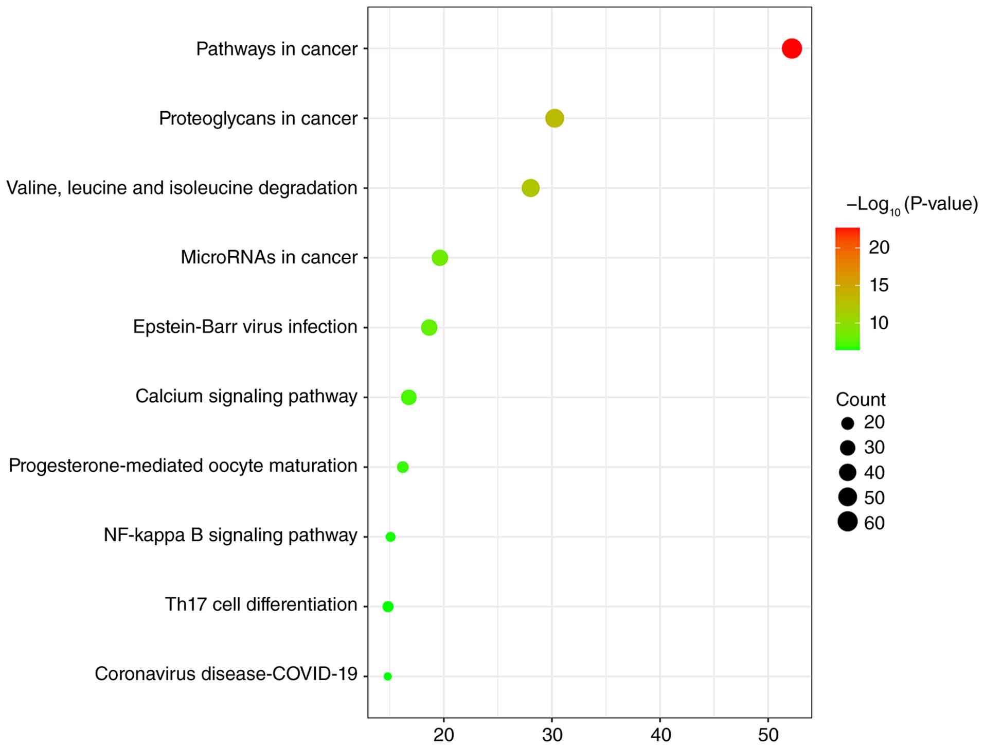
the therapeutic effects of AS-IV against breast cancer. KEGG
pathway enrichment analysis indicated that these core targets are
significantly enriched in pathways such as ‘pathways in cancer’,
‘proteoglycans in cancer’, ‘valine, leucine and isoleucine
degradation’, ‘microRNAs in cancer’ and ‘Epstein-Barr virus
infection’ (Fig. 3).
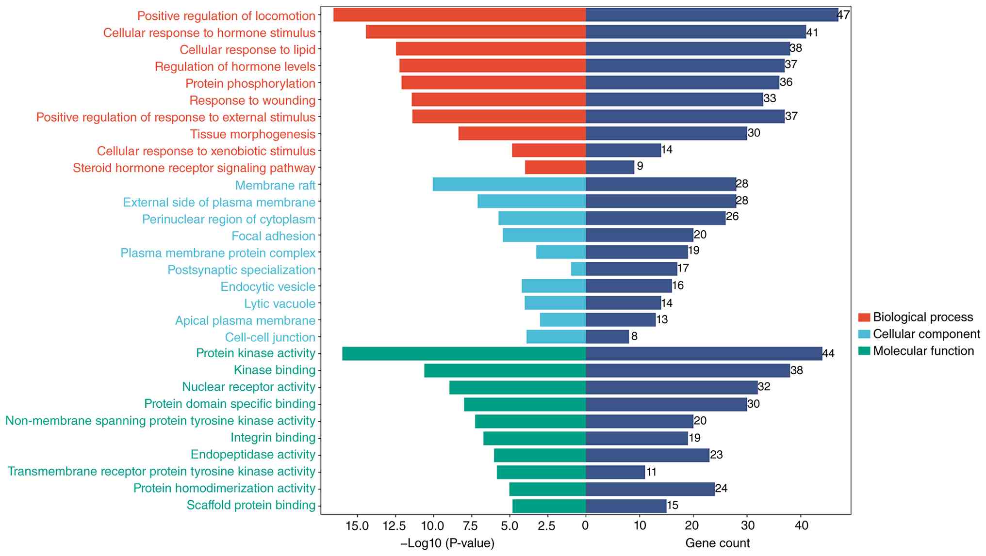
GO functional enrichment analysis suggested that
their biological processes mainly involve ‘positive regulation of
locomotion’, ‘cellular response to hormone stimulus’, ‘cellular
response to lipid’, ‘regulation of hormone levels’ and ‘protein
phosphorylation’. The analysis of cellular components primarily
highlighted ‘membrane raft’, ‘external side of plasma membrane’,
‘perinuclear region of cytoplasm’, ‘focal adhesion’ and ‘plasma
membrane protein complex’. In terms of molecular function, the key
enrichments included ‘protein kinase activity’, ‘kinase binding’,
‘nuclear receptor activity’, ‘protein domain specific binding’ and
‘non-membrane spanning protein tyrosine kinase activity’ (Fig. 4).
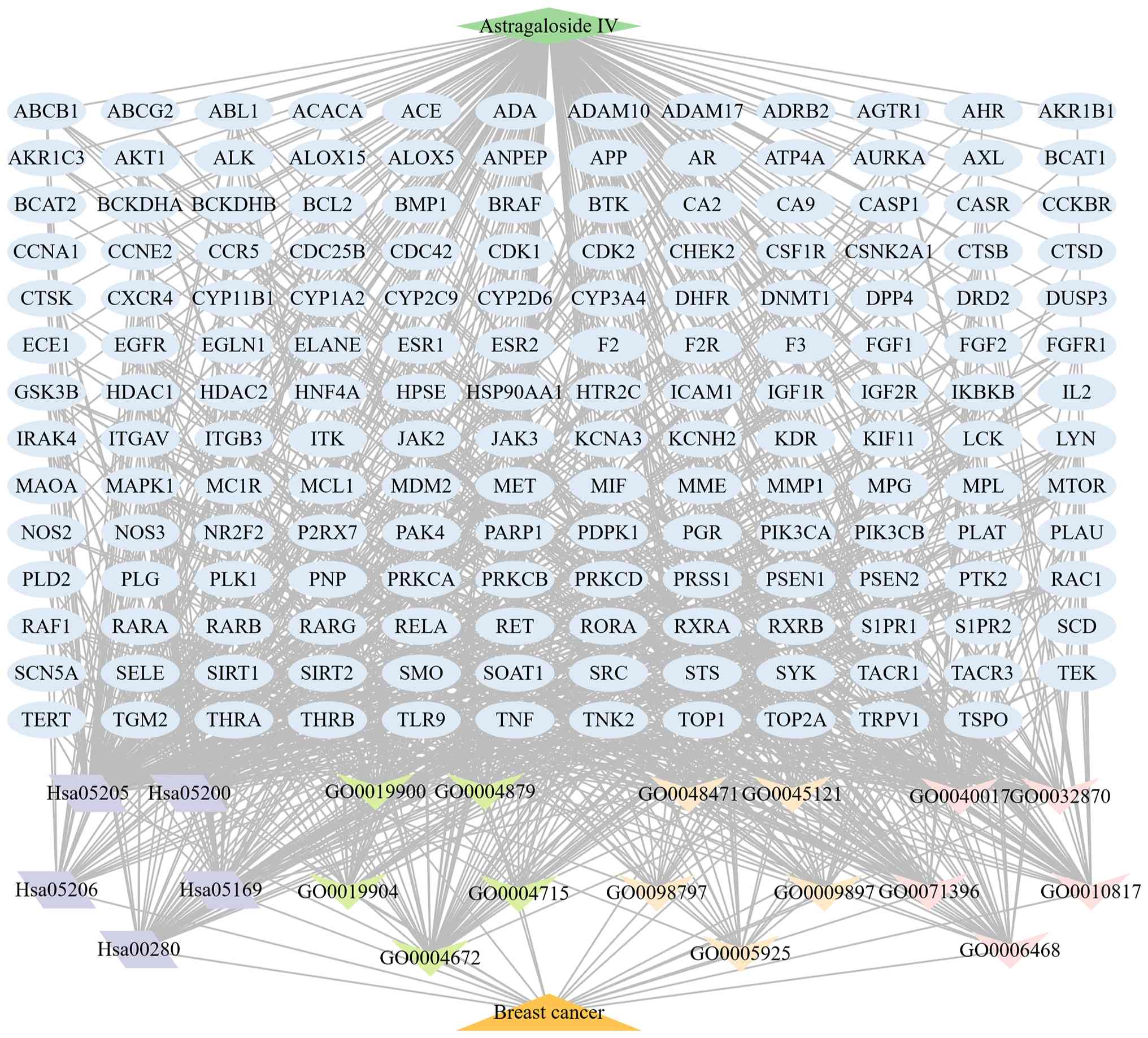
The further constructed comprehensive network
diagram ‘AS-IV-core targets-key pathways-breast cancer’ visually
illustrates the potential mechanism by which AS-IV exerts
synergistic effects through multiple targets and pathways. As
presented in Fig. 5, the network
comprises 189 nodes and 986 edges. This analysis further identified
key active components and core targets of AS-IV in the treatment of
breast cancer. Among these, BCAT1 and the BCAA metabolic pathway
emerged as key nodes within the network, providing a theoretical
basis and direction for subsequent experimental research targeting
BCAT1.
Establishment of BCAT1 KD and OE cell
models
To investigate whether AS-IV influences breast
cancer cell behavior through BCAT1, the present study transiently
knocked down or overexpressed BCAT1 in MDA-MB-231 TNBC cells using
siRNA and pcDNA3.1-BCAT1 plasmid, respectively. Transfection
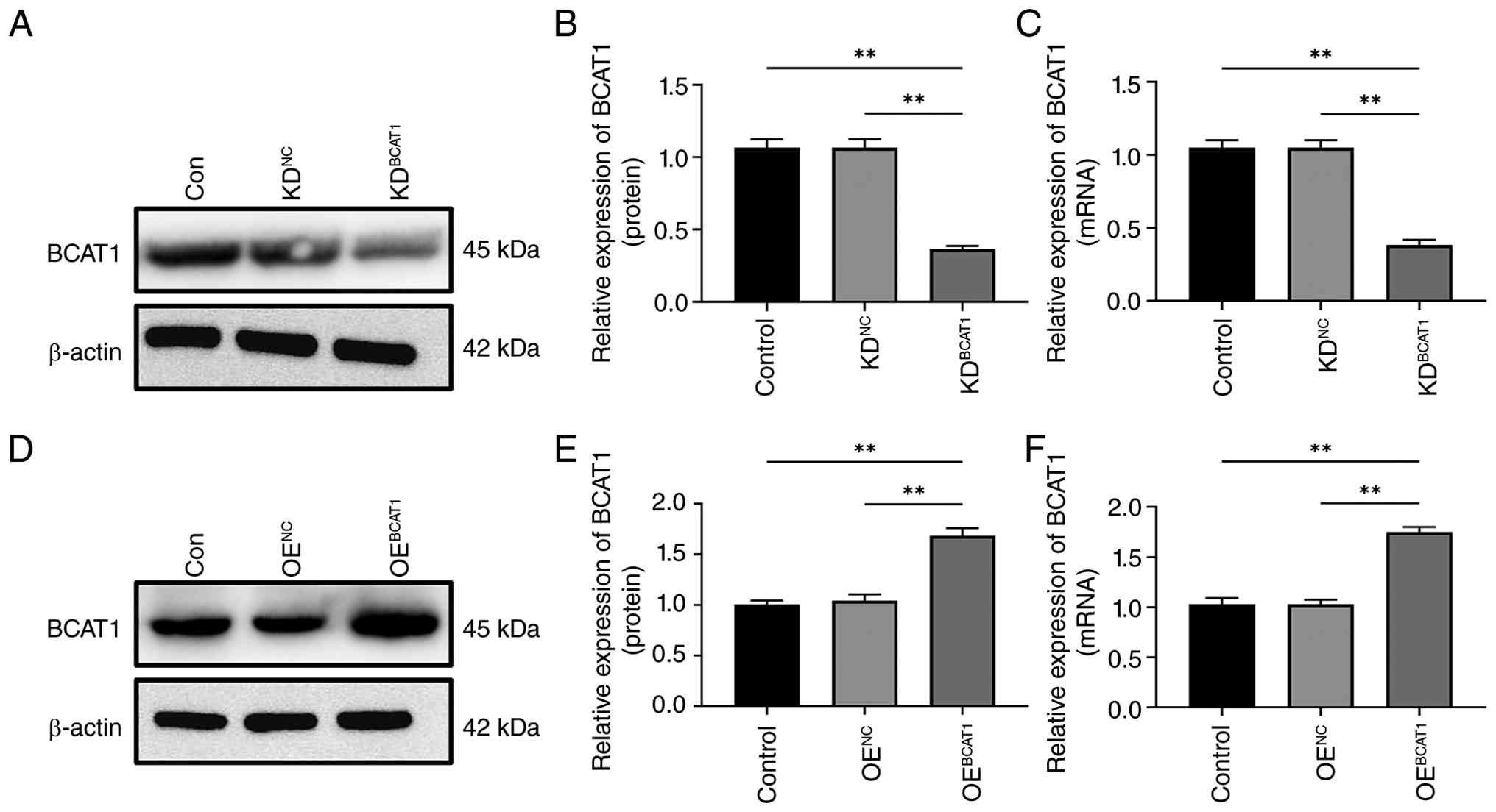
efficiency was validated 48 h post-transfection. Compared with the
control and KDNC groups (Fig. 6A-C), BCAT1 expression was
significantly decreased in the KDBCAT1 group
(P<0.01), indicating successful construction of the BCAT1-KD
transiently transfected cells. Similarly, compared with the control
and OENC groups (Fig.
6D-F), the OEBCAT1 group indicated a significant
increase in BCAT1 expression (P<0.01), confirming the successful
generation of the BCAT1-overexpressing transiently transfected
cells.
Evidence of direct interaction between
AS-IV and BCAT1
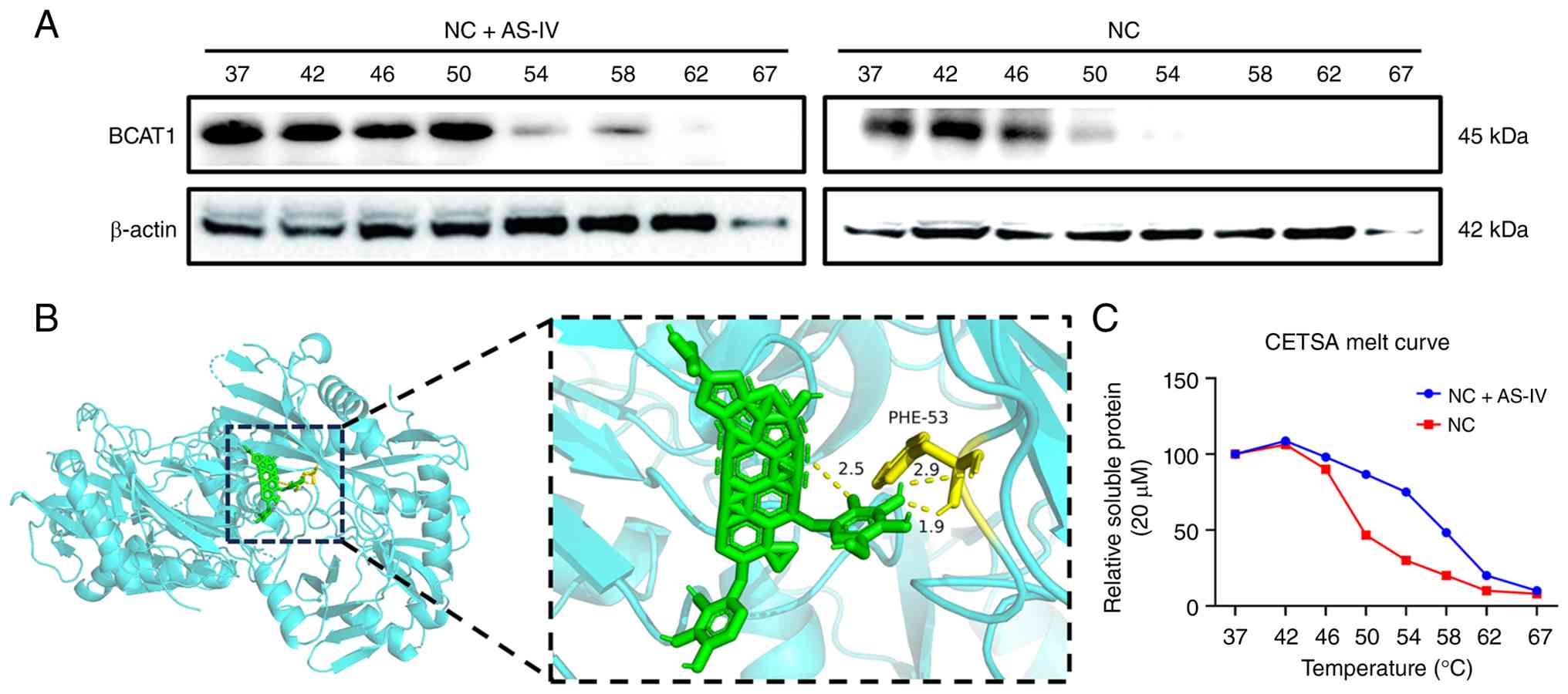
Molecular docking results demonstrated that AS-IV
stably binds to BCAT1 with a predicted binding free energy of 8.4
kcal/mol, as calculated by AutoDock, indicating a strong binding
affinity between the two molecules (Fig. 7B). The CETSA further revealed that
AS-IV treatment markedly enhanced the thermal stability of BCAT1.
As presented in Fig. 7A and C, in
the untreated control group (NC), BCAT1 underwent notable thermal
denaturation within the temperature range of 46–54°C, accompanied
by a marked decrease in solubility. By contrast, after treatment
with 10 µM AS-IV for 1 h, the denaturation curve of BCAT1 shifted
rightward and its melting temperature increased by 8°C compared
with the NC group (from 54 to 62°C). These results suggested that
AS-IV directly binds to BCAT1 under the investigated cellular
conditions and enhances its thermal stability. This finding
provided preliminary evidence of a physical interaction between
AS-IV and BCAT1, supporting the hypothesis that BCAT1 may be a
direct binding partner of AS-IV.
Effect of BCAT1 KD and OE on the
anti-migratory activity of AS-IV in MDA-MB-231 TNBC cells
As presented in Fig. 8A
and B (BCAT1 KD), two-way ANOVA revealed a significant main
effect of AS-IV concentration [F(3, 16)=510.8; P<0.001], a
significant main effect of BCAT1 modification [F(1, 16)=810.6;
P<0.001] and a significant interaction between AS-IV
concentration and BCAT1 modification [F(3, 16)=52.73; P<0.001]
on wound closure. AS-IV treatment significantly reduced the
relative wound area in both KDNC and KDBCAT1
groups in a dose-dependent manner (Bonferroni post hoc test; all
P<0.05). At each tested concentration, the KDBCAT1
group exhibited a more significant reduction in migration compared
with the KDNC group (P<0.001). As presented in
Fig. 8C and D (BCAT1 OE), two-way
ANOVA similarly demonstrated a significant main effect of AS-IV
concentration [F(3, 16)=199.3; P<0.001], a significant main
effect of BCAT1 modification [F(1, 16)=2667.5; P<0.001] and a
significant interaction [F(3, 16)=27.02; P<0.001] on wound
closure. AS-IV treatment decreased wound closure in both
OENC and OEBCAT1 groups in a
concentration-dependent manner (Bonferroni post hoc test; all
P<0.05). Notably, at each concentration tested, the
OEBCAT1 group demonstrated a significantly greater
reduction in migration compared with the OENC group
(P<0.05). These results demonstrated that AS-IV effectively
suppresses cell migration in a concentration-dependent manner under
both BCAT1 KD and OE conditions, albeit with altered efficacy
depending on BCAT1 expression levels.
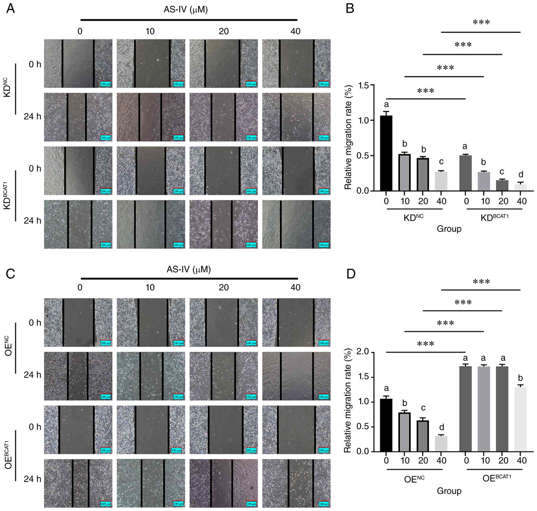 | Figure 8.BCAT1 knockdown and overexpression
modulate the anti-migratory effect of AS-IV in MDA-MB-231
triple-negative breast cancer cells. (A) Representative images of
wound closure in MDA-MB-231 breast cancer cells with BCAT1 KD after
treatment with different concentrations of AS-IV (0, 10, 20 and 40
µM) for 24 h. (B) Quantification of relative wound area in BCAT1 KD
cells. x-axis: AS-IV concentration (µM). (C) Representative images
of wound closure in MDA-MB-231 breast cancer cells with BCAT1 OE
after treatment with different concentrations of AS-IV (0, 10, 20
and 40 µM) for 24 h. (D) Quantification of relative wound area in
BCAT1 OE cells. x-axis: AS-IV concentration (µM). ‘a’, ‘b’, ‘c’ and
‘d’ represent the differences among different concentrations of
AS-IV. There were no significant differences among groups sharing
the same letters, while significant differences were observed
between groups containing different letters (P<0.05).
***P<0.001. AS-IV, astragaloside-IV; BCAT1, branched-chain amino
acid transaminase 1; OE, overexpression; NC, negative control; KD,
knockdown. |
Effect of BCAT1 KD and OE on the
anti-invasive activity of AS-IV in MDA-MB-231 TNBC cells
As presented in Fig. 9A
and B (BCAT1 KD), two-way ANOVA revealed a significant main
effect of AS-IV concentration [F(3, 16)=138.0; P<0.001], a
significant main effect of BCAT1 modification [F(1, 16)=421.4;
P<0.001] and a significant interaction between AS-IV
concentration and BCAT1 modification [F(3, 16)=20.9; P<0.001] on
cell invasion. AS-IV treatment significantly reduced the number of
invaded cells in both KDNC and KDBCAT1 groups
in a dose-dependent manner (Bonferroni post hoc test, all
P<0.05). At each concentration tested, the KDBCAT1
group exhibited a more significant decrease in cell invasion
compared with the KDNC group (P<0.05). As presented
in Fig. 9C and D (BCAT1 OE),
two-way ANOVA demonstrated a significant main effect of AS-IV
concentration [F(3, 16)=129.8; P<0.001], a significant main
effect of BCAT1 modification [F(1, 16)=160.0; P<0.001] and a
significant interaction between AS-IV concentration and BCAT1
modification [F(3, 16)=3.06; P<0.001] on cell invasion. AS-IV
treatment significantly decreased cell invasion in both
OENC and OEBCAT1 groups in a
concentration-dependent manner (Bonferroni post hoc test, all
P<0.05). At each concentration tested, the OEBCAT1
group demonstrated a significantly greater reduction in cell
invasion compared with the OENC group (P<0.05). These
results indicated that AS-IV potently suppresses the invasive
capacity of MDA-MB-231 TNBC cells in a concentration-dependent
manner, and that BCAT1 expression levels modulate the efficacy of
AS-IV.
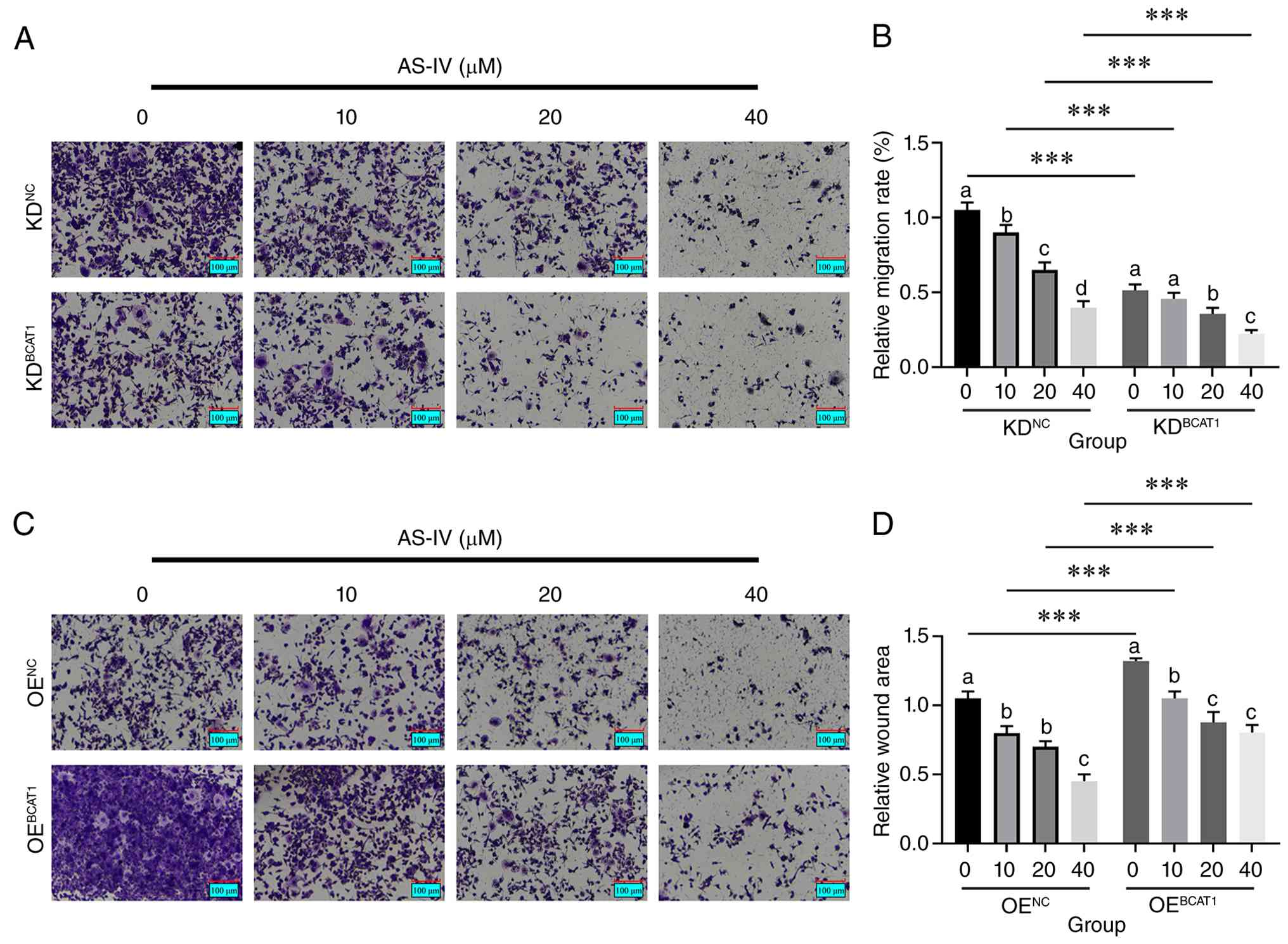 | Figure 9.Effects of BCAT1 KD and OE on
AS-IV-inhibited cell invasion by Transwell assay (scale bar, 100
µm). Representative images and statistical analysis of invading
cells through Matrigel in MDA-MB-231 breast cancer cells with BCAT1
KD (A and B) or OE (C and D) after treatment with different
concentrations of AS-IV (0, 10, 20 and 40 µM) for 24 h. For the
knockdown groups, the interaction was significant (P<0.001).
‘a’, ‘b’, ‘c’ and ‘d’ represent the differences among different
concentrations of AS-IV. There were no significant differences
among groups sharing the same letters, while significant
differences were observed between groups containing different
letters (P<0.05). ***P<0.001. AS-IV, astragaloside-IV; BCAT1,
branched-chain amino acid transaminase 1; OE, overexpression; NC,
negative control; KD, knockdown. |
Effects of AS-IV on the expression
levels of BCAT1 downstream and associated proteins in MDA-MB-231
TNBC cells
Following BCAT1 KD in MDA-MB-231 TNBC cells, AS-IV
treatment influenced the expression levels of BCAT1 downstream and
associated proteins, as illustrated in Fig. 10. The results revealed that BCAT1
KD induced compensatory upregulation of BCAT2 compared with the NC
(KDNC) group (P<0.001). Regarding the branched-chain
α-ketoacid dehydrogenase kinase (BCKDK)/branched-chain keto acid
dehydrogenase E1 subunit β (BCKDHB) pathway, BCAT1 KD significantly
reduced BCKDK activity compared with the KDNC group
(P<0.01), thereby alleviating its inhibitory effect on BCKDHB
and leading to significantly increased basal BCKDHB activity
compared with the KDNC group (P<0.001). AS-IV
intervention further enhanced this trend in a dose-dependent
manner, directly inhibiting BCKDK activity and more potently
activating BCKDHB.
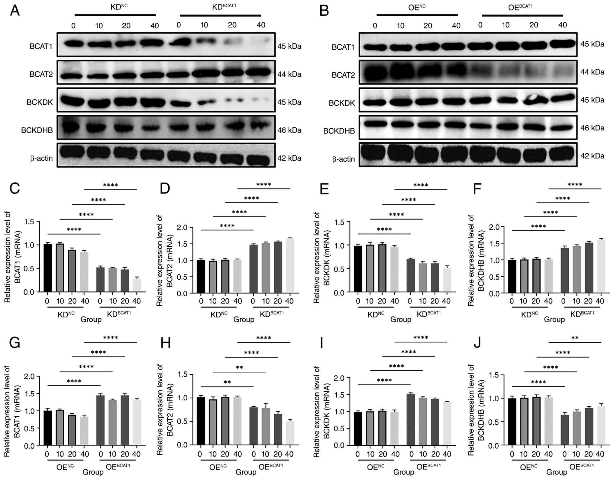 | Figure 10.Effects of AS-IV on the expression
levels of BCAT1 downstream and associated proteins in MDA-MB-231
TNBC cells. (A) Western blot analysis of BCAT2, BCKDK, BCKDHB and
β-actin protein expression in BCAT1 KD cells. (B) Western blot
analysis of BCAT2, BCKDK, BCKDHB and β-actin protein expression in
BCAT1 OE cells. (C) Relative expression level of BCAT2 mRNA in
BCAT1 KD cells. (D) Relative expression level of BCKDK mRNA in
BCAT1 KD cells. (E) Relative expression level of BCKDHB mRNA in
BCAT1 KD cells. (F) Relative expression level of BCAT1 mRNA in
BCAT1 KD cells. (G) Relative expression level of BCAT2 mRNA in
BCAT1 OE cells. (H) Relative expression level of BCKDK mRNA in
BCAT1 OE cells. (I) Relative expression level of BCKDHB mRNA in
BCAT1 OE cells. (J) Relative expression level of BCAT1 mRNA in
BCAT1 OE cells. β-actin was used as the internal control. Data are
presented as mean ± SD (n=3). **P<0.01 and ****P<0.0001
indicate significant differences compared with the respective
control group (0 µM AS-IV) within the same genetic background.
BCAT, branched-chain amino acid transaminase; OE, overexpression;
NC, negative control; KD, knockdown; BCKDK, branched-chain
α-ketoacid dehydrogenase kinase; BCKDHB, branched-chain keto acid
dehydrogenase E1 subunit β. |
OEBCAT1 resulted in compensatory
downregulation of BCAT2 compared with the OENC group
(P<0.001). Functionally, BCAT1 OE significantly enhanced BCKDK
activity compared with the OENC group, thereby
suppressing BCKDHB activity AS-IV treatment effectively
counteracted the hyperactivation of BCKDK in a dose-dependent
manner, partially reversing the inhibition of BCKDHB activity.
However, under BCAT1 OE conditions, the extent of BCKDHB activity
restoration by AS-IV did not reach the level observed in the NC
group, suggesting that the high branched-chain α-keto acids
environment resulting from OEBCAT1 partially attenuated
the efficacy of AS-IV.
AS-IV promotes BCAA catabolism by
modulating the BCAT1/BCKDK/BCKDH axis
The results indicated that KDBCAT1
significantly increased intracellular levels of BCAAs, including
leucine, isoleucine and valine, compared with the NC group
(P<0.001). This accumulation confirmed that BCAT1 is the
rate-limiting enzyme for BCAA transamination; its downregulation
creates a metabolic bottleneck upstream, impairing the initial step
of BCAA catabolism. All three BCAAs exhibited a consistent
dose-dependent accumulation (0, 10, 20 and 40 µM; P<0.01)
(Fig. 11).
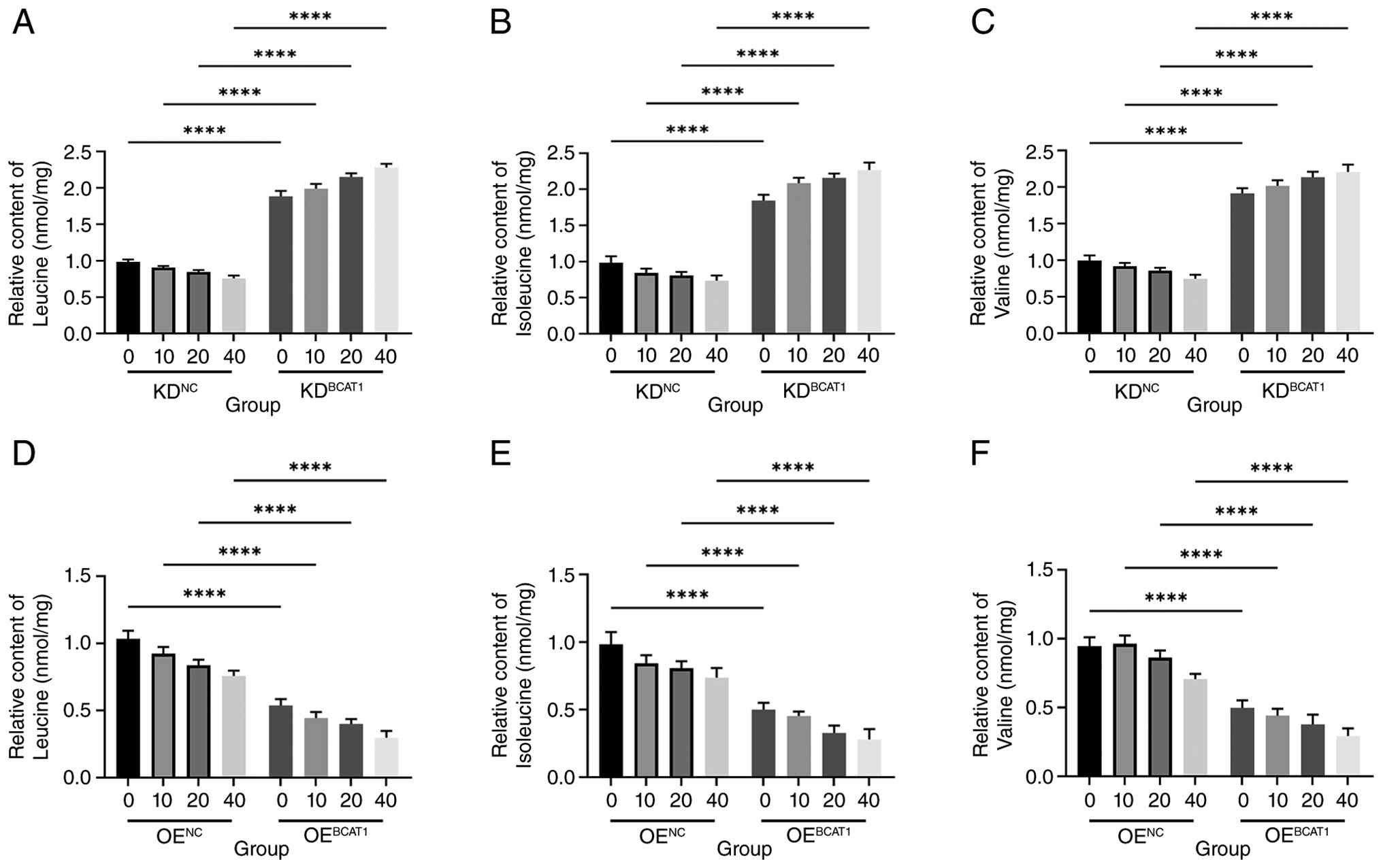 | Figure 11.Intracellular branched-chain amino
acid levels detected by liquid chromatography-mass spectrometry.
(A) Relative changes in the intracellular content of leucine in
MDA-MB-231 TNBC cells with BCAT1 KD after treatment with different
concentrations of AS-IV (0, 10, 20 and 40 µM) for 24 h. (B)
Relative changes in the intracellular content of isoleucine in
MDA-MB-231 TNBC cells with BCAT1 KD after treatment with different
concentrations of AS-IV (0, 10, 20 and 40 µM) for 24 h. (C)
Relative changes in the intracellular content of valine in
MDA-MB-231 TNBC cells with BCAT1 KD after treatment with different
concentrations of AS-IV (0, 10, 20 and 40 µM) for 24 h. (D)
Relative changes in the intracellular content of leucine in
MDA-MB-231 TNBC cells with BCAT1 OE after treatment with different
concentrations of AS-IV (0, 10, 20 and 40 µM) for 24 h. (E)
Relative changes in the intracellular content of isoleucine in
MDA-MB-231 TNBC cells with BCAT1 OE after treatment with different
concentrations of AS-IV (0, 10, 20 and 40 µM) for 24 h. (F)
Relative changes in the intracellular content of valine in
MDA-MB-231 TNBC cells with BCAT1 OE after treatment with different
concentrations of AS-IV (0, 10, 20 and 40 µM) for 24 h.
****P<0.0001 indicate significant differences compared with the
respective control group (0 µM AS-IV) within the same genetic
background. OE, overexpression; BCAT1, branched-chain amino acid
transaminase 1; NC, negative control; KD, knockdown; AS-IV,
astragaloside IV. |
By contrast, OEBCAT1 itself significantly
reduced the basal intracellular levels of leucine, isoleucine and
valine compared with the NC group (P<0.001). AS-IV further
enhanced BCAA degradation in a dose-dependent manner (0, 10, 20 and
40 µM; P<0.01) in both groups. Of note, in the OE group, due to
the higher abundance of the BCAT1 target protein, the efficacy of
AS-IV was markedly enhanced. The amino acid-lowering effect of
AS-IV was significantly stronger in the OE group compared with that
in the contemporaneous NC group (P<0.001), demonstrating a
notable synergistic effect (Fig.
11).
Discussion
The present study provided evidence for a mechanism
by which AS-IV inhibits the migration and invasion of MDA-MB-231
TNBC cells, potentially by targeting BCAT1 and modulating BCAA
metabolism. Notably, these findings were derived from a single cell
line model and thus represent preclinical evidence specific to this
TNBC cell type. The present study findings not only provide a novel
metabolic perspective on the antitumor effects of AS-IV but also
offer experimental evidence suggesting BCAT1 as a potential
therapeutic target for further investigation in oncology.
In recent years, numerous studies have highlighted
the key role of metabolic reprogramming in tumor progression, with
increasing attention focusing on aberrant amino acid metabolism
(37–41). As essential amino acids, BCAAs are
not only involved in protein synthesis but also function as
signaling molecules regulating pro-tumor pathways such as mTOR
complex 1 (42). BCAT1, the
rate-limiting enzyme in BCAA catabolism, has been reported to be
markedly expressed in various malignancies (for example, glioma and
acute myeloid leukemia) and promotes tumor growth by maintaining
intracellular BCAA levels (43).
However, the role and underlying mechanisms of BCAT1 in breast
cancer, particularly TNBC, remain to be elucidated. The present
study demonstrated that BCAT1 expression significantly influences
the migratory and invasive capabilities of MDA-MB-231 TNBC cells,
which is consistent with its reported pro-metastatic roles in other
malignancies. To the best of our knowledge, the present study
findings provide the first evidence that associates AS-IV, a
natural compound from traditional Chinese medicine, with the
regulation of BCAT1-mediated BCAA metabolism in breast cancer cells
(44).
A key finding of the present study was that AS-IV
binds to the BCAT1 protein and enhances its thermal stability,
indicating a possible direct interaction. Both molecular docking
and CETSA experiments consistently indicated a potential
interaction between AS-IV and BCAT1, with computational prediction
of high binding affinity and experimental observation of increased
protein thermal stability. It is key to note that while CETSA
provides evidence of target engagement in cell lysates and
molecular docking predicts potential binding modes, these
techniques are suggestive rather than definitive in establishing
direct physical binding. Further validation using orthogonal
approaches such as surface plasmon resonance (SPR) or isothermal
titration calorimetry (ITC) would be key to confirm the direct
interaction and determine binding kinetics. Furthermore, the
specific amino acid residues mediating the AS-IV-BCAT1 interaction
predicted by molecular docking have not been experimentally
validated. Future studies employing site-directed mutagenesis of
these key residues, combined with binding affinity measurements and
enzymatic activity assays, are warranted to definitively establish
the interaction interface and elucidate how AS-IV binding modulates
BCAT1 catalytic function. It is key to clarify the directionality
of this interaction and its functional consequences. The present
study data indicated that AS-IV binding to BCAT1 does not simply
‘activate’ the enzyme in a binary manner; by contrast, it modulates
the activity of the enzyme within the context of the entire BCAA
metabolic pathway. Specifically, AS-IV promotes the flux of BCAAs
through the catabolic pathway by influencing the downstream
BCKDK/BCKDH axis, leading to enhanced BCAA degradation and reduced
intracellular BCAA levels. This reduction in BCAA availability,
therefore, attenuates pro-migratory and -invasive signaling (for
example, the mTOR pathway), thereby suppressing malignant
phenotypes. Thus, AS-IV functions as a metabolic modulator that
redirects BCAA metabolism toward complete catabolism, rather than
increasing or decreasing BCAT1 enzymatic activity. This
identification provides a novel example of how natural compounds
can exert antitumor effects through direct modulation of metabolic
enzymes. While previous research has largely focused on the
immunomodulatory and apoptosis-inducing effects of AS-IV, the
present study revealed a novel mechanism by which it exerts
antitumor activity through targeting amino acid metabolic enzymes
(45,46).
At the functional level, the present study confirmed
that AS-IV markedly inhibits the migration and invasion of
MDA-MB-231 TNBC cells in a concentration-dependent manner. Analysis
of relative inhibition, normalized to the baseline of each group,
revealed that BCAT1 expression levels modulate AS-IV efficacy:
BCAT1 KD significantly attenuated the anti-migratory and -invasive
effects of AS-IV (as reflected by reduced percentage inhibition),
whereas BCAT1 OE enhanced AS-IV efficacy (increased percentage
inhibition). These findings supported the notion that BCAT1 is a
key mediator of the functional effects of AS-IV. Notably, AS-IV
still exhibited significant dose-dependent inhibitory effects in
BCAT1 KD cells, albeit to a lesser extent, suggesting that
additional mechanisms may also contribute to its anti-migratory
activity, consistent with its reported multi-target properties
(15,18,22,47–49).
Mechanistically, the present study revealed that
BCAT1 expression significantly influences the activity of the
downstream BCKDK/BCKDH pathway and intracellular BCAA accumulation.
AS-IV treatment further enhanced BCKDHB activation induced by BCAT1
KD and partially reversed BCKDHB suppression resulting from BCAT1
OE. This bidirectional regulatory capacity, enhancing BCKDHB
activity when it is basally low (as in KD cells) and partially
restoring it when it is suppressed (as in OE cells), suggests that
AS-IV acts as a modulator of pathway flux rather than a simple
agonist or antagonist of BCAT1 enzymatic activity. By promoting the
complete oxidation of BCAAs via the BCKDHB complex, AS-IV reduces
the intracellular pool of these amino acids, thereby limiting their
availability to promote pro-metastatic signaling pathways. Notably,
AS-IV demonstrated a stronger BCAA-lowering effect in
BCAT1-overexpressing cells compared with the NC (OENC)
group, indicating that its efficacy is closely associated with
BCAT1 protein levels. The present study finding is consistent with
the established central role of the BCAT1/BCKDK axis in amino acid
metabolism but proposes AS-IV as a positive modulator of this
pathway (50,51).
From the perspective of metabolic reprogramming,
BCAAs not only provide substrates for protein synthesis but also
act as nutrient signals that activate pro-tumor pathways such as
mTOR (52). The present study
suggested that by promoting BCAA degradation and reducing their
intracellular levels, AS-IV may indirectly suppress associated
oncogenic signaling pathways such as the mTOR pathway. This aligns
with previous studies on the role of the BCAA-mTOR axis in tumor
progression (53–56); however, to the best of our
knowledge, the present study is the first to directly associate a
natural monomer compound derived from traditional Chinese medicine
with this pathway, thereby providing a theoretical foundation for
the application of metabolic intervention strategies in breast
cancer treatment in the future.
However, the present study has certain limitations.
All experiments were conducted solely in cellular models and the
anti-metastatic effects of AS-IV mediated through BCAT1 have not
yet been validated in animal models. Furthermore, the expression
patterns and clinical significance of BCAT1 across different
molecular subtypes of breast cancer remain to be fully elucidated.
Future studies should focus on establishing breast cancer migration
animal models to evaluate the in vivo efficacy and potential
toxicity of AS-IV, in addition to conducting correlation analyses
based on clinical samples to facilitate its translational
applications. Second, the present study was conducted exclusively
in the MDA-MB-231 TNBC cell line. While TNBC represents an
aggressive subtype with high clinical relevance, the absence of
other breast cancer subtypes (for example, hormone
receptor+ MCF-7 or HER2-amplified SKBR3 cells) limits
the generalizability of the present study findings. BCAT1
expression and BCAA metabolic patterns may differ across breast
cancer molecular subtypes and whether AS-IV exerts similar effects
in these contexts remains to be investigated in future research.
Third, although the molecular docking and CETSA experiments in the
present study suggested a potential direct interaction between
AS-IV and BCAT1, the evidence remains indirect and preliminary. The
predicted binding mode has not been validated using site-directed
mutagenesis of key amino acid residues and the precise effect of
AS-IV binding on BCAT1 enzymatic activity has not been directly
measured. Such validation, including mutagenesis studies combined
with SPR or ITC to assess binding kinetics, as well as enzymatic
activity assays, is key to definitively establishing BCAT1 as a
direct molecular target of AS-IV and to understand the functional
consequences of their interaction.
In conclusion, the present study indicated that
AS-IV suppresses the migration and invasion of MDA-MB-231 TNBC
cells and that this effect may involve targeting BCAT1 and
modulating BCAA metabolism in this in vitro cell model.
AS-IV exhibits predicted high binding affinity to BCAT1 in
molecular docking experiments and enhances the thermal stability of
the protein in CETSA experiments, suggesting a potential direct
interaction. Functionally, AS-IV inhibits migration and invasion of
breast cancer cell in a concentration-dependent manner; although
this effect is attenuated under BCAT1 KD or OE conditions, it
remains statistically significant, indicating that BCAT1 is a key
target of AS-IV. Mechanistically, AS-IV promotes BCAA catabolism by
modulating BCKDK/BCKDH pathway activity, thereby lowering
intracellular BCAA levels and therefore, suppressing oncogenic
signaling cascades such as the mTOR axis. To the best of our
knowledge, the present study findings, for the first time,
associated an active component of traditional Chinese medicine with
a metabolic enzyme target and the malignant phenotype of MDA-MB-231
TNBC cells, not only expanding the antitumor mechanistic repertoire
of AS-IV but also offering a potential mechanistic rationale for
future exploration of BCAA-centric metabolic intervention in this
cell model. Further studies in diverse breast cancer models are
warranted to determine the generalizability of these observations.
Further investigations in pre-clinical animal models and clinical
specimens are warranted to validate the in vivo efficacy and
translational potential of this mechanism.
Supplementary Material
Supporting Data
Supporting Data
Acknowledgements
Not applicable.
Funding
The present study was supported by the Shaanxi Provincial
Department of Science and Technology (grant nos 2025JC-YBMS-903 and
2023-JC-QN-0995) and Yan'an Science and Technology Plan Project
(grant no. 2022SLSFGG-019).
Availability of data and materials
The data generated in the present study are not
publicly available due restrictions by the institutional data
management policy and the collaborative research agreement but may
be requested from the corresponding author.
Authors' contributions
YTL conceived and designed the research studies and
contributed markedly to data interpretation, manuscript writing and
final approval of the manuscript. LZ analyzed experimental data and
contributed to manuscript writing and revisions. XDS contributed to
data acquisition, analysis and interpretation, and critically
revised the manuscript for important intellectual content. All
authors read and approved the final manuscript. YTL and XDS confirm
the authenticity of all the raw data.
Ethics approval and consent to
participate
Not applicable.
Patient consent for publication
Not applicable.
Competing interests
The authors declare that they have no competing
interests.
Glossary
Abbreviations
Abbreviations:
|
BCAA
|
branched-chain amino acid
|
|
BCAT1
|
BCAA transaminase 1
|
|
AS-IV
|
astragaloside IV
|
|
BCKDK
|
branched-chain α-ketoacid
dehydrogenase kinase
|
|
BCKDHB
|
branched-chain keto acid dehydrogenase
E1 subunit β
|
|
CETSA
|
cellular thermal shift assay
|
|
TNBC
|
triple-negative breast cancer
|
References
|
1
|
Akizawa Y, Kanno T, Horibe Y, Shimizu Y,
Noguchi E, Yamamoto T, Okamoto T, Nagashima Y and Tabata T: Ovarian
metastasis from breast cancer mimicking a primary ovarian neoplasm:
A case report. Mol Clin Oncol. 15:1352021. View Article : Google Scholar : PubMed/NCBI
|
|
2
|
Kim J, Harper A, McCormack V, Sung H,
Houssami N, Morgan E, Mutebi M, Garvey G, Soerjomataram I and
Fidler-Benaoudia MM: Global patterns and trends in breast cancer
incidence and mortality across 185 countries. Nat Med.
31:1154–1162. 2025. View Article : Google Scholar : PubMed/NCBI
|
|
3
|
World Health Organization (WHO), . Breast
cancer. WHO fact sheet. WHO; Geneva: 2024
|
|
4
|
Kong F, Wang C, Zhang J, Wang X, Sun B,
Xiao X, Zhang H, Song Y and Jia Y: Chinese herbal medicines for
prostate cancer therapy: From experimental research to clinical
practice. Chin Herb Med. 15:485–495. 2023.PubMed/NCBI
|
|
5
|
Kong F, Wang C, Zhao L, Liao D, Wang X,
Sun B, Yang P and Jia Y: Traditional Chinese medicines for
non-small cell lung cancer: Therapies and mechanisms. Chin Herb
Med. 15:509–515. 2023.PubMed/NCBI
|
|
6
|
Tian Y, Ma B, Yu S, Li Y, Pei H, Tian S,
Zhao X, Liu C, Zuo Z and Wang Z: Clinical antitumor application and
pharmacological mechanisms of Dahuang Zhechong Pill. Chin Herb Med.
15:169–180. 2023.PubMed/NCBI
|
|
7
|
Li S, Liu Y, Hu W, Deng A, Ren X, Chen L,
Lu Y, Wu Y, Huang H, Cao J, et al: Protein lipoylation in cancer:
Metabolic reprogramming and therapeutic potential. Cell Death
Discov. 11:4202025. View Article : Google Scholar : PubMed/NCBI
|
|
8
|
Tanase DM, Valasciuc E, Costea CF,
Scripcariu DV, Ouatu A, Hurjui LL, Tarniceriu CC, Floria DE,
Ciocoiu M, Baroi LG and Floria M: Duality of branched-chain amino
acids in chronic cardiovascular disease: Potential biomarkers
versus active pathophysiological promoters. Nutrients. 16:19722024.
View Article : Google Scholar : PubMed/NCBI
|
|
9
|
Long L, Yang WS, Liu L, Tobias DK,
Katagiri R, Wu K, Jin L, Zhang FF, Luo X, Liu X, et al: Dietary
intake of branched-chain amino acids and survival after colorectal
cancer diagnosis. Int J Cancer. 148:2471–2480. 2021. View Article : Google Scholar : PubMed/NCBI
|
|
10
|
Song Y, Zhao B, Xu Y, Ren X, Lin Y, Zhou L
and Sun Q: Prognostic significance of branched-chain amino acid
transferase 1 and CD133 in triple-negative breast cancer. BMC
Cancer. 20:5842020. View Article : Google Scholar : PubMed/NCBI
|
|
11
|
Zhang L and Han J: Branched-chain amino
acid transaminase 1 (BCAT1) promotes the growth of breast cancer
cells through improving mTOR-mediated mitochondrial biogenesis and
function. Biochem Biophys Res Commun. 486:224–231. 2017. View Article : Google Scholar : PubMed/NCBI
|
|
12
|
Thewes V, Simon R, Hlevnjak M, Schlotter
M, Schroeter P, Schmidt K, Wu Y, Anzeneder T, Wang W, Windisch P,
et al: The branched-chain amino acid transaminase 1 sustains growth
of antiestrogen-resistant and ERα-negative breast cancer. Oncogene.
36:4124–4134. 2017. View Article : Google Scholar : PubMed/NCBI
|
|
13
|
Huang L, Li G, Zhang Y, Zhuge R, Qin S,
Qian J, Chen R, Kwan Wong Y, Tang H, Wang P, et al: Small-molecule
targeting BCAT1-mediated BCAA metabolism inhibits the activation of
SHOC2-RAS-ERK to induce apoptosis of Triple-negative breast cancer
cells. J Adv Res. 75:723–738. 2025. View Article : Google Scholar : PubMed/NCBI
|
|
14
|
Deng YP, Zhu H, Xing J, Gao J, Duan J, Liu
P, Zhong G and Cai X: The role of natural products in improving
lipid metabolism disorder-induced mitochondrial dysfunction of
diabetic kidney disease. Front Physiol. 16:16240772025. View Article : Google Scholar : PubMed/NCBI
|
|
15
|
Chen C, Bu X, Deng L, Xia J, Wang X, Chen
L, Li W, Huang J, Chen Q and Wang C: Astragaloside IV as a
promising therapeutic agent for liver diseases: Current landscape
and future perspectives. Front Pharmacol. 16:15741542025.
View Article : Google Scholar : PubMed/NCBI
|
|
16
|
Xiong BB, Zhuo YM, Wang H, Zheng QL, Tang
F, Huang Q and Yao M: Macrophage polarization in disease therapy:
Insights from astragaloside IV and cycloastragenol. Front
Pharmacol. 16:15980222025. View Article : Google Scholar : PubMed/NCBI
|
|
17
|
Lai Y, Liu R, Shao H, Zhan J, Ma Y, Zhou
L, Wan Z, Li S, Wang W, Jiang L and Shao Y: Mechanism of
Astragaloside-Brucea javanica oil nanoemulsion against oral
squamous cell carcinoma through CDK1/MTFR2: Network pharmacology,
bioinformatics, and experimental studies. PLoS One.
20:e03296222025. View Article : Google Scholar : PubMed/NCBI
|
|
18
|
Gao X, Hao W, Wang Y, Wu X, Zhu F and
Zhang Y: Molecular mechanisms of astragaloside-IV in hepatocellular
carcinoma therapy: A systematic review. BMC Cancer. 25:14072025.
View Article : Google Scholar : PubMed/NCBI
|
|
19
|
Zeng Y, Duan T, Huang J and Wang X:
Astragaloside IV inhibits nasopharyngeal carcinoma progression by
suppressing the SATB2/Wnt signaling axis. Toxicol Res (Camb).
14:tfaf0472025. View Article : Google Scholar : PubMed/NCBI
|
|
20
|
Yin W, Liao X, Sun J, Chen Q and Fan S:
Astragaloside IV inhibits the proliferation, migration, invasion,
and epithelial-mesenchymal transition of oral cancer cells by
aggravating autophagy. Tissue Cell. 90:1025242024. View Article : Google Scholar : PubMed/NCBI
|
|
21
|
Wang X, Gao S, Song L, Liu M, Sun Z and
Liu J: Astragaloside IV antagonizes M2 phenotype macrophage
polarization-evoked ovarian cancer cell malignant progression by
suppressing the HMGB1-TLR4 axis. Mol Immunol. 130:113–121. 2021.
View Article : Google Scholar : PubMed/NCBI
|
|
22
|
Hu C, Li Q, Gong SN, Zou XJ, Xu JY, Ying
HF and Zheng L: Astragaloside IV: A potential nemesis for gastric
cancer. Front Pharmacol. 16:16363412025. View Article : Google Scholar : PubMed/NCBI
|
|
23
|
Yang Y, Lu J, Zhu Y, Chen D, Tang J, Zhang
M, Lu J, Yang Y, Tian S and Zhao H: Astragaloside IV inhibits the
growth of obesity-associated triple-negative breast cancer by
activating FOXA1 transcription factor to regulate GAL3ST1-GalCer
signaling and remodel sphingolipid metabolism. Phytomedicine.
144:1569072025. View Article : Google Scholar : PubMed/NCBI
|
|
24
|
Hopkins AL: Network pharmacology: The next
paradigm in drug discovery. Nat Chem Biol. 4:682–690. 2008.
View Article : Google Scholar : PubMed/NCBI
|
|
25
|
Guan Y, Cheng J, Lv Q, Wei X, Jiang B and
Xiao P: Exploring new therapeutic potential of five commonly used
Pteris medicinal plants through pharmaphylogenomics and network
pharmacology. Chin Herb Med. 17:808–821. 2025.PubMed/NCBI
|
|
26
|
Gao J, Han C, Dai N, Wang W, Jin T, Du D
and Xia Q: Traditional Chinese medicine formulas alleviated acute
pancreatitis via improvement of microcirculation: A systematic
review and meta-analysis. Chin Herb Med. 17:584–600.
2024.PubMed/NCBI
|
|
27
|
Shang L, Wang Y, Li J, Zhou F, Xiao K, Liu
Y, Zhang M, Wang S and Yang S: Mechanism of Sijunzi Decoction in
the treatment of colorectal cancer based on network pharmacology
and experimental validation. J Ethnopharmacol. 302((Pt A)):
1158762023. View Article : Google Scholar : PubMed/NCBI
|
|
28
|
Zhai Y, Liu L, Zhang F, Chen X, Wang H,
Zhou J, Chai K, Liu J, Lei H, Lu P, et al: Network pharmacology: A
crucial approach in traditional Chinese medicine research. Chin
Med. 20:82025. View Article : Google Scholar : PubMed/NCBI
|
|
29
|
Li L, Yang L, Yang L, He C, He Y, Chen L,
Dong Q, Zhang H, Chen S and Li P: Network pharmacology: A bright
guiding light on the way to explore the personalized precise
medication of traditional Chinese medicine. Chin Med. 18:1462023.
View Article : Google Scholar : PubMed/NCBI
|
|
30
|
Li X, Liu Z, Liao J, Chen Q, Lu X and Fan
X: Network pharmacology approaches for research of Traditional
Chinese Medicines. Chin J Nat Med. 21:323–332. 2023.PubMed/NCBI
|
|
31
|
Tian JS, Wu ZN, Wu D, Yang C, Gao Y, Yan
DL and Qin XM: Combining network pharmacology and experimental
verification to reveal the mechanism of Chaigui granules in the
treatment of depression through PI3K/Akt/mTOR signaling pathways.
Metab Brain Dis. 38:2849–2864. 2023. View Article : Google Scholar : PubMed/NCBI
|
|
32
|
Paggi JM, Pandit A and Dror RO: The art
and science of molecular docking. Annu Rev Biochem. 93:389–410.
2024. View Article : Google Scholar : PubMed/NCBI
|
|
33
|
Ansari RM, Mundke RN, Agrawal YO, Goyal
SN, Nakhate KT and Rathod SS: Recent advances in molecular docking
techniques: Transforming perspectives in distinct drug targeting
and drug discovery approaches. Med Chem. Jul 21–2025.(Epub ahead of
print). View Article : Google Scholar : PubMed/NCBI
|
|
34
|
Abdul Kader S, Dib S, Achkar IW, Thareja
G, Suhre K, Rafii A and Halama A: Defining the landscape of
metabolic dysregulations in cancer metastasis. Clin Exp Metastasis.
39:345–362. 2022. View Article : Google Scholar : PubMed/NCBI
|
|
35
|
Biswas D, Slade L, Duffley L, Mueller N,
Dao KT, Mercer A, Pakkiriswami S, El Hiani Y, Kienesberger PC and
Pulinilkunnil T: Inhibiting BCKDK in triple negative breast cancer
suppresses protein translation, impairs mitochondrial function, and
potentiates doxorubicin cytotoxicity. Cell Death Discov. 7:2412021.
View Article : Google Scholar : PubMed/NCBI
|
|
36
|
Livak KJ and Schmittgen TD: Analysis of
relative gene expression data using real-time quantitative PCR and
the 2(−Delta Delta C(T)) Method. Methods. 25:402–408. 2001.
View Article : Google Scholar : PubMed/NCBI
|
|
37
|
Dong C, Zhang Y, Zeng J, Chong S, Liu Y,
Bian Z, Fan S and Chen X: FUT2 promotes colorectal cancer
metastasis by reprogramming fatty acid metabolism via YAP/TAZ
signaling and SREBP-1. Commun Biol. 7:129720242024. View Article : Google Scholar
|
|
38
|
Yu M, Yang D, Chen X, Yang Y, Zhang B,
Jiang X, Xing L, Yang Y, Sun Y and Li N: Metabolic reprogramming in
cancer: Dysregulation of glucose, lipid, and amino acid pathways
and therapeutic opportunities. Mol Biomed. 7:252026. View Article : Google Scholar : PubMed/NCBI
|
|
39
|
Huang K, Han Y, Chen Y, Shen H, Zeng S and
Cai C: Tumor metabolic regulators: Key drivers of metabolic
reprogramming and the promising targets in cancer therapy. Mol
Cancer. 24:72025. View Article : Google Scholar : PubMed/NCBI
|
|
40
|
Zhang J, Chen M, Yang Y, Liu Z, Guo W,
Xiang P, Zeng Z, Wang D and Xiong W: Amino acid metabolic
reprogramming in the tumor microenvironment and its implication for
cancer therapy. J Cell Physiol. 239:e313492024. View Article : Google Scholar : PubMed/NCBI
|
|
41
|
Brown ML, Cai X and Simon MC: Branched
chain amino acids and their aberrant metabolism in cancer. Trends
Cancer. 12:34–47. 2026. View Article : Google Scholar : PubMed/NCBI
|
|
42
|
Zhao Z, Dong J, Wang D, Zhao C, Tian X,
Meng Y, Zou Y, Zhao Y, Qin G, Wang T, et al: Metabolomic analysis
of rumen-protected branched-chain amino acids in primiparous dairy
cows. Front Immunol. 15:13858962024. View Article : Google Scholar : PubMed/NCBI
|
|
43
|
Ling ZN, Jiang YF, Ru JN, Lu JH, Ding B
and Wu J: Amino acid metabolism in health and disease. Signal
Transduct Target Ther. 8:3452023. View Article : Google Scholar : PubMed/NCBI
|
|
44
|
Wang M, Zhang Y, Ni S, Sun M, Wu Q, Wu X,
Chen Q and Wang S: The anti-cancer activity of Dioscin: An update
and future perspective. Med Oncol. 42:632025. View Article : Google Scholar : PubMed/NCBI
|
|
45
|
Xu F, Cui WQ, Wei Y, Cui J, Qiu J, Hu LL,
Gong WY, Dong JC and Liu BJ: Astragaloside IV inhibits lung cancer
progression and metastasis by modulating macrophage polarization
through AMPK signaling. J Exp Clin Cancer Res. 37:2072018.
View Article : Google Scholar : PubMed/NCBI
|
|
46
|
Tian L, Zhao JL, Kang JQ, Guo SB, Zhang N,
Shang L, Zhang YL, Zhang J, Jiang X and Lin Y: Astragaloside IV
alleviates the experimental DSS-Induced colitis by remodeling
macrophage polarization through STAT signaling. Front Immunol.
12:7405652021. View Article : Google Scholar : PubMed/NCBI
|
|
47
|
Cui Z and Shang Q: Mechanistic insights
into the antitumor effects of astragaloside IV and astragalus
polysaccharide in digestive system cancers. Front Pharmacol.
16:16910112025. View Article : Google Scholar : PubMed/NCBI
|
|
48
|
Liu S and Yu YW: Network pharmacology:
Changes the treatment mode of ‘one disease-one target’ in cancer
treatment. World J Gastrointest Oncol. 17:1015812025. View Article : Google Scholar : PubMed/NCBI
|
|
49
|
Li J, Qu Y, Zhang W, Yang Z, Zeng Y, Xu J,
Xie K and Liu Q: Astragaloside IV targets TUBB4B to inhibit
proliferation and promote apoptosis of pituitary tumor cells via
the STMN1/ERK pathway. Int J Mol Med. 57:1512026. View Article : Google Scholar : PubMed/NCBI
|
|
50
|
Ivanova OA, Predeus AV, Sorokina MY,
Ignatieva EV, Bobkov DE, Sukhareva KS, Kostareva AA and Dmitrieva
RI: LMNA R482L mutation causes impairments in C2C12 myoblasts
subpopulations, alterations in metabolic reprogramming during
differentiation, and oxidative stress. Sci Rep. 15:53582025.
View Article : Google Scholar : PubMed/NCBI
|
|
51
|
Jishi A, Hu D, Shang Y, Wang R, Gunzler SA
and Qi X: BCKDK loss impairs mitochondrial Complex I activity and
drives alpha-synuclein aggregation in models of Parkinson's
disease. Acta Neuropathol Commun. 12:1982024. View Article : Google Scholar : PubMed/NCBI
|
|
52
|
Holeček M: Branched-chain amino acids in
health and disease: Metabolism, alterations in blood plasma, and as
supplements. Nutr Metab (Lond). 15:332018. View Article : Google Scholar : PubMed/NCBI
|
|
53
|
Wang L, Shi F, Cao Y and Xie L: Multiple
roles of branched-chain amino acid metabolism in tumour
progression. J Biomed Sci. 32:412025. View Article : Google Scholar : PubMed/NCBI
|
|
54
|
He B, Li L, Liu Y, Hao M, Zhang L and He
R: BCAAs and related metabolic enzymes: Partners in crime driving
tumor development. Front Cell Dev Biol. 14:17485872026. View Article : Google Scholar : PubMed/NCBI
|
|
55
|
Xiong H, Liu R, Xu K, Chen X, Huang L,
Shou Y, Huang Y, Sheng H, Lu Y and Zhang H: Branched-chain amino
acid and cancer: Metabolism, immune microenvironment and
therapeutic targets. J Transl Med. 23:6362025. View Article : Google Scholar : PubMed/NCBI
|
|
56
|
Zhou Y, Kou J, Li W, Wang Y, Su X and
Zhang H: BCAA metabolism in cancer progression and therapy
resistance: The balance between fuel and cell signaling. Front
Pharmacol. 16:15951762025. View Article : Google Scholar : PubMed/NCBI
|