Introduction
Osteosarcoma is the most common primary malignant
bone tumor, predominantly affecting children and adolescents,
characterized by its highly aggressive nature and a propensity for
early metastasis, particularly to the lungs (1). Despite advancements in chemotherapy
regimens, the prognosis for patients, especially those with
metastatic disease or a poor response to chemotherapy, remains
dismal. Previous studies have elucidated the role of genetic
factors in modulating these clinical outcomes (2,3). Among
these, mutations in the tumor suppressor gene TP53 and
polymorphisms in the glutathione S-transferase P1 (GSTP1) gene have
been implicated.
p53, known as the ‘guardian of the genome,’ is a
tumor suppressor protein that plays a crucial role in preventing
cancer formation. Under normal conditions, p53 regulates the cell
cycle and promotes apoptosis or cell repair in response to DNA
damage (4,5). Mutations in the TP53 gene, which
encodes the p53 protein, are among the most common mutations found
in human cancers. These mutations often result in a loss of normal
p53 function and can confer oncogenic properties to the protein,
contributing to tumor progression and resistance to chemotherapy
(6,7). The missense hot-spot mutation R175H
was specifically selected for detailed analysis in the present
study as it is a canonical ‘hot-spot’ allele with well-documented
gain-of-function effects that promote chemoresistance in sarcoma
models, and since institutional molecular prescreening performed
between 2017 and 2022 identified R175H in 22% of sequenced
osteosarcoma samples, the highest single-allele frequency observed
locally (7), making it highly
relevant to the patient population of the present study (8–11).
GSTP1 is part of the GST family, which plays a
significant role in detoxification by conjugating glutathione to a
wide range of substrates, including chemotherapy agents. This
process can lead to the inactivation of drugs intended to kill
cancer cells, contributing to chemotherapy resistance (12). High levels of GSTP1 expression have
been associated with poor responses to chemotherapy. Furthermore,
GSTP1 expression is often upregulated in response to oxidative
stress and chemotherapy, which can further enhance resistance to
treatment (13,14). Whereas earlier pharmacogenetic
studies centred on the germline Ile105Val (rs1695) polymorphism,
protein-level assessment, such as that undertaken in the present
study, captures genotype-independent mechanisms, including somatic
copy-number gain, promoter methylation, nuclear factor erythroid
2-related factor 2 (NRF2)-driven transcription and
post-translational stabilisation (15,16).
This integrative read-out may better reflect the functional
detoxification capacity of the tumor.
Despite extensive investigation into the roles of
mutant p53 or GSTP1 expression in osteosarcoma, the findings have
been inconsistent and have often failed to account for the complex
interplay between a specific TP53 hot-spot and GSTP1 expression
(17,18). The present study, therefore,
investigates whether the R175H-mutant p53 and upregulation of GSTP1
are jointly associated with histological chemoresponse and
metastatic behaviour in osteosarcoma within a uniformly treated
cohort.
Materials and methods
Patients and samples
This cross-sectional study included tissue samples
from patients with osteosarcoma who had received neoadjuvant
chemotherapy as their first-line treatment at Cipto Mangunkusumo
General Hospital (Jakarta, Indonesia) between January 2019 and
December 2021. Ethical approval was obtained from the Institutional
Review Board (approval no. KET-420/UN2.FI/ETIK/PPM/00.02/2023).
Written informed consent was obtained from all participants, with
parental or guardian consent obtained for those <18 years old.
Participants included 36 patients with osteosarcoma [20 males
(55.6%) and 16 females (44.4%)] who received first-line
chemotherapy consisting of 120 mg/m2 cisplatin
administered on day 1 and 75 mg/m2 doxorubicin given as
25 mg/m2/day on days 1–3, repeated every 21 days for a
total of three cycles. The mean age was 18.14±8.37 years (age
range, 8–41 years). Exclusion criteria included patients with other
diagnosed malignancies that cause p53 mutant type and upregulated
GSTP1 expression. Each patient tissue sample was associated with
data about Enneking staging (Table
I) (19) and Huvos score
(Table II) (20). In addition, osteosarcoma cases were
classified according to the 2020 World Health Organization criteria
and staged using the AJCC 8th edition of the Tumor-Node-Metastasis
system, following Japanese Clinical Oncology Group recommendations
to ensure standardized reporting across cohorts (21,22).
Samples consisted of osteosarcoma tissues obtained during surgical
resection after completion of neoadjuvant chemotherapy. Tissue
samples were retrieved from the Department of Anatomical Pathology,
and formalin-fixed, paraffin-embedded (FFPE) resection specimens
were used for immunohistochemical (IHC) examination. All tissues
were fixed in 10% neutral buffered formalin at room temperature for
24 h before routine paraffin embedding.
 | Table I.Enneking staging for malignant bone
tumors (11). |
Table I.
Enneking staging for malignant bone
tumors (11).
| Stage | Grade | Site | Metastasis |
|---|
| IA | Low grade (G1) |
Intracompartmental | No |
| IB | Low grade (G1) |
Extracompartmental | No |
| IIA | High grade
(G2) |
Intracompartmental | No |
| IIB | High grade
(G2) |
Extracompartmental | No |
| III | Any grade | Any extent | Yes (regional or
distant) |
| Table II.Huvos grading system (score to
evaluate histopathological response to chemotherapy) (12). |
Table II.
Huvos grading system (score to
evaluate histopathological response to chemotherapy) (12).
| Grade | Percent
necrosis | Response |
|---|
| I | Little or no
evidence of necrosis (<50% tumor necrosis) | Poor |
| II | Partial response
with 50–90% necrosis | Poor |
| III | Good response with
90–99% necrosis | Good |
| IV | Complete response
with 100% necrosis (no viable tumor cells) | Good |
IHC analysis
Immunostaining of GSTP1 and mutant P53
FFPE biopsy samples were cut into 5-µm sections and
mounted onto glass slides before deparaffinization in xylene baths
and rehydration in descending ethanol concentrations. Antigen
retrieval was performed by heat-induced epitope retrieval using
EnVision FLEX Target Retrieval Solution (cat. no. DM828; Dako;
Agilent Technologies, Inc.) at 99°C for 40 min. The slides were
then allowed to cool to room temperature. Next, slides were placed
at room temperature to allow them to cool gradually. Endogenous
peroxidases were blocked with EnVision™ FLEX
Peroxidase-Blocking Reagent (cat. no. SM801; Dako; Agilent
Technologies, Inc.) for 5 min at room temperature. The IHC slides
were incubated with GSTP1 primary antibody (anti-GST3/GST pi
antibody; rabbit monoclonal; cat. no. ab138491; Abcam) or mutant
p53 (R175H) primary antibody (rabbit monoclonal; clone HL1129; cat.
no. GTX636395; GeneTex, Inc.) for 1 h (diluted 1:100). This
antibody was validated for use in immunohistochemistry-paraffin
[IHC-P] by: i) In-house optimisation (U2OS-R175H vs. SaOS-2 FFPE
pellets); ii) orthogonal confirmation with Sanger sequencing; and
iii) parallel staining with a pan-p53 DO-7 clone (data not shown).
Additionally, the same recombinant clone is listed as validated for
IHC-P by Abcam (cat. no. ab308342). Detection was performed using
the Mouse/Rabbit PolyVue Plus HRP/DAB Detection System (cat. no.
PVP100D; Diagnostic BioSystems, Inc.) according to the
manufacturer's instructions. Briefly, sections were incubated
sequentially with Mouse/Rabbit PolyVue Plus™ HRP/DAB Detection
System (catalog no. PVP250D; Diagnostic BioSystems, Inc.) for 10
min each at room temperature, followed by DAB/Plus chromogen for 5
min at room temperature. Slides were counterstained with Mayer's
hematoxylin for 2 min at room temperature, rinsed in running tap
water, dehydrated through graded ethanol, cleared in xylene and
mounted.
Evaluation of IHC slides was performed by a
musculoskeletal consultant or a histopathologist using light
microscopy. Semi-quantitative assessment of p53 mutant-type and
GSTP1 expression was performed using the Fedchenko and Reifenrath
immunoreactive scoring system (IRS), evaluating two parameters: The
intensity of immunostaining and the percentage of positive tumor
cells stained (Table III)
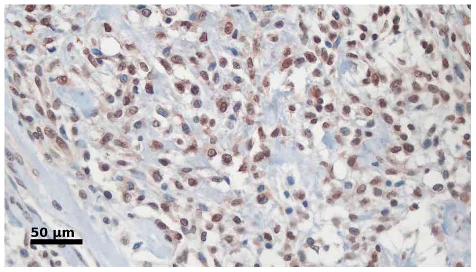
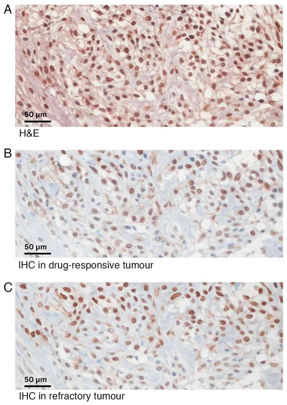
(23). Fig. 1 shows a representative
histopathology image from this study with an IRS score of 9 (strong
positive) and Fig. 2 shows a
representative image with an IRS score of 6 (moderate
positive).
| Table III.Immunoreactive scoring system
(13). |
Table III.
Immunoreactive scoring system
(13).
| Measurements | Score |
|---|
| SI |
|
| No
staining | 0 |
|
Weak | 1 |
|
Moderate | 2 |
|
Intense | 3 |
| PP |
|
| No
stained cells | 0 |
|
<10% | 1 |
|
10–50% | 2 |
|
51–80% | 3 |
|
>80% | 4 |
| IRSa |
|
|
Negative expression | 0-1 |
|
Positive, weak expression | 2-3 |
|
Positive, moderate
expression | 4-8 |
|
Positive, strong
expression | 9-12 |
Statistical analysis
Data were analyzed using SPSS software version 26.0
(IBM Corp.). Categorical variables, including patients with GSTP1
and mutant p53 expression, chemotherapy response (based on Huvos
grade) and metastatic status, are presented as frequencies and
percentages, and were analyzed using Fisher's exact test. This test
was chosen instead of the χ2 test since >20% of the
cells in several contingency tables had expected counts of ≤5,
violating the χ2 assumption. P<0.05 was considered to
indicate a statistically significant difference.
Results
Characteristics
The demographic and clinical characteristics of the
36 patients are summarized in Table
IV. The most common location for osteosarcoma was the distal
femur (52.8%). The majority of patients underwent surgical
interventions, with 23 patients (63.9%) receiving amputations and
13 patients (36.1%) undergoing limb salvage surgery. Notably, 16
patients (44.4%) presented with metastatic osteosarcoma, while 20
patients (55.6%) had non-metastatic disease.
| Table IV.Demographic characteristics. |
Table IV.
Demographic characteristics.
|
Characteristics | Value |
|---|
| Sex, n (%) |
|
|
Male | 20 (55.6) |
|
Female | 16 (44.4) |
| Mean age ± SD,
years | 18.14±8.37 |
| Location, n
(%) |
|
| Distal
femur | 19 (52.8) |
|
Proximal humerus | 4 (11.1) |
|
Proximal tibia | 7 (19.4) |
| Distal
tibia | 1 (2.8) |
|
Pelvis | 1 (2.8) |
|
Calcaneus | 1 (2.8) |
|
Talus | 1 (2.8) |
|
Proximal femur | 1 (2.8) |
| Distal
radius | 1 (2.8) |
| Subtype of
osteosarcoma, n (%) |
|
|
Osteoblastic | 21 (58.3) |
|
Chondroblastic | 2 (5.6) |
|
Fibroblastic | 3 (8.3) |
|
Osteoblastic and
chondroblastic | 7 (19.4) |
|
Fibroblastic and
chondroblastic | 2 (5.6) |
|
Osteoblastic and
fibroblastic | 1 (2.8) |
| Lung metastasis, n
(%) |
|
|
Metastasis | 16 (44.4) |
| Without
metastasis | 20 (55.6) |
| Type of surgery, n
(%) |
|
| Limb
salvage surgery | 13 (36.1) |
|
Amputation | 23 (63.9) |
Analysis of p53 mutant-type expression
and GSTP1 expression in association with chemotherapy response
p53 mutant-type and GSTP1 expression was
characterized by histopathology using brown staining and was found
in both the cell nucleus and cytoplasm. p53 mutant-type and GSTP1
immunoreactivity were evaluated using the IRS, and the analysis
results are displayed in Table
V.
| Table V.Fisher's exact test analysis of p53
mutant-type expression and GSTP1 expression with regard to
chemotherapy response. |
Table V.
Fisher's exact test analysis of p53
mutant-type expression and GSTP1 expression with regard to
chemotherapy response.
|
| Chemotherapy
response, n (%) |
|
|---|
|
|
|
|
|---|
| Variable | Good | Poor | P-value |
|---|
| GSTP1
expression |
|
| 0.024 |
|
Negative | 2 (33.3) | 0 (0.0) |
|
|
Positive | 4 (66.7) | 30 (100.0) |
|
| Mutant p53
expression |
|
| 0.024 |
|
Negative | 2 (33.3) | 0 (0.0) |
|
|
Positive | 4 (66.7) | 30 (100.0) |
|
In patients with a good chemotherapy response (n=6),
a lack of GSTP1 expression was found in 2 patients (33.3%), while
positive expression was found in 4 patients (66.7%). In those with
a poor chemotherapy response (n=30), all 30 patients exhibited
positive GSTP1 expression (100.0%). Using Fisher's exact test, the
association between GSTP1 expression and chemotherapy response was
determined to be significant (P=0.024).
A lack of mutant p53 expression was found in 2
patients (33.3%) with a good chemotherapy response, while positive
expression was exhibited in 4 patients (66.7%). In those with a
poor chemotherapy response, all 30 patients exhibited positive
mutant p53 expression (100.0%). Using Fisher's exact test, the
association between mutant p53 expression and chemotherapy response
was determined to be significant (P=0.024).
Analysis of p53 mutant-type expression
and GSTP1 expression in association with metastasis
According to Table
VI, only 1 patient (6.3%) with non-metastatic disease lacked
the expression of GSTP1, while positive expression was observed in
15 patients (93.8%) with non-metastatic disease. Meanwhile, in
patients with metastasis, a lack of GSTP1 expression was also
observed in 1 patient (5.0%), while positive expression was
exhibited by 19 patients (95.0%). Fisher's exact test revealed that
there was no significant association between GSTP1 expression and
the occurrence of metastasis (P>0.999).
| Table VI.Fisher's exact test analysis of p53
mutant-type expression and GSTP1 expression with regard to
metastasis. |
Table VI.
Fisher's exact test analysis of p53
mutant-type expression and GSTP1 expression with regard to
metastasis.
|
| Metastasis, n
(%) |
|
|---|
|
|
|
|
|---|
| Variable | No | Yes | P-value |
|---|
| GSTP1
expression |
|
| >0.999 |
|
Negative | 1 (6.3) | 1 (5.0) |
|
|
Positive | 15 (93.8) | 19 (95.0) |
|
| Mutant p53
expression |
|
| >0.999 |
|
Negative | 1 (6.3) | 1 (5.0) |
|
|
Positive | 15 (93.8) | 19 (95.0) |
|
There was a lack of p53 mutant-type expression in 1
non-metastatic patient (6.3%), while positive expression was
observed in 15 patients (93.8%). Meanwhile, in those with
metastasis, 1 patient (5.0%) was negative for mutant p53
expression, while positive expression was exhibited by 19 patients
(95.0%). Fisher's exact test revealed that there was no significant
association between the expression of mutant p53 and the occurrence
of metastasis (P>0.999).
Discussion
The present study findings reveal a significant and
robust correlation between GSTP1 expression and mutant p53
expression in chemotherapy response. The tumor suppressor protein
p53, encoded by the TP53 gene, plays a crucial role in regulating
cellular responses to stress, particularly DNA damage. p53 is often
referred to as the ‘guardian of the genome’ due to its role in
preventing genomic instability by inducing cell cycle arrest, DNA
repair, apoptosis or senescence in response to cellular stressors
(24–26). The correlation between p53 status
(wild-type vs. mutant-type) and response to chemotherapy in cancer
treatment is complex and multifaceted; its status in cancer cells
significantly affects the effectiveness of chemotherapy and
radiation therapy, two main pillars of cancer treatment (27,28).
The present study cohort consisted predominantly of
male adolescent cohort patients (mean age, 18 years), with tumors
chiefly in the distal femur. High amputation rates (63.9%)
reflected site-specific surgical constraints, yet the
histopathological markers, mutant p53 and GSTP1, proved far more
informative for treatment response than for metastatic behaviour.
Both proteins were upregulated in every case with a poor
chemotherapy response (IRS≥6), showing strong, significant
associations with chemoresistance. This dichotomy underscores that
chemo-protective mechanisms (e.g., defective apoptosis via mutant
p53 or drug detoxification via GSTP1) operate independently of the
biological pathways driving metastatic spread, highlighting the
value of these markers for predicting chemoresponsiveness across
clinical subgroups while signaling the need for additional
molecular profiling to clarify determinants of metastasis in
osteosarcoma.
The observation that mutant p53 and GSTP1 expression
specifically predicts chemoresponse but not metastasis is
consistent with the distinct prognostic roles of other established
histopathological parameters. Beyond the Huvos score, which remains
the standard measure of tumor necrosis, parameters such as
post-treatment mitotic index and microscopic vascular invasion
(MVI) are also prognostically significant. For example, a high
mitotic index (≥10 mitoses/10 high-power field) in the resection
specimen is an independent predictor of poor survival, reflecting a
failure to achieve cell cycle arrest post-treatment (29). This aligns with the present
molecular findings, as both indicate a chemoresistant phenotype.
The prognostic utility of mitotic rate appears specific to
post-treatment evaluation, as its value in pre-treatment biopsies
has been shown to be limited (30).
Conversely, and in contrast to the present p53 and
GSTP1 results, MVI is a powerful predictor of metastatic
progression; its presence is strongly associated with higher rates
of metastasis, local recurrence and mortality. This risk is
markedly amplified in patients who are also poor chemo-responders,
whose 5-year survival has been reported to be as low as 24%
(31). This comparison reinforces
the conclusion that the molecular mechanisms of chemoresistance,
indicated in the present cohort by mutant p53 and GSTP1 expression,
appear to be distinct from the biological pathways, such as MVI,
that primarily govern metastatic dissemination.
This lack of association contrasts sharply with a
recent study that estimated that the association of GSTP1
upregulation and mutant TP53 led to to a roughly two-fold increase
in metastatic risk; therefore, the null finding of the present
study most plausibly reflects the limited cohort size and
relatively short follow-up, which together reduce power to capture
late-emerging metastases (32).
Future multicentre studies with larger samples and extended
surveillance will be required to resolve this discrepancy.
Mutations in the TP53 gene often result in the loss
of wild-type p53 function, which disrupts the ability of a cell to
undergo apoptosis in response to DNA damage induced by chemotherapy
(33). This loss of function can
occur through various mechanisms, including disruptions in DNA
binding, which prevents p53 from activating its target genes
involved in cell cycle arrest and apoptosis (34,35).
Mutant p53 can modulate the tumor microenvironment to create
conditions that support tumor survival and growth, as well as
resistance to chemotherapy (36).
This includes promoting angiogenesis, immune evasion and the
secretion of factors, such as IL-6, IL-8, HGF or IGF-1, that
support tumor cell survival (37,38).
p53 mutations have been linked to increased
expression of multidrug resistance proteins, such as
P-glycoprotein, which can actively efflux chemotherapy agents from
cancer cells, reducing their effectiveness (39). Additionally, p53 mutations can
modulate the expression of genes involved in DNA repair, allowing
cancer cells to survive and proliferate despite the genotoxic
effects of chemotherapy (25).
Mutant p53 proteins often acquire gain-of-function
(GOF) properties that contribute to tumor progression and
chemotherapy resistance. These GOF properties include enhanced drug
efflux, metabolism, promotion of survival, inhibition of apoptosis,
increased DNA repair, suppression of autophagy, enhanced
microenvironmental resistance and induction of a stem-like
phenotype. For example, specific p53 mutations, such as R175H and
R273H, have been shown to increase resistance to chemotherapeutic
agents, such as cisplatin and etoposide, by disrupting the normal
p53-mediated apoptosis pathway (40,41).
These mutations can also affect the expression of
various genes involved in metabolism and drug effects, further
contributing to resistance. Specifically, in osteosarcoma, the
R270C p53 mutant does not suppress the transcriptional activation
function of wild-type p53, leading to resistance to doxorubicin, a
common chemotherapy agent (42).
Additionally, the expression of mutant p53 is associated with
increased expression of ONZIN (Plac8), which contributes to
osteosarcoma metastasis through the CXCL5-MAPK signaling pathway
(43).
GSTP1 is a critical enzyme involved in the
detoxification of xenobiotics and carcinogens by catalyzing the
conjugation of glutathione to reactive intermediates. This enzyme
plays a crucial role in cellular defense mechanisms against toxic
and carcinogenic compounds. However, its expression in cancer cells
has been associated with resistance to chemotherapy, affecting the
efficacy of treatment various types of malignancies, including
breast, ovarian, lung, colorectal, prostate, gastric and hepatic
cancers, as well as leukemias, where elevated GSTP1 levels have
been consistently linked to reduced sensitivity to commonly used
chemotherapeutic agents (44,45).
GSTP1 is a phase II metabolic enzyme that catalyzes
the conjugation of reduced glutathione to various substrates,
including chemotherapy agents, leading to their detoxification and
excretion. Upregulation of GSTP1 in osteosarcoma cells has been
associated with an increased capacity to deactivate chemotherapy
drugs, thereby reducing their cytotoxicity and effectiveness
(46). A study indicated that
osteosarcoma cells with increased GSTP1 expression exhibit
resistance to various chemotherapeutic drugs, including
doxorubicin, cisplatin and methotrexate, which are commonly used in
the treatment of osteosarcoma (47).
The GSTP1 mechanism contributes to chemotherapy drug
resistance by its ability to conjugate glutathione to chemotherapy
drugs, thereby neutralizing their cytotoxic effects. This
conjugation increases the solubility of the drugs, leading to their
rapid elimination from cells and reducing the effects of their
cytotoxic chemotherapy. For example, GSTP1 has been shown to
detoxify platinum-based compounds, such as cisplatin and
oxaliplatin, which are commonly used in the treatment of various
types of cancer (13,48). Additionally, GSTP1 can modulate
signaling pathways associated with cell survival and apoptosis. For
instance, GSTP1 has been shown to inhibit the pathway of Jun
N-terminal kinase, a protein involved in critical mediation of
apoptosis or pro-apoptotic signaling, thereby promoting the
survival of cancer cells from chemotherapy (49).
By contrast, pharmacogenetic investigations have
centred on the germline Ile105Val (rs1695) polymorphism of GSTP1,
yet its association with chemoresponse and survival has been
inconsistent (50). Measuring GSTP1
at the protein level provides a more integrative read-out, as
expression reflects not only such coding variants but also somatic
copy-number changes, promoter methylation, NRF2-driven
transcription and post-translational stabilisation during oxidative
stress; this broader regulatory capture may explain why expression
status, but not Ile105Val genotype alone, was robustly associated
with chemoresistance.
In addition, in the present study, contrary results
were found between GSTP1 expression and mutant p53 expression
regarding metastasis events in patients with osteosarcoma at Cipto
Mangunkusumo General Hospital, showing no significant associations.
Several studies have explored the genetic and molecular
underpinnings of GSTP1 expression in osteosarcoma, particularly its
potential role in metastasis. A comprehensive study conducted by Li
et al (50) investigated the
role of genes involved in metabolic and transport pathways,
including GSTP1, in osteosarcoma survival after chemotherapy. The
findings revealed no significant association between GSTP1
expression and metastatic disease in patients with osteosarcoma.
Similarly, a pharmacogenetic analysis of primary and metastatic
osteosarcoma failed to identify GSTP1 among the genes whose
expression is associated with metastasis (51).
The mechanistic role of GSTP1 in cancer metastasis
remains unclear. Although its detoxification function could
theoretically influence metastatic potential by altering the tumor
microenvironment or modulating responses to chemotherapy, current
studies have not established a direct mechanistic link between
GSTP1 expression and metastatic behavior in osteosarcoma cells.
Some investigations have examined GSTP1 in relation to
chemoresistance and apoptosis, but they have not directly addressed
its impact on metastasis. Although GSTP1 participates in
detoxification and has been implicated in chemotherapy resistance,
its direct contribution to osteosarcoma metastasis remains
uncertain as available evidence has not demonstrated a clear
association between GSTP1 expression and metastatic progression.
The complexity of metastasis, which involves multiple genetic and
environmental factors, likely diminishes the influence that any
single gene, including GSTP1, may exert on metastatic outcomes
(46,52).
The complexity of cancer metastasis, which involves
many genetic and environmental factors, likely diminishes the
potential impact of a single gene, such as GSTP1, on metastasis
outcomes. Other studies suggest various mechanisms and pathways
that could be more directly involved in the osteosarcoma metastasis
process. For example, research has highlighted the importance of
signaling pathways such as PI3K/AKT/mTOR and their roles in tumor
growth and metastasis in osteosarcoma. These pathways may offer a
more direct link to metastatic behavior than GSTP1 (53,54).
Several studies have investigated the correlation
between p53 mutations and metastasis in osteosarcoma, with varying
conclusions. For example, a study by Wunder et al (55) found no evidence that p53 mutations
predict the development of metastases in patients with high-grade
osteosarcoma. The p53 mutation status did not differentiate between
patients who presented with localized or metastatic disease.
Similarly, research by Overholtzer et al (56) reported that the p53 mutation status
was concordant between primary tumors and matched metastases in
patients with osteosarcoma, suggesting that p53 mutations are early
events in tumorigenesis rather than drivers of metastasis.
Mutant p53 plays a critical role in osteosarcoma
metastasis. The R270C mutant p53, equivalent to human R273C, has
been shown to bind to chromatin near the transcription start sites
of various genes, altering their expression. This mutant form
exhibits a different binding profile from wild-type p53 and does
not suppress the transcriptional activation function of the
wild-type protein in osteosarcoma cells. Although the deletion of
this mutant p53 reduces tumor growth, it does not affect invasion
or prevent metastasis in vivo. This suggests that while
mutant p53 contributes to tumor growth, it may not directly affect
the metastatic ability of osteosarcoma cells (42).
The clinical implication based on the results of the
present study is that the expression of mutant p53 and GSTP1 can be
used as for diagnostic purposes in patients with osteosarcoma,
thereby serving as a predictor of chemotherapy response, prognosis
and decision-making in the management of affected patients.
The present study focused on a specific population
of patients with osteosarcoma, providing valuable insights within a
defined geographic and demographic context, thus yielding relevant
data for that population. Additionally, this study was more
feasible to implement due to its cross-sectional design, which
eliminates the need for long-term follow-up. Conducted at a single
point in time, this study offers a faster and more cost-effective
approach compared with longitudinal studies.
A key limitation of this study is its
cross-sectional design, which involves a single data collection
point, thereby providing only a snapshot of the population at a
particular moment. This limitation is particularly pertinent in
research involving dynamic biological processes, such as
chemotherapy response or metastasis development, where longitudinal
changes and trends may be crucial to understanding the underlying
mechanisms. Moreover, the relatively small, single-centre cohort
and the retrospective design may reduce statistical power,
introduce selection bias and limit the generalisability of the
study findings.
In conclusion, the present study found a significant
association between mutant p53 expression and GSTP1 expression and
poor chemotherapy response in patients with osteosarcoma.
Meanwhile, the association between mutant p53 expression and GSTP1
expression and the occurrence of metastasis was not significant in
patients with osteosarcoma. Further research should focus on
longitudinal studies that track the expression levels of GSTP1 and
mutant p53 from initial diagnosis to the development of metastasis
and the treatments provided. Further research requires
comprehensive genomic profiling studies to explore the interactions
of GSTP1 and mutant P53 with other genes, as well as the signaling
pathways involved in metastasis and chemotherapy response in
osteosarcoma, to provide a more comprehensive understanding of the
biology of this disease.
Acknowledgements
Not applicable.
Funding
Funding: No funding was received.
Availability of data and materials
The data generated in the present study may be
requested from the corresponding author.
Authors' contributions
BP, AFK, EK, YP, AK, NCS, MNSBI, RM, IGEW and III
were responsible for study design. BP, AFK and EK were responsible
for data analysis. BP, AFK and EK wrote the manuscript. BP, AFK and
EK confirm the authenticity of all the raw data. All authors have
read and approved the manuscript.
Ethics approval and consent to
participate
Ethical approval was obtained from the Ethics
Committee of the Cipto Mangunkusumo General Hospital, Faculty of
Medicine, University of Indonesia (Jakarta, Indonesia; approval no.
KET-420/UN2.FI/ETIK/PPM/00.02/2023). Written informed consent was
obtained from all participants, with parental or guardian consent
obtained for those <18 years old.
Patient consent for publication
Not applicable.
Competing interests
The authors declare that they have no competing
interests.
Glossary
Abbreviations
Abbreviations:
|
GSTP1
|
glutathione S-transferase P1
|
|
FFPE
|
formalin-fixed paraffin-embedded
|
|
IRS
|
immunoreactive scoring system
|
|
CC
|
contingency coefficient
|
|
GOF
|
gain-of-function
|
References
|
1
|
Soares do Brito J, Santos R, Sarmento M,
Fernandes P and Portela J: Chemotherapy regimens for non-metastatic
conventional appendicular osteosarcoma: A literature review based
on the outcomes. Curr Oncol. 30:6148–6165. 2023. View Article : Google Scholar : PubMed/NCBI
|
|
2
|
Xin S and Wei G: Prognostic factors in
osteosarcoma: A study level meta-analysis and systematic review of
current practice. J Bone Oncol. 21:1002812020. View Article : Google Scholar : PubMed/NCBI
|
|
3
|
Morrow JJ and Khanna C: Osteosarcoma
genetics and epigenetics: Emerging biology and candidate therapies.
Crit Rev Oncog. 20:173–197. 2015. View Article : Google Scholar : PubMed/NCBI
|
|
4
|
Ozaki T and Nakagawara A: Role of p53 in
cell death and human cancers. Cancers (Basel). 3:994–1013. 2011.
View Article : Google Scholar : PubMed/NCBI
|
|
5
|
Marei HE, Althani A, Afifi N, Hasan A,
Caceci T, Pozzoli G, Morrione A, Giordano A and Cenciarelli C: p53
signaling in cancer progression and therapy. Cancer Cell Int.
21:7032021. View Article : Google Scholar : PubMed/NCBI
|
|
6
|
Chen X, Zhang T, Su W, Dou Z, Zhao D, Jin
X, Lei H, Wang J, Xie X, Cheng B, et al: Mutant p53 in cancer: From
molecular mechanism to therapeutic modulation. Cell Death Dis.
13:9742022. View Article : Google Scholar : PubMed/NCBI
|
|
7
|
Monti P, Menichini P, Speciale A, Cutrona
G, Fais F, Taiana E, Neri A, Bomben R, Gentile M, Gattei V, et al:
Heterogeneity of TP53 mutations and P53 protein residual function
in cancer: Does it matter? Front Oncol. 10:5933832020. View Article : Google Scholar : PubMed/NCBI
|
|
8
|
Chiang YT, Chien YC, Lin YH, Wu HH, Lee DF
and Yu YL: The function of the mutant p53-R175H in cancer. Cancers
(Basel). 13:40882021. View Article : Google Scholar : PubMed/NCBI
|
|
9
|
Durairaj G, Demir Ö, Lim B, Baronio R,
Tifrea D, Hall LV, DeForest JC, Lauinger L, Jebril Fallatah MM, Yu
C, et al: Discovery of compounds that reactivate p53 mutants in
vitro and in vivo. Cell Chem Biol. 29:1381–1395.e13. 2022.
View Article : Google Scholar : PubMed/NCBI
|
|
10
|
Rickel K, Fang F and Tao J: Molecular
genetics of osteosarcoma. Bone. 102:69–79. 2017. View Article : Google Scholar : PubMed/NCBI
|
|
11
|
Mizuarai S, Yamanaka K and Kotani H:
Mutant p53 induces the GEF-H1 oncogene, a guanine nucleotide
exchange factor-H1 for RhoA, resulting in accelerated cell
proliferation in tumor cells. Cancer Res. 66:6319–6326. 2006.
View Article : Google Scholar : PubMed/NCBI
|
|
12
|
Mazari AMA, Zhang L, Ye ZW, Zhang J, Tew
KD and Townsend DM: The multifaceted role of glutathione
S-Transferases in health and disease. Biomolecules. 13:6882023.
View Article : Google Scholar : PubMed/NCBI
|
|
13
|
Sawers L, Ferguson M, Ihrig BR, Young HC,
Chakravarty P, Wolf CR and Smith G: Glutathione S-transferase P1
(GSTP1) directly influences platinum drug chemosensitivity in
ovarian tumour cell lines. Br J Cancer. 111:1150–1158. 2014.
View Article : Google Scholar : PubMed/NCBI
|
|
14
|
Miyake T, Nakayama T, Naoi Y, Yamamoto N,
Otani Y, Kim SJ, Shimazu K, Shimomura A, Maruyama N, Tamaki Y and
Noguchi S: GSTP1 expression predicts poor pathological complete
response to neoadjuvant chemotherapy in ER-negative breast cancer.
Cancer Sci. 103:913–920. 2012. View Article : Google Scholar : PubMed/NCBI
|
|
15
|
Hattinger CM, Biason P, Iacoboni E, Gagno
S, Fanelli M, Tavanti E, Vella S, Ferrari S, Roli A, Roncato R, et
al: Candidate germline polymorphisms of genes belonging to the
pathways of four drugs used in osteosarcoma standard chemotherapy
associated with risk, survival and toxicity in non-metastatic
high-grade osteosarcoma. Oncotarget. 7:61970–61987. 2016.
View Article : Google Scholar : PubMed/NCBI
|
|
16
|
Pu F, Chen F, Chen S, Wang B, Liu J and
Shao Z: Association between GSTP1 polymorphisms and prognosis of
osteosarcoma in patients treated with chemotherapy: A
meta-analysis. Onco Targets Ther. 8:1835–1842. 2015.PubMed/NCBI
|
|
17
|
Hassanain O, Alaa M, Khalifa MK, Kamal N,
Albagoury A and El Ghoneimy AM: Genetic variants associated with
osteosarcoma risk: A systematic review and meta-analysis. Sci Rep.
14:38282024. View Article : Google Scholar : PubMed/NCBI
|
|
18
|
Fengfeng W, Ruqing Y and Juntao X: GSTP1
A>G polymorphism and chemosensitivity of osteosarcoma: A
meta-analysis. Open Med (Wars). 11:101–105. 2016. View Article : Google Scholar : PubMed/NCBI
|
|
19
|
Jawad MU and Scully SP: In brief:
Classifications in brief: Enneking classification: Benign and
malignant tumors of the musculoskeletal system. Clin Orthop Relat
Res. 468:2000–2002. 2010. View Article : Google Scholar : PubMed/NCBI
|
|
20
|
Prabowo Y, Setiawan I, Kamal AF, Kodrat E
and Labib Zufar ML: Correlation between prognostic factors and the
histopathological response to neoadjuvant chemotherapy in
osteosarcoma: A retrospective study. Int J Surg Oncol.
2021:88433252021.PubMed/NCBI
|
|
21
|
WHO, . In: Soft Tissue and Bone Tumours.
5th edition. Board Co TE: WHO; Geneva, Switzerland: 2020
|
|
22
|
Amin MB, Edge SB, Greene FL, Byrd DR,
Brookland RK, Kay Washington M, Gershenwald JE, Compton CC, Hess
KR, Sullivan DC, et al: AJCC Cancer Staging Manual. 8th edition.
New York; Springer: 2017
|
|
23
|
Fedchenko N and Reifenrath J: Different
approaches for interpretation and reporting of immunohistochemistry
analysis results in the bone tissue-a review. Diagn Pathol.
9:2212014. View Article : Google Scholar : PubMed/NCBI
|
|
24
|
Feroz W and Sheikh AMA: Exploring the
multiple roles of guardian of the genome: P53. Egypt J Med Hum
Genet. 21:492020. View Article : Google Scholar
|
|
25
|
Williams AB and Schumacher B: p53 in the
DNA-damage-repair process. Cold Spring Harb Perspect Med.
6:a0260702016. View Article : Google Scholar : PubMed/NCBI
|
|
26
|
Ljungman M: Dial 9-1-1 for p53: Mechanisms
of p53 activation by cellular stress. Neoplasia. 2:208–225. 2000.
View Article : Google Scholar : PubMed/NCBI
|
|
27
|
Carlsen L and El-Deiry WS: Differential
p53-Mediated cellular responses to DNA-damaging therapeutic agents.
Int J Mol Sci. 22:118282021. View Article : Google Scholar : PubMed/NCBI
|
|
28
|
Abuetabh Y, Wu HH, Chai C, Al Yousef H,
Persad S, Sergi CM and Leng R: DNA damage response revisited: The
p53 family and its regulators provide endless cancer therapy
opportunities. Exp Mol Med. 54:1658–1669. 2022. View Article : Google Scholar : PubMed/NCBI
|
|
29
|
Chui M, Kandel R, Wong M, Griffin A, Bell
R, Blackstein M, Wunder J and Dickson B: Histopathologic features
of prognostic significance in high-grade osteosarcoma. Arch Pathol
Lab Med. 140:1231–1238. 2016. View Article : Google Scholar : PubMed/NCBI
|
|
30
|
Cates JM and Dupont WD: Cytologic
anaplasia is a prognostic factor in osteosarcoma biopsies, but
mitotic rate or extent of spontaneous tumor necrosis are not: A
critique of the College of American Pathologists bone biopsy
template. Mod Pathol. 30:52–59. 2017. View Article : Google Scholar : PubMed/NCBI
|
|
31
|
Anderson ME, Wu JS and Vargas SO: CORR
Tumor board: Is microscopic vascular invasion in tumor specimens
associated with worse prognosis in patients with high-grade
localized osteosarcoma? Clin Orthop Relat Res. 478:2814–2818. 2020.
View Article : Google Scholar : PubMed/NCBI
|
|
32
|
Wang G, Cao Y, Hu T, Cai Z, Chen C, Geng
Q, Luo X, Liu Y, Wang W, Jin J and Sheng W: A mutual interaction
between GSTP1 and p53 improves the drug resistance and malignant
biology of pancreatic cancer. Cancer Sci. 116:1268–1281. 2025.
View Article : Google Scholar : PubMed/NCBI
|
|
33
|
Tsuda Y, Tsoi K, Stevenson JD, Parry M,
Fujiwara T, Sumathi VP and Jeys LM: Is microscopic vascular
invasion in tumor specimens associated with worse prognosis in
patients with high-grade localized osteosarcoma? Clin Orthop Relat
Res. 478:1253–1262. 2020. View Article : Google Scholar : PubMed/NCBI
|
|
34
|
Babamohamadi M, Babaei E, Ahmed Salih B,
Babamohammadi M, Jalal Azeez H and Othman G: Recent findings on the
role of wild-type and mutant p53 in cancer development and therapy.
Front Mol Biosci. 9:9030752022. View Article : Google Scholar : PubMed/NCBI
|
|
35
|
Kennedy MC and Lowe SW: Mutant p53: It's
not all one and the same. Cell Death Differ. 29:983–987. 2022.
View Article : Google Scholar : PubMed/NCBI
|
|
36
|
Zhu Kl, Su F, Yang JR, Xiao RW, Wu RY, Cao
MY, Ling XL and Zhang T: TP53 to mediate immune escape in tumor
microenvironment: An overview of the research progress. Mol Biol
Rep. 51:2052024. View Article : Google Scholar : PubMed/NCBI
|
|
37
|
Carlsen L, Zhang S, Tian X, De La Cruz A,
George A, Arnoff TE and El-Deiry WS: The role of p53 in anti-tumor
immunity and response to immunotherapy. Front Mol Biosci.
10:11483892023. View Article : Google Scholar : PubMed/NCBI
|
|
38
|
Liu S, Liu T, Jiang J, Guo H and Yang R:
p53 mutation and deletion contribute to tumor immune evasion. Front
Genet. 14:10884552023. View Article : Google Scholar : PubMed/NCBI
|
|
39
|
Linn S, Honkoop A, Hoekman K, van der Valk
P, Pinedo HM and Giaccone G: p53 and P-glycoprotein are often
co-expressed and are associated with poor prognosis in breast
cancer. Br J Cancer. 74:63–68. 1996. View Article : Google Scholar : PubMed/NCBI
|
|
40
|
Alvarado-Ortiz E, de la Cruz-López KG,
Becerril-Rico J, Sarabia-Sánchez MA, Ortiz-Sánchez E and
García-Carrancá A: Mutant p53 Gain-of-Function: Role in cancer
development, progression, and therapeutic approaches. Front Cell
Dev Biol. 8:6076702021. View Article : Google Scholar : PubMed/NCBI
|
|
41
|
Do PM, Varanasi L, Fan S, Li C, Kubacka I,
Newman V, Chauhan K, Daniels SR, Boccetta M, Garrett MR, et al:
Mutant p53 cooperates with ETS2 to promote etoposide resistance.
Genes Dev. 26:830–845. 2012. View Article : Google Scholar : PubMed/NCBI
|
|
42
|
Shimizu T, Sugihara E, Takeshima H,
Nobusue H, Yamaguchi R, Yamaguchi-Iwai S, Fukuchi Y, Ushijima T,
Muto A and Saya H: Depletion of R270C Mutant p53 in osteosarcoma
attenuates cell growth but does not prevent invasion and metastasis
in vivo. Cells. 11:36142022. View Article : Google Scholar : PubMed/NCBI
|
|
43
|
Zhang Y, Hu Q, Li G, Li L, Liang S, Zhang
Y, Liu J, Fan Z, Li L, Zhou B, et al: ONZIN upregulation by mutant
p53 contributes to osteosarcoma metastasis through the CXCL5-MAPK
signaling pathway. Cell Physiol Biochem. 48:1099–1111. 2018.
View Article : Google Scholar : PubMed/NCBI
|
|
44
|
Ogino S, Konishi H, Ichikawa D, Matsubara
D, Shoda K, Arita T, Kosuga T, Komatsu S, Shiozaki A, Okamoto K, et
al: Glutathione S-transferase Pi 1 is a valuable predictor for
cancer drug resistance in esophageal squamous cell carcinoma.
Cancer Sci. 110:795–804. 2019. View Article : Google Scholar : PubMed/NCBI
|
|
45
|
Bocedi A, Noce A, Marrone G, Noce G,
Cattani G, Gambardella G, Di Lauro M, Di Daniele N and Ricci G:
Glutathione transferase P1-1 an enzyme useful in biomedicine and as
biomarker in clinical practice and in environmental pollution.
Nutrients. 11:17412019. View Article : Google Scholar : PubMed/NCBI
|
|
46
|
Cui J, Li G, Yin J, Li L, Tan Y, Wei H,
Liu B, Deng L, Tang J and Chen Y and Chen Y: GSTP1 and cancer:
Expression, methylation, polymorphisms and signaling (review). Int
J Oncol. 56:867–878. 2020.PubMed/NCBI
|
|
47
|
Huang G, Mills L and Worth LL: Expression
of human glutathione S-transferase P1 mediates the chemosensitivity
of osteosarcoma cells. Mol Cancer Ther. 6:1610–1619. 2007.
View Article : Google Scholar : PubMed/NCBI
|
|
48
|
Yang SJ, Wang DD, Li J, Xu HZ, Shen HY,
Chen X, Zhou SY, Zhong SL, Zhao JH and Tang JH: Predictive role of
GSTP1-containing exosomes in chemotherapy-resistant breast cancer.
Gene. 623:5–14. 2017. View Article : Google Scholar : PubMed/NCBI
|
|
49
|
Pljesa-Ercegovac M, Savic-Radojevic A,
Matic M, Coric V, Djukic T, Radic T and Simic T: Glutathione
transferases: Potential targets to overcome chemoresistance in
solid tumors. Int J Mol Sci. 19:37852018. View Article : Google Scholar : PubMed/NCBI
|
|
50
|
Li JZ, Tian ZQ, Jiang SN and Feng T:
Effect of variation of ABCB1 and GSTP1 on osteosarcoma survival
after chemotherapy. Genet Mol Res. 13:3186–3192. 2014. View Article : Google Scholar : PubMed/NCBI
|
|
51
|
Trujillo-Paolillo A, Tesser-Gamba F,
Seixas Alves MT, Filho RJG, Oliveira R, Petrilli AS and Toledo SRC:
Pharmacogenetics of the primary and metastatic osteosarcoma: Gene
expression profile associated with outcome. Int J Mol Sci.
24:56072023. View Article : Google Scholar : PubMed/NCBI
|
|
52
|
Chatterjee A and Gupta S: The multifaceted
role of glutathione S-transferases in cancer. Cancer Lett.
433:33–42. 2018. View Article : Google Scholar : PubMed/NCBI
|
|
53
|
Xiang Y, Yang Y, Liu J and Yang X:
Functional role of MicroRNA/PI3K/AKT axis in osteosarcoma. Front
Oncol. 13:12192112023. View Article : Google Scholar : PubMed/NCBI
|
|
54
|
Zhang J, Yu XH, Yan YG, Wang C and Wang
WJ: PI3K/Akt signaling in osteosarcoma. Clin Chim Acta.
444:182–192. 2015. View Article : Google Scholar : PubMed/NCBI
|
|
55
|
Wunder JS, Gokgoz N, Parkes R, Bull SB,
Eskandarian S, Davis AM, Beauchamp CP, Conrad EU, Grimer RJ, Healey
JH, et al: TP53 mutations and outcome in osteosarcoma: A
prospective, multicenter study. J Clin Oncol. 23:1483–1490. 2005.
View Article : Google Scholar : PubMed/NCBI
|
|
56
|
Overholtzer M, Rao PH, Favis R, Lu XY,
Elowitz MB, Barany F, Ladanyi M, Gorlick R and Levine AJ: The
presence of p53 mutations in human osteosarcomas correlates with
high levels of genomic instability. Proc Natl Acad Sci USA.
100:11547–1152. 2003. View Article : Google Scholar : PubMed/NCBI
|