Introduction
In 1887, Wegner (1)
classified lymphangiomas into three histological types, namely,
simple, cavernous and cystic, a classification that remains widely
accepted today. The pathogenesis of lymphangiomas remains
incompletely understood, although they are generally regarded as
developmental anomalies (2,3). Consequently, most patients are
diagnosed during early childhood. Lymphangiomas account for 4–6% of
benign soft tissue tumors in children. The head and neck region
(~75%) and axillary region (~20%) are the most common sites of
involvement, whereas intra-abdominal lymphangiomas are relatively
rare, accounting for <5% of all lymphangiomas (2,3). Among
these, mesenteric cavernous lymphangiomas (MCLs) represent a
particularly rare and underrecognized disease entity. Clinically,
the more common abdominal cystic lymphangioma is usually discovered
incidentally during asymptomatic physical examinations of
middle-aged women (4,5). By contrast, according to published
case reports, mesenteric cavernous lymphangioma seems to be more
prevalent among male patients, and is often accompanied by clinical
manifestations such as abdominal pain and abdominal distension.
Owing to its locally infiltrative growth pattern, MCL often
contains abundant fibrous stroma and adipose tissue, resulting in
imaging features that may mimic malignant or intermediate-grade
mesenteric tumors, including liposarcoma, desmoid tumors,
inflammatory pseudotumors and extra-gastrointestinal stromal
tumors, thereby increasing the difficulty of preoperative
diagnosis. The etiology of MCL in adults may be associated with
acquired factors, such as inflammation, trauma, surgery or
radiotherapy. Owing to its lack of specific clinical
manifestations, MCL is frequently misdiagnosed, which may result in
unnecessarily extensive surgery or delayed treatment (6–9).
However, MCL has received limited attention in the
surgical and pathological literature Since Viar et al
(10) first described this lesion
in 1961, the available literature has remained largely limited to
isolated case reports, with no comprehensive analysis of its
clinical and imaging characteristics. Accordingly, the current
study presents a case of MCL and conducts a comprehensive review of
the relevant literature to synthesize and analyze its demographic,
clinical and imaging characteristics. The aim of the present study
is to provide evidence that may facilitate the accurate
preoperative diagnosis of MCL.
Case report
Patient presentation
A 55-year-old woman presented to the Zhuji People's
Hospital (Zhuji, China) in July 2021 with a 3-day history of right
upper quadrant abdominal pain that had progressively worsened over
the preceding 13 h. The patient's medical history was notable for
surgery for benign intestinal lesions 25 years previously. Upon
physical examination, the patient's vital signs were stable. The
abdomen was soft and distended, with no mildly palpable masses.
Laboratory investigations revealed a normal white
blood cell count (3.5–9.5×10⁹/l) and a mildly elevated
C-reactive protein (CRP) level (45.32 mg/l; reference range, <5
mg/l). Serum tumor markers, including α-fetoprotein (AFP; reference
range, 0–8.1 ng/ml), carcinoembryonic antigen (CEA; reference
range, 0–5.0 ng/ml), cancer antigen 125 (CA125; reference range,
0–35 U/ml) and CA19-9 (reference range, 0–37 U/ml), were all within
the normal ranges. Follow-up laboratory tests demonstrated that the
serum total protein and albumin levels had decreased from 71.3 and
42.4 g/l at admission to 64.4 and 38.9 g/l, respectively.
Imaging findings
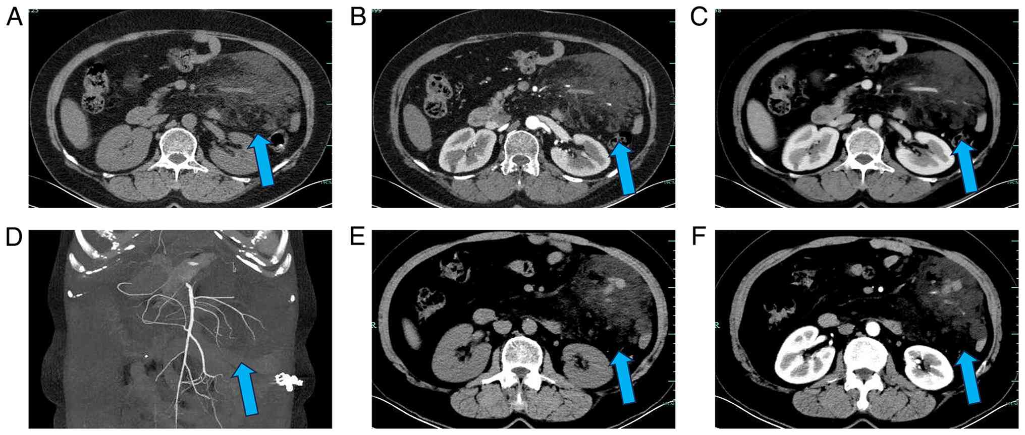
Contrast-enhanced abdominal computed tomography (CT)
performed on admission revealed an irregular, well-defined
low-attenuation mass in the left mid-abdomen containing multiple
traversing vascular structures. The lesion showed no appreciable
contrast enhancement (Fig. 1A-D).
Based on these imaging findings, the initial radiological diagnosis
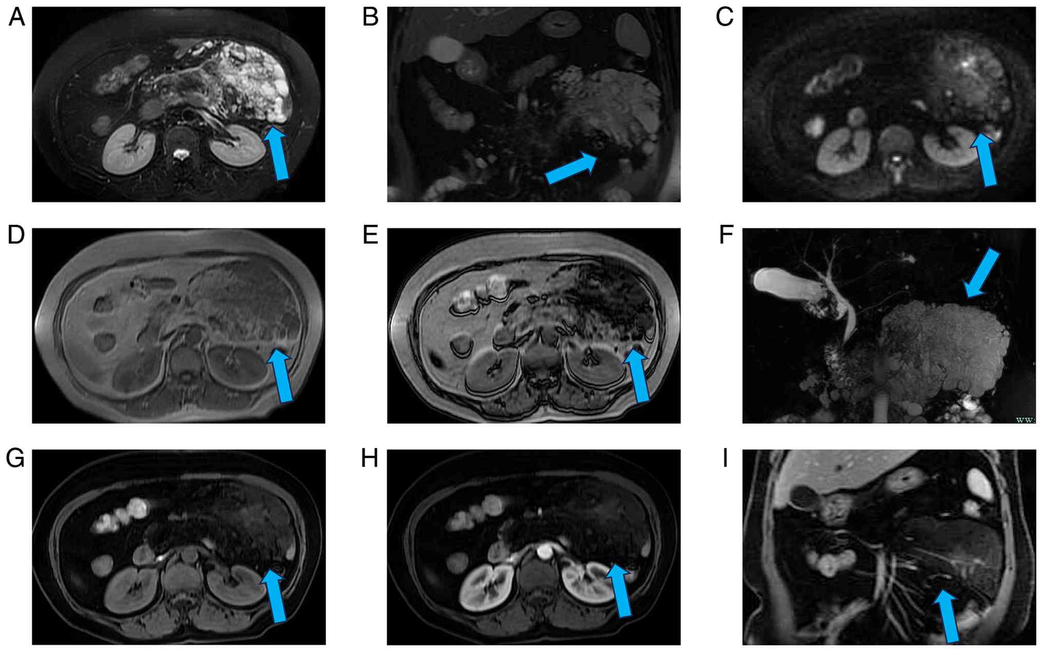
was liposarcoma. Upper abdominal MRI performed 3 days later
demonstrated that the lesion was hypointense on T1-weighted imaging
(T1WI) and hyperintense on T2WI (Fig.
2A and B), with patchy signal loss on in-phase imaging
(Fig. 2D-F). DWI shows patchy
hyperintense signals (Fig. 2C). The
lesion showed no significant enhancement after contrast
administration (Fig. 2G-I). A
follow-up CT performed during hospitalization demonstrated new
intralesional hemorrhagic foci (Fig. 1E
and F). The location of the newly emerged hemorrhagic focus is
consistent with the high-signal area on DWI (Fig. 2C).
Treatment and pathological
findings
At 9 days post-admission, the patient underwent an
exploratory laparotomy, a complete resection of the mesenteric
mass, a partial jejunal resection and a jejunojejunostomy.
Intraoperatively, a soft, milky-white mass measuring ~10×15 cm was
identified within the jejunal mesentery. The lesion involved ~30 cm
of the jejunum and extended to a previous intestinal anastomosis.
Milky fluid exuded from the cut surface of the specimen.
Intraoperative frozen-section examination revealed vascular-like
structures and scattered lymphocytes within proliferating adipose
tissue. The resected specimen was initially fixed using 10% neutral
buffered formalin (ready-to-use) at 25°C for 12 h. Subsequently, 3
µm-thick sections were prepared. The sections were stained with
hematoxylin and eosin (H&E) at 25°C for 45 min and examined
using a Zeiss Axio Lab A1 bright-field light microscope.
Representative images were captured at ×100 magnification.
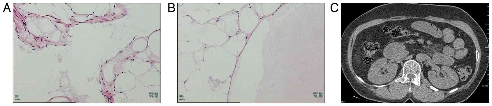
Definitive histopathological examination confirmed the diagnosis of
mesenteric cavernous lymphangioma (Fig.
3).
Postoperative course and
follow-up
Postoperatively, the patient received antimicrobial
therapy, fluid replacement and nutritional support. The patient's
clinical condition remained stable during the first 4 postoperative
days. On postoperative day 1 (POD1), 210 ml of pale red fluid was
drained. On POD2, the drainage fluid became clear, gastrointestinal
decompression was discontinued. On POD3, the drainage volume
decreased to 70 ml. On POD5, the patient developed a fever of 39°C,
and bilious fluid was noted in the abdominal drain. On POD6, the
patient resumed bowel function, with the passage of flatus and
stool; however, the drainage volume increased to 650 ml, consistent
with a high-output intestinal fistula. On POD7, the drainage fluid
appeared enteric in nature, with a volume of 250 ml. On POD8, the
patient developed a fever of 38.4°C, and the drainage fluid became
tinged with blood. On POD9, the patient developed lower abdominal
pain and vomited blood-tinged fluid. The drainage fluid became
turbid and blood-tinged, with a total drainage volume of 250 ml.
Following the development of these postoperative complications,
conservative management was initiated, including fasting,
gastrointestinal decompression, parenteral nutritional support,
fluid and electrolyte management, and anti-infective therapy. Due
to persistent bloody drainage and suspected active intra-abdominal
bleeding, the patient was transferred to a higher-level medical
center for further evaluation. Mesenteric angiography revealed
contrast extravasation from a branch of the superior mesenteric
artery, and transcatheter arterial embolization was successfully
performed. Following the intervention, the drainage volume
gradually decreased, inflammatory markers improved, and the
intestinal fistula was managed conservatively. Subsequent small
bowel contrast examination demonstrated no communication between
the intestinal lumen and the drainage tract, allowing removal of
the drainage tube. The patient was discharged after 36 days of
hospitalization and remained clinically stable without recurrence
during 4.5 years of follow-up. The detailed postoperative clinical
course and management strategies are summarized in Table I.
 | Table I.Course of patient treatment. |
Table I.
Course of patient treatment.
| Time | General
condition | Imaging study | Invasive medical
procedure | Nursing and
medication management | Drainage fluid
color/volume |
|---|
| Admission | CRP, 45.32 mg/l; TP,
77.4 g/l; ALB, 43.1 g/l | Whole-abdomen
contrast-enhanced CT | - | Anti-infective
therapy (cefotiam 2.0 g IV infusion twice daily) | - |
| Hospital day 1 | CRP, 24.7 mg/l; TP,
71.3 g/L; ALB, 42.4 g/l | Non-contrast chest
CT | - | Anti-infective
therapy |
|
| Hospital day 3 | - | Upper abdominal
contrast-enhanced MRI | - |
|
|
| Hospital day 8 | TP, 64.4 g/l; ALB,
38.9 g/l | Upper abdominal
contrast-enhanced CT |
|
|
|
| Hospital day 9 | - |
| Exploratory
laparotomy | Level I nursing care,
anti-infective therapy (piperacillin/tazobactam 4.5 g), analgesic
therapy, hemostatic therapy and fluid replacement therapy |
|
| Hospital day 10;
postoperative day 1 | - |
|
| Anti-infective
therapy, analgesic therapy, hemostatic therapy and fluid
replacement therapy | Light red fluid/210
ml |
| Hospital day 11;
postoperative day 2 | - |
|
| Hemostatic therapy
was discontinued. The patient was downgraded to level II nursing
care, and gastrointestinal decompression was discontinued | Light red fluid/350
ml |
| Hospital day 12;
postoperative day 3 | - |
|
|
| Light red fluid/70
ml |
| Hospital day 14;
postoperative day 5 | Body temperature
39°C | Whole-abdomen
non-contrast CT |
| The patient was
upgraded to level I nursing care. Somatostatin therapy,
anti-infective therapy (piperacillin/tazobactam 4.5 g every 8 h),
and fluid replacement therapy were administered | Bilious fluid |
| Hospital day 15;
postoperative day 6 |
| Whole-abdomen
non-contrast CT |
| Treatment was
continued without modification | Bilious fluid/650
ml |
| Hospital day 16;
postoperative day 7 |
|
|
|
| Enteric-appearing
fluid/250 ml |
| Hospital day 17;
postoperative day 8 | Body temperature
38.4°C |
|
|
| Light red fluid |
| Hospital day 18;
postoperative day 9 | The patient presented
with lower abdominal pain and hematemesis |
| Superior mesenteric
artery angiography with embolization |
| Turbid bloody
fluid/250 ml |
| Hospital day 19;
post-intervention day 1 | CRP, 83.4 mg/l; TP,
56.7 g/l; ALB, 35.3 g/l |
|
| Treatment was
continued without modification | Dark red fluid/530
ml |
| Hospital day 21;
post-intervention day 3 |
|
|
| Level II nursing
care, anti-infective therapy, somatostatin therapy,
acid-suppressive therapy with gastric mucosal protection, blood
transfusion, fluid replacement therapy and nutritional support | Brown drainage
fluid/200 ml |
| Hospital day 24;
post-intervention day 6 | CRP, 61.9 mg/l; TP,
62.7 g/l; ALB, 38.4 g/l; |
|
| Treatment was
continued without modification |
|
| Discharge |
|
|
|
| Bilious fluid/50-200
ml |
| ~2 weeks after
discharge | - |
|
| - | Intermittent bilious
fluid/5-50 ml |
| ~1 month after
discharge |
| Abdominal drainage
catheter contrast study | Abdominal drainage
catheter removal | - | Intermittent bilious
fluid/5-10 ml |
| ~5 weeks after
discharge |
| Upper abdominal
contrast-enhanced CT |
| - |
|
The patient was subsequently transferred to the
First Affiliated Hospital, Zhejiang University School of Medicine
(Hangzhou, China) for further management. Telephone follow-up and a
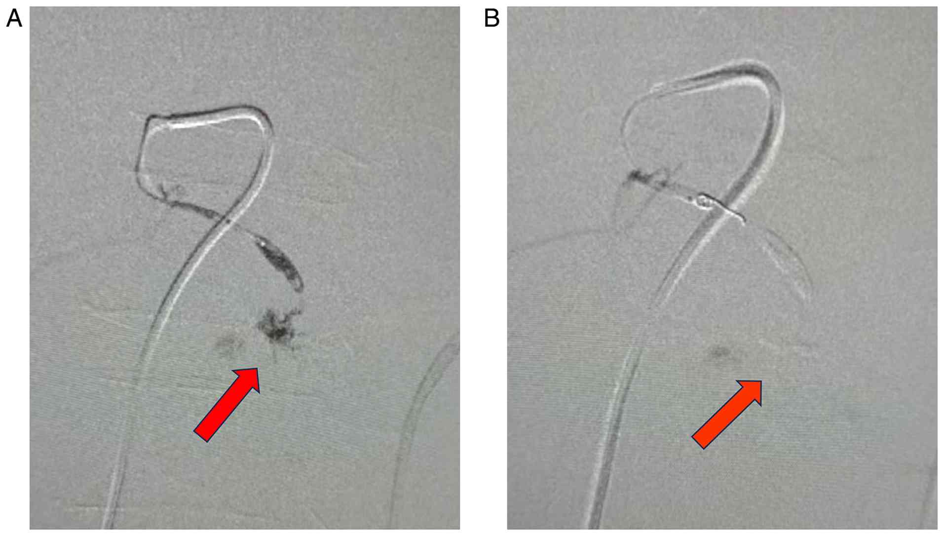
review of the patient's medical records revealed that, on POD9,
superior mesenteric angiography demonstrated contrast extravasation
from a branch of the superior mesenteric artery. Emergency
transcatheter arterial embolization was subsequently performed
(Fig. 4). The fistula was
considered likely to have originated from the jejunal anastomotic
site. However, imaging studies were unable to precisely localize
the fistulous opening. Hemostasis was successfully achieved
following transcatheter arterial embolization.
Following the interventional procedure, the
abdominal drainage output gradually decreased, with the fistula
subsequently transitioning to a low-output fistula. On the first
day after the interventional procedure, 530 ml of dark red fluid
was drained. Laboratory tests showed that the serum total protein
and albumin levels had decreased to 56.7 g/l (reference ranges,
65–85 g/l) and 35.3 g/l (reference ranges, 40–55 g/l),
respectively, while the CRP level had increased to 83.4 mg/l
(reference range, <5 mg/l). The patient received nutritional
support, correction of the electrolyte imbalance and conservative
management, including pharmacological therapy to reduce
gastrointestinal secretions, acid suppression, gastric mucosal
protection and antimicrobial treatment. By the third day after the
interventional procedure, the drainage volume had decreased to 200
ml. Follow-up laboratory tests performed on the sixth day after the
procedure showed that the serum total protein and albumin levels
had increased to 62.7 and 38.4 g/l, respectively, while the CRP
level had decreased to 61.9 mg/l. Conservative management was
continued, consisting of supportive care with fluid replacement,
nutritional support, infection control, suppression of
gastrointestinal secretions and acid suppression.
The patient underwent abdominal CT and contrast
studies to evaluate postoperative complications and fistula
healing. Although no definite intra-abdominal abscess was
identified on imaging, recurrent fever and persistently elevated
CRP levels suggested a localized postoperative inflammatory or
infectious process. Sequential broad-spectrum antimicrobial therapy
was therefore administered during the treatment course, including
cefotiam, piperacillin/tazobactam, cefoperazone/sulbactam and
cefuroxime. At the end of August 2021, a small bowel contrast study
demonstrated the unobstructed passage of contrast medium through
the small intestine. After 36 days of bowel rest, enteral nutrition
was gradually resumed. The patient was discharged after 36 days of
hospitalization at the tertiary referral hospital. At the
outpatient follow-up visit at the end of September 2021, the
abdominal drain continued to intermittently discharge bilious
fluid, with a daily output of 5–50 ml. In October 2021, a contrast
study via the abdominal drainage catheter demonstrated no passage
of contrast medium into the intestinal lumen and no communication
between the drainage catheter and the bowel, confirming fistula
closure. The abdominal drainage catheter was subsequently removed.
During 4.5 years of postoperative follow-up, no recurrence was
observed, and serial CT examinations demonstrated stable
postoperative findings (Fig.
3).
Discussion
MCL usually presents with an insidious onset; the
most common presenting symptom is abdominal pain, followed by
abdominal distension (6). However,
its clinical manifestations are non-specific. Xu et al
(11) reported a case of MCL
presenting with chronic gastrointestinal hemorrhage and persistent
anemia. The patient lost ~10 kg over the course of 1 year and
underwent two esophagogastroduodenoscopies and one colonoscopy;
however, none of these procedures identified the source of the
bleeding. Consequently, the patient required monthly blood
transfusions. Given its clinical spectrum, which ranges from
asymptomatic lesions detected incidentally to acute abdominal
emergencies, the true prevalence of MCL is likely underestimated
due to misdiagnosis and underdiagnosis (4,5).
A systematic search was performed in the following
electronic databases: PubMed (https://pubmed.ncbi.nlm.nih.gov/), the China National
Knowledge Infrastructure (CNKI) (https://www.cnki.net/) and the Chinese Biomedical
Literature Database (https://www.sinomed.ac.cn/). The search included case
reports and case series of mesenteric cavernous lymphangioma (MCL)
published up to November 2025. The search strategy included
combinations of the following keywords: ‘mesenteric cavernous
lymphangioma’, ‘cavernous lymphangioma’, ‘mesenteric lymphangioma’,
‘mesenteric tumor’, ‘lymphatic malformation’ and ‘lymphangioma’.
The reference lists of the included studies were also manually
reviewed to identify additional eligible publications.
The inclusion criteria were as follows: i) Cases
with a definitive pathological diagnosis of MCL; and ii) studies
reporting relevant clinical information, including demographic
characteristics, clinical manifestations, imaging findings and
treatment strategies. The exclusion criteria were as follows: i)
Studies without pathological confirmation of MCL; ii) lymphangiomas
originating from sites other than the mesentery; iii) duplicate
publications; and iv) studies lacking sufficient clinical, imaging
or treatment-related information. After excluding duplicate cases,
26 cases identified from the literature (2,7–26) and
1 case from Zhuji People's Hospital (Zhuji, China) were included,
yielding a total of 27 cases for descriptive analysis. The majority
of patients were of East Asian ethnicity (70.4%), with a
male-to-female ratio of 1.7:1. The patients ranged in age from 3.5
to 71 years, and only 3 (11.1%) were younger than 18 years. A total
of 25 patients (92.6%) were symptomatic, with abdominal pain
(63.0%) and abdominal distension (25.9%) being the most common
presenting symptoms. A total of 4 patients (14.8%) had decreased
serum albumin and total protein levels at the time of hospital
admission after developing clinical symptoms (Table II). The small bowel mesentery was
the most common site of involvement (77.8%), and lesions frequently
extended into adjacent bowel segments (77.8%). Detailed demographic
and clinical characteristics are presented in Table II.
| Table II.Demographic and clinical
characteristics of 27 patients with mesenteric cavernous
lymphangiomata. |
Table II.
Demographic and clinical
characteristics of 27 patients with mesenteric cavernous
lymphangiomata.
| Characteristics | Value |
|---|
| Age, years |
|
| Mean | 39 |
|
Range | 3.5–71 |
| F/M ratio, n | 1:1.7 |
| Ethnicity, n |
|
| East
Asian | 19 |
|
White | 8 |
| Clinical
presentation, % |
|
|
Asymptomatic | 7.4 |
|
Symptomatic | 92.6 |
| Symptoms, % |
|
|
Abdominal pain | 63.0 |
|
Abdominal distension | 25.9 |
|
Increased abdominal girth | 14.8 |
|
Self-palpable mass | 29.6 |
|
Nausea | 7.4 |
|
Vomiting | 11.1 |
| Poor
appetite | 3.7 |
|
Diarrhea | 7.4 |
| Back
pain | 3.7 |
|
Constipation | 7.4 |
| Bloody
stool | 14.8 |
|
Fever | 7.4 |
|
Difficulty breathing | 7.4 |
|
Difficulty in urination | 11.1 |
| Weight
loss | 11.1 |
| Site of onset,
% |
|
|
Mesentery of the small
intestine | 77.8 |
|
Mesentery of the colon | 7.4 |
|
Involvement of surrounding
intestines | 77.8 |
| Other
complications of abdominal organs | 7.4 |
Overall, 23 patients underwent CT and 4 underwent
MRI. On contrast-enhanced imaging, most lesions demonstrated either
no enhancement or enhancement confined to the internal septations.
The vascular traversal sign was identified on contrast-enhanced
imaging in 11 patients (40.7%). On pathological examination,
chylous fluid was the most common cystic fluid content, occurring
in 59.3% of cases. Detailed imaging and pathological
characteristics are summarized in Table III.
| Table III.Summary of imaging findings,
pathological features, treatment and outcomes in 27 patients with
mesenteric cavernous lymphangiomata. |
Table III.
Summary of imaging findings,
pathological features, treatment and outcomes in 27 patients with
mesenteric cavernous lymphangiomata.
| Parameter | Variable |
|---|
| Inspection method,
n |
|
| CT | 23 |
| Low
density | 11 |
| Mixed
density or mixed echo | 11 |
|
Solidity | 1 |
|
MRI | 4 |
| TIWI
low-signal and T2WI hyperintensity | 4 |
| Strengthening
method, n |
|
| No
reinforcement | 7 |
| Mild
reinforcement | 1 |
|
Moderate reinforcement | 1 |
|
Partition sample
strengthening | 2 |
| Blood vessel
traversing, n |
|
|
Intralesional vascular
traversing | 11 |
| Not
mentioned | 16 |
| Content of the
cyst, n (%) |
|
|
Serous | 1 (3.7) |
|
Chylous | 16 (59.3) |
|
Hemorrhagic | 4 (14.8) |
|
Hybridity | 3 (11.1) |
| Not
mentioned | 3 (11.1) |
| Laboratory
examination, n |
|
| Tumor
marker-negative | 6 |
| CA125
increased | 1 |
| Albumin
decreased; total protein decreased | 4 |
| Treatment, n |
|
|
Surgical resection | 27 |
| Follow-up status,
n |
|
|
Stable | 22 |
|
Death | 1 (day 6 pulmonary
embolism) |
| Not
mentioned | 4 |
All patients underwent surgical resection. Overall
postoperative outcomes were favorable; however, 1 patient died on
POD6 from a pulmonary embolism. In this cohort, 1 patient (the
present case) developed a postoperative intestinal fistula. The
serum tumor markers of the 6 patients were within the normal range
Notably, 4 patients exhibited progressively decreasing serum
albumin and total protein levels (Table III). Whether these laboratory
abnormalities are directly associated with MCL remains unclear and
warrants further investigation.
Histologically, MCL is characterized by
proliferating, disorganized lymphatic channels embedded within
connective tissue, containing serous fluid, blood or chylous fluid,
with chylous fluid being the most common component. This finding is
considered to result from communication between the abnormal
lymphatic channels of cavernous lymphangiomas and the adjacent
normal lymphatic system, allowing chylous fluid to leak into cystic
spaces (6). Unlike cystic
lymphangioma, cavernous lymphangioma often infiltrates the
intestinal wall despite its benign histological nature. This
infiltrative growth pattern may cause atrophy of the surrounding
tissues and subsequent functional impairment, resulting in
hemorrhage, necrosis, and acute inflammatory changes involving the
mesentery and intestine (3,7,8,10).
From an imaging perspective, CT is generally the
first-line imaging modality; however, it may have limited ability
to delineate the cystic components of MCL, particularly when the
lesion is predominantly microcystic (3,12). By
contrast, MRI offers superior soft-tissue contrast and greater
sensitivity to tissue water content, providing improved lesion
characterization compared with CT. In predominantly microcystic
lesions, CT may misinterpret the lesion as a solid mass, whereas
MRI typically demonstrates homogeneous high signal intensity on
T2WI, with no enhancement or only mild septal enhancement after
contrast administration. Differences in signal intensity on T1WI
and T2WI may also help characterize the composition of the cystic
contents. MRI also facilitates assessment of bowel wall
involvement. In some cases, multiple submucosal cystic lesions can
be identified, further increasing diagnostic confidence (13).
Based on the present case and the literature review,
we propose that the combination of ‘no enhancement or only septal
enhancement’ and the ‘vascular traversal sign’ may represent a
characteristic imaging feature of MCL. This imaging appearance is
attributable to the characteristic histopathological architecture
of MCL, which comprises clusters of dilated cavernous lymphatic
channels embedded within lymphoid stroma. These dilated lymphatic
spaces surround or are traversed by normal mesenteric vessels,
rather than containing abundant neovascularization. Consequently,
the lesion itself demonstrates little or no enhancement on
contrast-enhanced imaging (3,7). These
imaging features may help distinguish MCL from hypervascular tumors
and liposarcomas, both of which can present as low-attenuation
masses on CT. Furthermore, the 2024 Chinese Society of Clinical
Oncology (CSCO) Clinical Practice Guidelines recommend that
positron emission tomography (PET)/CT may serve as an adjunctive
imaging modality for differentiating MCL from malignant mesenteric
tumors by assessing glucose metabolism and the extent of systemic
involvement (27). However, PET/CT
findings were not reported in either the cases included in the
present study or the published cases identified in the literature
review.
Complete surgical resection remains the standard
treatment for MCL; however, preoperative misdiagnosis may result in
unnecessarily extensive surgery. In the present case, the
preoperative diagnosis of suspected liposarcoma led to an extensive
surgical resection, which was complicated by a postoperative
intestinal fistula and required >2 months of conservative
management. Previous studies have reported that incomplete
resection is associated with an increased risk of local recurrence
(14). Although complete resection
remains the optimal treatment goal, residual disease may remain
stable over long-term follow-up when complete excision is not
feasible (2,28). In the present case, the main tumor
mass was completely resected. Postoperatively, persistent fluid
accumulation was observed at the mesenteric root. This gradually
resolved during follow-up, and the residual lesion has remained
radiologically stable upon follow-up. Accurate preoperative
recognition of the characteristic imaging features of MCL may
facilitate a more precise and limited surgical resection, thereby
reducing the risk of postoperative complications.
The present study has several limitations. First, it
is a retrospective analysis based on published case reports and
case series. Second, not all relevant data, including clinical
presentation, imaging findings, gross pathological findings,
histopathological features and immunohistochemical results, were
comprehensively reported in the included studies.
In conclusion, although MCL is a rare entity, it
exhibits characteristic imaging features that may facilitate its
diagnosis. MCL should be considered when CT or MRI demonstrates a
well-defined mesenteric mass with locally infiltrative growth, no
enhancement or only septal enhancement on contrast-enhanced
imaging, and the vascular traversal sign, particularly in patients
presenting with non-specific abdominal pain or hypoproteinemia.
Contrast-enhanced abdominal MRI is recommended to further
characterize the lesion, evaluate the extent of disease and
facilitate preoperative surgical planning.
Acknowledgements
Not applicable.
Funding
Funding: No funding was received.
Availability of data and materials
The data generated in the present study may be
requested from the corresponding author.
Authors' contributions
ZZ and EC designed the study and participated in the
literature search. ZZ and TZ collected, reviewed and analyzed the
medical images, contributed to the literature review, and prepared
the draft manuscript. JF and TZ critically revised the manuscript
for important intellectual content and provided general
supervision. ZZ, EC, JF, and LG were instrumental in revising the
manuscript, participating in data analysis and providing treatment
recommendations for the patient. TZ and LG confirm the authenticity
of all the raw data. All authors read and approved the final
version of the manuscript.
Ethics approval and consent to
participate
The study was conducted in accordance with the
Declaration of Helsinki. The present study was approved by the
Ethics Committee of Zhuji People's Hospital [Zhuji, China; approval
no. (2026) MedEthics no. (0120)].
Patient consent for publication
Written consent for publication was obtained from
the patient.
Competing interests
The authors declare that they have no competing
interests.
References
|
1
|
Wegner G: Ueber Lymphangiome. Archiv für
klinische Chirurgie. 20:6411877.(In German).
|
|
2
|
Jeong WK, Kim Y, Song SY, Heo JN and Park
CK: Cavernous mesenteric lymphangioma (2006: 4b). Eur Radiol.
16:1625–1628. 2006. View Article : Google Scholar : PubMed/NCBI
|
|
3
|
Francavilla ML, White CL, Oliveri B, Lee
EY and Restrepo R: Intraabdominal lymphatic malformations: Pearls
and pitfalls of diagnosis and differential diagnoses in pediatric
patients. AJR Am J Roentgenol. 208637–649. 2017.
|
|
4
|
Mede A, Chotai PN, Huh WJ and Tan M:
Intra-abdominal cystic lymphangiomas: The Vanderbilt experience. J
Surg Res. 285:197–204. 2023. View Article : Google Scholar : PubMed/NCBI
|
|
5
|
Liu J, Zhong ZH and Zheng SS:
Characteristics of adult retroperitoneal lymphangioma: A
single-center Chinese cohort study of 15 cases. Discov Oncol.
15:2622024. View Article : Google Scholar : PubMed/NCBI
|
|
6
|
Saxena A, Kakodkar P, Zhang D, Poulin A,
Shekari N, Kanthan SC and Kanthan R: Intra-abdominal multi cystic
lymphangiomas: A case series with adult and pediatric literature
review. Arch Clin Gastroenterol. 10:27–39. 2024. View Article : Google Scholar
|
|
7
|
Cab-Serrano BP, Ayuso-Diaz VM,
Torres-Valdes EA, Chacon-Pacho RE and Moreno-Enriquez A: Giant
mesenteric cavernous lymphangioma in an adult as a cause of chronic
intestinal subocclusion: A case report. Cureus.
17:e789342025.PubMed/NCBI
|
|
8
|
Zhu D, Li M, Wang S, Shen R and Wang X:
Pancreaticoduodenectomy for a giant retroperitoneal cavernous
lymphangioma: A case report. Chin J Oper Proc Gen Surg (Electronic
Edition),. 9:300–301. 2015.(In Chinese).
|
|
9
|
Shi L, Li W and Yu J: A case of
intraperitoneal hemorrhage caused by lymphangiomatosis of the
ileocecal mesentery. Chinese Clinical Case Results Database.
7:E0909. 2025.(In Chinese).
|
|
10
|
Viar WN, Scott WF Jr and Donald JM:
Mesenteric cavernous lymphangiomata: Brief review and report of two
cases. Ann Surg. 153:157–160. 1961. View Article : Google Scholar : PubMed/NCBI
|
|
11
|
Xu X, Liu W and Zheng C: A rare cause of
repeated gastrointestinal bleeding. Mesenteric cavernous
lymphangioma. Gastroenterology. 146:e11–e13. 2014. View Article : Google Scholar : PubMed/NCBI
|
|
12
|
Fan R, You L, Yu H and Li B: Mesenteric
cavernous lymphangioma in adult: Report of one case. J Chin Clin
Med Imaging. 34:677–678. 2023.(In Chinese).
|
|
13
|
Zheng W and Chen Y: A case of jejunal
lymphangioma misdiagnosed on MR enterography. Radiol Practice.
35:115–116. 2020.(In Chinese).
|
|
14
|
Iqbal J, Isaacs P, Sissons M, Bury R,
Khurshid M and Akhtar M: A cause for concern? An asymptomatic
mesenteric lesion: Cavernous lymphangioma. Gut. 58:1184–1225. 2009.
View Article : Google Scholar : PubMed/NCBI
|
|
15
|
Dang MH, Li ZC and Yang JL: Imaging
diagnosis of mesenteric cavernous hemolymphangioma. J Pract Radiol.
9:1204–1206. 12382007.(In Chinese).
|
|
16
|
Estébanez-Ferrero B, Rico-Morales MDM,
Lorenzo-Liñán MA, Teruel-Lillio I and Reina-Duarte Á: Giant
cavernous mesenteric lymphangioma in an adult. Rev Esp Enferm Dig.
114:631–632. 2022.PubMed/NCBI
|
|
17
|
Zhang Y, Yang P and Chen M: Cavernous
mesenteric lymphangioma presenting as intra-abdominal malignancy.
Asian J Surg. 46:4855–4856. 2023. View Article : Google Scholar : PubMed/NCBI
|
|
18
|
Christofi N and Hextall A: Cystic
cavernous lymphangioma of the mesentery in a patient with Cowden
syndrome. J Obstet Gynaecol. 27:329–330. 2007. View Article : Google Scholar : PubMed/NCBI
|
|
19
|
Meng Y, Li Y, Tang HB and David M: One
case of multiple mesenteric lymphangio-cavernoma. J Dalian Med
Univ. 27:12005.(In Chinese).
|
|
20
|
Sun YQ and Ren JW: A case of cavernous
lymphangioma of the ileal mesentery. J Clin Radiol. 24:5482005.(In
Chinese).
|
|
21
|
Ma XT, Sun HB and Zhao MY: The cavernous
lymphangioma occurred in child mesojejunum: A case report. Chin J
Clin Anat. 25:1552007.(In Chinese).
|
|
22
|
Peng J and He D: A case of cavernous
lymphangioma of the small bowel mesentery complicated by volvulus.
Sichuan Med J. 25:9252004.(In Chinese).
|
|
23
|
Alhabil Y, Dola QN and Khader A:
Interrelation of cancers of unknown primary, chylous ascites, and
cavernous mesenteric lymphangioma in a 57-year-old female patient:
A case report. J Surg Case Rep. 2024:rjae1292024. View Article : Google Scholar : PubMed/NCBI
|
|
24
|
Vennarecci G, Ceribelli C, Laurenzi A,
Moroni E and Ettorre GM: Giant cavernous mesenteric lymphangioma in
adult. Updates Surg. 65:317–319. 2013. View Article : Google Scholar : PubMed/NCBI
|
|
25
|
Chai H and Jin XY: A case of giant
mesenteric cavernous lymphangioma combined with teratoma. Chin J
Rural Med Pharm. 27:512020.(In Chinese).
|
|
26
|
Jiao FL: A case of giant mesenteric
cavernous lymphangioma misdiagnosed. Sichuan Med J. 23:7692002.(In
Chinese).
|
|
27
|
Wang F, Chen G, Zhang Z, Yuan Y, Wang Y,
Gao YH, Sheng W, Wang Z, Li X, Yuan X, et al: The Chinese society
of clinical oncology (CSCO): Clinical guidelines for the diagnosis
and treatment of colorectal cancer, 2024 update. Cancer Commun
(Lond). 45:332–379. 2025. View Article : Google Scholar : PubMed/NCBI
|
|
28
|
Mansour S, Kluger Y and Khuri S: Adult
primary retroperitoneal lymphangioma: Updated facts. World J Oncol.
14:15–20. 2023. View Article : Google Scholar : PubMed/NCBI
|