Gynecological malignancies represent a major threat
to health, with ovarian, endometrial cancer and cervical cancer
ranking among the most prevalent and lethal neoplasms of the female
reproductive tract (1–3). Statistics indicate that >1.3
million new cases of gynecological malignancies and nearly 500,000
associated deaths are reported globally each year. Ovarian cancer,
in particular, is characterized by insidious onset and challenges
in early diagnosis, with >70% of patients presenting at an
advanced stage (International Federation of Gynecology and
Obstetrics stage III/IV) at the time of diagnosis (4,5). While
primary debulking surgery combined with platinum-based chemotherapy
can achieve initial remission, the postoperative recurrence rate of
ovarian cancer is >70%, with a 5-year survival rate of only
~30%. Platinum resistance stands as the primary bottleneck
contributing to treatment failure (6,7).
Endometrial cancer has witnessed a steady rise in incidence, with
obesity-associated type I endometrial cancer (Bokhman
classification) accounting for >80% of cases and conferring a
poor prognosis (8). For advanced or
recurrent endometrial cancer, therapeutic options remain limited,
as immune checkpoint inhibitors only benefit a subset of patients
with mismatch repair-deficient subtypes (9). Although early intervention for
cervical cancer is feasible through human papillomavirus (HPV)
vaccination and screening, high-risk subtypes associated with
persistent HPV infection (such as HPV16/18) still pose a risk of
disease progression (10–12). Chemoradiotherapy resistance and
lymph node metastasis remain critical factors limiting patient
outcomes. Collectively, the interplay between subtype
heterogeneity, therapeutic resistance and metabolic dysregulation
constitutes the core impediment to favorable clinical outcomes in
gynecological malignancies (13,14).
Metabolic reprogramming, a hallmark of malignant
tumors, is closely linked to tumor initiation, progression,
metastasis and drug resistance (15–17).
As a key component of metabolic reprogramming, lipid metabolic
dysregulation has been identified as a critical biological feature
of gynecological malignancies (18). Beyond providing energy and
structural building blocks for rapid tumor cell proliferation,
lipid metabolism exerts multifaceted effects on tumor progression
through metabolite-mediated signal transduction and tumor
microenvironment (TME) remodeling (19). Studies have demonstrated that
gynecological cancer cells actively rewire their lipid metabolic
networks, encompassing lipid uptake, synthesis, storage and
catabolism, to adapt to stressors such as nutrient deprivation and
hypoxia within the TME (20,21).
Notably, distinct subtypes of gynecological cancers exhibit marked
phenotypic heterogeneity in lipid metabolism due to differences in
genetic background, etiological factors (for example, obesity
association and HPV infection) and TME characteristics (22,23).
For instance, obesity-associated endometrial cancer is
characterized by sterol regulatory element-binding protein 1
(SREBP1)-driven enhanced lipogenesis, while ovarian clear cell
carcinoma (OCCC) is marked by abnormal lipid droplet accumulation
and reliance on exogenous fatty acid uptake (24,25).
By contrast, HPV-positive cervical cancer displays increased
low-density lipoprotein receptor (LDLR)-mediated cholesterol uptake
(26). This subtype-specific lipid
metabolic reprogramming not only contributes to malignant
transformation but also closely associates with therapeutic
resistance; for example, platinum-resistant ovarian cancer cells
exhibit disrupted cholesterol homeostasis and activated fatty acid
oxidation (FAO), while progesterone resistance in endometrial
cancer correlates with fatty acid synthase (FASN)-mediated enhanced
fatty acid synthesis (27–29).
In recent years, lipid metabolism-targeted
therapeutic strategies have emerged as a research focus in
oncology. Several inhibitors targeting key lipid metabolic enzymes
[for example, FASN, 3-hydroxy-3-methylglutaryl-CoA reductase
(HMGCR) and stearoyl-CoA desaturase 1 (SCD1)] have advanced to
preclinical or early-phase clinical trials, with some demonstrating
notable antitumor activity and reversal of drug resistance in
gynecological cancer models (30,31).
However, significant gaps remain in current research: The
subtype-specific regulatory networks of lipid metabolism in
gynecological malignancies are not fully elucidated, the crosstalk
mechanisms between lipid metabolism and other oncogenic pathways
(for example, PI3K/Akt/mTOR and Hippo-YAP) require further
investigation, and precision targeting strategies based on lipid
metabolic profiles have not been widely translated into clinical
practice (32). Therefore,
systematically summarizing the subtype-specific features of lipid
metabolic dysregulation in gynecological malignancies, elaborating
on the underlying molecular regulatory mechanisms, and synthesizing
the progress of lipid metabolism-targeted interventions and their
clinical translation hold theoretical and clinical importance for
advancing precision medicine and improving patient outcomes. The
present review focuses on the subtype-specific characteristics of
lipid metabolism, core regulatory mechanisms, associations with
malignant phenotypes and targeted intervention strategies in
gynecological cancers, aiming to provide insights for basic
research and clinical translation in this field.
Distinct gynecological malignancies and even
different subtypes within the same tumor exhibit marked
heterogeneity in lipid metabolic reprogramming patterns (33). This heterogeneity stems from
intrinsic differences in genetic background, TME characteristics
and etiological mechanisms and exerts a direct impact on tumor
biological behavior, therapeutic sensitivity and patient
prognosis.
Ovarian cancer encompasses diverse pathological
subtypes, among which high-grade serous ovarian cancer (HGSOC),
OCCC and endometrioid carcinoma are the three most clinically
prevalent subtypes. Each subtype displays distinct divergence in
lipid metabolic profiles (34,35).
HGSOC accounts for >70% of all ovarian cancer cases, with
enhanced FAO as its core metabolic phenotype. Tumor cells adapt to
nutrient deprivation and hypoxic stress in the ascites
microenvironment by activating the FAO pathway to efficiently
utilize fatty acids for energy production (36). Studies have confirmed that the
expression of carnitine palmitoyltransferase 1 (CPT1), a key
rate-limiting enzyme in FAO, is significantly upregulated in HGSOC
cells (37). CPT1 facilitates the
translocation of long-chain fatty acids across the mitochondrial
membrane and initiates their oxidative breakdown; silencing CPT1
directly inhibits tumor cell proliferation, colony formation and
peritoneal metastatic capacity (38). Additionally, HGSOC exhibits aberrant
cholesterol uptake: High expression of the LDLR positively
correlates with a poor patient prognosis. Serum levels of LDL and
total cholesterol in platinum-resistant patients are markedly
higher than those in sensitive populations. Enhanced LDLR-mediated
exogenous cholesterol uptake may maintain cell membrane stability
and reduce drug-induced lipid peroxidation damage, thereby
contributing to the development of a resistant phenotype (39,40).
OCCC is characterized by a hallmark feature of
abnormal lipid storage: Lipid droplets accumulate extensively
within tumor cells, and high expression of the lipid
droplet-associated protein adipophilin is correlated with shortened
progression-free survival in patients (41). Mechanistic investigations reveal
that acyl-CoA cholesterol acyltransferase 1 (ACAT1) is upregulated
in OCCC cells, promoting the esterification of free cholesterol for
storage in lipid droplets (42,43).
This process not only avoids the cytotoxicity of free cholesterol
but also enables rapid breakdown for energy supply under nutrient
deficiency, conferring a metabolic adaptive advantage. Meanwhile,
OCCC cells exhibit low expression of FASN and rely more heavily on
exogenous fatty acid uptake, with enhanced CD36-mediated free fatty
acid (FFA) transport playing a pivotal regulatory role in tumor
proliferation and invasion (44–46).
The endometrioid ovarian cancer subtype shares a
strong association with obesity, with enhanced lipogenesis as its
core lipid metabolic feature. As a master transcription factor
(47), SREBP1 drives the activation
of fatty acid synthesis pathways, upregulating the expression of
key enzymes such as FASN and acetyl-CoA carboxylase (ACC) (48,49).
This promotes de novo fatty acid synthesis, providing
membrane structural building blocks and energy support for rapid
tumor cell proliferation (50).
Collectively, the three major ovarian cancer subtypes exhibit
distinct lipid metabolic dependencies: HGSOC relies on FAO-mediated
energy production and exogenous cholesterol uptake for survival in
the ascites microenvironment; OCCC is characterized by extensive
lipid droplet accumulation and dependence on exogenous fatty acid
uptake; while endometrioid ovarian cancer shows SREBP1-driven
enhanced de novo lipogenesis associated with obesity. These
divergent metabolic phenotypes directly contribute to their
differences in biological behavior, therapeutic sensitivity and
patient prognosis.
Endometrial cancer is categorized into two subtypes
based on etiological features: Type I (estrogen-dependent) and type
II (estrogen-independent), and these two subtypes exhibit striking
divergence in patterns of lipid metabolic dysregulation. Type I
endometrial cancer accounts for ~80% of all endometrial cancer
cases and shows a strong association with metabolic syndrome,
including obesity, diabetes mellitus and hyperlipidemia (51,52). A
core feature of this subtype is enhanced de novo lipogenesis
as the primary lipid metabolic abnormality. Studies have
demonstrated that type I tumors exhibit markedly elevated
expression of SREBP1, a master transcription factor that directly
binds to the promoter regions of key lipogenic enzymes such as FASN
and SCD1, thereby activating de novo fatty acid synthesis
(53). The hyperestrogenic
microenvironment induced by obesity enhances the nuclear
localization and transcriptional activity of SREBP1, forming a
positive regulatory loop termed the ‘estrogen-SREBP1-lipogenesis
axis’ that continuously drives tumor progression. Additionally,
type I endometrial cancer is accompanied by cholesterol metabolic
disorders: Upregulated expression of HMGCR promotes endogenous
cholesterol synthesis, while statins, by inhibiting HMGCR activity,
can markedly suppress tumor cell proliferation and induce apoptosis
(54).
Type II endometrial cancer, predominantly composed
of serous carcinoma, displays lipid metabolic traits that are
distinctly different from those of type I (55). This subtype exhibits substantially
enhanced lipid uptake capacity, with upregulated expression of CD36
and fatty acid transport protein 2 (FATP2) enabling efficient
scavenging of FFAs from the tumor microenvironment to meet
metabolic demands. Concurrently, the level of FAO in type II tumor
cells is markedly higher than in type I tumor cells, and increased
expression of CPT1A correlates with a poor patient prognosis
(56,57). Inhibition of FAO has been shown to
enhance the sensitivity of these tumor cells to chemotherapeutic
agents. Furthermore, studies have revealed that type II endometrial
cancer exhibits reduced expression of ATP-binding cassette (ABC)
subfamily A member 1, a protein involved in cholesterol efflux.
This downregulation leads to abnormal intracellular cholesterol
accumulation, which activates the PI3K/Akt signaling pathway to
augment the tumor's invasive and metastatic potential (58–60).
In summary, type I and type II endometrial cancers exhibit
fundamentally different lipid metabolic profiles: Type I tumors are
characterized by SREBP1-mediated enhanced de novo
lipogenesis driven by obesity and hyperestrogenism, while type II
tumors rely on increased lipid uptake and FAO for energy supply.
These metabolic differences underlie their distinct responses to
hormonal therapy and chemotherapy.
Cervical cancer development is associated with
persistent HPV infection, with HPV16 and HPV18 as the primary
high-risk subtypes (61,62). Abnormal lipid metabolism in these
tumors is specifically regulated by HPV oncoproteins, featuring a
distinct signature centered on cholesterol metabolic reprogramming.
Mechanistic studies demonstrate that the HPV E7 oncoprotein
upregulates LDLR expression at the transcriptional level while
suppressing the transcription of ABC sub-family G member 1, a gene
critical for cholesterol efflux. This dual regulation leads to
abnormal intracellular cholesterol accumulation, which in turn
activates lipid raft-associated c-Src/AKT/FAK signaling pathways to
enhance tumor cell migration and invasion (63,64).
Additionally, HPV-positive cervical cancer cells exhibit markedly
elevated expression of FASN; enhanced fatty acid synthesis driven
by FASN promotes lymphangiogenesis, thereby augmenting the
potential for lymph node metastasis (65).
Based on histological features, cervical cancer is
classified into squamous cell carcinoma and adenocarcinoma
subtypes, each with distinct lipid metabolic traits (66,67).
Squamous cell carcinoma relies more heavily on FAO for energy
production: Upregulated expression of acyl-CoA synthetase
long-chain family member 3 (ACSL3) facilitates the activation of
long-chain fatty acids and their translocation into mitochondrial
oxidative pathways, supplying the energy required for tumor
proliferation (68). By contrast,
adenocarcinoma is characterized by enhanced lipogenesis, with
markedly higher expression of SREBP1 and FASN compared with that in
squamous cell carcinoma; targeted inhibition of FASN effectively
suppresses its proliferative activity (69). Due to the absence of HPV oncoprotein
regulation, HPV-negative cervical cancer exhibits a lipid metabolic
pattern analogous to other solid tumors, with enhanced lipid uptake
as a core feature. This allows tumor cells to efficiently utilize
exogenous lipids to meet their metabolic demands (70–72).
Notably, the two major histological subtypes of cervical cancer
display divergent lipid metabolic traits: Squamous cell carcinoma
depends on ACSL3-mediated FAO for energy production, while
adenocarcinoma is characterized by SREBP1-driven enhanced
lipogenesis. HPV-negative cervical cancer, lacking the regulation
of viral oncoproteins, exhibits a metabolic pattern dominated by
enhanced lipid uptake, similar to other solid tumors.
To facilitate a clear understanding of the
heterogeneous lipid metabolic profiles across gynecological
malignancies, a comprehensive table comparing the core metabolic
phenotypes, key regulatory molecules, functional impacts and
potential targeted agents for each major subtype is presented as
Table I.
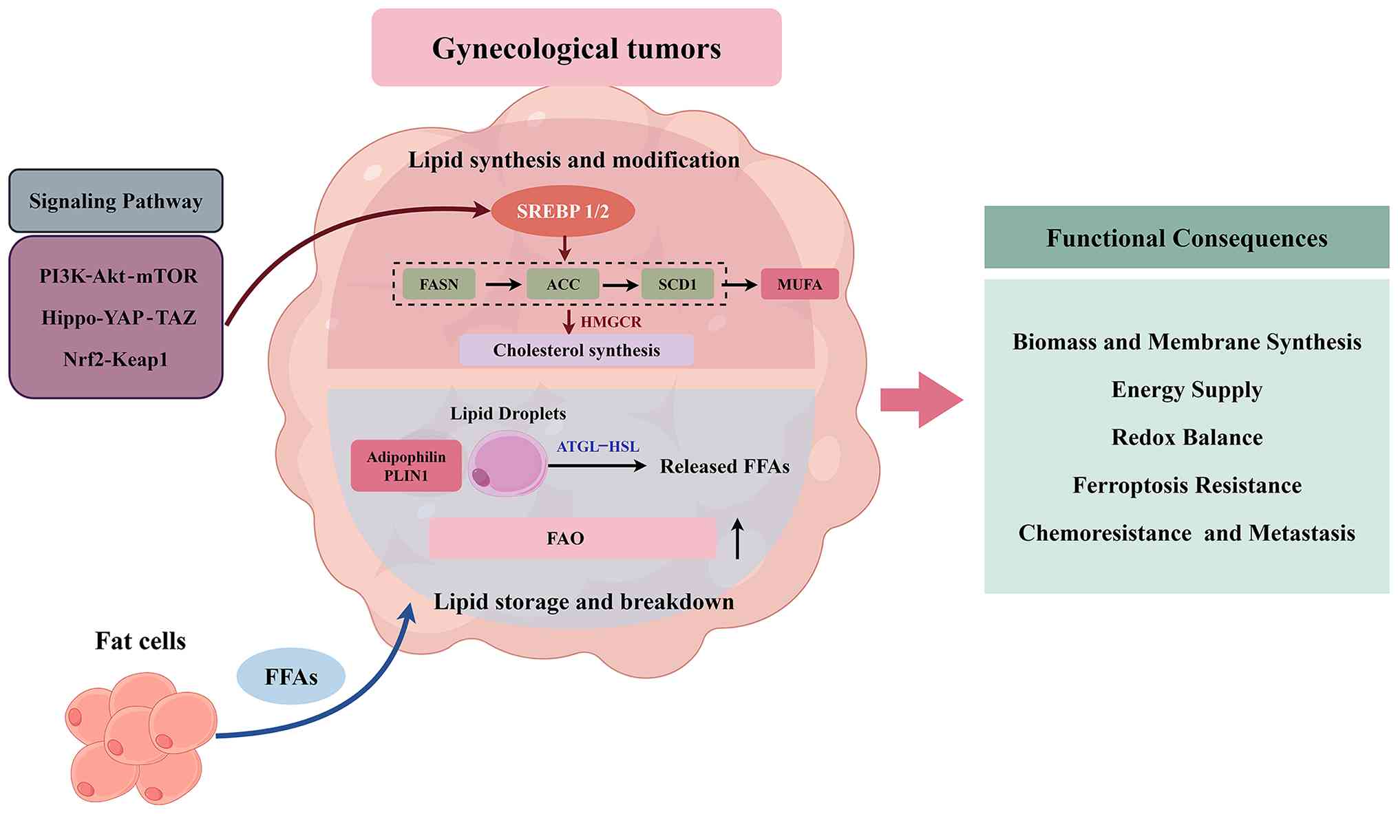
Gynecological cancer cells actively rewire their
lipid metabolic networks by selectively modulating core processes
such as lipid uptake, synthesis, storage and catabolism, thereby
meeting the demands for malignant proliferation, TME adaptation and
therapeutic resistance (73). These
abnormally activated metabolic processes are not isolated; instead,
they are synergistically regulated by multiple signaling pathways
and transcription factors, forming an interconnected and complex
molecular regulatory network (74,75).
Lipid uptake serves as the initiating and
rate-limiting step in tumor cell lipid metabolism. Gynecological
cancer cells selectively upregulate the expression and function of
lipid transporters to markedly boost the efficiency of exogenous
lipid uptake, providing abundant substrates for subsequent
metabolic reprogramming and laying a material foundation for
malignant tumor progression (76,77).
CD36, a key molecule mediating transmembrane fatty
acid transport, is commonly overexpressed in gynecological
malignancies such as ovarian and cervical cancer. CD36 specifically
mediates the selective uptake of FFAs, particularly unsaturated
fatty acids (78). In ovarian
cancer, direct contact between omental adipocytes and tumor cells
can induce CD36 upregulation in tumor cells via paracrine
signaling, enhancing fatty acid uptake efficiency to support energy
metabolism during peritoneal metastasis (79). VT1021, a specific CD36 inhibitor,
has entered early-phase clinical trials, aiming to inhibit tumor
progression by blocking lipid uptake (80).
Abnormal LDLR-mediated cholesterol uptake is one of
the key mechanisms underlying platinum resistance in ovarian
cancer: Platinum-resistant ovarian cancer cells exhibit markedly
elevated LDLR expression, while the expression of HMGCR, a key
enzyme in endogenous cholesterol synthesis, is downregulated. This
forms a metabolic adaptation pattern characterized by ‘enhanced
exogenous cholesterol uptake and reduced endogenous synthesis’,
which maintains cell membrane stability and sustains the resistant
phenotype (81,82). Studies have confirmed that silencing
tumor necrosis factor receptor-associated protein 1 can mimic this
resistant phenotype by upregulating LDLR to enhance cholesterol
uptake while suppressing the transcriptional activity of
cholesterol synthesis-related genes (83). Additionally, proprotein convertase
subtilisin/kexin type 9 (PCSK9) binds to the extracellular domain
of LDLR and promotes its intracellular degradation; reduced PCSK9
expression in platinum-resistant ovarian cancer cells further
prolongs the cell membrane residence time of LDLR, boosting its
cholesterol uptake efficiency (82).
The FATP family exhibits distinct subtype-specific
expression patterns in gynecological cancer: FATP2 is upregulated
in ovarian cancer cells, and its specific inhibitor lipofermata can
markedly suppress tumor cell proliferation and invasion by blocking
long-chain fatty acid uptake. In obesity-associated subtypes of
endometrial cancer, FATP1 expression is elevated, which is closely
linked to the phenotype of enhanced lipid metabolism and serves as
a potential metabolic biomarker for this subtype (84). Fatty acid-binding proteins (FABPs)
are responsible for the intracellular targeted transport of lipids.
Among them, FABP4 is highly expressed in ovarian cancer, mediating
the efficient translocation of lipids from the cell membrane to
lipid droplets (85,86). This not only avoids the accumulation
of lipid toxicity but also improves the efficiency of tumor cell
utilization of TME lipids, providing metabolic support for tumor
progression.
Enhanced lipogenesis stands as the most distinctive
metabolic aberration in gynecological malignancies, primarily
involving two core pathways, fatty acid synthesis and cholesterol
synthesis, both under the pivotal regulation of the SREBP family of
transcription factors (87). Among
these, SREBP1 primarily governs fatty acid synthesis and is
aberrantly upregulated in gynecological cancers such as endometrial
and ovarian cancer. SREBP1 specifically binds to the promoter
regions of downstream target genes, activating the transcription
and expression of key enzymes, including FASN, ACC and SCD1
(88). As the rate-limiting enzyme
in de novo fatty acid synthesis, FASN is markedly
upregulated in gynecological malignancy, such as cervical cancer
(89). FASN catalyzes the
condensation of acetyl-CoA and malonyl-CoA to produce palmitic
acid, a core building block for tumor cell membrane synthesis and
signaling molecule production (90). TVB-2640, a targeted inhibitor of
FASN, has demonstrated notable therapeutic activity in HGSOC; when
combined with poly(ADP-ribose) polymerase (PARP) inhibitors, it
exerts a synergistic effect by suppressing metabolic reprogramming
and DNA damage repair, further enhancing antitumor efficacy
(91).
SCD1, a key enzyme in fatty acid desaturation,
converts saturated fatty acids into monounsaturated fatty acids
(MUFAs); its upregulation in ovarian cancer promotes tumor
progression by enhancing cell membrane fluidity and attenuating
ferroptosis-associated lipid peroxidation (92,93).
Inhibition of SCD1 markedly reduces MUFA content in membrane
phospholipids, decreases the production of the antioxidant molecule
coenzyme Q10, disrupts cellular redox homeostasis, and ultimately
induces the accumulation of lipid peroxidation and ferroptosis.
A939572, a specific SCD1 inhibitor, has shown promising antitumor
effects in preclinical ovarian cancer models, offering a novel
direction for the treatment of drug-resistant tumors (94). In the cholesterol synthesis pathway,
HMGCR acts as the rate-limiting enzyme; its abnormal expression is
closely associated with the malignant progression of cervical and
ovarian cancer. Statins, commonly used clinical lipid-lowering
agents, can block cholesterol synthesis by inhibiting HMGCR
activity, which not only markedly suppresses tumor cell
proliferation but also enhances the cytotoxicity of
chemotherapeutic drugs, exerting a synergistic therapeutic effect
(95).
The aberrant activation of lipid synthesis is
further subject to hierarchical regulation by multiple oncogenic
signaling pathways (96).
Additionally, TP53 mutations can enhance the transcriptional
activity of SREBP1 while downregulating the expression of fatty
acid degradation-related genes (97,98),
leading to abnormal lipid accumulation in ovarian cancer cells and
the formation of a metabolic addiction phenotype (99).
Lipid droplets serve as core intracellular
organelles for lipid storage and metabolic regulation, and exhibit
an abnormal accumulation phenomenon in gynecological cancer cells
(100,101). The functions of lipid droplets
extend beyond mere lipid storage, as they actively participate in
regulating tumor progression by dynamically adjusting lipid
metabolic balance (102). In
ovarian cancer, the expression level of the lipid
droplet-associated marker adipophilin is markedly elevated, and
this upregulation correlates positively with a poor patient
prognosis (103). Inhibitors
targeting ACAT1 and HMGCR can block cholesterol esterification and
synthesis, respectively, thereby suppressing lipid droplet
accumulation in ovarian cancer cells and inhibiting tumor growth.
The formation of lipid droplets is associated with cholesterol
esterification. ACAT2 is highly expressed in serous ovarian cancer;
it converts free cholesterol into cholesterol esters for storage in
lipid droplets, thus avoiding the cytotoxicity induced by free
cholesterol (104). Lipid droplets
form dynamic contact sites with mitochondria to facilitate direct
fatty acid transfer for oxidation. This interaction is particularly
prominent in cells, supporting their survival under nutrient stress
by enhancing FAO efficiency (105,106). The expression level of ACAT2 is
positively correlated with chemoresistance in ovarian cancer.
Additionally, the phosphatidylcholine synthesis mediated by choline
kinase α (CHKα) can promote lipid droplet formation by regulating
membrane lipid composition, while dehydrogenase/reductase SDR
family member 2 inhibits lipid droplet accumulation and peritoneal
metastasis in ovarian cancer cells by downregulating CHKα
expression (107,108).
FAO is the core pathway of lipid catabolism and
serves an indispensable role in enabling adaptation to the TME and
sustaining survival advantages in gynecological cancers (109). Ovarian cancer cells exhibit a
distinct feature of enhanced FAO in the ascites microenvironment
(110). The translocation of
long-chain fatty acids across the mitochondrial membrane, mediated
by CPT1, is the rate-limiting step of FAO (111). Etomoxir, a CPT1 inhibitor, can
block this process, markedly suppressing the proliferation and
survival of ovarian cancer cells under the nutrient-depleted
conditions of ascites. Mitochondrial dynamics (fusion and fission)
directly regulate FAO efficiency by modulating the assembly of
oxidative enzyme complexes. Enhanced mitochondrial fusion in
ovarian cancer cells promotes FAO flux and confers chemoresistance,
while mitochondrial fission impairs lipid oxidation and induces
lipid accumulation (112,113). Studies have confirmed that
adipocytes within the ovarian cancer ascites microenvironment
secrete FFAs to serve as metabolic substrates for tumor cells
(114,115). Tumor cells efficiently utilize
these fatty acids by enhancing FAO, while simultaneously activating
the AMPK signaling pathway to drive downstream oncogenic signal
transduction, forming a vicious cycle of TME lipids-tumor
metabolism-signal activation. In endometrial cancer, the
upregulated expression of ACSL1 facilitates the activation of
long-chain fatty acids and their entry into the FAO pathway,
providing sufficient energy for tumor cells. Silencing ACSL1 can
significantly inhibit tumor proliferation and metastatic capacity
by suppressing FAO activity (116–118).
The dynamic balance between lipid storage and
catabolism is dynamically regulated by the nutritional status of
the TME. Under nutrient deprivation, tumor cells can rapidly
decompose triglycerides and cholesterol esters stored in lipid
droplets through lipolysis, releasing FFAs for energy supply.
Adipose triglyceride lipase (ATGL) and hormone-sensitive lipase are
the core enzymes mediating lipolysis, and both are highly expressed
in ovarian cancer (119,120). Inhibiting ATGL can markedly reduce
fatty acid release, impairing the survival ability of tumor cells
under nutrient-deficient conditions. Furthermore, autophagy
(lipophagy) participates in lipid catabolism through the selective
degradation of lipid droplets (121). Ovarian cancer cells can activate
lipophagy under stress conditions such as hypoxia and nutrient
deprivation, converting degraded lipids into energy and metabolic
raw materials to provide necessary support for tumor progression
(122,123).
The aberrant reprogramming of lipid metabolism is
orchestrated by the synergistic regulation of multiple oncogenic
signaling pathways, with distinct subtype-specific effects. As a
classic oncogenic pathway, the PI3K/Akt/mTOR pathway modulates
lipid metabolism via multi-layered, multi-target mechanisms
(124,125). Upon activation, it phosphorylates
and activates SREBP1, promoting its nuclear translocation and
initiating the transcription of lipid synthesis-related genes
(126,127). In ovarian cancer, aberrant
activation of the PI3K/Akt/mTOR pathway concurrently upregulates
the expression of FASN and SCD1, enhancing fatty acid synthesis
efficiency while inhibiting the activity of lipolysis-related
enzymes (128). This leads to
abnormal lipid accumulation, providing both material and energy
support for tumor proliferation.
The Hippo-YAP/TAZ pathway directly regulates lipid
metabolism by targeting SREBP1 and key metabolic enzymes. YAP/TAZ
can bind to the promoter region of SREBP1, facilitating its
transcriptional activation and thereby enhancing fatty acid
synthesis (129,130). In ovarian cancer cells, aberrant
YAP activation upregulates ATP-citrate lyase, which promotes the
conversion of citrate to acetyl-CoA, the core precursor for lipid
synthesis, while simultaneously boosting the tumor's invasive and
metastatic capacity (131,132). Furthermore, YAP regulates the
expression of lipid droplet-associated genes (such as perilipin 1),
promoting lipid droplet accumulation and strengthening the
resistance of tumor cells to chemotherapeutic agents (133).
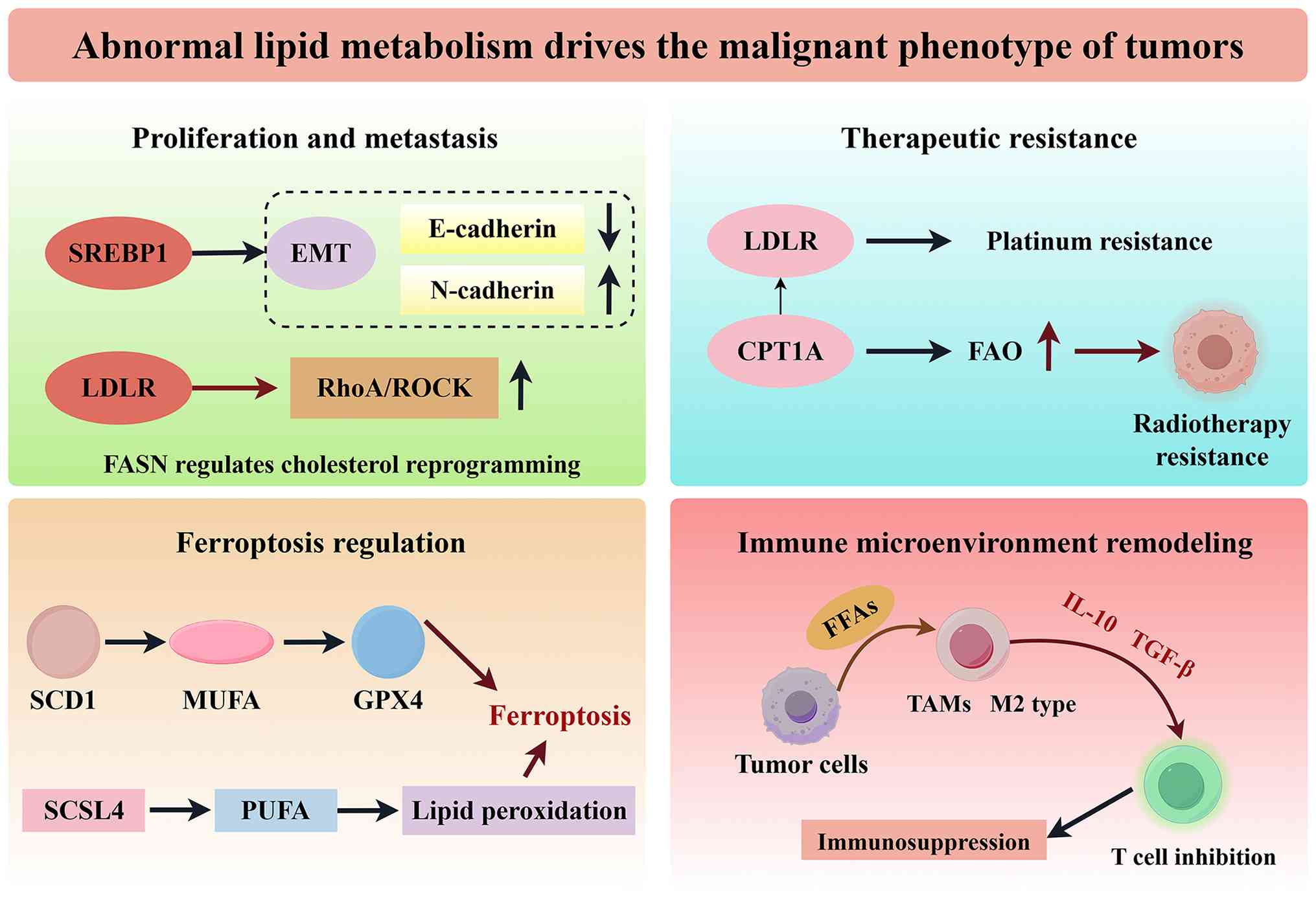
Dysregulated lipid metabolism in gynecological
malignancies is not merely a consequence of metabolic adaptation;
instead, it is deeply involved in regulating core malignant
phenotypes, including proliferation, metastasis, therapeutic
resistance, ferroptosis resistance and immune evasion, through
multiple mechanisms such as providing energy substrates, mediating
signaling pathway activation, regulating cell death modes and
remodeling the tumor immune microenvironment (139,140). This association exhibits distinct
tumor subtype specificity, offering clear theoretical basis and
therapeutic targets for precision targeted intervention.
Aberrant lipid metabolism fuels the malignant
proliferation, invasion and metastasis of gynecological cancer
cells via the dual mechanisms of ‘energy supply-signal regulation’
(141). At the level of fatty acid
synthesis, palmitic acid generated by enhanced lipogenesis can
directly activate the PI3K/Akt signaling pathway, promoting the
transition of ovarian cancer cells from the G1 phase to
the S phase of the cell cycle and accelerating proliferation
(142). By contrast, FASN
inhibitors block this signaling axis by reducing palmitic acid
production, arresting tumor cells in the G1 phase and
suppressing proliferative activity. In endometrial cancer,
SREBP1-mediated enhanced lipogenesis activates the
epithelial-mesenchymal transition process by promoting the
degradation of E-cadherin and upregulating the expression of
N-cadherin, thereby enhancing the invasive capacity of tumor cells
(143,144).
Abnormal cholesterol metabolism, in turn, promotes
metastasis by regulating membrane-associated signaling pathways
(145). In ovarian cancer cells,
enhanced LDLR-mediated cholesterol uptake enriches lipid raft
structures in the cell membrane, activating the RhoA/ROCK signaling
pathway to facilitate actin cytoskeleton reorganization and improve
cell motility and invasiveness (146). In cervical cancer, FASN regulates
cholesterol reprogramming to exert dual effects: On the one hand,
it activates the lipid raft-associated c-Src/AKT/FAK signaling
pathway; on the other hand, it promotes the secretion of
platelet-derived growth factor-AA and insulin-like growth
factor-binding protein 3, inducing lymphangiogenesis and markedly
boosting lymph node metastasis capacity (147). Furthermore, lipid metabolites (for
example, oleic acid and cholesterol esters) can upregulate the
expression of vascular endothelial growth factor (VEGF) to promote
tumor angiogenesis, establishing a nutrient transport network that
supports proliferation and metastasis (148,149).
Lipid metabolic reprogramming serves as a central
driver underlying resistance to multiple therapeutic modalities in
gynecological malignancies, with distinct subtype-specific
metabolic aberrations corresponding to different types of treatment
resistance (150,151). For platinum resistance in ovarian
cancer, disrupted cholesterol homeostasis plays a pivotal role:
Platinum-resistant cells exhibit markedly upregulate LDLR
expression, which enhances exogenous cholesterol uptake. This
metabolic adaptation maintains cell membrane fluidity and
stability, reducing DNA cross-linking damage induced by
platinum-based drugs. Statins, by inhibiting HMGCR activity, can
lower intracellular cholesterol levels and reverse the
platinum-resistant phenotype (152,153). Additionally, FASN-mediated
enhanced fatty acid synthesis is linked to PARP inhibitor
resistance in ovarian cancer. When the FASN inhibitor TVB-2640 is
combined with PARP inhibitors, it exerts a synergistic effect by
co-suppressing metabolic reprogramming and DNA damage repair
pathways, markedly improving the therapeutic response rate in
resistant patients (154–156).
Chemoradiotherapy resistance in cervical cancer is
predominantly driven by enhanced FAO; chemoradiotherapy induces
upregulated CPT1A expression in cervical cancer cells, activating
the FAO pathway to augment energy supply and enhance cell survival
under therapeutic stress (157–159). Combining the FAO inhibitor
etomoxir with chemoradiotherapy blocks this metabolic adaptation,
markedly boosting treatment sensitivity. By contrast, hormone
therapy resistance in endometrial cancer is associated with
exaggerated lipogenesis: Progesterone-resistant cells display
abnormally elevated expression of SREBP1 and FASN (160). Enhanced lipogenesis weakens the
growth-inhibitory effect of progesterone by activating the AKT/mTOR
signaling pathway, while targeted inhibition of lipogenesis can
restore tumor cell sensitivity to progesterone. Mechanistically,
lipid metabolic reprogramming mediates drug resistance through
three core mechanisms: Augmenting the activity of DNA damage repair
enzymes, suppressing the activation of apoptotic signaling pathways
and sustaining energy metabolic homeostasis (161–163). Mitochondrial lipid metabolism is a
key driver of therapeutic resistance. Enhanced FAO provides
sufficient ATP for DNA damage repair and increases mitochondrial
antioxidant production. Targeting CPT1A, the rate-limiting enzyme
of mitochondrial FAO, effectively reverses platinum resistance in
cells (164,165).
Ferroptosis is an iron-dependent, lipid
peroxidation-mediated form of regulated cell death, and lipid
metabolic dysregulation serves as a central regulator in
ferroptosis modulation, with this regulatory effect exhibiting
distinct tumor subtype specificity (166). SCD1, a key fatty acid desaturase,
is upregulated in ovarian cancer (167). The MUFAs catalyzed by SCD1 enhance
tumor cell resistance to ferroptosis by decreasing lipid
peroxidation accumulation and maintaining cell membrane stability
(168). By contrast, SCD1
inhibitors promote ferroptosis induction by increasing
polyunsaturated fatty acid (PUFA) accumulation and disrupting redox
homeostasis. Studies have confirmed that tumor cells derived from
ovarian cancer ascites show markedly elevated expression levels of
SCD1 and fatty acid desaturase 2; combined inhibition of these two
enzymes downregulates GPX4 expression, triggering an outbreak of
lipid peroxidation and initiating ferroptosis (169–171).
Lipid peroxidation is the core event driving
ferroptosis, and intracellular PUFA accumulation markedly enhances
lipid peroxidation sensitivity, thereby promoting ferroptosis
(172,173). In endometrial cancer cells,
upregulated expression of ACSL4 facilitates PUFA activation and
integration into membrane phospholipids, increasing the risk of
lipid peroxidation (174).
Consequently, patients with high ACSL4 expression are more
sensitive to ferroptosis inducers. By contrast, lipid droplet
accumulation reduces lipid peroxidation by sequestering PUFAs.
OCCC, characterized by abundant lipid droplets, exhibits inherent
resistance to ferroptosis inducers; targeting diacylglycerol
acyltransferase 1, a key gene involved in lipid droplet formation,
can diminish the sequestration of PUFAs by lipid droplets,
enhancing ferroptosis sensitivity (175). This close association between
lipid metabolism and ferroptosis offers a novel direction for
gynecological cancer treatment. For instance, the combination of
SCD1 inhibitors and cisplatin can enhance the cytotoxicity of
cisplatin against ovarian cancer by inducing ferroptosis, thereby
improving therapeutic outcomes (176).
Abnormal lipid metabolism shapes an
immunosuppressive phenotype by regulating immune cell function and
altering the composition of the tumor immune microenvironment,
ultimately facilitating tumor immune evasion (177). FFAs secreted by gynecological
cancer cells are specifically taken up by tumor-associated
macrophages (TAMs), inducing their polarization toward the M2
phenotype. M2-type TAMs suppress effector T-cell function by
secreting anti-inflammatory cytokines such as interleukin-10 and
transforming growth factor-β. In the ovarian cancer
microenvironment, lipid-enriched TAMs also abnormally express
programmed death ligand 1 (PD-L1), directly inhibiting the
cytotoxic activity of CD8+ T cells via the programmed
cell death protein 1 (PD-1)/PD-L1 pathway and impairing the
antitumor immune response (178,179).
T-cell function is highly regulated by lipid
metabolism: Excess fatty acids in the tumor microenvironment are
taken up by CD8+ T cells through CD36-mediated pathways,
leading to abnormal intracellular lipid accumulation (180,181). This accumulation inhibits T-cell
proliferation and the secretion of cytotoxic molecules (such as
perforin and granzyme B), inducing T-cell exhaustion. In cervical
cancer, enhanced CD36-mediated fatty acid uptake is a key driver of
T-cell exhaustion; combining CD36 inhibitors with PD-1 inhibitors
can reduce T-cell lipid accumulation, reverse the exhausted
phenotype and markedly enhance antitumor immune efficacy (182,183). Additionally, abnormal lipid
metabolism in regulatory T cells (Tregs) contributes to
immunosuppression: The fatty acid synthesis pathway in Tregs within
the ovarian cancer microenvironment is significantly enhanced,
sustaining their survival and immunosuppressive function to weaken
antitumor immunity (184,185). Targeting key enzymes in fatty acid
synthesis can markedly attenuate the immunosuppressive activity of
Tregs and improve the immune microenvironment (Fig. 2).
Tumor cell lipid metabolism abnormalities also
regulate immune evasion through other mechanisms: FASN inhibitors
can downregulate PD-L1 expression on the tumor cell surface,
enhancing the recognition and killing of tumor cells by
CD8+ T cells (186,187). Abnormal cholesterol accumulation
in ovarian cancer inhibits the infiltration and activity of natural
killer (NK) cells, while statins can improve NK cell function by
regulating cholesterol metabolism, boosting innate immune responses
(188,189).
Given the pivotal role of lipid metabolism in the
malignant progression and therapeutic resistance of gynecological
malignancies, lipid metabolism-targeted intervention strategies
have emerged as a research focus in oncology (190,191). These strategies encompass key
metabolic enzyme inhibitors, signaling pathway-targeted agents and
multi-modal combination therapies, with several approaches, such as
FASN inhibitors and statins, having advanced from preclinical
research to clinical translation, providing novel technical
pathways and clinical evidence for the precision treatment of
gynecological cancer (192,193).
Specific inhibitors against key lipid metabolic
enzymes represent the most direct intervention strategy. By
blocking core processes such as lipid uptake, synthesis and
catabolism, these agents disrupt tumor cell metabolic homeostasis
to exert antitumor effects. To date, multiple inhibitors have
completed preclinical validation in gynecological cancers, with
some progressing to clinical trials (194,195).
SCD1 inhibitors exhibit unique advantages in ovarian
cancer treatment. Specific inhibitors, such as A939572 and E6446,
block SCD1-mediated monounsaturated fatty acid synthesis, promoting
polyunsaturated fatty acid accumulation and triggering lipid
peroxidation and ferroptosis. These agents significantly suppress
tumor growth in ovarian cancer xenograft models, with notable
efficacy particularly against ferroptosis-resistant subtypes
(201). HMGCR inhibitors
(statins), commonly used as lipid-lowering drugs in clinical
practice, have been extensively validated for their antitumor
potential: Simvastatin inhibits ovarian cancer cell proliferation
and invasion, and enhances chemosensitivity when combined with
cisplatin (202). Multiple
large-scale epidemiological studies have consistently shown that
long-term statin use is associated with reduced incidence of
ovarian cancer and improved overall survival in patients with
endometrial cancer. Real-world clinical evidence also indicates
that statin use combined with platinum-based chemotherapy can
prolong progression-free survival in patients with advanced ovarian
cancer, particularly in those with high LDLR expression, supporting
the potential repurposing value of statins in gynecological cancer
treatment (203,204).
Inhibitors targeting FAO are also under active
development. Etomoxir, a CPT1 inhibitor, blocks the entry of
long-chain fatty acids into the mitochondria, reducing FAO-mediated
energy supply and enhancing the sensitivity of gynecological cancer
cells to chemotherapy (205). ACAT
inhibitors (such as avasimibe and K604) suppress cholesterol
esterification and lipid droplet accumulation, exerting antitumor
effects by disrupting cholesterol homeostasis in preclinical
ovarian cancer models. Additionally, liver X receptor (LXR)
agonists (such as GW3965) promote cholesterol efflux and regulate
lipid metabolic balance. When combined with anti-VEGF agents, they
exert a synergistic effect via ‘metabolic regulation-vascular
inhibition’ enhancing therapeutic outcomes in ovarian cancer
(206,207).
Drugs targeting lipid metabolism regulatory pathways
can indirectly rewire the lipid metabolic networks of tumor cells,
thereby achieving antitumor effects. These agents typically
modulate core transcription factors or signaling axes to
concurrently affect multiple lipid metabolic pathways, featuring a
broad range of action and potent synergistic effects.
SREBP pathway inhibitors are core representatives:
Fatostatin blocks the activation and nuclear translocation of
SREBP, concomitantly suppressing both fatty acid and cholesterol
synthesis (208,209). In endometrial cancer, it markedly
downregulates the expression of downstream target genes such as
FASN and ACC, reduces intracellular lipid accumulation and inhibits
tumor growth (210).
PI3K/Akt/mTOR pathway inhibitors indirectly
interfere with lipid metabolism by regulating the activation status
of SREBP1. As a VEGF inhibitor, bevacizumab enhances the antitumor
efficacy of lipid metabolism inhibitors by indirectly suppressing
PI3K/Akt pathway activity. Everolimus, an mTOR inhibitor, directly
inhibits lipid synthesis in ovarian cancer cells and exerts a
synergistic antitumor effect when combined with FASN inhibitors
(211,212).
Hippo-YAP/TAZ pathway inhibitors exert their
functions by regulating lipid metabolic networks: Verteporfin, a
YAP inhibitor, downregulates SREBP1 expression, inhibits fatty acid
synthesis and lipid droplet accumulation in ovarian cancer cells,
and concomitantly impairs the chemoresistance of tumor cells
(213,214). Inhibitors of Nrf2, such as ML385,
disrupt cellular redox homeostasis by suppressing the Nrf2/Keap1
pathway, increasing lipid peroxidation levels and promoting
ferroptosis. When combined with SCD1 inhibitors, they markedly
enhance the therapeutic efficacy against ovarian cancer through the
combined effect of metabolic inhibition and oxidative stress
(215,216).
Single-agent targeted therapy against lipid
metabolism often yields limited effects, making multi-modal
combination therapy an emerging core direction to break treatment
bottlenecks and reverse drug resistance (217). By combining lipid metabolism
inhibitors with chemotherapy, targeted therapy or immunotherapy,
synergistic efficacy can be achieved through mechanism
complementarity, while simultaneously covering tumor cell
populations with diverse metabolic phenotypes (218,219).
The combination of lipid metabolism inhibitors and
chemotherapy effectively reverses metabolism-adaptive resistance.
For instance, the combination of SCD1 inhibitors and cisplatin
enhances cisplatin-induced DNA damage by inducing ferroptosis,
reversing platinum resistance in ovarian cancer (220). Statins combined with paclitaxel
strengthen the cytotoxicity of chemoradiotherapy in cervical cancer
by inhibiting cholesterol synthesis, thereby improving treatment
sensitivity. Mechanistically, such combinations regulate lipid
metabolic balance, promote the accumulation of drug-induced DNA
damage and activate apoptotic signaling, breaking the metabolic
adaptive advantage of tumor cells (221,222).
The combination of lipid metabolism inhibitors and
immunotherapy represents a highly promising approach. FASN
inhibitors downregulate the expression level of PD-L1 on tumor cell
surfaces, reducing immune checkpoint-mediated immune evasion; when
combined with PD-1 inhibitors, they enhance the cytotoxic activity
of CD8+ T cells and markedly improve therapeutic
outcomes in ovarian cancer (223,224). CD36 inhibitors combined with PD-L1
inhibitors reduce lipid accumulation and exhaustion in T cells,
remodel the cervical cancer immune microenvironment and exhibit
synergistic antitumor activity (225). Additionally, LXR agonists improve
the polarization phenotype of TAMs by promoting cholesterol efflux,
thereby boosting the antitumor effects of immunotherapy (226,227).
The combination of lipid metabolism inhibitors and
PARP inhibitors targets the ‘metabolic reprogramming-DNA damage
repair’ pathway. The combination of TVB-2640 and PARP inhibitors
enhances the synthetic lethal effect induced by PARP inhibitors by
suppressing fatty acid synthesis, markedly improving the
therapeutic response rate in HGSOC (228,229). A preclinical study demonstrated
that this combination effectively inhibits the proliferation of
drug-resistant cells and extends the survival time of tumor-bearing
mice, providing a novel therapeutic option for tumors with abnormal
DNA damage repair pathways (230).
Lipid metabolism-targeted intervention strategies
have achieved phased progress in clinical translation, with
multiple agents advancing to clinical trials at various stages, and
a biomarker-guided precision treatment model taking shape.
The clinical translation of the FASN inhibitor
TVB-2640 has been notable: Phase I clinical trials have confirmed
its favorable safety and tolerability in patients with advanced
solid tumors, with partial disease remission observed in some cases
(231–233). A phase II trial is evaluating its
efficacy in combination with bevacizumab for recurrent high-grade
astrocytoma, providing valuable reference data for analogous
combination therapies in gynecological malignancies (234,235). Statins demonstrate distinct
advantages in clinical translation: Numerous epidemiological
studies have linked statin use to a reduced incidence risk of
ovarian and endometrial cancer (236). A phase II clinical trial is
assessing the efficacy of atorvastatin combined with progestogens
in early-stage endometrial cancer, and preliminary findings
indicate that this combination regimen markedly reduces tumor Ki-67
proliferation index (237). A
separate phase II trial investigating rosuvastatin + progestogens
for early-stage endometrial cancer is also underway to validate the
feasibility and safety of this regimen as fertility-sparing
therapy, with preclinical mechanistic evidence supporting the
biological rationale for this combination strategy (238).
Overall, biomarker-guided precision targeted
therapy has emerged as the core direction for future development.
Lipid metabolism-related molecules, including FASN, fatty acid
transporter CD36 and LDLR, have been validated as prognostic
biomarkers and predictors of treatment response. Studies have found
that patients with ovarian cancer with high FASN expression are
more sensitive to TVB-2640 treatment, while patients with cervical
cancer with elevated CD36 expression are more likely to benefit
from the combination of CD36 inhibitors and immunotherapy. Future
clinical research should further expand sample sizes to validate
the clinical predictive value of these biomarkers, establish a
precision treatment decision-making system based on lipid metabolic
profiles, and realize individualized therapy centered on metabolic
subtyping and targeted intervention. Several lipid
metabolism-related biomarkers have been validated in clinical
settings for prognostic prediction and treatment response
stratification. For example, high FASN expression in tumor tissues
is an independent poor prognostic factor for ovarian cancer and
predicts better response to TVB-2640 treatment. Serum LDL
cholesterol levels have been shown to correlate with platinum
resistance in patients with ovarian cancer. Additionally, CD36
expression in tumor-infiltrating CD8+ T cells predicts
the efficacy of anti-PD-1 immunotherapy in patients with cervical
cancer, providing a potential biomarker for patient selection and
individualized treatment decision-making.
Compared with previously published reviews focusing
on a single tumor type or general lipid metabolic phenotypes, the
present review provides a subtype-centric systematic overview
across three major gynecological malignancies. The review
integrates a full regulatory axis from upstream oncogenic signaling
to downstream malignant phenotypes, incorporates emerging
mechanisms of ferroptosis regulation and immune microenvironment
remodeling, and further summarizes the clinical translation
progress of subtype-matched targeted interventions. This aims to
offer a more refined theoretical framework for precision metabolic
intervention in gynecological malignancies. While this review
systematically summarizes the subtype-specific lipid metabolic
reprogramming mechanisms and emerging targeted intervention
strategies in gynecological malignancies, several critical evidence
gaps remain that limit the clinical feasibility of current research
plans. Most mechanistic insights are derived from preclinical cell
line and animal models, which cannot fully recapitulate the
complexity of the human tumor microenvironment and interpatient
metabolic heterogeneity. Large-scale, subtype-enriched clinical
trials are still lacking to validate the efficacy and safety of
lipid metabolism-targeted therapies, and there is no standardized
biomarker system to guide patient selection and dynamic efficacy
monitoring.
Building on these limitations, despite the
groundbreaking advancements in lipid metabolism-targeted research,
its clinical translation and widespread application in
gynecological malignancies face multiple interconnected challenges
that require systematic investigations to address (239). First, the network complexity of
lipid metabolism and tumor subtype specificity represent the core
constraint for targeted therapy. Notable heterogeneity in lipid
metabolic profiles exists across distinct gynecological cancer
subtypes and even among patients within the same subtype (240). For instance, the serous subtype of
ovarian cancer is characterized by enhanced FAO, while the clear
cell subtype relies on lipid storage and exogenous uptake (241). A single therapeutic regimen
struggles to cover all metabolic phenotypes, hampering the
implementation of precision intervention. Second, the insufficient
target specificity of lipid metabolism inhibitors poses a critical
issue. Lipid metabolism is a fundamental physiological process in
normal cells, and existing inhibitors cannot fully distinguish the
metabolic differences between tumor and normal cells. This tends to
disrupt lipid homeostasis in normal tissues (such as liver and
adipose tissues), leading to off-target effects and adverse
reactions. For example, statins may induce muscle injury, while
FASN inhibitors can cause weight loss and metabolic disorders
(242). Additionally, extensive
crosstalk between metabolic pathways increases the risk of
therapeutic resistance (243).
Lipid metabolism is intricately regulated through cross-talk with
glucose metabolism, amino acid metabolism and oncogenic signaling
pathways. Targeting lipid metabolism alone is prone to inducing
tumor cells to switch their metabolic dependency via reprogramming
(for example, shifting from lipogenesis to glycolysis), resulting
in adaptive therapeutic resistance and compromised long-term
efficacy (244,245).
Future research should focus on the following core
directions to optimize and advance lipid metabolism-targeted
strategies: i) Systematically decipher the subtype-specific
regulatory networks of lipid metabolism in gynecological
malignancies. Integrate multi-omics technologies (for example,
transcriptomics, lipidomics and metabolomics) with single-cell
sequencing to identify subtype-specific and patient-specific
metabolic targets, providing a molecular classification basis for
precision therapy. ii) Develop highly selective lipid metabolism
inhibitors and leverage novel drug delivery systems (such as
liposomes and polymeric nanoparticles) to achieve tumor-specific
drug accumulation. This approach can minimize damage to normal
tissues while improving drug bioavailability and tumor penetration.
iii) Elucidate the cross-talk mechanisms between lipid metabolism
and oncogenic pathways such as PI3K/Akt/mTOR and Hippo-YAP. Develop
multi-targeted combination therapy regimens that exert dual effects
of ‘metabolic inhibition + signal blocking’ to overcome therapeutic
resistance caused by single-target interventions. iv) Identify and
validate lipid metabolism-related biomarkers (for example, FASN,
CD36 and LDLR) through multi-center, large-cohort studies.
Establish individualized treatment prediction models incorporating
metabolic features and clinicopathological parameters to guide
treatment selection and dynamic efficacy assessment. v) Conduct
subtype-enriched clinical trials targeting specific populations
(such as HGSOC and obesity-associated endometrial cancer). Validate
the efficacy, safety and dosage optimization of lipid
metabolism-targeted therapies to expedite their clinical
translation.
With the in-depth elucidation of lipid metabolism
regulatory mechanisms in gynecological malignancies, lipid
metabolism-targeted strategies are expected to become an integral
component of precision therapy for these diseases. They may provide
a novel breakthrough for improving the prognosis of advanced and
drug-resistant patients, ultimately achieving the closed-loop of
metabolic subtyping-precision intervention-efficacy prediction in
individualized therapy.
Not applicable.
Funding: No funding was received.
Not applicable.
YR conceived the study, conducted the systematic
literature retrieval and screening, extracted the relevant research
data, drafted the full manuscript and critically revised it. Data
authentication is not applicable. The author has read and approved
the final manuscript.
Not applicable.
Not applicable.
The author declares that they have no competing
interests.
|
1
|
Zhao X, Ran J, Li S and Chen J: Advances
and obstacles of T cell-based immunotherapy in gynecological
malignancies. Mol Cancer. 24:2072025. View Article : Google Scholar : PubMed/NCBI
|
|
2
|
Fotopoulou C, Eriksson AG, Baiocchi G and
Zivanovic O: Surgery for gynecological cancers in the era of
personalized medicine: A novel paradigm. Int J Gynecol Cancer.
34:3512024. View Article : Google Scholar : PubMed/NCBI
|
|
3
|
Movahed F, Ourang Z, Neshat R, Hussein WS,
Saihood AS, Alarajy MS and Zareii D: PROTACs in gynecological
cancers: Current knowledge and future potential as a treatment
strategy. Pathol Res Pract. 263:1556112024. View Article : Google Scholar : PubMed/NCBI
|
|
4
|
Morice P, Scambia G, Abu-Rustum NR, Acien
M, Arena A, Brucker S, Cheong Y, Collinet P, Fanfani F, Filippi F,
et al: Fertility-sparing treatment and follow-up in patients with
cervical cancer, ovarian cancer, and borderline ovarian tumours:
Guidelines from ESGO, ESHRE, and ESGE. Lancet Oncol. 25:e602–e610.
2024. View Article : Google Scholar : PubMed/NCBI
|
|
5
|
Gupta R, Kumar R, Penn CA and Wajapeyee N:
Immune evasion in ovarian cancer: Implications for immunotherapy
and emerging treatments. Trends Immunol. 46:166–181. 2025.
View Article : Google Scholar : PubMed/NCBI
|
|
6
|
Xie W, Tang S, Tang R, Li L and Liu X:
Global, regional, and national burden of ovarian cancer, 1990–2021,
and projections to 2050: A cross-sectional analysis of the Global
Burden of Disease Study 2021. Int J Surg. 112:226–238. 2026.
View Article : Google Scholar : PubMed/NCBI
|
|
7
|
Zhu B, Gu H, Mao Z, Beeraka NM, Zhao X,
Anand MP, Zheng Y, Zhao R, Li S, Manogaran P, et al: Global burden
of gynaecological cancers in 2022 and projections to 2050. J Glob
Health. 14:041552024. View Article : Google Scholar : PubMed/NCBI
|
|
8
|
Huang P, Fan X, Yu H, Zhang K, Li H, Wang
Y and Xue F: Glucose metabolic reprogramming and its therapeutic
potential in obesity-associated endometrial cancer. J Transl Med.
21:942023. View Article : Google Scholar : PubMed/NCBI
|
|
9
|
Crosbie EJ, Kitson SJ, McAlpine JN,
Mukhopadhyay A, Powell ME and Singh N: Endometrial cancer. Lancet.
399:1412–1428. 2022. View Article : Google Scholar : PubMed/NCBI
|
|
10
|
Huang R, Liu Z, Sun T and Zhu L:
Cervicovaginal microbiome, high-risk HPV infection and cervical
cancer: Mechanisms and therapeutic potential. Microbiol Res.
287:1278572024. View Article : Google Scholar : PubMed/NCBI
|
|
11
|
Branda F, Pavia G, Ciccozzi A, Quirino A,
Marascio N, Gigliotti S, Matera G, Romano C, Locci C, Azzena I, et
al: Human papillomavirus (HPV) vaccination: Progress, challenges,
and future directions in global immunization strategies. Vaccines
(Basel). 12:12932023. View Article : Google Scholar
|
|
12
|
Xu M, Cao C, Wu P, Huang X and Ma D:
Advances in cervical cancer: Current insights and future
directions. Cancer Commun (Lond). 45:77–109. 2025. View Article : Google Scholar : PubMed/NCBI
|
|
13
|
Mayadev JS, Ke G, Mahantshetty U, Pereira
MD, Tarnawski R and Toita T: Global challenges of radiotherapy for
the treatment of locally advanced cervical cancer. Int J Gynecol
Cancer. 32:436–445. 2022. View Article : Google Scholar : PubMed/NCBI
|
|
14
|
Small W Jr, Bacon MA, Bajaj A, Chuang LT,
Fisher BJ, Harkenrider MM, Jhingran A, Kitchener HC, Mileshkin LR,
Viswanathan AN and Gaffney DK: Cervical cancer: A global health
crisis. Cancer. 123:2404–2412. 2017. View Article : Google Scholar : PubMed/NCBI
|
|
15
|
Ma Y, Temkin SM, Hawkridge AM, Guo C, Wang
W, Wang X and Fang X: Fatty acid oxidation: An emerging facet of
metabolic transformation in cancer. Cancer Lett. 435:92–100. 2018.
View Article : Google Scholar : PubMed/NCBI
|
|
16
|
Li J, Yang H, Zhang L, Zhang S and Dai Y:
Metabolic reprogramming and interventions in endometrial carcinoma.
Biomed Pharmacother. 161:1145262023. View Article : Google Scholar : PubMed/NCBI
|
|
17
|
Li B and Sui L: Metabolic reprogramming in
cervical cancer and metabolomics perspectives. Nutr Metab (Lond).
18:932021. View Article : Google Scholar : PubMed/NCBI
|
|
18
|
Žalytė E: Ferroptosis, metabolic rewiring,
and endometrial cancer. Int J Mol Sci. 25:752023. View Article : Google Scholar : PubMed/NCBI
|
|
19
|
Zhang J, Ouyang F, Gao A, Zeng T, Li M, Li
H, Zhou W, Gao Q, Tang X, Zhang Q, et al: ESM1 enhances fatty acid
synthesis and vascular mimicry in ovarian cancer by utilizing the
PKM2-dependent warburg effect within the hypoxic tumor
microenvironment. Mol Cancer. 23:942024. View Article : Google Scholar : PubMed/NCBI
|
|
20
|
Hwang S, Awasthi D, Jeong J, Sandoval TA,
Chae C, Ramos Y, Tan C, Marin Falco M, Salvagno C, Emmanuelli A, et
al: Transgelin 2 guards T cell lipid metabolism and antitumour
function. Nature. 635:1010–1018. 2024. View Article : Google Scholar : PubMed/NCBI
|
|
21
|
Zhang C, Wang Y, He M, Wang C, Cao K,
Zhong Y, Wang X, Yang M, Zhang G, Lu J, et al: Mannose Enhances
Immunotherapy Efficacy in Ovarian Cancer by Modulating Gut
Microbial Metabolites. Cancer Res. 85:2468–2484. 2025. View Article : Google Scholar : PubMed/NCBI
|
|
22
|
Dyck L, Prendeville H, Raverdeau M, Wilk
MM, Loftus RM, Douglas A, McCormack J, Moran B, Wilkinson M, Mills
EL, et al: Suppressive effects of the obese tumor microenvironment
on CD8 T cell infiltration and effector function. J Exp Med.
219:e202100422022. View Article : Google Scholar : PubMed/NCBI
|
|
23
|
Guo C, Qu X, Tang X, Song Y, Wang J, Hua K
and Qiu J: Spatiotemporally deciphering the mysterious mechanism of
persistent HPV-induced malignant transition and immune remodelling
from HPV-infected normal cervix, precancer to cervical cancer:
Integrating single-cell RNA-sequencing and spatial transcriptome.
Clin Transl Med. 13:e12192023. View Article : Google Scholar : PubMed/NCBI
|
|
24
|
Shafiee MN, Mongan N, Seedhouse C, Chapman
C, Deen S, Abu J and Atiomo W: Sterol regulatory element binding
protein-1 (SREBP1) gene expression is similarly increased in
polycystic ovary syndrome and endometrial cancer. Acta Obstet
Gynecol Scand. 96:556–562. 2017. View Article : Google Scholar : PubMed/NCBI
|
|
25
|
Yano H, Fujiwara Y and Komohara Y:
Cholesterol metabolism and lipid droplet vacuoles; a potential
target for the therapy of aggressive lymphoma. J Clin Exp Hematop.
62:190–194. 2022. View Article : Google Scholar : PubMed/NCBI
|
|
26
|
Wang Y, Han S, You X, Shi X, Liu L, Sun Y,
Ma Y, Qian Q, Liu H, Cui B and Zhang Y: The role of low density
lipoprotein receptor-related protein 11 as a tumor promoter in
cervical cancer. Cancer Manag Res. 11:8081–8093. 2019. View Article : Google Scholar : PubMed/NCBI
|
|
27
|
Liu S, Zhang X, Wang W, Li X, Sun X, Zhao
Y, Wang Q, Li Y, Hu F and Ren H: Metabolic reprogramming and
therapeutic resistance in primary and metastatic breast cancer. Mol
Cancer. 23:2612024. View Article : Google Scholar : PubMed/NCBI
|
|
28
|
Russo S, Kwiatkowski M, Govorukhina N,
Bischoff R and Melgert BN: Meta-Inflammation and metabolic
reprogramming of macrophages in diabetes and obesity: The
importance of metabolites. Front Immunol. 12:7461512021. View Article : Google Scholar : PubMed/NCBI
|
|
29
|
Wang L, Zhu H, Shi Z, Chen B, Huang H, Lin
G, Li J, Yu H, Xu S, Chen G, et al: MK8722 initiates early-stage
autophagy while inhibiting late-stage autophagy via FASN-dependent
reprogramming of lipid metabolism. Theranostics. 14:75–95. 2024.
View Article : Google Scholar : PubMed/NCBI
|
|
30
|
Fu Y, Francés R, Monge C, Desterke C,
Marchio A, Pineau P, Chang-Marchand Y and Mata-Garrido J: Metabolic
and epigenetic mechanisms in hepatoblastoma: Insights into tumor
biology and therapeutic targets. Genes (Basel). 15:13582024.
View Article : Google Scholar : PubMed/NCBI
|
|
31
|
Tufail M, Jiang CH and Li N: Altered
metabolism in cancer: Insights into energy pathways and therapeutic
targets. Mol Cancer. 23:2032024. View Article : Google Scholar : PubMed/NCBI
|
|
32
|
Yi J, Zhu J, Wu J, Thompson CB and Jiang
X: Oncogenic activation of PI3K-AKT-mTOR signaling suppresses
ferroptosis via SREBP-mediated lipogenesis. Proc Natl Acad Sci USA.
117:31189–31197. 2020. View Article : Google Scholar : PubMed/NCBI
|
|
33
|
Luo M, Yan J, Hu X, Li H, Li H, Liu Q,
Chen Y and Zou Z: Targeting lipid metabolism for ferroptotic cancer
therapy. Apoptosis. 28:81–107. 2023. View Article : Google Scholar : PubMed/NCBI
|
|
34
|
Xu J, Peng JJ, Yang W, Fu K and Zhang Y:
Vaginal microbiomes and ovarian cancer: A review. Am J Cancer Res.
10:743–756. 2020.PubMed/NCBI
|
|
35
|
Caruso G, Weroha SJ and Cliby W: Ovarian
cancer: A review. JAMA. 334:1278–1291. 2025. View Article : Google Scholar : PubMed/NCBI
|
|
36
|
Huang Y, Du Y, Zheng Y, Wen C, Zou H,
Huang J, Zhou H, Zhao H and Wu L: Ct-OATP1B3 promotes high-grade
serous ovarian cancer metastasis by regulation of fatty acid
beta-oxidation and oxidative phosphorylation. Cell Death Dis.
13:5562022. View Article : Google Scholar : PubMed/NCBI
|
|
37
|
Huang D, Chowdhury S, Wang H, Savage SR,
Ivey RG, Kennedy JJ, Whiteaker JR, Lin C, Hou X, Oberg AL, et al:
Multiomic analysis identifies CPT1A as a potential therapeutic
target in platinum-refractory, high-grade serous ovarian cancer.
Cell Rep Med. 2:1004712021. View Article : Google Scholar : PubMed/NCBI
|
|
38
|
Sawyer BT, Qamar L, Yamamoto TM, McMellen
A, Watson ZL, Richer JK, Behbakht K, Schlaepfer IR and Bitler BG:
Targeting fatty acid oxidation to promote anoikis and inhibit
ovarian cancer progression. Mol Cancer Res. 18:1088–1098. 2020.
View Article : Google Scholar : PubMed/NCBI
|
|
39
|
Kolb S, Hoffmann I, Monjé N, Dragomir MP,
Jank P, Bischoff P, Keunecke C, Pohl J, Kunze CA, Marchenko S, et
al: LRP1B-a prognostic marker in tubo-ovarian high-grade serous
carcinoma. Hum Pathol. 141:158–168. 2023. View Article : Google Scholar : PubMed/NCBI
|
|
40
|
van Baal JOAM, Lok CAR, Jordanova ES,
Horlings H, van Driel WJ, Amant FC and Van de Vijver KK: The effect
of the peritoneal tumor microenvironment on invasion of peritoneal
metastases of high-grade serous ovarian cancer and the impact of
NEOADJUVANT chemotherapy. Virchows Arch. 477:535–544. 2020.
View Article : Google Scholar : PubMed/NCBI
|
|
41
|
Koizume S, Takahashi T and Miyagi Y: Lipid
droplets: A candidate new research field for epithelial ovarian
cancer. Front Pharmacol. 15:14371612024. View Article : Google Scholar : PubMed/NCBI
|
|
42
|
Luo H, She X, Zhang Y, Xie B, Zhang S, Li
Q, Zhou Y, Guo S, Zhang S, Jiang Y, et al: PLIN2 promotes lipid
accumulation in ascites-associated macrophages and ovarian cancer
progression by HIF1α/SPP1 signaling. Adv Sci (Weinh).
12:e24113142025. View Article : Google Scholar : PubMed/NCBI
|
|
43
|
Zheng C, Tan H, Niu G, Huang X, Lu J, Chen
S, Li H, Zhu J, Zhou Z, Xu M, et al: ACAT1-Mediated ME2 acetylation
drives chemoresistance in ovarian cancer by linking glutaminolysis
to lactate production. Adv Sci (Weinh). 12:e24164672025. View Article : Google Scholar : PubMed/NCBI
|
|
44
|
Ahmad N, Moton S, Kuttikrishnan S, Prabhu
KS, Masoodi T, Ahmad S and Uddin S: Fatty acid synthase: A key
driver of ovarian cancer metastasis and a promising therapeutic
target. Pathol Res Pract. 260:1554652024. View Article : Google Scholar : PubMed/NCBI
|
|
45
|
Lombardi S, Goldman AR, Tang HY, Kossenkov
AV, Liu H, Zhou W, Herlyn M, Lin J and Zhang R: Targeting fatty
acid reprogramming suppresses CARM1-expressing ovarian cancer.
Cancer Res Commun. 3:1067–1077. 2023. View Article : Google Scholar : PubMed/NCBI
|
|
46
|
Jiang L, Fang X, Wang H, Li D and Wang X:
Ovarian cancer-intrinsic fatty acid synthase prevents anti-tumor
immunity by disrupting tumor-infiltrating dendritic cells. Front
Immunol. 9:29272018. View Article : Google Scholar : PubMed/NCBI
|
|
47
|
Sung S, Hong Y, Kim BG, Choi JY, Kim JW,
Park SY, Kim JM, Kim YJ, Lee JM, Kim TJ, et al: Stratifying the
risk of ovarian cancer incidence by histologic subtypes in the
Korean epithelial ovarian cancer study (Ko-EVE). Cancer Med.
12:8742–8753. 2023. View Article : Google Scholar : PubMed/NCBI
|
|
48
|
Wu R, Li N, Huang W, Yang Y, Zang R, Song
H, Shi J, Zhu S and Liu Q: Melittin suppresses ovarian cancer
growth by regulating SREBP1-mediated lipid metabolism.
Phytomedicine. 137:1563672025. View Article : Google Scholar : PubMed/NCBI
|
|
49
|
Ji Z, Shen Y, Feng X, Kong Y, Shao Y, Meng
J, Zhang X and Yang G: Deregulation of lipid metabolism: The
critical factors in ovarian cancer. Front Oncol. 10:5930172020.
View Article : Google Scholar : PubMed/NCBI
|
|
50
|
Mukherjee A, Wu J, Barbour S and Fang X:
Lysophosphatidic acid activates lipogenic pathways and de novo
lipid synthesis in ovarian cancer cells. J Biol Chem.
287:24990–25000. 2012. View Article : Google Scholar : PubMed/NCBI
|
|
51
|
Tian W, Teng F, Zhao J, Gao J, Gao C, Sun
D, Liu G, Zhang Y, Yu S, Zhang W, et al: Estrogen and insulin
synergistically promote type 1 endometrial cancer progression.
Cancer Biol Ther. 18:1000–1010. 2017. View Article : Google Scholar : PubMed/NCBI
|
|
52
|
Kim DS and Scherer PE: Obesity, diabetes,
and increased cancer progression. Diabetes Metab J. 45:799–812.
2021. View Article : Google Scholar : PubMed/NCBI
|
|
53
|
Qiu C, Dongol S, Lv QT, Gao X and Jiang J:
Sterol regulatory element-binding protein-1/fatty acid synthase
involvement in proliferation inhibition and apoptosis promotion
induced by progesterone in endometrial cancer. Int J Gynecol
Cancer. 23:1629–1634. 2013. View Article : Google Scholar : PubMed/NCBI
|
|
54
|
Dang C, Wang X, Liu P, Liu J and Yu X:
Genetically proxied therapeutic effect of lipid-lowering drugs use,
breast cancer, and endometrial cancer's risk: A drug target-based
mendelian Randomization study. Int J Womens Health. 16:2033–2041.
2024. View Article : Google Scholar : PubMed/NCBI
|
|
55
|
Galant N, Krawczyk P, Monist M, Obara A,
Gajek U, Grenda A, Nico M, Kalinka E and Milanowski J: Molecular
classification of endometrial cancer and its impact on therapy
selection. Int J Mol Sci. 25:58932024. View Article : Google Scholar : PubMed/NCBI
|
|
56
|
Garikapati KK, Ammu VVVR, Krishnamurthy
PT, Chintamaneni PK and Pindiprolu SKSS: Type-II endometrial
cancer: Role of adipokines. Arch Gynecol Obstet. 300:239–249. 2019.
View Article : Google Scholar : PubMed/NCBI
|
|
57
|
Wysham WZ, Roque DR, Han J, Zhang L, Guo
H, Gehrig PA, Zhou C and Bae-Jump VL: Effects of Fatty acid
synthase inhibition by orlistat on proliferation of endometrial
cancer cell lines. Target Oncol. 11:763–769. 2016. View Article : Google Scholar : PubMed/NCBI
|
|
58
|
Garg P, Ramisetty S, Nair M, Kulkarni P,
Horne D, Salgia R and Singhal SS: Strategic advancements in
targeting the PI3K/AKT/mTOR pathway for Breast cancer therapy.
Biochem Pharmacol. 236:1168502025. View Article : Google Scholar : PubMed/NCBI
|
|
59
|
Hao C, Wei Y, Meng W, Zhang J and Yang X:
PI3K/AKT/mTOR inhibitors for hormone receptor-positive advanced
breast cancer. Cancer Treat Rev. 132:1028612025. View Article : Google Scholar : PubMed/NCBI
|
|
60
|
Roncolato F, Lindemann K, Willson ML,
Martyn J and Mileshkin L: PI3K/AKT/mTOR inhibitors for advanced or
recurrent endometrial cancer. Cochrane Database Syst Rev.
10:CD0121602019.PubMed/NCBI
|
|
61
|
Kyrgiou M and Moscicki AB: Vaginal
microbiome and cervical cancer. Semin Cancer Biol. 86((Pt 3)):
189–198. 2022. View Article : Google Scholar : PubMed/NCBI
|
|
62
|
Bhatla N and Singhal S: Primary HPV
screening for cervical cancer. Best Pract Res Clin Obstet Gynaecol.
65:98–108. 2020. View Article : Google Scholar : PubMed/NCBI
|
|
63
|
Pal A and Kundu R: Human Papillomavirus E6
and E7: The cervical cancer hallmarks and targets for therapy.
Front Microbiol. 10:31162020. View Article : Google Scholar : PubMed/NCBI
|
|
64
|
Hu C, Liu T, Han C, Xuan Y, Jiang D, Sun
Y, Zhang X, Zhang W, Xu Y, Liu Y, et al: HPV E6/E7 promotes aerobic
glycolysis in cervical cancer by regulating IGF2BP2 to stabilize
m(6)A-MYC expression. Int J Biol Sci. 18:507–521. 2022. View Article : Google Scholar : PubMed/NCBI
|
|
65
|
Wang S, Dang L, Liu S, Rezhake R, Yan H,
Duan X, Zhang L, Zhang L, Zhang L, Su M, et al: Cervical cancer
screening via visual inspection with acetic acid and lugol iodine
for triage of HPV-Positive women. JAMA Netw Open. 7:e2440902024.
View Article : Google Scholar : PubMed/NCBI
|
|
66
|
Al-Qurayshi Z, Foggia MJ, Pagedar N, Lee
GS, Tufano R and Kandil E: Thyroid cancer histological subtypes
based on tumor size: National perspective. Head Neck. 42:2257–2266.
2020. View Article : Google Scholar : PubMed/NCBI
|
|
67
|
Wang M, Huang K, Wong MCS, Huang J, Jin Y
and Zheng ZJ: Global cervical cancer incidence by histological
subtype and implications for screening methods. J Epidemiol Glob
Health. 14:94–101. 2024. View Article : Google Scholar : PubMed/NCBI
|
|
68
|
Liu C, Zhang M, Yan X, Ni Y, Gong Y, Wang
C, Zhang X, Wan L, Yang H, Ge C, et al: Single-cell dissection of
cellular and molecular features underlying human cervical squamous
cell carcinoma initiation and progression. Sci Adv. 9:eadd89772023.
View Article : Google Scholar : PubMed/NCBI
|
|
69
|
Cibula D, Dostálek L, Jarkovsky J, Mom CH,
Lopez A, Falconer H, Scambia G, Ayhan A, Kim SH, Isla Ortiz D, et
al: Post-recurrence survival in patients with cervical cancer.
Gynecol Oncol. 164:362–369. 2022. View Article : Google Scholar : PubMed/NCBI
|
|
70
|
Ilhan ZE, Aniewski P, Thomas N, Roe DJ,
Chase DM and Herbst-Kralovetz MM: Deciphering the complex interplay
between microbiota, HPV, inflammation and cancer through
cervicovaginal metabolic profiling. EBioMedicine. 44:675–690. 2019.
View Article : Google Scholar : PubMed/NCBI
|
|
71
|
Williamson A: Recent developments in human
papillomavirus (HPV) vaccinology. Viruses. 15:14402023. View Article : Google Scholar : PubMed/NCBI
|
|
72
|
Baba SK, Alblooshi SSE, Yaqoob R, Behl S,
Al Saleem M, Rakha EA, Malik F, Singh M, Macha MA, Akhtar MK, et
al: Human papilloma virus (HPV) mediated cancers: An insightful
update. J Transl Med. 23:4832025. View Article : Google Scholar : PubMed/NCBI
|
|
73
|
Du H, Xu T, Yu S, Wu S and Zhang J:
Mitochondrial metabolism and cancer therapeutic innovation. Signal
Transduct Target Ther. 10:2452025. View Article : Google Scholar : PubMed/NCBI
|
|
74
|
Gopinath P, Natarajan A, Sathyanarayanan
A, Veluswami S and Gopisetty G: The multifaceted role of
Matricellular Proteins in health and cancer, as biomarkers and
therapeutic targets. Gene. 815:1461372022. View Article : Google Scholar : PubMed/NCBI
|
|
75
|
Corrêa LH, Heyn GS and Magalhaes KG: The
impact of the adipose organ plasticity on inflammation and cancer
progression. Cells. 8:6622019. View Article : Google Scholar : PubMed/NCBI
|
|
76
|
Slattery K, Yao CH, Mylod E, Scanlan J,
Scott B, Crowley JP, McGowan O, McManus G, Brennan M, O'Brien K, et
al: Uptake of lipids from ascites drives NK cell metabolic
dysfunction in ovarian cancer. Sci Immunol. 10:eadr47952025.
View Article : Google Scholar : PubMed/NCBI
|
|
77
|
Kouba S, Ouldamer L, Garcia C, Fontaine D,
Chantome A, Vandier C, Goupille C and Potier-Cartereau M: Lipid
metabolism and Calcium signaling in epithelial ovarian cancer. Cell
Calcium. 81:38–50. 2019. View Article : Google Scholar : PubMed/NCBI
|
|
78
|
Jin N, Qian YY, Jiao XF, Wang Z, Li X, Pan
W, Jiang JK, Huang P, Wang SY, Jin P, et al: Niraparib restricts
intraperitoneal metastases of ovarian cancer by eliciting
CD36-dependent ferroptosis. Redox Biol. 80:1035282025. View Article : Google Scholar : PubMed/NCBI
|
|
79
|
Ladanyi A, Mukherjee A, Kenny HA, Johnson
A, Mitra AK, Sundaresan S, Nieman KM, Pascual G, Benitah SA, Montag
A, et al: Adipocyte-induced CD36 expression drives ovarian cancer
progression and metastasis. Oncogene. 37:2285–2301. 2018.
View Article : Google Scholar : PubMed/NCBI
|
|
80
|
Huang Z, Zhang X, Huang J, Liu X, Liang M,
Yang F, Gao T, Huang J, Xia G, Cao J, et al: Elevated CD36 drives
lymph node metastasis of breast cancer by activating Hippo-YAP
signaling-mediated anoikis resistance. Commun Biol. 9:802025.
View Article : Google Scholar : PubMed/NCBI
|
|
81
|
Tang S, Chen K, Zheng F, Fu Z, Niu Y, Liu
X, Ni H, Yuan X, Cui Z, Lu W, et al: High serum LDL promotes EMT
and stemness through LDLR/FOXQ1/NF-κB1 pathway in epithelial
ovarian cancer. Oncogene. 44:4587–4600. 2025. View Article : Google Scholar : PubMed/NCBI
|
|
82
|
Wang M, Zhao D, Xu L, Guo W, Nie L, Lei Y,
Long Y, Liu M, Wang Y, Zhang X, et al: Role of PCSK9 in lipid
metabolic disorders and ovarian dysfunction in polycystic ovary
syndrome. Metabolism. 94:47–58. 2019. View Article : Google Scholar : PubMed/NCBI
|
|
83
|
Criscuolo D, Avolio R, Calice G, Laezza C,
Paladino S, Navarra G, Maddalena F, Crispo F, Pagano C, Bifulco M,
et al: Cholesterol homeostasis modulates platinum sensitivity in
human ovarian cancer. Cells. 9:8282020. View Article : Google Scholar : PubMed/NCBI
|
|
84
|
Knapp P, Chabowski A, Posmyk R and Górski
J: Expression of the energy substrate transporters in uterine
fibroids. Prostaglandins Other Lipid Mediat. 123:9–15. 2016.
View Article : Google Scholar : PubMed/NCBI
|
|
85
|
Grunt TW, Lemberger L, Colomer R, López
Rodríguez ML and Wagner R: The pharmacological or genetic blockade
of endogenous de novo fatty acid synthesis does not increase the
uptake of exogenous lipids in ovarian cancer cells. Front Oncol.
11:6108852021. View Article : Google Scholar : PubMed/NCBI
|
|
86
|
Lemberger L, Wagner R, Heller G, Pils D
and Grunt TW: Pharmacological inhibition of lipid import and
transport proteins in ovarian cancer. Cancers (Basel). 14:60042022.
View Article : Google Scholar : PubMed/NCBI
|
|
87
|
Wang Q, Hu R, Li W, Tai Y, Gu W, Das BC,
Yang F, Ji J, Wang C and Zhou J: BF175 inhibits endometrial
carcinoma through SREBP-regulated metabolic pathways in vitro. Mol
Cell Endocrinol. 523:1111352021. View Article : Google Scholar : PubMed/NCBI
|
|
88
|
Abdul-Maksoud RS, Zidan HE, Saleh HS and
Amer SA: Visfatin and SREBP-1c mRNA expressions and serum levels
among egyptian women with polycystic ovary syndrome. Genet Test Mol
Biomarkers. 24:409–419. 2020. View Article : Google Scholar : PubMed/NCBI
|
|
89
|
Du Q, Liu P, Zhang C, Liu T, Wang W, Shang
C, Wu J, Liao Y, Chen Y, Huang J, et al: FASN promotes lymph node
metastasis in cervical cancer via cholesterol reprogramming and
lymphangiogenesis. Cell Death Dis. 13:4882022. View Article : Google Scholar : PubMed/NCBI
|
|
90
|
Gao S, Shi Z, Li X, Li W, Wang Y, Liu Z
and Jiang J: Fatostatin suppresses growth and enhances apoptosis by
blocking SREBP-regulated metabolic pathways in endometrial
carcinoma. Oncol Rep. 39:1919–1929. 2018.PubMed/NCBI
|
|
91
|
Wang X, Du Q, Mai Q, Zou Q, Wang S, Lin X,
Chen Q, Wei M, Chi C, Peng Z, et al: Targeting FASN enhances
cisplatin sensitivity via SLC7A11-mediated ferroptosis in cervical
cancer. Transl Oncol. 56:1023962025. View Article : Google Scholar : PubMed/NCBI
|
|
92
|
Liu Y, Liu X, Wang H, Ding P and Wang C:
Agrimonolide inhibits cancer progression and induces ferroptosis
and apoptosis by targeting SCD1 in ovarian cancer cells.
Phytomedicine. 101:1541022022. View Article : Google Scholar : PubMed/NCBI
|
|
93
|
Katoh Y, Yaguchi T, Kubo A, Iwata T, Morii
K, Kato D, Ohta S, Satomi R, Yamamoto Y, Oyamada Y, et al:
Inhibition of stearoyl-CoA desaturase 1 (SCD1) enhances the
antitumor T cell response through regulating β-catenin signaling in
cancer cells and ER stress in T cells and synergizes with anti-PD-1
antibody. J Immunother Cancer. 10:e0046162022. View Article : Google Scholar : PubMed/NCBI
|
|
94
|
Ikeda T, Katoh Y, Hino H, Seta D, Ogawa T,
Iwata T, Nishio H, Sugawara M and Hirai S: FADS2 confers SCD1
inhibition resistance to cancer cells by modulating the ER stress
response. Sci Rep. 14:131162024. View Article : Google Scholar : PubMed/NCBI
|
|
95
|
Su T, Li G, Guan Y, Tian Q, Qi Y, Zhang Y,
Wei W, Duan X, Rui J, Zhu H, et al: PROTAC?Mediated HMGCR depletion
reprograms lipid metabolism in breast cancer to potentiate
photoimmunotherapy via ferroptosis. Adv Sci (Weinh). 13:e215252026.
View Article : Google Scholar : PubMed/NCBI
|
|
96
|
Mei X, Xiong J, Liu J, Huang A, Zhu D,
Huang Y and Wang H: DHCR7 promotes lymph node metastasis in
cervical cancer through cholesterol reprogramming-mediated
activation of the KANK4/PI3K/AKT axis and VEGF-C secretion. Cancer
Lett. 584:2166092024. View Article : Google Scholar : PubMed/NCBI
|
|
97
|
Zhang L, Zhao J, Su C, Wu J, Jiang L, Chi
H and Wang Q: Organoid models of ovarian cancer: Resolving immune
mechanisms of metabolic reprogramming and drug resistance. Front
Immunol. 16:15736862025. View Article : Google Scholar : PubMed/NCBI
|
|
98
|
Valdivia A, Isac AM, Cardenas H, Zhao G,
Zhang Y, Huang H, Wei JJ, Cuello-Fredes M, Kato S, Gómez-Valenzuela
F, et al: Complement activation at the interface between adipocytes
and cancer cells drives tumor progression. JCI Insight.
10:e1849352025. View Article : Google Scholar : PubMed/NCBI
|
|
99
|
Wang W, Li X, Gao Y, Zheng H and Gao M: A
nomogram prediction model for the TP53mut subtype in endometrial
cancer based on preoperative noninvasive parameters. BMC Cancer.
23:7202023. View Article : Google Scholar : PubMed/NCBI
|
|
100
|
Shang C, Qiao J and Guo H: The dynamic
behavior of lipid droplets in the pre-metastatic niche. Cell Death
Dis. 11:9902020. View Article : Google Scholar : PubMed/NCBI
|
|
101
|
Pan Y, Chen L, Shen J, Hong S, Guan X, Ma
X, Tang R, Lu M, Sun F, Shang S, et al: GANT61 modulates autophagy
and lipid metabolism in ovarian cancer. Cell Prolif. 58:e700512025.
View Article : Google Scholar : PubMed/NCBI
|
|
102
|
Cadenas C, Vosbeck S, Edlund K, Grgas K,
Madjar K, Hellwig B, Adawy A, Glotzbach A, Stewart JD, Lesjak MS,
et al: LIPG-promoted lipid storage mediates adaptation to oxidative
stress in breast cancer. Int J Cancer. 145:901–915. 2019.
View Article : Google Scholar : PubMed/NCBI
|
|
103
|
Deswal B, Nallanthighal S, Nikpayam E,
Minhas Z, Paul A, Goo YH and Cheon DJ: Lipid droplet-associated
hydrolase (LDAH) knockdown enhances TAG hydrolysis and promotes
ovarian cancer progression and chemoresistance. Oncogenesis.
14:222025. View Article : Google Scholar : PubMed/NCBI
|
|
104
|
Ayyagari VN, Wang X, Diaz-Sylvester PL,
Groesch K and Brard L: Assessment of acyl-CoA cholesterol
acyltransferase (ACAT-1) role in ovarian cancer progression-An in
vitro study. PLoS One. 15:e02280242020. View Article : Google Scholar : PubMed/NCBI
|
|
105
|
Shi H, Pang B, Zhang F, Guo Z, Xu W, Zheng
M, You Y, Liu G, Nie Y, Liang J and Chang X: A novel
ligustrazine-based nanodelivery system protects against
doxorubicin-induced cardiotoxicity by targeting the SIRT5-DUSP1
axis for mitochondrial repair. J Nanobiotechnology. 23:6812025.
View Article : Google Scholar : PubMed/NCBI
|
|
106
|
Pang B, Dong G, Pang T, Sun X, Liu X, Nie
Y and Chang X: Emerging insights into the pathogenesis and
therapeutic strategies for vascular endothelial injury-associated
diseases: focus on mitochondrial dysfunction. Angiogenesis.
27:623–639. 2024. View Article : Google Scholar : PubMed/NCBI
|
|
107
|
Rizzo A, Satta A, Garrone G, Cavalleri A,
Napoli A, Raspagliesi F, Figini M, De Cecco L, Iorio E, Tomassetti
A, et al: Choline kinase alpha impairment overcomes TRAIL
resistance in ovarian cancer cells. J Exp Clin Cancer Res.
40:52021. View Article : Google Scholar : PubMed/NCBI
|
|
108
|
Li Z, Tan Y, Li X, Quan J, Bode AM, Cao Y
and Luo X: DHRS2 inhibits cell growth and metastasis in ovarian
cancer by downregulation of CHKα to disrupt choline metabolism.
Cell Death Dis. 13:8452022. View Article : Google Scholar : PubMed/NCBI
|
|
109
|
Yuan L, Jiang H, Jia Y, Liao Y, Shao C,
Zhou Y, Li J, Liao Y, Huang H, Pan Y, et al: Fatty acid oxidation
supports lymph node metastasis of cervical cancer via
Acetyl-CoA-Mediated stemness. Adv Sci (Weinh). 11:e23084222024.
View Article : Google Scholar : PubMed/NCBI
|
|
110
|
Huang M, Wang S, Huang H, Li L and Wang T:
CAR-NK cell for gynecological cancers: Immune microenvironment
remodeling and immunotherapeutic strategies. Front Immunol.
16:17053542025. View Article : Google Scholar : PubMed/NCBI
|
|
111
|
Park JH, Vithayathil S, Kumar S, Sung PL,
Dobrolecki LE, Putluri V, Bhat VB, Bhowmik SK, Gupta V, Arora K, et
al: Fatty acid oxidation-driven src links mitochondrial energy
reprogramming and oncogenic properties in triple-negative breast
cancer. Cell Rep. 14:2154–2165. 2016. View Article : Google Scholar : PubMed/NCBI
|
|
112
|
Wang J, Zhuang H, Li C, Cai R, Shi H, Pang
B, Guo Z, Ong S, Nie Y, Du Y, et al: Ligustrazine nano-drug
delivery system ameliorates doxorubicin-mediated myocardial injury
via piezo-type mechanosensitive ion channel component 1-prohibitin
2-mediated mitochondrial quality surveillance. J Nanobiotechnology.
23:3832025. View Article : Google Scholar : PubMed/NCBI
|
|
113
|
Wang J, Zhuang H, Jia L, He X, Zheng S, Ji
K, Xie K, Ying T, Zhang Y, Li C and Chang X: Nuclear receptor
subfamily 4 group A member 1 promotes myocardial
ischemia/reperfusion injury through inducing mitochondrial fission
factor-mediated mitochondrial fragmentation and inhibiting FUN14
domain containing 1-depedent mitophagy. Int J Biol Sci.
20:4458–4475. 2024. View Article : Google Scholar : PubMed/NCBI
|
|
114
|
Duarte Mendes A, Freitas AR, Vicente R,
Vitorino M, Vaz Batista M, Silva M and Braga S: Adipocyte
microenvironment in ovarian cancer: A critical contributor? Int J
Mol Sci. 24:165892023. View Article : Google Scholar : PubMed/NCBI
|
|
115
|
Almeida-Nunes DL, Mendes-Frias A,
Silvestre R, Dinis-Oliveira RJ and Ricardo S: Immune tumor
microenvironment in ovarian cancer ascites. Int J Mol Sci.
23:106922022. View Article : Google Scholar : PubMed/NCBI
|
|
116
|
Takahashi Y, Serada S, Ohkawara T,
Fujimoto M, Hiramatsu K, Ueda Y, Kimura T, Takemori H and Naka T:
LSR promotes epithelial ovarian cancer cell survival under energy
stress through the LKB1-AMPK pathway. Biochem Biophys Res Commun.
537:93–99. 2021. View Article : Google Scholar : PubMed/NCBI
|
|
117
|
Yung MMH, Siu MKY, Ngan HYS, Chan DW and
Chan KKL: Orchestrated Action of AMPK activation and combined
VEGF/PD-1 blockade with lipid metabolic tunning as multi-target
therapeutics against ovarian cancers. Int J Mol Sci. 23:68572022.
View Article : Google Scholar : PubMed/NCBI
|
|
118
|
Pakhira S and Roy SS: Altered fatty acid
oxidation via CPT1A promotes epithelial-to-mesenchymal transition
in ovarian cancer. FEBS J. 292:6283–6306. 2025. View Article : Google Scholar : PubMed/NCBI
|
|
119
|
Awad D, Cao PHA, Pulliam TL, Spradlin M,
Subramani E, Tellman TV, Ribeiro CF, Muzzioli R, Jewell BE, Pakula
H, et al: Adipose triglyceride lipase is a therapeutic target in
advanced prostate cancer that promotes metabolic plasticity. Cancer
Res. 84:703–724. 2024. View Article : Google Scholar : PubMed/NCBI
|
|
120
|
Li L and Ling Z: Mechanisms of cancer
cachexia and targeted therapeutic strategies. Biochim Biophys Acta
Rev Cancer. 1879:1892082024. View Article : Google Scholar : PubMed/NCBI
|
|
121
|
Park M, Choe S, Shin M, Kim A, Mo K, Kwon
H and Yoon H: Potential therapeutic targets in ovarian cancer:
Autophagy and metabolism. Front Biosci (Landmark Ed). 28:472023.
View Article : Google Scholar : PubMed/NCBI
|
|
122
|
Ashraf R and Kumar S: Mfn2-mediated
mitochondrial fusion promotes autophagy and suppresses ovarian
cancer progression by reducing ROS through AMPK/mTOR/ERK signaling.
Cell Mol Life Sci. 79:5732022. View Article : Google Scholar : PubMed/NCBI
|
|
123
|
Yang Y, Rui L, Zhang B, Huang T and Shao
G: Autophagy regulation in ovarian cancer chemoresistance:
Molecular mechanisms and emerging frontiers. Life Sci.
380:1239662025. View Article : Google Scholar : PubMed/NCBI
|
|
124
|
Cheaib B, Auguste A and Leary A: The
PI3K/Akt/mTOR pathway in ovarian cancer: Therapeutic opportunities
and challenges. Chin J Cancer. 34:4–16. 2015. View Article : Google Scholar : PubMed/NCBI
|
|
125
|
Mabuchi S, Kuroda H, Takahashi R and
Sasano T: The PI3K/AKT/mTOR pathway as a therapeutic target in
ovarian cancer. Gynecol Oncol. 137:173–179. 2015. View Article : Google Scholar : PubMed/NCBI
|
|
126
|
Ediriweera MK, Tennekoon KH and Samarakoon
SR: Role of the PI3K/AKT/mTOR signaling pathway in ovarian cancer:
Biological and therapeutic significance. Semin Cancer Biol.
59:147–160. 2019. View Article : Google Scholar : PubMed/NCBI
|
|
127
|
Ghoneum A and Said N: PI3K-AKT-mTOR and
NFkappaB pathways in ovarian cancer: Implications for targeted
therapeutics. Cancers (Basel). 11:9492019. View Article : Google Scholar : PubMed/NCBI
|
|
128
|
Agrawal M, Agrawal SK and Chopra K:
Overcoming drug resistance in ovarian cancer through PI3K/AKT
signaling inhibitors. Gene. 948:1493522025. View Article : Google Scholar : PubMed/NCBI
|
|
129
|
Wang D, He J, Dong J, Meyer TF and Xu T:
The HIPPO pathway in gynecological malignancies. Am J Cancer Res.
10:610–629. 2020.PubMed/NCBI
|
|
130
|
Shen X, Li Q, Sun Y, Chen L, Xue F, Tian W
and Wang Y: The Hippo pathway in endometrial cancer: A potential
therapeutic target? Front Oncol. 13:12733452023. View Article : Google Scholar : PubMed/NCBI
|
|
131
|
Tao YP, Zhu HY, Shi QY, Wang CX, Hua YX,
Hu HY, Zhou QY, Zhou ZL, Sun Y, Wang XM, et al: S1PR1 regulates
ovarian cancer cell senescence through the PDK1-LATS1/2-YAP
pathway. Oncogene. 42:3491–3502. 2023. View Article : Google Scholar : PubMed/NCBI
|
|
132
|
Wang M, Yan Q, Song Y, Zhang Z, Chen X,
Gao K and Wan X: Loss-of-function mutations of SOX17 lead to
YAP/TEAD activation-dependent malignant transformation in
endometrial cancer. Oncogene. 42:322–334. 2023. View Article : Google Scholar : PubMed/NCBI
|
|
133
|
Wen X, Wan J, He Q, Wang M, Li S, Jiang M,
Qian Z, Liu B, Lu W, Wang K, et al: p190A inactivating mutations
cause aberrant RhoA activation and promote malignant transformation
via the Hippo-YAP pathway in endometrial cancer. Signal Transduct
Target Ther. 5:812020. View Article : Google Scholar : PubMed/NCBI
|
|
134
|
Tossetta G and Marzioni D: Targeting the
NRF2/KEAP1 pathway in cervical and endometrial cancers. Eur J
Pharmacol. 941:1755032023. View Article : Google Scholar : PubMed/NCBI
|
|
135
|
Wang H, Liu J, Zhang Z, Peng J, Wang Z,
Yang L, Wang X, Hu S and Hong L: β-Sitosterol targets ASS1 for Nrf2
ubiquitin-dependent degradation, inducing ROS-mediated apoptosis
via the PTEN/PI3K/AKT signaling pathway in ovarian cancer. Free
Radic Biol Med. 214:137–157. 2024. View Article : Google Scholar : PubMed/NCBI
|
|
136
|
Tossetta G, Fantone S, Montanari E,
Marzioni D and Goteri G: Role of NRF2 in ovarian cancer.
Antioxidants (Basel). 11:6632022. View Article : Google Scholar : PubMed/NCBI
|
|
137
|
Zhu X, Chen X, Qiu L, Zhu J and Wang J:
Norcantharidin induces ferroptosis via the suppression of NRF2/HO-1
signaling in ovarian cancer cells. Oncol Lett. 24:3592022.
View Article : Google Scholar : PubMed/NCBI
|
|
138
|
Cho H, Kim K, Kim Y, Kim H and No JH.:
Expression patterns of Nrf2 and Keap1 in ovarian cancer cells and
their prognostic role in disease recurrence and patient survival.
Int J Gynecol Cancer. 27:412–419. 2017. View Article : Google Scholar : PubMed/NCBI
|
|
139
|
Zhan S, Yung MMH, Siu MKY, Jiao P, Ngan
HYS, Chan DW and Chan KKL: New insights into ferroptosis initiating
therapies (FIT) by targeting the rewired lipid metabolism in
ovarian cancer peritoneal metastases. Int J Mol Sci. 23:152632022.
View Article : Google Scholar : PubMed/NCBI
|
|
140
|
Liu C, Liu W, Huang J, Wu Z, Li W, Chen B,
Yang Y, Lin H and Xu Y: Metabolic reprogramming shapes the
progression and therapeutic landscape of ovarian cancer. Cancer
Manag Res. 17:1707–1722. 2025. View Article : Google Scholar : PubMed/NCBI
|
|
141
|
Zhou X, Zhang J, Lv W, Zhao C, Xia Y, Wu Y
and Zhang Q: The pleiotropic roles of adipocyte secretome in
remodeling breast cancer. J Exp Clin Cancer Res. 41:2032022.
View Article : Google Scholar : PubMed/NCBI
|
|
142
|
Wei S, Zhang J, Zhao R, Shi R, An L, Yu Z,
Zhang Q, Zhang J, Yao Y, Li H and Wang H: Histone lactylation
promotes malignant progression by facilitating USP39 expression to
target PI3K/AKT/HIF-1α signal pathway in endometrial carcinoma.
Cell Death Discov. 10:1212024. View Article : Google Scholar : PubMed/NCBI
|
|
143
|
Xie W, Yu J, Yin Y, Zhang X, Zheng X and
Wang X: OCT4 induces EMT and promotes ovarian cancer progression by
regulating the PI3K/AKT/mTOR pathway. Front Oncol. 12:8762572022.
View Article : Google Scholar : PubMed/NCBI
|
|
144
|
Ji Z, Li X, Gao W, Xia Q and Li J: ALOX5
induces EMT and promotes cell metastasis via the LTB4/BLT2/PI3K/AKT
pathway in ovarian cancer. Cell Signal. 124:1114042024. View Article : Google Scholar : PubMed/NCBI
|
|
145
|
Tomi Nagli D, Mandi AA, Milankov A,
Pejakovi SA, Jani IS, Vukovi N, Bajkin I, Ičin T, Manojlovi M and
Stoki E: Metabolic dysregulation in obese women and the
carcinogenesis of gynecological tumors: A review. Biomol Biomed.
24:787–797. 2024. View Article : Google Scholar : PubMed/NCBI
|
|
146
|
Wei X, Lou H, Zhou D, Jia Y, Li H, Huang
Q, Ma J, Yang Z, Sun C, Meng Y, et al: TAGLN mediated
stiffness-regulated ovarian cancer progression via RhoA/ROCK
pathway. J Exp Clin Cancer Res. 40:2922021. View Article : Google Scholar : PubMed/NCBI
|
|
147
|
Zhang P, Zhu J, Zheng Y, Zhang H, Sun H
and Gao S: miRNA-574-3p inhibits metastasis and chemoresistance of
epithelial ovarian cancer (EOC) by negatively regulating epidermal
growth factor receptor (EGFR). Am J Transl Res. 11:4151–4165.
2019.PubMed/NCBI
|
|
148
|
Mishra Y, Chattaraj A, Mishra V, Ranjan A
and Tambuwala MM: Aptamers versus vascular endothelial growth
factor (VEGF): A new battle against ovarian cancer. Pharmaceuticals
(Basel). 16:8492023. View Article : Google Scholar : PubMed/NCBI
|
|
149
|
He Y, OuYang Z, Liu W, Chen Y and Zhang Q:
TARDBP promotes ovarian cancer progression by altering vascular
endothelial growth factor splicing. Oncogene. 42:49–61. 2023.
View Article : Google Scholar : PubMed/NCBI
|
|
150
|
Shi Y, Zheng H, Wang T, Zhou S, Zhao S, Li
M and Cao B: Targeting KRAS: From metabolic regulation to cancer
treatment. Mol Cancer. 24:92025. View Article : Google Scholar : PubMed/NCBI
|
|
151
|
Bäumer N, Tiemann J, Scheller A, Meyer T,
Wittmann L, Suburu MEG, Greune L, Peipp M, Kellmann N, Gumnior A,
et al: Targeted siRNA nanocarrier: A platform technology for cancer
treatment. Oncogene. 41:2210–2224. 2022. View Article : Google Scholar : PubMed/NCBI
|
|
152
|
Zheng B, Zhang W, Xie S, Han Y, Liu G, Liu
Y, Gao M, Wang S and Liu Q: Selinexor as a therapeutic target:
Advances in non-small cell and small cell lung cancer treatment
strategies. Recent Pat Anticancer Drug Discov. 20:274–284. 2025.
View Article : Google Scholar : PubMed/NCBI
|
|
153
|
Liu L, Sun Y, An R, Cheng R, Li N and
Zheng J: LDLR promotes autophagy-mediated cisplatin resistance in
ovarian cancer associated with the PI3K/AKT/mTOR signaling pathway.
Kaohsiung J Med Sci. 39:779–788. 2023. View Article : Google Scholar : PubMed/NCBI
|
|
154
|
Ma Q, Liu Z, Wang T, Zhao P, Liu M, Wang
Y, Zhao W, Yuan Y and Li S: Resensitizing paclitaxel-resistant
ovarian cancer via targeting lipid metabolism key enzymes CPT1A,
SCD and FASN. Int J Mol Sci. 24:165032023. View Article : Google Scholar : PubMed/NCBI
|
|
155
|
Liu C, Li J, Xu F, Chen L, Ni M, Wu J,
Zhao H, Wu Y, Li J, Wu X and Chen X: PARP1-DOT1L transcription axis
drives acquired resistance to PARP inhibitor in ovarian cancer. Mol
Cancer. 23:1112024. View Article : Google Scholar : PubMed/NCBI
|
|
156
|
Yang L, Xie HJ, Li YY, Wang X, Liu XX and
Mai J: Molecular mechanisms of platinum?based chemotherapy
resistance in ovarian cancer (Review). Oncol Rep. 47:822022.
View Article : Google Scholar : PubMed/NCBI
|
|
157
|
An S, Han A, Lin Z and Quan H: Targeting
ferroptosis in cervical cancer: Mechanistic insights and
therapeutic opportunities. Cell Signal. 136:1121222025. View Article : Google Scholar : PubMed/NCBI
|
|
158
|
Muhammad N, Ruiz F, Stanley J, Rashmi R,
Cho K, Jayachandran K, Zahner MC, Huang Y, Zhang J, Markovina S, et
al: Monounsaturated and diunsaturated fatty acids sensitize
cervical cancer to radiation therapy. Cancer Res. 82:4515–4527.
2022. View Article : Google Scholar : PubMed/NCBI
|
|
159
|
Xu X, Ping P, Zhang Z and Zou L: Plasma
free fatty acid levels in cervical cancer: Concurrent
chemoradiotherapy improves abnormal profile. Front Pharmacol.
15:13521012024. View Article : Google Scholar : PubMed/NCBI
|
|
160
|
Shi Z, Zhou Q, Gao S, Li W, Li X, Liu Z,
Jin P and Jiang J: Silibinin inhibits endometrial carcinoma via
blocking pathways of STAT3 activation and SREBP1-mediated lipid
accumulation. Life Sci. 217:70–80. 2019. View Article : Google Scholar : PubMed/NCBI
|
|
161
|
Lin Q, Jiang Y, Zhou F and Zhang Y: Fatty
acid synthase (FASN) inhibits the cervical squamous cell carcinoma
(CESC) progression through the Akt/mTOR signaling pathway. Gene.
851:1470232023. View Article : Google Scholar : PubMed/NCBI
|
|
162
|
Xu L, Li J, Ma J and Hasim A: Combined
spatially resolved metabolomics and spatial transcriptomics reveal
the mechanism of RACK1-mediated fatty acid synthesis. Mol Oncol.
19:1668–1686. 2025. View Article : Google Scholar : PubMed/NCBI
|
|
163
|
Yi SA, Kim GW, Yoo J, Han JW and Kwon SH:
HP1γ sensitizes cervical cancer cells to cisplatin through the
suppression of UBE2L3. Int J Mol Sci. 21:59762020. View Article : Google Scholar : PubMed/NCBI
|
|
164
|
Wang J, Chang X, Li C, Gao J, Guo Z,
Zhuang H, Wang L, Huang Y, Wang W, Li C and He Q: DNA-PKcs-Driven
YAP1 phosphorylation and nuclear translocation: A key regulator of
ferroptosis in hyperglycemia-induced cardiac dysfunction in type 1
diabetes. Adv Sci (Weinh). 12:e24126982025. View Article : Google Scholar : PubMed/NCBI
|
|
165
|
Chang X, Zhou H, Hu J, Ge T, He K, Chen Y,
Zou R and Fan X: Targeting mitochondria by lipid-selenium conjugate
drug results in malate/fumarate exhaustion and induces
mitophagy-mediated necroptosis suppression. Int J Biol Sci.
20:5793–5811. 2024. View Article : Google Scholar : PubMed/NCBI
|
|
166
|
Tang S and Chen L: The recent advancements
of ferroptosis of gynecological cancer. Cancer Cell Int.
24:3512024. View Article : Google Scholar : PubMed/NCBI
|
|
167
|
Tang S, Shen Y, Wei X, Shen Z, Lu W and Xu
J: Olaparib synergizes with arsenic trioxide by promoting apoptosis
and ferroptosis in platinum-resistant ovarian cancer. Cell Death
Dis. 13:8262022. View Article : Google Scholar : PubMed/NCBI
|
|
168
|
Samovich SN, Mikulska-Ruminska K, Dar HH,
Tyurina YY, Tyurin VA, Souryavong AB, Kapralov AA, Amoscato AA,
Beharier O, Karumanchi SA, et al: Strikingly high activity of
15-lipoxygenase towards di-polyunsaturated
arachidonoyl/adrenoyl-phosphatidylethanolamines generates
peroxidation signals of ferroptotic cell death. Angew Chem Int Ed
Engl. 63:e2023147102024. View Article : Google Scholar : PubMed/NCBI
|
|
169
|
Luo N, Zhang K, Li X, Hu Y and Guo L:
Tanshinone IIA destabilizes SLC7A11 by regulating PIAS4-mediated
SUMOylation of SLC7A11 through KDM1A, and promotes ferroptosis in
breast cancer. J Adv Res. 69:313–327. 2025. View Article : Google Scholar : PubMed/NCBI
|
|
170
|
Glibetic N and Weichhaus M: Metabolic
regulation of ferroptosis in breast cancer. Int J Mol Sci.
26:96862025. View Article : Google Scholar : PubMed/NCBI
|
|
171
|
Liu Y, Wan Y, Jiang Y, Zhang L and Cheng
W: GPX4: The hub of lipid oxidation, ferroptosis, disease and
treatment. Biochim Biophys Acta Rev Cancer. 1878:1888902023.
View Article : Google Scholar : PubMed/NCBI
|
|
172
|
Liu M, Wu K and Wu Y: The emerging role of
ferroptosis in female reproductive disorders. Biomed Pharmacother.
166:1154152023. View Article : Google Scholar : PubMed/NCBI
|
|
173
|
Hu Q, Dai J, Zhang Z, Yu H, Zhang J, Zhu
X, Qin Y, Zhang L and Zhang P: ASS1-Mediated reductive
carboxylation of cytosolic glutamine confers ferroptosis resistance
in cancer cells. Cancer Res. 83:1646–1665. 2023. View Article : Google Scholar : PubMed/NCBI
|
|
174
|
Jiang Y, Li J, Wang T, Gu X, Li X, Liu Z,
Yue W and Li M: VIPAS39 confers ferroptosis resistance in
epithelial ovarian cancer through exporting ACSL4. EBioMedicine.
114:1056462025. View Article : Google Scholar : PubMed/NCBI
|
|
175
|
Wang Y, Hu M, Cao J, Wang F, Han JR, Wu
TW, Li L, Yu J, Fan Y, Xie G, et al: ACSL4 and polyunsaturated
lipids support metastatic extravasation and colonization. Cell.
188:412–429.e27. 2025. View Article : Google Scholar : PubMed/NCBI
|
|
176
|
Sugi T, Katoh Y, Ikeda T, Seta D, Iwata T,
Nishio H, Sugawara M, Kato D, Katoh K, Kawana K, et al: SCD1
inhibition enhances the effector functions of CD8(+) T cells via
ACAT1-dependent reduction of esterified cholesterol. Cancer Sci.
115:48–58. 2024. View Article : Google Scholar : PubMed/NCBI
|
|
177
|
Li W, Xu Y, Zeng X, Tan J, Wang Y, Wu H,
Li M and Yi C: Etiological relationship between lipid metabolism
and endometrial carcinoma. Lipids Health Dis. 22:1162023.
View Article : Google Scholar : PubMed/NCBI
|
|
178
|
Guo F, Kong W, Li D, Zhao G, Anwar M, Xia
F, Zhang Y, Ma C and Ma X: M2-type tumor-associated macrophages
upregulated PD-L1 expression in cervical cancer via the PI3K/AKT
pathway. Eur J Med Res. 29:3572024. View Article : Google Scholar : PubMed/NCBI
|
|
179
|
Li X, Fu Y, Yang B, Guo E, Wu Y, Huang J,
Zhang X, Xiao R, Li K, Wang B, et al: BRD4 inhibition by AZD5153
promotes antitumor immunity via depolarizing M2 macrophages. Front
Immunol. 11:892020. View Article : Google Scholar : PubMed/NCBI
|
|
180
|
Liao P, Wang W, Wang W, Kryczek I, Li X,
Bian Y, Sell A, Wei S, Grove S, Johnson JK, et al: CD8(+) T?cells
and fatty acids orchestrate tumor ferroptosis and immunity via
ACSL4. Cancer Cell. 40:365–378.e6. 2022. View Article : Google Scholar : PubMed/NCBI
|
|
181
|
Luu M, Riester Z, Baldrich A, Reichardt N,
Yuille S, Busetti A, Klein M, Wempe A, Leister H, Raifer H, et al:
Microbial short-chain fatty acids modulate CD8(+) T cell responses
and improve adoptive immunotherapy for cancer. Nat Commun.
12:40772021. View Article : Google Scholar : PubMed/NCBI
|
|
182
|
Ma S, Dahabieh MS, Mann TH, Zhao S,
McDonald B, Song WS, Chung HK, Farsakoglu Y, Garcia-Rivera L,
Hoffmann FA, et al: Nutrient-driven histone code determines
exhausted CD8(+) T cell fates. Science. 387:eadj30202025.
View Article : Google Scholar : PubMed/NCBI
|
|
183
|
Launonen IM, Niemiec I, Hincapié-Otero M,
Erkan EP, Junquera A, Afenteva D, Falco MM, Liang Z, Salko M,
Chamchougia F, et al: Chemotherapy induces myeloid-driven spatially
confined T?cell exhaustion in ovarian cancer. Cancer Cell.
42:2045–2063.e10. 2024. View Article : Google Scholar : PubMed/NCBI
|
|
184
|
Xia X, Wu H, Chen Y, Peng H and Wang S:
Ferroptosis of T cell in inflammation and tumour immunity. Clin
Transl Med. 15:e702532025. View Article : Google Scholar : PubMed/NCBI
|
|
185
|
Sun Y, Jing J, Xu H, Xu L, Hu H, Tang C,
Liu S, Wei Q, Duan R, Guo J and Yang L: N-cadherin inhibitor
creates a microenvironment that protect TILs from immune
checkpoints and Treg cells. J Immunother Cancer. 9:e0021382021.
View Article : Google Scholar : PubMed/NCBI
|
|
186
|
Liu JY, Yao J, Liu JJ, He T, Wang FJ, Xie
TY, Cui JX and Yang XD: LASTR is a novel prognostic biomarker and
predicts response to cancer immunotherapy in gastric cancer. Front
Oncol. 12:10202552022. View Article : Google Scholar : PubMed/NCBI
|
|
187
|
Wang W, Green M, Choi JE, Gijón M, Kennedy
PD, Johnson JK, Liao P, Lang X, Kryczek I, Sell A, et al: CD8(+) T
cells regulate tumour ferroptosis during cancer immunotherapy.
Nature. 569:270–274. 2019. View Article : Google Scholar : PubMed/NCBI
|
|
188
|
Correia AL, Guimaraes JC, Auf Der Maur P,
De Silva D, Trefny MP, Okamoto R, Bruno S, Schmidt A, Mertz K,
Volkmann K, et al: Hepatic stellate cells suppress NK
cell-sustained breast cancer dormancy. Nature. 594:566–571. 2021.
View Article : Google Scholar : PubMed/NCBI
|
|
189
|
Schaar B, Krishnan V, Tallapragada S,
Chanana A and Dorigo O: Cell-based immunotherapy in gynecologic
malignancies. Curr Opin Obstet Gynecol. 31:43–48. 2019. View Article : Google Scholar : PubMed/NCBI
|
|
190
|
Wang X, Zhu L, Deng Y, Zhang Q, Li R and
Yang L: Screening of potential targets and small-molecule drugs
related to lipid metabolism in ovarian cancer based on
bioinformatics. Biochem Biophys Res Commun. 733:1506732024.
View Article : Google Scholar : PubMed/NCBI
|
|
191
|
Mikhael S and Daoud G: Navigating
metabolic challenges in ovarian cancer: Insights and innovations in
drug repurposing. Cancer Med. 14:e706812025. View Article : Google Scholar : PubMed/NCBI
|
|
192
|
Zhang W, Li Q and Yin R: Targeting WEE1
kinase in gynecological malignancies. Drug Des Devel Ther.
18:2449–2460. 2024. View Article : Google Scholar : PubMed/NCBI
|
|
193
|
Ghanaatgar-Kasbi S, Khazaei M,
Rastgar-Moghadam A, Ferns GA, Hassanian SM and Avan A: The
therapeutic potential of MEK1/2 inhibitors in the treatment of
gynecological cancers: Rational strategies and recent progress.
Curr Cancer Drug Targets. 20:417–428. 2020. View Article : Google Scholar : PubMed/NCBI
|
|
194
|
Lai Y, Gao Y, Lin J, Liu F, Yang L, Zhou
J, Xue Y, Li Y, Chang Z, Li J, et al: Dietary elaidic acid boosts
tumoral antigen presentation and cancer immunity via ACSL5. Cell
Metab. 36:822–838.e8. 2024. View Article : Google Scholar : PubMed/NCBI
|
|
195
|
Lin F, Li X and Wang X, Sun H, Wang Z and
Wang X: Stanniocalcin 1 promotes metastasis, lipid metabolism and
cisplatin chemoresistance via the FOXC2/ITGB6 signaling axis in
ovarian cancer. J Exp Clin Cancer Res. 41:1292022. View Article : Google Scholar : PubMed/NCBI
|
|
196
|
Bauerschlag DO, Maass N, Leonhardt P,
Verburg FA, Pecks U, Zeppernick F, Morgenroth A, Mottaghy FM, Tolba
R, Meinhold-Heerlein I and Bräutigam K: Fatty acid synthase
overexpression: Target for therapy and reversal of chemoresistance
in ovarian cancer. J Transl Med. 13:1462015. View Article : Google Scholar : PubMed/NCBI
|
|
197
|
Xu LX, Hao LJ, Ma JQ, Liu JK and Hasim A:
SIRT3 promotes the invasion and metastasis of cervical cancer cells
by regulating fatty acid synthase. Mol Cell Biochem. 464:11–20.
2020. View Article : Google Scholar : PubMed/NCBI
|
|
198
|
Han S, Dwivedi P, Mangrio FA, Dwivedi M,
Khatik R, Cohn DE, Si T and Xu RX: Sustained release
paclitaxel-loaded core-shell-structured solid lipid microparticles
for intraperitoneal chemotherapy of ovarian cancer. Artif Cells
Nanomed Biotechnol. 47:957–967. 2019. View Article : Google Scholar : PubMed/NCBI
|
|
199
|
Liu J, Ning J, Wang Y, He X, Wang D, Zhou
J and Wang J: FOXA2 sensitizes endometrial carcinoma to
progestin-mediated conservative therapy by triggering PR
transcriptional activation. Oncogene. 44:4145–4158. 2025.
View Article : Google Scholar : PubMed/NCBI
|
|
200
|
Song J, Ruan X, Gu M, Wang L, Wang H and
Mueck AO: Effect of orlistat or metformin in overweight and obese
polycystic ovary syndrome patients with insulin resistance. Gynecol
Endocrinol. 34:413–417. 2018. View Article : Google Scholar : PubMed/NCBI
|
|
201
|
Guo Z, Li Y, Guo Y, Zhang A, Huo X, Song
Y, Li B, Tang Y, He T, Liu T, et al: Oleic acid activates
TGFβ-Smad3 signaling to promote ovarian cancer progression. J
Ovarian Res. 18:1802025. View Article : Google Scholar : PubMed/NCBI
|
|
202
|
Gao A, Zou J, Zeng T, Qin M, Tang X, Yi T,
Song G, Zhong J, Zeng Y, Zhou W, et al: IGF2BP3/ESM1/KLF10/BECN1
positive feedback loop: A novel therapeutic target in ovarian
cancer via lipid metabolism reprogramming. Cell Death Dis.
16:3082025. View Article : Google Scholar : PubMed/NCBI
|
|
203
|
Hafizz AMHA, Zin RRM, Aziz NHA, Kampan NC
and Shafiee MN: Beyond lipid-lowering: Role of statins in
endometrial cancer. Mol Biol Rep. 47:8199–8207. 2020. View Article : Google Scholar : PubMed/NCBI
|
|
204
|
Ahmad A, Javed S and Kiran S: Statin as
repurposed drug in ovarian cancer: A comprehensive review. Curr
Pharm Des. 29:2116–2123. 2023. View Article : Google Scholar : PubMed/NCBI
|
|
205
|
Guo X, Liu Z, Li X, Zhou B and Chen J:
Metabolic vulnerabilities in ovarian cancer decoding the nexus
between nutrient adaptation and therapy resistance. J Adv Res.
S2090-1232(26)00072-X. 2026, View Article : Google Scholar
|
|
206
|
Scoles DR, Xu X, Wang H, Tran H,
Taylor-Harding B, Li A and Karlan BY: Liver X receptor agonist
inhibits proliferation of ovarian carcinoma cells stimulated by
oxidized low density lipoprotein. Gynecol Oncol. 116:109–116. 2010.
View Article : Google Scholar : PubMed/NCBI
|
|
207
|
Gibson DA, Collins F, Cousins FL, Esnal
Zufiaurre A and Saunders PTK: The impact of 27-hydroxycholesterol
on endometrial cancer proliferation. Endocr Relat Cancer.
25:381–391. 2018. View Article : Google Scholar : PubMed/NCBI
|
|
208
|
Yang J and Stack MS: Lipid regulatory
proteins as potential therapeutic targets for ovarian cancer in
obese women. Cancers (Basel). 12:34692020. View Article : Google Scholar : PubMed/NCBI
|
|
209
|
Yao L, Chen S and Li W: Fatostatin
inhibits the development of endometrial carcinoma in endometrial
carcinoma cells and a xenograft model by targeting lipid
metabolism. Arch Biochem Biophys. 684:1083272020. View Article : Google Scholar : PubMed/NCBI
|
|
210
|
Razghonova Y, Mika A, Czapiewska M,
Stanczak A, Zygowska P, Wydra DG, Sledzinski T and Abacjew-Chmylko
A: Endometrial cancer is associated with altered metabolism and
composition of fatty acids. Int J Mol Sci. 26:33222025. View Article : Google Scholar : PubMed/NCBI
|
|
211
|
Barra F, Evangelisti G, Ferro Desideri L,
Di Domenico S, Ferraioli D, Vellone VG, De Cian F and Ferrero S:
Investigational PI3K/AKT/mTOR inhibitors in development for
endometrial cancer. Expert Opin Investig Drugs. 28:131–142. 2019.
View Article : Google Scholar : PubMed/NCBI
|
|
212
|
Huang TT, Lampert EJ, Coots C and Lee JM:
Targeting the PI3K pathway and DNA damage response as a therapeutic
strategy in ovarian cancer. Cancer Treat Rev. 86:1020212020.
View Article : Google Scholar : PubMed/NCBI
|
|
213
|
Yang X, Liu Z, Wang X, Tian W, Zhao T,
Yang Q, Li W, Yang L, Yang H and Jia Y: Anti-cancer effects of
nitazoxanide in epithelial ovarian cancer in-vitro and in-vivo.
Chem Biol Interact. 400:1111762024. View Article : Google Scholar : PubMed/NCBI
|
|
214
|
Han T, Chen T, Chen L, Li K, Xiang D, Dou
L, Li H and Gu Y: HLF promotes ovarian cancer progression and
chemoresistance via regulating Hippo signaling pathway. Cell Death
Dis. 14:6062023. View Article : Google Scholar : PubMed/NCBI
|
|
215
|
Teng K, Ma H, Gai P, Zhao X and Qi G:
SPHK1 enhances olaparib resistance in ovarian cancer through the
NFκB/NRF2/ferroptosis pathway. Cell Death Discov. 11:292025.
View Article : Google Scholar : PubMed/NCBI
|
|
216
|
Xuan Y, Wang H, Yung MM, Chen F, Chan WS,
Chan YS, Tsui SK, Ngan HY, Chan KK and Chan DW: SCD1/FADS2 fatty
acid desaturases equipoise lipid metabolic activity and
redox-driven ferroptosis in ascites-derived ovarian cancer cells.
Theranostics. 12:3534–3552. 2022. View Article : Google Scholar : PubMed/NCBI
|
|
217
|
Su M, Qin H, Shen J, An H and Cao Y:
Beyond the tumor: Enhancing pancreatic cancer therapy through
glutamine metabolism and innovative drug delivery. J Cell Commun
Signal. 19:e700332025. View Article : Google Scholar : PubMed/NCBI
|
|
218
|
Paddock MN, Field SJ and Cantley LC:
Treating cancer with phosphatidylinositol-3-kinase inhibitors:
Increasing efficacy and overcoming resistance. J Lipid Res.
60:747–752. 2019. View Article : Google Scholar : PubMed/NCBI
|
|
219
|
Zhao D, Wu L and Li Y: Targeting lipid
metabolism to enhance cancer immunotherapy. Biochim Biophys Acta
Rev Cancer. 1880:1894162025. View Article : Google Scholar : PubMed/NCBI
|
|
220
|
Zhao Y, Li X, Xu S, Yang Y, Chen Q, Li J,
Tian W, Zhang Q, Hou H and Li D: The anthraquinone derivative KA-4s
reduces energy metabolism and enhances the sensitivity of ovarian
cancer cells to cisplatin. Mol Carcinog. 63:2090–2102. 2024.
View Article : Google Scholar : PubMed/NCBI
|
|
221
|
Kobayashi Y, Takeda T, Kunitomi H, Chiwaki
F, Komatsu M, Nagai S, Nogami Y, Tsuji K, Masuda K, Ogiwara H, et
al: Response predictive markers and synergistic agents for drug
repositioning of statins in ovarian cancer. Pharmaceuticals
(Basel). 15:1242022. View Article : Google Scholar : PubMed/NCBI
|
|
222
|
Guo L, He Y and Xin H: GDF15 promotes the
resistance of epithelial ovarian cancer cells to gemcitabine via
DHCR24-mediated cholesterol metabolism to elevate ABCB1 and ABCC1
levels in lipid rafts. Am J Cancer Res. 15:452–469. 2025.
View Article : Google Scholar : PubMed/NCBI
|
|
223
|
Zhao T, Chen X, Wu J, Chen S, Gan Y, Liu
C, Ha X, Wu Y, Zhou X, Wu Y and Wu X: YY1-induced USP43 drives
ferroptosis suppression by FASN stabilization and subsequent
activation of SLC7A11 in ovarian cancer. Cell Death Dis.
16:5892025. View Article : Google Scholar : PubMed/NCBI
|
|
224
|
Cai H, Yan L, Liu N, Xu M and Cai H: IFI16
promotes cervical cancer progression by upregulating PD-L1 in
immunomicroenvironment through STING-TBK1-NF-kB pathway. Biomed
Pharmacother. 123:1097902020. View Article : Google Scholar : PubMed/NCBI
|
|
225
|
Ma X, Xiao L, Liu L, Ye L, Su P, Bi E,
Wang Q, Yang M, Qian J and Yi Q: CD36-mediated ferroptosis dampens
intratumoral CD8(+) T cell effector function and impairs their
antitumor ability. Cell Metab. 33:1001–1012.e5. 2021. View Article : Google Scholar : PubMed/NCBI
|
|
226
|
Griffett K and Burris TP: Development of
LXR inverse agonists to treat MAFLD, NASH, and other metabolic
diseases. Front Med (Lausanne). 10:11024692023. View Article : Google Scholar : PubMed/NCBI
|
|
227
|
Carbó JM, León TE, Font-Díaz J, De la Rosa
JV, Castrillo A, Picard FR, Staudenraus D, Huber M, Cedó L,
Escolà-Gil JC, et al: Pharmacologic activation of LXR alters the
expression profile of tumor-associated macrophages and the
abundance of regulatory T cells in the tumor microenvironment.
Cancer Res. 81:968–985. 2021. View Article : Google Scholar : PubMed/NCBI
|
|
228
|
Ribeiro CF, Rodrigues S, Bastos DC,
Fanelli GN, Pakula H, Foiani M, Zadra G and Loda M: Blocking lipid
synthesis induces DNA damage in prostate cancer and increases cell
death caused by PARP inhibition. Sci Signal. 17:eadh19222024.
View Article : Google Scholar : PubMed/NCBI
|
|
229
|
Kim JH, Rho JR and Choi JH: Marine
sponge-derived gukulenin a sensitizes ovarian cancer cells to PARP
inhibition via ferroptosis induction. Mar Drugs. 23:1382025.
View Article : Google Scholar : PubMed/NCBI
|
|
230
|
Berckmans Y, Ene HM, Ben-Meir K,
Martinez-Conde A, Wouters R, Van den Ende B, Van Mechelen S, Monin
R, Frechtel-Gerzi R, Gabay H, et al: Tumor Treating Fields
(TTFields) induce homologous recombination deficiency in ovarian
cancer cells, thus mitigating drug resistance. Front Oncol.
14:14028512024. View Article : Google Scholar : PubMed/NCBI
|
|
231
|
Menendez JA, Cuyàs E, Encinar JA, Vander
Steen T, Verdura S, Llop-Hernández À, López J, Serrano-Hervás E,
Osuna S, Martin-Castillo BA and Lupu R: Fatty acid synthase (FASN)
signalome: A molecular guide for precision oncology. Mol Oncol.
18:479–516. 2024. View Article : Google Scholar : PubMed/NCBI
|
|
232
|
Singh S, Karthikeyan C and Moorthy NSHN:
Recent advances in the development of fatty acid synthase
inhibitors as anticancer agents. Mini Rev Med Chem. 20:1820–1837.
2020. View Article : Google Scholar : PubMed/NCBI
|
|
233
|
Nuzzo PV, Rodrigues S, Ribeiro CF,
Teixeira IF, Fanelli GN, Bleve S, Ravera F, Pakula H, Pederzoli F,
Nanus DM and Loda M: Targeting cancer metabolism: Therapeutic
potential of the fatty acid synthase (FASN) inhibitors. Crit Rev
Oncol Hematol. 214:1049102025. View Article : Google Scholar : PubMed/NCBI
|
|
234
|
Falchook G, Infante J, Arkenau HT, Patel
MR, Dean E, Borazanci E, Brenner A, Cook N, Lopez J, Pant S, et al:
First-in-human study of the safety, pharmacokinetics, and
pharmacodynamics of first-in-class fatty acid synthase inhibitor
TVB-2640 alone and with a taxane in advanced tumors.
EClinicalMedicine. 34:1007972021. View Article : Google Scholar : PubMed/NCBI
|
|
235
|
Franz A, Coscia F, Shen C, Charaoui L,
Mann M and Sander C: Molecular response to PARP1 inhibition in
ovarian cancer cells as determined by mass spectrometry based
proteomics. J Ovarian Res. 14:1402021. View Article : Google Scholar : PubMed/NCBI
|
|
236
|
Xia L, Ding S, Wang X, Zhang X, Zhu L,
Zhang H and Li H: Advances in ovarian cancer treatment using a
combination of statins with other drugs. Front Pharmacol.
13:10484842023. View Article : Google Scholar : PubMed/NCBI
|
|
237
|
Jones HM, Fang Z, Sun W, Clark LH, Stine
JE, Tran AQ, Sullivan SA, Gilliam TP, Zhou C and Bae-Jump VL:
Atorvastatin exhibits anti-tumorigenic and anti-metastatic effects
in ovarian cancer in vitro. Am J Cancer Res. 7:2478–2490.
2017.PubMed/NCBI
|
|
238
|
Osataphan S, Patell R, Babuta M, Rosovsky
RP, Hisada Y, Edmiston C, Elavalakanar P, Pfeffer MA, Bregar A,
Ramos A, et al: The efficacy of rosuvastatin to reduce circulating
tissue factor extracellular vesicles after ovarian cancer surgery.
Blood Adv. 9:4472–4476. 2025. View Article : Google Scholar : PubMed/NCBI
|
|
239
|
Yoon H and Lee S: Fatty acid metabolism in
ovarian cancer: Therapeutic implications. Int J Mol Sci.
23:21702022. View Article : Google Scholar : PubMed/NCBI
|
|
240
|
Wang X, Xie W, Zhao D, Liu M, Li W, Wang
R, Cao L and Yu H: Molecular subtypes of ovarian cancer based on
lipid metabolism and glycolysis reveals potential therapeutic
targets. Front Biosci (Landmark Ed). 28:2532023. View Article : Google Scholar : PubMed/NCBI
|
|
241
|
Niemi RJ, Braicu EI, Kulbe H, Koistinen
KM, Sehouli J, Puistola U, M Enp JU and Hilvo M: Ovarian tumours of
different histologic type and clinical stage induce similar changes
in lipid metabolism. Br J Cancer. 119:847–854. 2018. View Article : Google Scholar : PubMed/NCBI
|
|
242
|
Zeleznik OA, Irvin SR, Samimi G and
Trabert B: The role of statins in the prevention of ovarian and
endometrial cancers. Cancer Prev Res (Phila). 16:191–197. 2023.
View Article : Google Scholar : PubMed/NCBI
|
|
243
|
Feng WW, Zuppe HT and Kurokawa M: The role
of CD36 in cancer progression and its value as a therapeutic
target. Cells. 12:16052023. View Article : Google Scholar : PubMed/NCBI
|
|
244
|
Li H, Luo F, Jiang X, Zhang W, Xiang T,
Pan Q, Cai L, Zhao J, Weng D, Li Y, et al: CircITGB6 promotes
ovarian cancer cisplatin resistance by resetting tumor-associated
macrophage polarization toward the M2 phenotype. J Immunother
Cancer. 10:e0040292022. View Article : Google Scholar : PubMed/NCBI
|
|
245
|
Ding L, Wang Q, Martincuks A, Kearns MJ,
Jiang T, Lin Z, Cheng X, Qian C, Xie S, Kim HJ, et al: STING
agonism overcomes STAT3-mediated immunosuppression and adaptive
resistance to PARP inhibition in ovarian cancer. J Immunother
Cancer. 11:e0056272023. View Article : Google Scholar : PubMed/NCBI
|