Introduction
Oncogene-addicted lung cancer typically affects
young patients without a history of tobacco exposure. In metastatic
non-small cell lung cancers (NSCLC), molecular diagnosis is
recommended for treatment decisions. Anaplastic lymphoma kinase
(ALK) gene fusions are observed in 3-6% of NSCLC (1) and are associated with increased tumor
cell proliferation and survival properties. About 90 different
fusion partners have been described for ALK gene (2), Echinoderm microtubule-associated
protein-like 4 (EML4)-ALK gene fusions represent the most
frequent ALK rearrangement (accounting for 85% of ALK
rearrangements) (3). These fusions
confer sensitivity to ALK kinase inhibitors such as crizotinib,
alectinib, ceritinib, brigatinib, lorlatinib or ensartinib
(4,5). Treatment with alectinib has been
associated with a prolonged progression-free survival (PFS) of 48.2
months in the first-line setting, establishing it as a recommended
option (6). However, based on the
CROWN study, which reported a 60% PFS rate at 5 years, lorlatinib
is now considered the preferred first-line treatment (7). During anti-ALK treatment, acquired
resistance mechanisms inevitably occur. Liquid biopsy represents an
attractive and non-invasive alternative to tissue biopsy for
resistance mutations identification or switch treatment (8).
Here, we report the case of a 36-year-old man with
an ALK-positive metastatic NSCLC, which develops six
different Kirsten rat sarcoma virus (KRAS) mutations at
progression after treatment with alectinib.
Case report
A 36-year-old, never-smoker, Caucasian male
presented to the emergency department of Nancy's university
hospital in France (CHRU de Nancy) in September 2023, with symptoms
of dyspnea, cough, and lateral thoracic pain persisting for five
weeks. He had no significant medical history or treatment and was
an active sportsman. There was no known exposure to asbestos.
A computed tomography (CT) scan revealed two masses
in the left upper lobe, measuring 22 and 46 mm, associated with a
tumor infiltration in the right perihilar region, encasing the
right main bronchus, and in the left hilar region, encasing the
right pulmonary artery and right pulmonary veins. Multiple
mediastinal lymphadenopathies and a suspicious left adrenal nodule
were also identified. Magnetic resonance imaging of the head
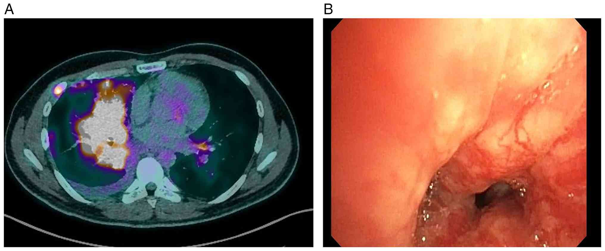
revealed two brain metastases of 4 and 5 mm. A positron emission
tomography (18FDG-PET scan) scan revealed hyperfixation
in the right upper lobe mass (Fig.
1A), and several nodules in the left lung, the hilar regions,
and the right lower and middle lobes, associated with a minor right
pleural effusion. Bilateral supraclavicular and mediastinal
lymphadenopathies were also observed, along with hyperfixation in
the left adrenal gland. Bronchoscopy identified a tumoral
infiltration in the right lower and middle lobes (Fig. 1B). Ten biopsies were collected for
anatomopathological examination. The pathologist diagnosed a
non-small cell lung adenocarcinoma. Immunohistochemical staining
showed ALK expression in 80% of the tumor cells, and Programmed
death-ligand 1 (PD-L1) expression in 60% of the tumor cells, ROS1
expression was negative (Fig.
2).
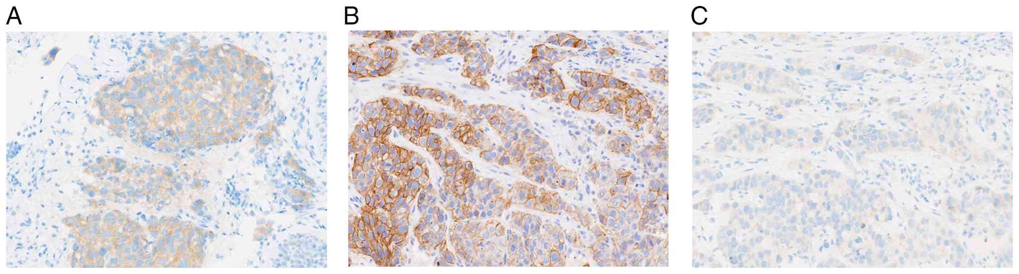 | Figure 2.ALK, PD-L1 and ROS1
immunohistochemistry images. (A) ALK expression was assessed by
immunohistochemistry. Moderate granular cytoplasm staining was
observed for ALK, with a score of 2+ (magnification, ×200). The
fixative used was 10% buffered formalin. Immunohistochemistry was
performed using the VENTANA Benchmark automated platform (Roche
Tissue Diagnostics). Anti-ALK antibody (clone 5A4; Abcam) was
diluted at 1:100. Antigen retrieval was performed using CC1 for 32
min. The duration of incubation with primary antibody was 1 h.
Detection was performed using OptiView DAB. (B) PD-L1 expression
was assessed by immunohistochemistry. Diffuse membrane staining was
observed for PD-L1 (tumor proportion score, 80%; magnification,
×200). The fixative used was 10% buffered formalin.
Immunohistochemistry was performed using an Agilent OMNIS automated
platform (Agilent Technologies, Inc.). PD-L1 antibody (prediluted;
clone 22C3; Agilent Technologies, Inc.) was used. Antigen retrieval
was performed under low pH conditions for 40 min. The duration of
incubation with primary antibody was 40 min. Detection was
performed using EnVision FLEX+Mouse DAB Enhancer. (C) ROS1
expression was examined using immunohistochemistry. Weak cytoplasm
and membrane staining was observed for ROS1, with a score of 1+
(magnification, ×200). The fixative used was 10% buffered formalin.
Immunohistochemistry was performed using the VENTANA Benchmark
automated platform. Anti-ROS1 antibody (SP384; ready to use) was
used. Antigen retrieval was performed using CC1 for 64 min. The
duration of incubation with primary antibody was 16 min. Detection
was performed using OptiView DAB. ALK, anaplastic lymphoma kinase;
CC1, Cell Conditioning 1; DAB, 3,3′-diaminobenzidine; PD-L1,
programmed death-ligand 1. |
Molecular diagnosis first included the research of
hotspots EGFR variants using Idylla® EGFR
mutation assay (Biocartis, Mechelen, Belgium) on formalin-fixed and
paraffin-embedded tumor sample. This PCR-based assay did not detect
any mutation in the EGFR gene. RNA-based sequencing (RNASeq)
was then performed but provided uninterpretable results due to the
low quality of RNA extracted from the sample. A liquid biopsy
sample was collected a few days later and targeted 51-gene
DNA-based sequencing (DNASeq, custom Solid Tumor Solution, Sophia
Genetics, Saint Sulpice, Switzerland) (9), it revealed a pathogenic c.524G>A
R175H TP53 gene mutation (NM_000546.6) with an 7.2% allele
frequency (AF) and copy number variations (CNV) in the KRAS
gene (CNV value of 8.5). To confirm the presence of ALK
rearrangement as suspected by immunohistochemistry, another
bronchoscopy was conducted, and eight novel biopsies were obtained.
Analysis of these biopsies by RNASeq revealed a rearrangement
between EML4 (exon 13) and ALK (exon 20) genes, ALK
(NM_0043304.5), fusion breakpoints: chr2:42522656; chr2:29446394,
AF: 97%).
Based on this, treatment with alectinib (600 mg
daily) was initiated, following national guidelines for ALK-NSCLC
as first line therapy at that time Based on these findings,
first-line treatment with alectinib (600 mg twice daily) was
initiated, in accordance with the French national guidelines (IFCT,
January 2023 version) (10) and the
European Society for Medical Oncology (ESMO) 2023 clinical practice
guidelines (4) for ALK-rearranged
NSCLC. At the 1-month clinical evaluation, an improvement in
dyspnea, cough, and general health status was observed. However,
the patient exhibited biological adverse events in the first four
months of treatment, including grade-2 hyperbilirubinemia, and
grade-1 creatine phosphokinase increase. After 3 months of
treatment, he had a complete resolution of dyspnea and cough but
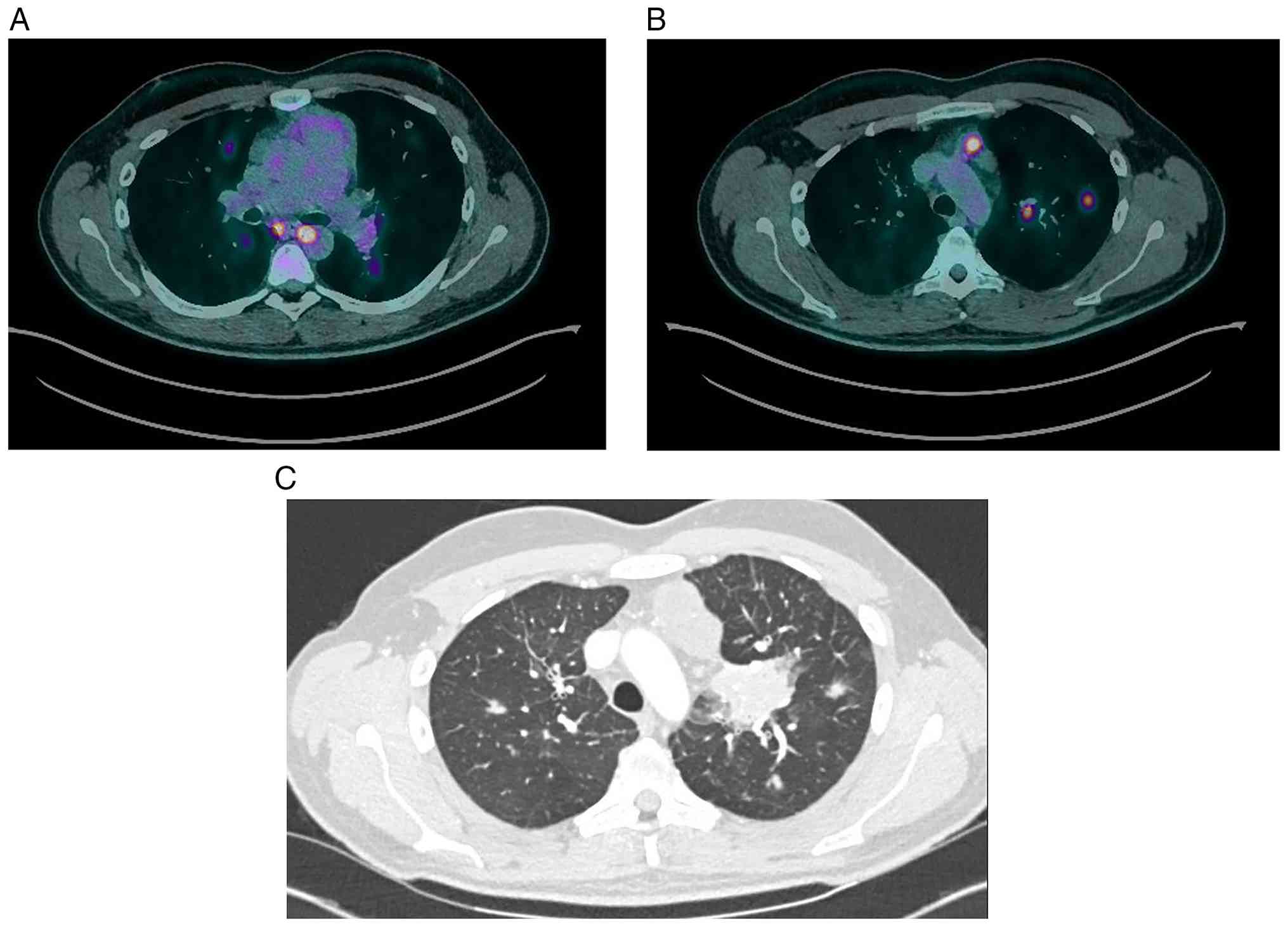
presented a grade-1 asthenia. A 18FDG-PET scan performed
3 months after treatment initiation revealed regression of all
pulmonary and adrenal lesions (Fig.
3A).
The 18FDG-PET scan, performed 7 months
after the initiation of alectinib, revealed disease progression,
with increase in size of known bilateral pulmonary nodules and
eight novel pulmonary lesions (Fig.
3B). The thoracic tumor board confirmed disease progression and
recommended switching from alectinib to lorlatinib. The analysis by
DNASeq of a second liquid biopsy collected at the time of treatment
switch detected the known R175H TP53 variant as well as six
novels pathogenic KRAS variants (L19F, G12V, G12D, G13C, and
G12C variants with AF ranging from 2 to 3% and Q61H variant at
23.3% AF). Copy number gain of the KRAS gene was also
detected (CNV value: 3). One month later, another liquid biopsy was
collected and analyzed by a new approach named Hedera Profiling 2
ctDNA panel (Hedera Dx, Epalinges, Switzerland) which allows the
detection of both single nucleotide variations, CNV, microsatellite
instability, and specific gene fusions in 32 genes of interest.
This technique confirmed the presence of the TP53 and
KRAS genes variants with no ALK rearrangement.
Following a 7-day washout period after the last dose
of alectinib, the patientinitiated treatment with second-line
lorlatinib (100 mg daily). This therapeutic strategy was
administered as standard-of-care therapy, based on the clinical
evidence established by the CROWN trial (7) and in accordance with the IFCT-2023
(10) and ESMO-2023 (4) guidelines for ALK-positive NSCLC. The
lorlatinib dosage was set at 100 mg daily. At this time, the
patient experienced symptoms of dyspnea, cough, and mild
hemoptysis. He presented under treatment an adverse event of
depressive mood, which resolved spontaneously after a few weeks.
The patient also presented hypercholesterolemia, treated with
rosuvastatin.
A follow-up CT scan was performed 3 months after
lorlatinib initiation, it detected multiple progression in the
lungs and adenopathies (Fig. 3C).
Therefore, a bronchoscopy was performed and culmen biopsies were
obtained for DNASeq and RNASeq analyses. We retrieved one known
KRAS mutation (G13C, 28.7% AF), one known TP53
mutation (R175H, 14% AF) and the EML4 (exon 13)::ALK
(exon 20) rearrangement (AF 91%) associated with KRAS copy
number gain (CNV value: 9.1). A phylogenetic tree resuming
genetic evolution can be found in Fig.
4. A switch to chemo-immunotherapy was then decided, before
resistance to targeted therapy and multiple resistance genetic
alterations, following national guidelines for first line
immunochemotherapy for NSCLC. The patient passed away in March 2025
due to disease progression.
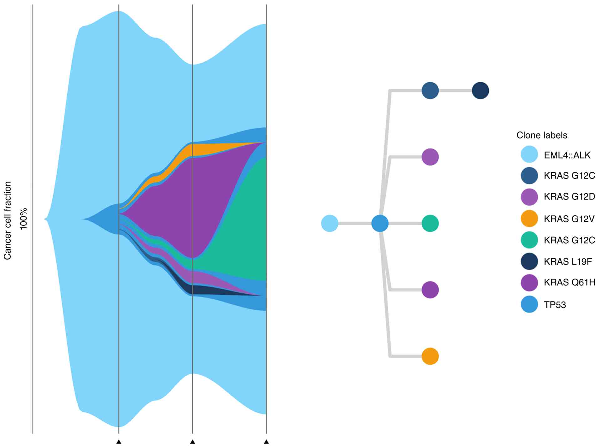 | Figure 4.Phylogenetic tree illustrating the
evolution of genetic mutations in the patient's cancer: Hypotheses
on divergent clonal lineages based on next-generation sequencing
results. The allele frequencies were: i) At diagnosis (second
vertical bar): TP53, 7.2%; EML4::ALK, 97.0%; ii) at first
progression (third vertical bar): KRAS L19F, 2.1%; KRAS G12C, 2.1%;
KRAS G12V, 2.8%; KRAS G12D, 2.8%; KRAS G13C, 2.1%; KRAS Q61H,
23.3%; TP53, 2.9%; and iii) at second progression (fourth vertical
bar): KRAS G13C, 28.7%; TP53, 14.0%; EML4::ALK, 91.0%. The figure
was generated using the ClevRvis package (v.1.8.0; http://github.com/sandmanns/clevRvis)
and R (v.4.3.1; R Core Team; http://www.r-project.org/). ctDNA, circulating tumour
DNA. |
Discussion
Our case depicts a real-life resistance to alectinib
used in the first-line setting for a patient with ALK+
NSCLC. It highlights the importance of reevaluating the tumor's
molecular characteristics at clinical progression as resistance
mutations can occur during treatment. Various mechanisms have been
explored to explain acquired resistance to ALK inhibition. Gainor
et al (11) studied
resistance mechanisms in 103 ALK+ NSCLC patients and found
that the most frequent resistance mechanism to second-generation
ALK inhibitors was the emergence of secondary ALK mutations,
especially the G1202R variant, they did not report any KRAS
mutations in their cohort. Lin et al (12) confirmed their findings in their
25-patient population. A review from Toyokawa and Seto evaluated
the resistance mechanisms to crizotinib, alectinib, and ceritinib
(13), showing that a mutation or a
copy number gain in the ALK gene, or the reactivation of
bypass tracks (including KRAS variants) could explain
resistance. They found three cases of patients presenting
resistance to crizotinib due to KRAS mutation, with one
presenting a primary resistance due to co-occurring ALK and
KRAS alterations at diagnosis.
The emergence of KRAS mutations as a
resistance mechanism is biologically plausible, as they activate
the MAPK/ERK signaling pathway downstream of the ALK receptor,
inducing cancer progression independently of the ALK pathway
(13). Other clinical cases
reported KRAS mutations as a resistance mechanism to
ALK inhibitors in ALK+ NSCLC, often showing initial
responses followed by rapid progression (14–21).
In these previous reports, patients were usually treated with
crizotinib, a first-generation ALK inhibitor, and presented
with a KRAS mutation at progression, or at diagnosis with
primary resistance. Progression usually occurred within 1 to 3
months, but in three cases out of 21, it happened after 6 months of
treatment. To our knowledge, we described here for the first time
the emergence of multiple KRAS mutations in a single
case.
Although specific mutations (G12C, Q22K, G12V, G13D,
A146V and Q61H) have been observed, this case is, to our knowledge,
the first to describe extensive KRAS mosaicism under
anti-ALK treatment. The technical validity of these findings is
supported by the high Variant Allele Frequency (VAF) of the Q61H
mutation (23.3%) detected at progression. This high VAF, combined
with the use of Unique Molecular Identifiers (UMIs) in our liquid
biopsy assays (limit of detection, LOD, 0.5%), allows us to exclude
laboratory artifacts [detailed technical specifications of the NGS
platforms and gene panels are provided in Data S1 and Table SI, Table SII, Table SIII, and detailed mapping of the
five primary datasets (SRX31979634 to SRX31979638) according to
sample type and collection date is provided in Table SIV] (9,22,23).
Regarding the timing of these alterations, our case suggests a dual
resistance process. While the KRAS amplification and
TP53 mutation present at diagnosis likely acted as primary
resistance factors contributing to a reduced initial sensitivity,
the KRAS mosaicism was not detectable at baseline even with
high-sensitivity NGS analysis (LOD 0.5%). This supports the
hypothesis that these specific KRAS variants were acquired
or selected under the selective pressure of alectinib, driving
progression as an acquired bypass mechanism.
Hua et al (24) described a potential resistance
mechanism to ALK inhibitor mediated by KRAS amplification in
one of their cases. In our case, the patient presented KRAS
amplification at diagnosis, which may have contributed to
resistance to both ALK inhibitors. An interesting piece of
information is the reevaluation of the EML4::ALK fusion,
which was not found at first progression. Gainor et al
(11) stated that ALK fusion
and KRAS mutation could not coexist; however, our case
report clearly demonstrates the contrary, joining other reports of
concomitant ALK rearrangements and KRAS mutations
(16–19,25,26).
This discrepancy could be explained by tumor heterogeneity, as
suggested by Tang et al (27). This evolution illustrates that the
molecular profile is not static; a selection process eliminates
cells with the targeted alteration and selects resistant clones. In
our case, the biopsy at second progression revealed KRAS
G13C, TP53 R175H, and EML4::ALK alterations. This
finding suggests that either this clone was present at first
progression but not detected, or the fusion remained below the
detection threshold while the mutations dominated.
The rapid progression observed under ALK inhibitors
in our case could also be partly explained by the pathogenic TP53
variant, a marker of poor prognosis associated with reduced
response rates to targeted therapies (28).
Finally, optimal therapeutic strategies for patients
harboring concomitant ALK and KRAS alterations remain
undefined. According to the ESMO Clinical Practice Guidelines
(4) and recent management updates
(5), the treatment of progression
on second-generation TKIs such as alectinib is guided by the nature
of the resistance mechanism. While third-generation TKIs such as
lorlatinib are highly effective against secondary mutations
occurring directly within the ALK kinase domain (on-target
resistance) (7), they are generally
ineffective when resistance arises from the activation of
alternative signaling pathways that bypass the ALK inhibition
(off-target resistance). In our case, the emergence of KRAS
variants represents a bypass track that allows the tumor to
maintain downstream MAPK/ERK signaling even if the ALK fusion
protein is successfully blocked by the TKI.
As highlighted by Toyokawa and Seto (13), the reactivation of downstream
signaling through KRAS variants bypasses the ALK inhibition,
rendering sequential TKI therapy potentially ineffective. In our
case, the extensive KRAS mosaicism poses a unique challenge
for specific targeted therapies. Consequently, for such
heterogeneous resistance, platinum-based chemotherapy represents a
more robust and reliable option to address the diverse tumor cell
populations (4,5). This underscores the necessity of
repeat molecular profiling to distinguish between
ALK-dependent resistance and complex bypass mechanisms like
the mosaicism described here, ensuring that patients are not
maintained on ineffective targeted therapies.
Supplementary Material
Supporting Data
Supporting Data
Acknowledgements
Not applicable.
Funding
Funding: No funding was received.
Availability of data and materials
The DNA and RNA sequencing data generated in the
present study may be found in the National Center for Biotechnology
Information BioProject database under accession number PRJNA1416035
or at the following URL:
https://www.ncbi.nlm.nih.gov/bioproject/?term=PRJNA1416035. The
other data generated in the present study may be requested from the
corresponding author.
Authors' contributions
AS made substantial contributions to the conception
of the study, analyzed the data, and wrote the manuscript. GT was
involved in the acquisition and interpretation of the clinical
data. PG, JLM and AH assisted in the study design and revised the
manuscript critically for important intellectual content. AS and AH
created the figures. GG and AL performed the pathological analysis
and provided the histopathological slides. GP, MH, IH, PG and AH
contributed to the genomic analysis and data interpretation. PG, AH
and JLM confirm the authenticity of all the raw data. All authors
have read and approved the final version of the manuscript.
Ethics approval and consent to
participate
Written informed consent for both participation and
the publication of the patient's individual data, including all
text and images, was obtained from the patient. This consent was
obtained in accordance with the principles of the Declaration of
Helsinki. Following institutional policies for retrospective case
reports, the study was deemed not to meet the definition of human
subjects research requiring formal review by the Institutional
Review Board of the Regional University Hospital Center of Nancy
(Nancy, France).
Patient consent for publication
The patient was informed about the use of their
biological, imaging and clinical data for research purposes, and
written informed consent was obtained.
Competing interests
The authors declare that they have no competing
interests.
Use of artificial intelligence tools
During the preparation of this work, artificial
intelligence tools (Gemini 3) were used to improve the readability
and language of the manuscript, and subsequently, the authors
revised and edited the content produced by the artificial
intelligence tools as necessary, taking full responsibility for the
ultimate content of the present manuscript.
Glossary
Abbreviations
Abbreviations:
|
NSCLC
|
non-small cell lung cancer
|
|
PFS
|
progression-free survival
|
|
CT
|
computed tomography
|
|
PET
|
positron emission tomography
|
|
RNASeq
|
RNA-sequencing
|
|
DNASeq
|
DNA-sequencing
|
|
CNV
|
copy number variation
|
References
|
1
|
Zhao F, Xu M, Lei H, Zhou Z, Wang L, Li P,
Zhao J and Hu P: Clinicopathological characteristics of patients
with non-small-cell lung cancer who harbor EML4-ALK fusion gene: A
meta-analysis. PLoS One. 10:e01173332015. View Article : Google Scholar : PubMed/NCBI
|
|
2
|
Ou SHI, Zhu VW and Nagasaka M: Catalog of
5′ fusion partners in ALK-positive NSCLC circa 2020. JTO Clin Res
Rep. 1:1000152020.PubMed/NCBI
|
|
3
|
Tao H, Shi L, Zhou A, Li H, Gai F, Huang
Z, Che N and Liu Z: Distribution of EML4-ALK fusion variants and
clinical outcomes in patients with resected non-small cell lung
cancer. Lung Cancer. 149:154–161. 2020. View Article : Google Scholar : PubMed/NCBI
|
|
4
|
Hendriks LE, Kerr KM, Menis J, Mok TS,
Nestle U, Passaro A, Peters S, Planchard D, Smit EF, Solomon BJ, et
al: Oncogene-addicted metastatic non-small-cell lung cancer: ESMO
clinical practice guideline for diagnosis, treatment and follow-up.
Ann Oncol. 34:339–357. 2023. View Article : Google Scholar : PubMed/NCBI
|
|
5
|
Jeon H, Wang S, Song J, Gill H and Cheng
H: Update 2025: Management of non-small-cell lung cancer. Lung.
203:532025. View Article : Google Scholar : PubMed/NCBI
|
|
6
|
Mok T, Camidge DR, Gadgeel SM, Rosell R,
Dziadziuszko R, Kim DW, Pérol M, Ou SI, Ahn JS, Shaw AT, et al:
Updated overall survival and final progression-free survival data
for patients with treatment-naive advanced ALK-positive
non-small-cell lung cancer in the ALEX study. Ann Oncol.
31:1056–1064. 2020. View Article : Google Scholar : PubMed/NCBI
|
|
7
|
Solomon BJ, Liu G, Felip E, Mok TSK, Soo
RA, Mazieres J, Shaw AT, de Marinis F, Goto Y, Wu YL, et al:
Lorlatinib versus crizotinib in patients with advanced ALK-positive
non-small cell lung cancer: 5-year outcomes from the phase III
CROWN study. J Clin Oncol. 42:3400–3409. 2024. View Article : Google Scholar : PubMed/NCBI
|
|
8
|
Canale M, Pasini L, Bronte G, Delmonte A,
Cravero P, Crinò L and Ulivi P: Role of liquid biopsy in
oncogene-addicted non-small cell lung cancer. Transl Lung Cancer
Res. 8 (Suppl 3):S265–S279. 2019. View Article : Google Scholar : PubMed/NCBI
|
|
9
|
Gilson P, Levy J, Rouyer M, Demange J,
Husson M, Bonnet C, Salleron J, Leroux A, Merlin JL and Harlé A:
Evaluation of 3 molecular-based assays for microsatellite
instability detection in formalin-fixed tissues of patients with
endometrial and colorectal cancers. Sci Rep. 10:163862020.
View Article : Google Scholar : PubMed/NCBI
|
|
10
|
Institut Francophone de Cancérologie
Thoracique (IFCT), . French national guidelines for molecular
testing and precision medicine in thoracic oncology. IFCT; Paris:
2023, https://www.ifct.fr/images/Indication_des_tests_mol%C3%A9culaires_traitements_de_pr%C3%A9cision_Poumon_1.pdfApril
2–2026
|
|
11
|
Gainor JF, Dardaei L, Yoda S, Friboulet L,
Leshchiner I, Katayama R, Dagogo-Jack I, Gadgeel S, Schultz K,
Singh M, et al: Molecular mechanisms of resistance to first- and
second-generation ALK inhibitors in ALK-rearranged lung cancer.
Cancer Discov. 6:1118–1133. 2016. View Article : Google Scholar : PubMed/NCBI
|
|
12
|
Lin YT, Yu CJ, Yang JCH and Shih JY:
Anaplastic lymphoma kinase (ALK) kinase domain mutation following
ALK inhibitor(s) failure in advanced ALK positive non-small-cell
lung cancer: Analysis and literature review. Clin Lung Cancer.
17:e77–e94. 2016. View Article : Google Scholar : PubMed/NCBI
|
|
13
|
Toyokawa G and Seto T: Updated evidence on
the mechanisms of resistance to ALK inhibitors and strategies to
overcome such resistance: Clinical and preclinical data. Oncol Res
Treat. 38:291–298. 2015. View Article : Google Scholar : PubMed/NCBI
|
|
14
|
Doebele RC, Pilling AB, Aisner DL,
Kutateladze TG, Le AT, Weickhardt AJ, Kondo KL, Linderman DJ,
Heasley LE, Franklin WA, et al: Mechanisms of resistance to
crizotinib in patients with ALK gene rearranged non-small cell lung
cancer. Clin Cancer Res. 18:1472–1482. 2012. View Article : Google Scholar : PubMed/NCBI
|
|
15
|
Rossing HH, Grauslund M, Urbanska EM,
Melchior LC, Rask CK, Costa JC, Skov BG, Sørensen JB and
Santoni-Rugiu E: Concomitant occurrence of EGFR (epidermal growth
factor receptor) and KRAS (V-Ki-ras2 Kirsten rat sarcoma viral
oncogene homolog) mutations in an ALK (anaplastic lymphoma
kinase)-positive lung adenocarcinoma patient with acquired
resistance to crizotinib: A case report. BMC Res Notes. 6:4892013.
View Article : Google Scholar : PubMed/NCBI
|
|
16
|
Campos-Gomez S, Lara-Guerra H, Routbort
MJ, Lu X and Simon GR: Lung adenocarcinoma with concurrent KRAS
mutation and ALK rearrangement responding to crizotinib: Case
report. Int J Biol Markers. 30:e254–e257. 2015. View Article : Google Scholar : PubMed/NCBI
|
|
17
|
Azzato EM, Deshpande C, Aikawa V, Aggarwal
C, Alley E, Jacobs B, Morrissette J and Daber R: Rare complex
mutational profile in an ALK inhibitor-resistant non-small cell
lung cancer. Anticancer Res. 35:3007–3012. 2015.PubMed/NCBI
|
|
18
|
Mengoli MC, Barbieri F, Bertolini F and
Tiseo M: K-RAS mutations indicating primary resistance to
crizotinib in ALK-rearranged adenocarcinomas of the lung: Report of
two cases and review of the literature. Lung Cancer. 93:55–58.
2016. View Article : Google Scholar : PubMed/NCBI
|
|
19
|
Zhu YC, Wan B, Wu LX, Li XL, Wang WX, Xu
CW, Zhuang W, Wei JG and Du KQ: Dual drive coexistence of ALK
rearrangement and KRAS mutation advanced lung adenocarcinoma and
response to crizotinib. Transl Cancer Res. 8:1630–1634. 2019.
View Article : Google Scholar : PubMed/NCBI
|
|
20
|
Kemper M, Evers G, Schulze AB, Sperveslage
J, Schülke C, Lenz G, Herold T, Hartmann W, Schildhaus HU and
Bleckmann A: Polyclonal on- and off-target resistance mutations in
an EML4-ALK positive non-small cell lung cancer patient under ALK
inhibition. Oncotarget. 12:1946–1952. 2021. View Article : Google Scholar : PubMed/NCBI
|
|
21
|
Huang N, Qu T, Zhang C and Li J: Case
report: Successful treatment of advanced pulmonary sarcomatoid
carcinoma with BUBIB-ALK rearrangement and KRAS G12C mutation by
sintilimab combined with anlotinib. Front Oncol. 14:12691482024.
View Article : Google Scholar : PubMed/NCBI
|
|
22
|
Lescuyer G, Harlé A, Kumar HS,
Constantoulakis P, Pfarr N, Heitzer E, Michon C, Russo G, Speel EM,
Piecyk M, et al: Analytical validation of a pan-cancer
next-generation sequencing assay for in-house liquid biopsy
testing: An international multicenter study. J Mol Diagn.
27:1174–1188. 2025. View Article : Google Scholar : PubMed/NCBI
|
|
23
|
Gilson P, Pouget C, Belmonte R, Fadil S,
Demange J, Rouyer M, Lacour J, Betz M, Dardare J, Witz A, et al:
Validation of the Idylla GeneFusion assay to detect fusions and MET
exon-skipping in non-small cell lung cancers. Sci Rep.
13:129092023. View Article : Google Scholar : PubMed/NCBI
|
|
24
|
Hua G, Zhang X, Zhang M, Wang Q, Chen X,
Yu R, Bao H, Liu J, Wu X, Shao Y, et al: Real-world circulating
tumor DNA analysis depicts resistance mechanism and clonal
evolution in ALK inhibitor-treated lung adenocarcinoma patients.
ESMO Open. 7:1003372022. View Article : Google Scholar : PubMed/NCBI
|
|
25
|
Ju L, Han M, Zhao C and Li X: EGFR, KRAS
and ROS1 variants coexist in a lung adenocarcinoma patient. Lung
Cancer. 95:94–97. 2016. View Article : Google Scholar : PubMed/NCBI
|
|
26
|
Bazhenova L, Hodgson JG, Camidge DR,
Langer CJ, Huber RM, Kim DW, Reckamp KL, Ahn MJ, Tan DSW, Patel JD,
et al: Activity of brigatinib in patients with crizotinib-resistant
ALK-positive non-small-cell lung cancer according to ALK fusion and
mutation status. Clin Lung Cancer. 26:e503–e513.e11. 2025.
View Article : Google Scholar : PubMed/NCBI
|
|
27
|
Tang Q, Li T, Ren F, Li X, Cao W, Yu H,
Mao F, Cao C, Zu L and Xu S: Response of a novel KANK1::ALK fusion
to alectinib in an advanced lung adenocarcinoma: A case report. J
Natl Compr Canc Netw. 22:e2371072024. View Article : Google Scholar : PubMed/NCBI
|
|
28
|
Jiang B, Hu L, Dong D, Guo Z, Wei W, Wang
C, Shao W, Ma T, Chen Y, Li Q and Hu W: TP53 or CDKN2A/B
covariation in ALK/RET/ROS1-rearranged NSCLC is associated with a
high TMB, tumor immunosuppressive microenvironment and poor
prognosis. J Cancer Res Clin Oncol. 149:10041–10052. 2023.
View Article : Google Scholar : PubMed/NCBI
|