Introduction
Breast cancer (BC) is the most prevalent malignant
neoplasm among women. The most recent data from 2025 indicates that
32% of new cancer cases diagnosed in women are attributable to BC
(1). BC can be classified based on
molecular subtypes, namely luminal A, luminal B, HER2+
and triple-negative BC (TNBC) (2).
In the context of BC, the TNBC molecular type, characterized by the
absence of the expression of estrogen receptor (ER), progesterone
receptor and HER2, has been identified as a high-risk category with
regard to the degrees of metastasis and recurrence (3). For early-stage BC, the primary
treatment modality is typically breast-conserving surgery combined
with radiation therapy (4). In the
cohort of patients diagnosed with hormone receptor-positive cancer,
endocrine therapy constitutes 80% of all treatment modalities.
Nevertheless, not all patients respond positively to this
therapeutic approach (1). The
heterogeneity of BC presents notable therapeutic challenges.
Nonetheless, progress in the fields of targeted therapy and
immunotherapy has generated a renewed optimism regarding the
management of the disease (5). The
dynamic interaction between tumor progression and immune
microenvironment modulation serves a key role in BC development and
therapeutic response (6).
Therefore, it is imperative to conduct in-depth research into the
mechanism of BC to discover novel biomarkers for providing novel
targeted treatment options.
DEAH-box helicase 15 (DHX15) is a prominent member
of the DEAD-box RNA-unwinding subfamily of the DEAD/H helicase
family (7). DHX15 is traditionally
recognized as an RNA unwinding enzyme that promotes mRNA maturation
and ribosome assembly (7). However,
studies have revealed multiple functions of DHX15 that were
previously unknown. DHX15 deficiency in intestinal epithelial cells
has been shown to increase tumorigenicity (8). In addition, DHX15 can fulfill a role
in innate immunity through its capacity to act as a nucleic acid
sensor. DHX15 has been reported to promote B cell survival and
proliferation. Conditional knockout of DHX15 in B cells leads to B
cell depletion and impaired humoral immune responses (9). DHX15 has also been identified as a
regulator of natural killer (NK) cell homeostasis and function
(10). Furthermore, DHX15 is
involved in tumorigenesis, acting as an oncogenic driver in acute
lymphoblastic leukemia, lung cancer, colorectal cancer and prostate
cancer. By contrast, it has been documented to act as a tumor
suppressor in glioma, hepatocellular carcinoma and gastric cancer
(11–14). Previous studies have indicated that
these helicases are essential for diverse cellular and
physiological processes, including cell proliferation,
hematopoiesis, inflammation, cancer pathogenesis, embryonic
development and the regulation of autoimmune diseases (11–14).
In addition, DEAD-Box helicase 5 and 17 play a role in
transcriptional coactivation by interacting with transcription
factors such as estrogen receptor α (ERα) and p53 in in
vitro cellular models, which is involved in BC development
(15). These molecules act as
primary regulators of the estrogen signaling pathway by controlling
transcription and splicing processes upstream and downstream of the
ER (16). Therefore, a possible
association exists between the therapeutic potential of targeting
DHX15 in BC and its underlying tumorigenic functions. Nevertheless,
the mechanisms of action, biological functions and regulatory
patterns of DHX15 expression in BC remain to be fully
elucidated.
The present study investigated the expression of
DHX15 in BC and its effect on tumorigenesis, progression,
development and prognosis. Moreover, the role of DHX15 in BC was
examined by constructing a co-expression gene protein-protein
interaction (PPI) network and performing pathway enrichment
analysis to predict the underlying molecular mechanisms.
Furthermore, the impact of DHX15 on tumor-infiltrating lymphocytes
was analyzed. Finally, by downregulating DHX15 expression in
MDA-MB-231 cells, knockdown efficiency was validated and the
biological functions of DHX15 were assessed. The purpose of the
present study is to reveal the potential diagnostic and prognostic
value of DHX15 in BC, while also investigating its relationship
with immune infiltration.
Materials and methods
Cell culture and reagents
Human BC MDA-MB-231 (cat. no. 240528I) and MCF-7
(cat. no. 20220817-01) cell lines were provided by Shanghai FuHeng
Biotechnology Co. Ltd. All cells used in the present experimental
procedure were cultured in DMEM (cat. no. KGL1206-500; Jiangsu
KeyGen Biotech Co., Ltd.) supplemented with 10% FBS (Invitrogen;
Thermo Fisher Scientific, Inc.) and 1% penicillin/streptomycin.
Cells were incubated at 37°C in a constant-temperature incubator
with 5% CO2.
Data download and analysis
The Cancer Genome Atlas (TCGA)-BRCA data, which
consisted entirely of data on invasive breast cancer, were obtained
from the publicly accessible Xena database (https://xenabrowser.net). Briefly, the cohort keyword
TCGA Breast Cancer was used for retrieval, and the HTSeq-FPKM
normalized RNA-seq expression dataset was selected. Tumor Immune
Estimation Resource (TIMER1.0) (cistrome.shinyapps.io/timer)
(17) was utilized for a
comprehensive investigation of the molecular characterization of
tumor-immune interactions. The ‘Diff Exp’ module was employed to
investigate the differential expression of DHX15 between tumor
tissue and adjacent normal tissue across all TCGA tumor samples;
the paired normal tissues for TCGA-BRCA used in TIMER 1.0 were
collected at least 2 cm from the tumor boundary, following the
standard TCGA sample acquisition criteria, and verified as
histologically normal tissue. For correlation analysis between
DHX15 expression and immune cell abundance, the ‘Gene’ module was
applied in the BRCA cohort, with purity adjustment and Spearman
correlation calculation for six immune subsets (B cells,
CD8+ T cells, CD4+ T cells, macrophages,
neutrophils and dendritic cells). The ‘Survival’ module was used to
explore the combined prognostic effect of DHX15 expression and
immune infiltration by Cox regression analysis within TCGA-BRCA
dataset. The University of Alabama at Birmingham Cancer (UALCAN)
database (18) was utilized to
validate the differential expression levels of DHX15 across
distinct BC subtypes and its association with signaling pathways
(http://ualcan.path.uab.edu). The Pathway
Enrichment module of UALCAN was utilized to retrieve the top
enriched biological signaling pathways significantly correlated
with DHX15 expression in BC samples. The built-in statistical
algorithm of UALCAN automatically calculated correlation
coefficients and P-values for pathway enrichment.
Stable transfection using lentiviral
infection
A 3rd generation lentiviral system was used to
establish stable knockdown or overexpression cell lines. According
to the manufacturer's instructions, 293T cells were used for
lentivirus production, purification and subsequent infection
(LentiPac™ HIV Expression Kit; GeneCopoeia, Inc). 293T cells were
co-transfected with 2.5 µg of lentiviral expression plasmid and 2.5
µg of Lenti-Pac mixed packaging plasmid (containing packaging and
envelope components) at 37°C with 5% CO2. At 48 h
post-transfection, viral supernatants were collected, centrifuged
(3,000 × g for 10 min at 4°C) and filtered. Target cells were
infected at a multiplicity of infection of 3. After 14 days of
selection with puromycin (4 µg/ml) (cat. no. HY-B1743;
MedChemExpress), stably transduced cells were maintained in medium
containing the same antibiotic concentrations and used for
subsequent experiments. The plasmids encoding DHX15 short hairpin
RNAs (cat. no. HSH152517-LVRU6GP), an sh-control (a non-targeting
sequence) (cat. no. CSHCTR001-LVRU6GP), an overexpression plasmid
(cat. no. EX-T8229-Lv122) and a control plasmid (cat. no.
EX-NEG-Lv122) were provided by GeneCopoeia, Inc. The sequence of
sh-DHX15 was as follows: Sense,
5′-CCGGGTGGAGTACATGCGATCATTACTCGAGTAATGATCGCATGTACTCCACTTTTTG-3′
and antisense,
5′-AATTCAAAAAGTGGAGTACATGCGATCATTACTCGAGTAATGATCGCATGTACTCCAC-3′.
The sequence of sh-control was as follows: Sense,
5′-CCGGCGGCATGGACGAGCTGTACAATTTTTG-3′; and antisense, 5′-
AATTCAAAAATTGTACAGCTCGTCCATGCCG-3′. Following transfection of 293T
cells with the plasmids, the virus-containing supernatant culture
was collected at 48 h post-transfection. At 48 h post-transfection,
viral supernatants were collected, centrifuged (3,000 × g for 10
min at 4°C) and filtered, and added to Polybrene. Polybrene can
neutralize the electrostatic repulsion between the negatively
charged lentiviral particles and cell membranes, notably improving
the efficiency of viral adsorption and infection into target BC
cells. To obtain stable control cell lines, infected cells were
subjected to puromycin (4 µg/ml) (cat. no. HY-B1743;
MedChemExpress) selection for ≥1 weeks. Stably transduced cells
were maintained in medium containing the same antibiotic
concentrations and used for subsequent experiments. Additionally,
cell lines infected with lentivirus vectors were established.
Stable cell lines were selected with puromycin (4 µg/ml) for 14
days before being used in subsequent experiments.
Western blotting analysis
Transfected cells were harvested and lysed using
RIPA buffer (cat. no. R0020; Beijing Solarbio Science &
Technology Co., Ltd.) supplemented with 1 mM PMSF (cat. no.
HY-B0496MedChemExpress), 10 mM DTT (cat. no.
HY-15917MedChemExpress), and 10 µM protein kinase inhibitor (cat.
no. P1006; Beyotime Biotechnology) on ice for 30 min. Equal amounts
of protein (20 µg) quantified by BCA (cat. no. PC0020; Beijing
Solarbio Science & Technology Co., Ltd.) were separated by
SDS-PAGE (4-20% gradient gel; cat. no. ET15420L; ACE Biotechnology)
and transferred to 0.2-µm pore size PVDF membranes (cat. no.
ISEQ00010; Merck Sharp & Dohme-Hoddesdon). Following the
blocking of non-specific binding sites using 5% skimmed milk (cat.
no. P0216; Beyotime Biotechnology) or bovine serum albumin (cat.
no. NGP0028A; Beyotime Biotechnology) for 1 h at room temperature,
the membranes were incubated with primary antibodies (incubation
overnight at 4°C) against DHX15 (cat. no. sc-271686; 1:1,000) and
GAPDH (cat. no. sc-47724; 1:1,000; Santa Cruz Biotechnology, Inc.),
and subsequently incubated with the corresponding secondary
antibodies (dilution 1:1,500) for 2 h at room temperature. The
secondary antibodies used were horseradish peroxidase
(HRP)-conjugated goat anti-rabbit (cat. no. ZB-2301; Beijing
Zhongshan Jinqiao Biotechnology Co., Ltd.) and HRP-conjugated goat
anti-mouse (cat. no. ZB-2305; Beijing Zhongshan Jinqiao
Biotechnology Co., Ltd.). Chemiluminescence was detected using
WesternBright ECL HRP substrate (cat. no. R-03031-D2; Advansta
Inc.). Bands were visualized using the C-DiGit Blot Scanner (LI-COR
Biosciences), and a densitometric analysis was performed using
ImageJ software (version: 1.52a) (National Institutes of Health).
Three independent replicates were performed for each experimental
condition.
Diagnostic and prognostic value of
DHX15
The Kaplan-Meier plotter (19) database (http://kmplot.com/analysis/) was utilized to explore
the association between DHX15 expression and clinical outcome
(overall survival), in addition to predicting chemotherapy
responses (20), in patients with
BC. ROC analysis to evaluate the predictive value of DHX15 for
chemotherapy pathological response was conducted via the ‘ROC
Plotter for Breast Cancer’ module of KM Plotter. The endpoint was
set as pathological response, the cohort was restricted to patients
with TNBC who received any chemotherapy, and the ROC metrics were
calculated using the platform's default parameters after entering
the gene symbol DHX15.
Co-expression and gene enrichment
analysis
The LinkedOmics database (http://www.linkedomics.org/login.php) is a web-based
platform designed for the analysis of TCGA cancer-associated
multi-dimensional data (21).
Statistical analysis of DHX15 co-expression was performed using
Pearson's correlation coefficient and visualized using a volcano
plot. The ‘LinkedOmics’ functional module was used to analyze Gene
Ontology (GO) biological processes and Kyoto Encyclopedia of Genes
and Genomes (KEGG) pathway enrichment, through gene set enrichment
analysis (GSEA). GSEA-based GO and KEGG enrichment analyses were
conducted using the LinkedOmics database. GO and KEGG predefined
gene sets were tested against the full ranked gene list correlated
with DHX15. Terms with false discovery rate (FDR)<0.05 (500
permutations) were considered significantly enriched, with a
minimum gene set cutoff of 2 members. GO enrichment analysis was
performed, as well as PPI network analysis of DHX15-associated
genes using Metascape (http://metascape.org/gp/index.html) (22).
Immune infiltration analysis
The proportion of tumor-infiltrating immune cells in
TCGA-BC samples was calculated using the CIBERSORT algorithm. The
correlation between the infiltration levels of 28
tumor-infiltrating immune cell types and DHX15 expression was
calculated using single-sample GSEA (ssGSEA). Single-sample gene
set enrichment analysis (ssGSEA) and CIBERSORT immune cell
infiltration profiling were performed using the Xiantao Academic
web tool (https://www.xiantao.love). Standard
built-in reference signatures and default algorithm parameters were
applied throughout the analysis. The CIBERSORT algorithm was
invoked via the dedicated immune infiltration module of Xiantao
Academic. The relative proportions of all immune cell populations
within the BC samples were calculated using the platform's default
parameters. For ssGSEA, the GSVA package with a KS-like
non-parametric test and MSigDB immune signatures were utilized, and
Wilcoxon rank-sum tests were used for subgroup comparisons after
BH-FDR adjustment. In addition, the TIMER database was utilized to
construct a multi-factor Cox proportional hazards model to
investigate the effect of immune cell infiltration on the survival
rate of patients with BC. Finally, correlations between DHX15
expression and specific invasive immune markers were estimated
using the TIMER1.0 database ‘Correlation’ module.
Single-cell level analysis
The Human Proteome Atlas (HPA) database (https://www.proteinatlas.org) can be used to explore
the expression of DHX15 in different tumor pathology samples and
across various single-cell types (23). Using the CancerSEA database
(http://biocc.hrbmu.edu.cn/CancerSEA/), the biological
roles of DHX15 in BC were investigated at the single-cell level
(24). DHX15 was queried in the BC
cohort, and Pearson correlation coefficients were calculated to
evaluate its association with 14 classic malignant functional
signatures using the platform's default built-in algorithm.
Immunohistochemistry (IHC) images from
the HPA database
IHC staining images of DHX15 protein expression in
normal and BC tissues were obtained from the HPA database
(https://www.proteinatlas.org/) (23). The staining patterns and expression
levels were evaluated based on the annotation provided by the HPA
consortium. DHX15 protein expression levels from HPA IHC images
were semi-quantitatively scored using the immunoreactive score
(IRS) system, calculated as the product of staining intensity and
the percentage of positively stained tumor cells. Samples were
stratified into high and low expression groups based on the final
IRS values. Staining intensity was scored from 0 to 3, multiplied
by the percentage of positive stained cells (scored from 0-3),
generating a total score ranging from 0 to 9. These H-scores were
dichotomized into high and low expression groups using the median
H-score of all BC samples as the cutoff value.
Wound-healing assays
All cell groups, including sh-control, sh-DHX15,
MCF-7 and EX-DHX15, were seeded into 6-well plates. To eliminate
the interference of cell proliferation on migration results, cells
were subjected to serum starvation treatment for 12 h before
scratching. Once the cells reached full confluence (~100%), a wound
was created using a 100-µl sterile pipette tip, before the samples
were imaged (0 h). After creating the artificial wound, the culture
medium was replaced with serum-free basal DMEM without fetal bovine
serum, and cells were incubated under standard cell culture
conditions for the subsequent observation period. The gap closure
rate was measured at 24, 48 and 96 h. In total, three independent
replicates were performed for each experiment. Cells were
continuously incubated at 37°C in a humidified incubator containing
5% CO2 during the entire wound healing observation
period. Images were captured using a light microscope (80i; Nikon
Corporation). Migration rate (%)=Width at time t/initial width
×100.
Cell invasion and migration
experiments
The migration assay was performed in a 24-well
plate. DHX15-transfected MDA-MB-231 cells and MCF-7 cells were
placed in the upper chamber and suspended in serum-free medium,
whereas the lower chamber was filled with DMEM supplemented with
10% FBS. For the Transwell migration assays, 8-µm Transwell
chambers (cat. no. 725321; Wuhan NEST Biotechnology Co., Ltd.) were
used. Serum-starved cells (1×10⁴ cells/well) were plated
in the upper chambers with 100 µl FBS-free medium, and 400 µl 10%
FBS medium was added to the lower chambers. The cells that migrated
through the membrane were counted and images captured after 24 h.
Following an overnight incubation period at 37°C with 5%
CO2 in a humidified incubator, the cells were fixed with
100% methanol for 30 min at room temperature, and stained with 0.1%
crystal violet for a duration of 20 min at room temperature.
Invasion assays were performed following the same protocol as the
migration assays; however, before cell seeding, the Transwell
chambers were coated with Matrigel. For the Transwell invasion
assays, 4×10⁴ cells in 100 µl FBS-free medium were
plated in the top chamber precoated with Matrigel (cat. no.
0827045; Xiamen Mogengel Biotechnology Co., Ltd.). Matrigel was
kept chilled on ice throughout all preparation steps. First, frozen
Matrigel was thawed overnight at 4°C in a refrigerator, then the
Matrigel was diluted with chilled serum-free medium at a ratio of
1:2 (Matrigel:medium). Finally, the plates were placed in a
humidified incubator at 37°C for 60 min to allow the Matrigel to
form a gel. Cell counting was performed using an inverted light
microscope (Nikon Corporation). Each experiment was performed in
triplicate.
Plate colony formation assays
A colony formation assay was performed to evaluate
clonogenic survival by seeding single-cell suspensions at
1×10³ cells/well in 6-well plates. This experiment was
conducted in the DHX15-downregulated group and the control group of
MDA-MB-231 cells. After 14 days, the colonies were fixed with 100%
methanol for 10 min at room temperature. Following fixation,
colonies were stained with 0.1% crystal violet solution for 20 min
at room temperature. Colonies were defined as clusters containing
≥50 cells or visible colonies measuring >0.1 mm in diameter.
Colonies were counted manually and imaged under an inverted
microscope (Nikon Corporation).
Data analysis
The statistical analysis was conducted using
GraphPad Prism 9.1.0 (Dotmatics) and R software (version 4.1.2;
Posit Software, PBC). Data are presented as the mean ± SD.
Comparisons between two groups were performed using unpaired
Student's t-tests (for independent samples), whereas multi-group
comparisons were performed using one-way ANOVA, and Tukey's test
was used for pairwise comparisons between groups. Survival
probabilities were calculated using Kaplan-Meier analysis and
differences in survival curves were assessed using the log-rank
test. Gene expression correlations were quantified using Spearman's
rank coefficients. All experiments were performed in triplicate.
P<0.05 was considered to indicate a statistically significant
difference.
Results
Upregulation of DHX15 mRNA expression
in BC
The expression levels of DHX15 mRNA were first
estimated across multiple malignant tumor types using the TIMER
database. Results indicated that DHX15 mRNA expression was
upregulated in BC, cholangiocarcinoma, colon adenocarcinoma,
esophageal cancer, head-neck squamous cell carcinoma, liver
hepatocellular carcinoma, lung adenocarcinoma, lung squamous cell
carcinoma, prostate adenocarcinoma and stomach adenocarcinoma but
was downregulated in kidney renal clear cell carcinoma, kidney
renal papillary cell carcinoma and thyroid carcinoma (Fig. 1A). The Kaplan-Meier database results
indicated that DHX15 mRNA expression levels were significantly
higher in the BC group compared with those in the normal group
(Fig. 1B). Receiver operating curve
(ROC) analysis revealed that DHX15 exhibited an area under the
curve (AUC) value of 0.638 (P<0.05) in BC. The AUC of 0.638 was
>0.5, demonstrating the preliminary diagnostic potential of
DHX15 for breast cancer identification. (Fig. 1C). Patients were divided into high
and low DHX15 expression groups according to the median expression
level (50% percentile) for Kaplan-Meier survival analysis.
Kaplan-Meier analysis demonstrated that patients with high DHX15
mRNA expression in the TCGA-BRCA dataset had a worse prognosis
[hazard ratio (HR)=1.55; P<0.01; Fig. 1D]. The Kaplan-Meier database was
further employed to estimate the association between DHX15
expression and patient prognosis with BC and TNBC. The present
study revealed that patients with high DHX15 expression had a worse
prognosis in BC (HR=1.22; P<0.001; Fig. 1E) and in TNBC (HR=1.5; P<0.001;
Fig. 1F). ROC curves were next
generated to validate the ability of DHX15 ability to predict
chemotherapy response. Results indicated that DHX15 expression was
higher in non-responders compared with that in responders amongst
patients with TNBC (Fig. 1G)
(P<0.05). Fig. 1H shows that the
AUC was 0.598 (P<0.05), indicating a modest predictive trend of
DHX15 for chemotherapy response in patients with TNBC.
Consequently, these findings suggest that DHX15 may influence the
prognosis of BC and could be used to develop novel biomarkers for
BC.
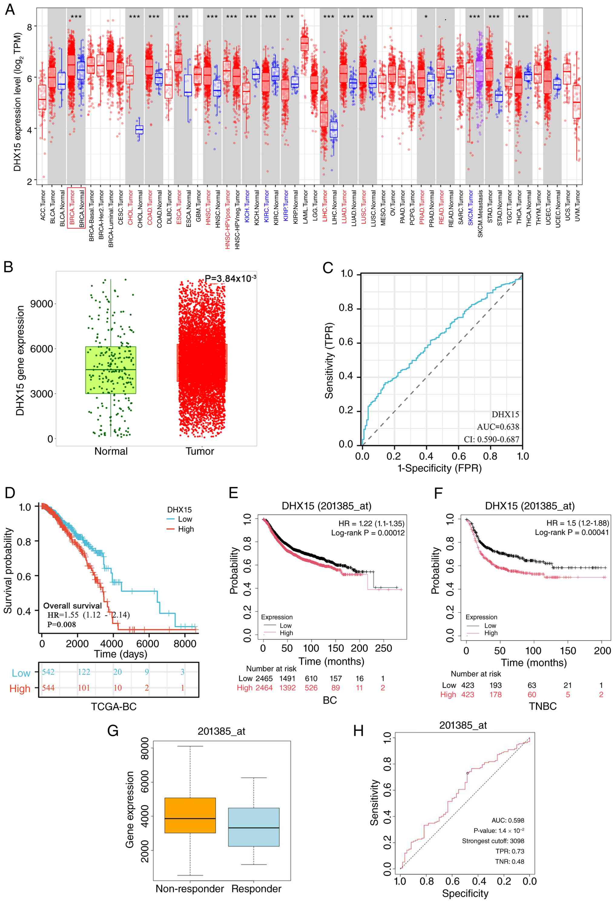 | Figure 1.Upregulation of DHX15 mRNA expression
in BC. (A) The expression level of DHX15 in different types of
tumor tissues and normal tissues in the Tumor Immune Estimation
Resource database. (B) Analysis using the Kaplan-Meier database
indicating upregulated DHX15 mRNA in BC (P=0.00384). (C) ROC curve
analysis of DHX15 for distinguishing patients with BC from normal
(healthy cancer-free) donors (AUC=0.638; CI: 0.590-0.687). (D)
Survival curves stratified by DHX15 expression levels in patients
from TCGA-BRCA database (P=0.008; n=1,086). Patients were divided
into high-expression and low-expression groups based on the median
expression value (50%) of DHX15. The Kaplan-Meier database displays
survival curves for the differentially expressed DHX15 gene in (E)
patients with BC (P=0.00012; n=4,924) and (F) patients with TNBC
(P=0.00041; n=846). (G) DHX15 expression levels in
chemotherapy-responsive and non-responsive TNBC cases. (H) ROC
curves validating the predictive value of DHX15 for chemotherapy
response in patients with TNBC (AUC=0.598; P=0.014). *P<0.05,
**P<0.01 and ***P<0.001. BC, breast cancer; DHX15, DEAH-box
helicase 15; TPR, true positive rate; TPM, transcript per million;
FPR, false positive rate; HR, hazard ratio; TNBC, triple-negative
BC; ROC, receiver operating characteristic; AUC, area under the
curve. |
DHX15 expression level is associated
with different clinical-pathological variables and pathways among
patients with BC
According to the UALCAN database, among all BC
subtypes, DHX15 expression was significantly higher in the luminal,
HER2+ and TNBC types compared with that in normal
tissues (P<0.001; Fig. 2B).
Expression levels were significantly higher in tumor stages II and
III compared with those in the normal group (P<0.001), with
those in stage II being higher compared with those in stage III
(P<0.05; Fig. 2A). Pathway
analysis revealed higher expression levels of DHX15 in the p53/Rb,
Hippo, WNT, Myc/Mycn, chromatin modifier and mTOR pathways compared
with those in the normal group. Pathway activity changes were
evaluated by Z-values, calculated as standardized differences
between BC samples and normal breast tissues. Positive Z-values
indicated elevated activation of all six pathways in BC compared
with normal controls. (P<0.001; Fig.
2C-H). The DHX15 expression levels of N0 (no lymph node
metastasis), N1 and N2 were significantly higher compared with
those of normal tissues (cancer-free normal breast tissues), as
indicated by the presence of lymph node metastasis (P<0.001;
Fig. 2I). The prognostic relevance
of DHX15 in patients with BC was also explored. The results
suggested that amongst patients in stage T2, T3 and T4, as well as
those in stages II and III, shorter survival times were associated
with higher levels of DHX15 expression (both P<0.01; Fig. 2L and N). Additionally, Fig. 2J, K and M displayed the IHC staining
results of DHX15 in the high-expression group and low-expression
group (P<0.001). HPA IHC images and semi-quantitative scoring
validated the upregulation of DHX15 at the protein level,
consistent with the mRNA-level bioinformatics results.
Representative high/low staining pictures directly visualized DHX15
expression heterogeneity in clinical BC tissues, and the
statistical comparison further supported that DHX15 upregulation is
a common feature in BC, preliminarily supporting its potential
value as a pathological biomarker.
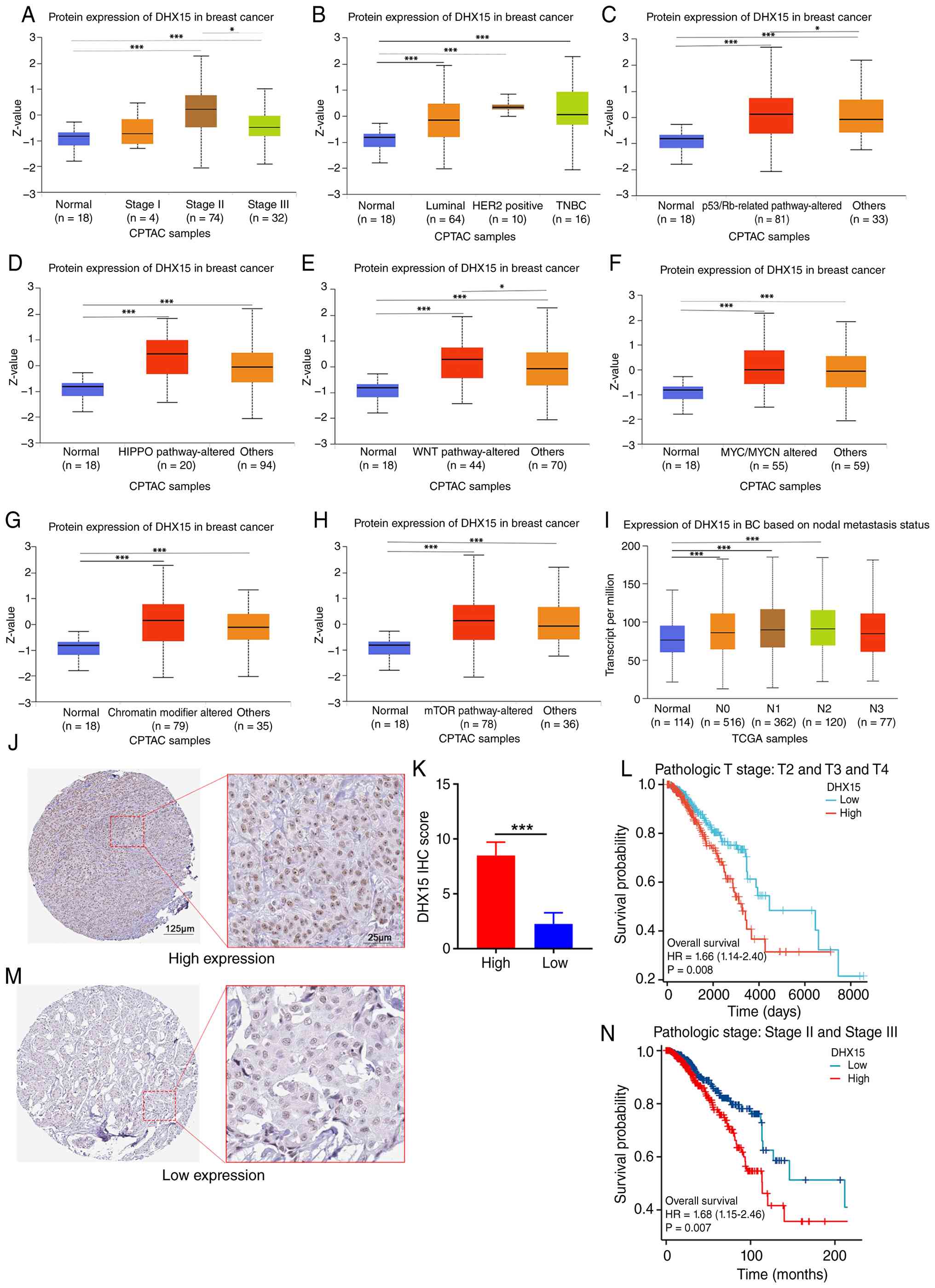 | Figure 2.DHX15 expression levels are
associated with different clinicopathological variables and
pathways among patients with BC. In the University of Alabama at
Birmingham Cancer database, an association was observed between
DHX15 protein expression and (A) stage, (B) subtypes, (C) p53/Rb,
(D) Hippo, (E) WNT, (F) Myc/Mycn, (G) chromatin modifier and (H)
mTOR pathways. (I) Association between DHX15 mRNA expression and
lymph node metastasis status. (L) Patients with T2, T3 and T4
stages (P=0.008) and (N) patients with stage II and III disease
(P=0.007) Representative IHC images of DHX15 protein expression in
(J) high and (M) low expression BC tissues, with (K) unpaired
Student's t-tests (n=12; P<0.001). Images (J) and (M) were
obtained from the Human Protein Atlas database (https://www.proteinatlas.org/). (J and M)
Association of DHX15 expression with the prognosis of patients with
BC at different clinical stages. *P<0.05 and ***P<0.001. BC,
breast cancer; CPTAC, clinical proteomic tumor analysis consortium;
DHX15, DEAH-box helicase 15; TNBC, triple-negative BC; TCGA, The
Cancer Genome Atlas; IHC, immunohistochemistry. |
DHX15 co-expression gene and pathway
enrichment in BC
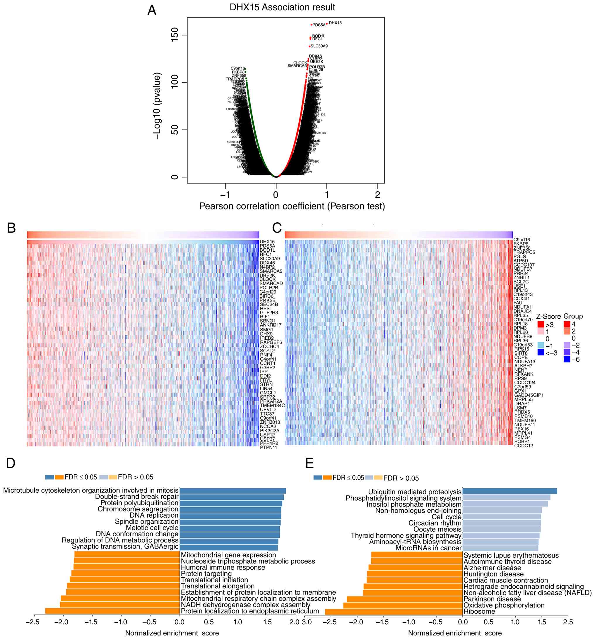
Genes co-expressed with DHX15 were screened and
their expression patterns were visualized via a volcano plot in BC,
thereby advancing understanding of the DHX15 mechanism of action
(Fig. 3A). The 50 most significant
genes positively or negatively correlated with DHX15 are also
displayed (Fig. 3B and C).
Subsequently, pathway enrichment analysis was employed to further
investigate the role of co-expressed genes with DHX15 in BRCA. GO
enrichment analysis revealed that genes co-expressed with DHX15
were primarily enriched in ‘microtubule cytoskeleton organization
involved in mitosis’, ‘double-strand break repair’ and ‘protein
polyubiquitination’ (Fig. 3D). KEGG
enrichment analysis indicated that these genes were primarily
enriched in the ‘ubiquitin-mediated proteolysis’ pathway (Fig. 3E).
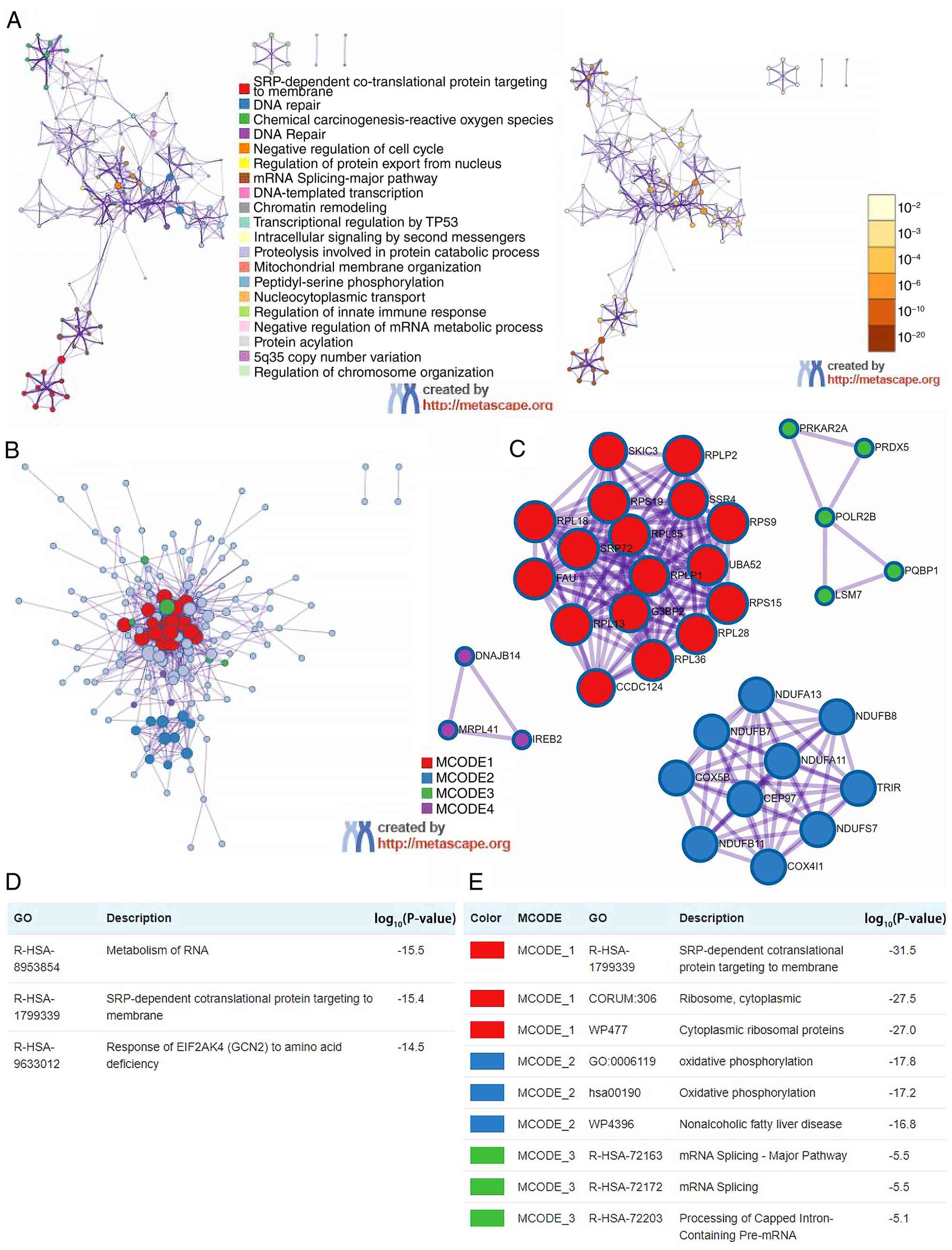
Metascape analysis indicated that the top 50
positive and top 50 negative genes with DHX15 were mainly clustered
in the ‘signal recognition particle (SRP)-dependent
co-translational protein targeting to the rough endoplasmic
reticulum’, ‘DNA repair’ and ‘chemical carcinogenesis-reactive
oxygen species’ (Fig. 4A). The PPI
network of the first 100 genes co-expressed with DHX15 were
primarily enriched in RNA metabolism, ‘SRP-dependent
co-translational protein targeting to the membrane’ and the
‘response of EIF2AK4 (GCN2) to amino acid deficiency’ (Fig. 4B and D). Furthermore, MCODE
profiling indicated that DHX15 and its neighboring genes may
influence ‘SRP-dependent co-translational protein targeting to
membrane’, ‘ribosome, cytoplasmic’ and ‘cytoplasmic ribosomal
proteins’ (Fig. 4C and E). In
summary, these findings indicate that genes co-expressed with DHX15
are primarily involved in translational protein localization and
DNA repair processes within BC. Based on these findings, it is
hypothesized that DHX15 participates in regulating cellular
transcription processes.
Analysis of DHX15 at the single cell
level in BC
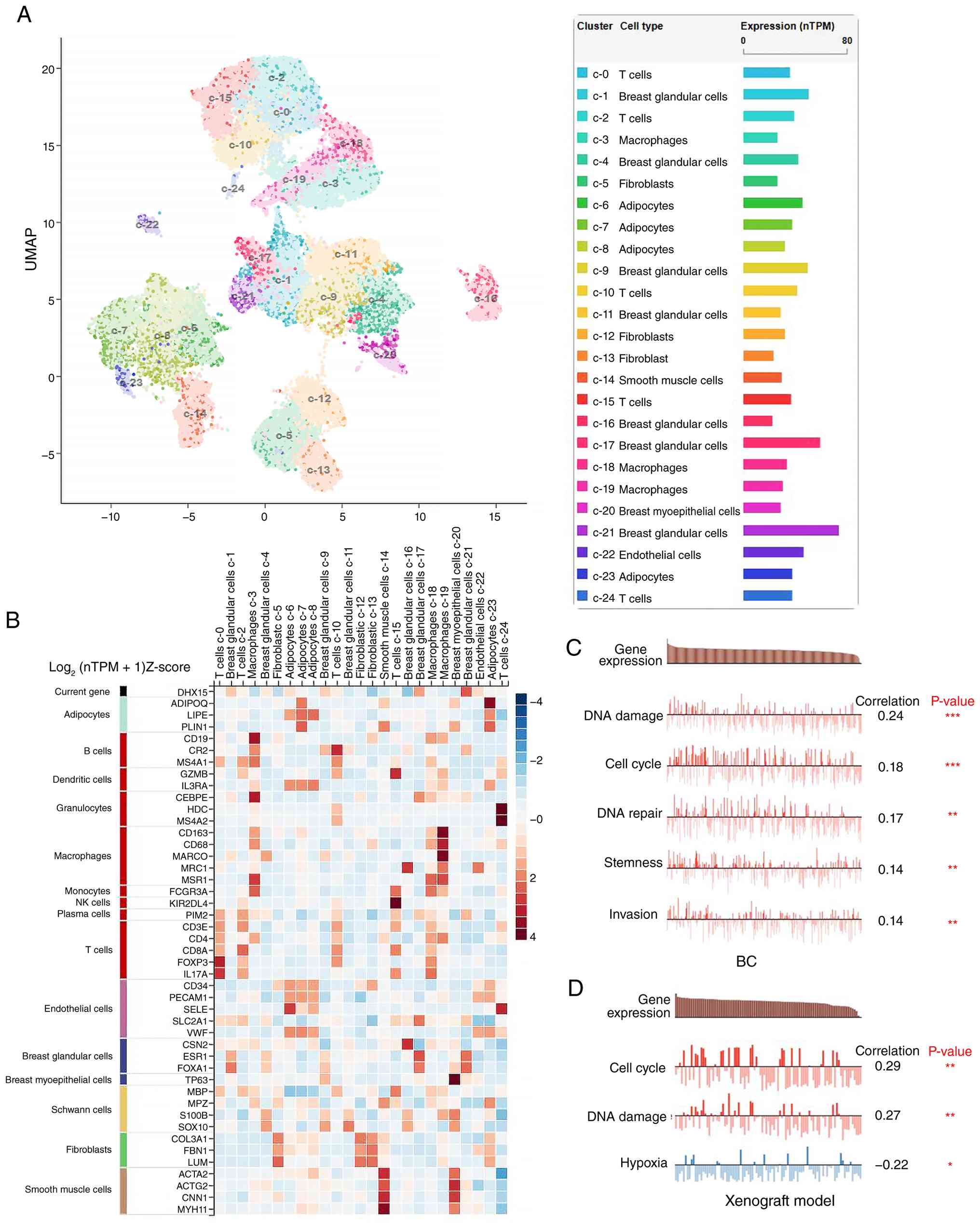
The HPA database revealed the three most prominent
single cells exhibiting DHX15 expression in BC are breast glandular
cells, T cells and macrophages. While DHX15 expression is moderate
in endothelial cells and adipocytes, these cell types account for a
relatively small proportion of the total. DHX15 is predominantly
expressed in breast glandular cells, with a mean of 73.2
standardized transcripts per million protein-coding genes across
all cell lines (Fig. 5A).
Furthermore, the differential expression levels of DHX15
facilitated the clustering of cells into distinct populations.
DHX15 is mainly expressed in the c-21 cell cluster (breast
glandular cells). Fig. 5B presents
the varying expressions levels of DHX15 and established cell type
markers across the distinct BC cell clusters. In breast glandular
cells, clusters demonstrated higher expression of ESR1 and forkhead
box A1 (FOXA1), which is positively correlated with DHX15 (Fig. 5B). Subsequently, the CancerSEA
database was utilized to analyze the association between DHX15
expression levels and individual BC single-cell biological
functions. The results indicated that DHX15 expression was
positively associated with DNA damage, the cell cycle, DNA repair,
stemness and invasion (Fig. 5C).
Xenograft models from the database were used to further validate
these biological functions. Results indicated that DHX15 expression
was positively associated with the cell cycle and DNA damage, but
negatively associated with hypoxia (Fig. 5D). These findings further reveal the
potential biological functions of DHX15 in BRCA, particularly at
the single-cell level.
Correlation between DHX15 and immune
infiltration in BC through bioinformatics analysis
The present study investigated the correlation
between DHX15 expression and immune cell infiltration in patients
with BC. ssGSEA analysis and CIBERSORT analysis were conducted to
examine this correlation. The ssGSEA results indicated a
significant positive association between DHX15 expression and T
helper cells (R=0.413; P<0.001), and central memory T cell (Tcm)
(R=0.34; P<0.001), and DHX15 expression had a weak positive
association with T helper 2 (Th2) cell infiltration (R=0.263;
P<0.001). A negative significant correlations were found with
plasmacytoid dendritic cells(pDC) (R=−0.408; P<0.001), and a
weak negative association with cytotoxic cells (R=−0.266;
P<0.001), DCs (R=−0.234; P<0.001), NK cells (R=−0.206;
P<0.001), CD8+ T cell infiltration (R=−0.196;
P<0.001) and B cells (R=−0.122; P<0.001; Fig. 6A). The CIBERSORT results indicated a
weak positive association between DHX15 expression and resting
CD4+ memory T cells (R=0.217; P<0.001) and M2
macrophages (R=0.152; P<0.001), with negative weak associations
with regulatory T cells [(Tregs); R=−0.295; P<0.001],
CD8+ T cells (R=−0.163; P<0.001), activated NK cells
(R=−0.134; P<0.001) and memory B cells (R=−0.119; P<0.001;
Fig. 6B). Consistent positive
correlations were observed in two independent algorithms: ssGSEA
revealed a positive association between DHX15 and TCM signatures,
while CIBERSORT further confirmed this trend in resting CD4 memory
T cells. Univariate Cox regression analysis was initially performed
for all candidate variables. DHX15 expression showed significant
prognostic value (P<0.05); Stage III, Stage IV and age also
showed statistical significance. Several immune cell variables
exhibited P>0.05 in univariate analysis, but they were still
incorporated into the multivariate model due to their
well-established clinical prognostic relevance in breast cancer
(Fig. 6C). In addition, the Cox
proportional hazard model was applied for DHX15 expression, six
tumor-infiltrating immune cell types, stage II–IV and age in BC. As
presented in Fig. 6D, stage II
(P<0.05), stage III (P<0.001), stage IV (P<0.001), age
(P<0.001) and DHX15 (P<0.05) were revealed to be
significantly associated with overall survival in patients with BC.
Furthermore, the TIMER1.0 database was utilized to conduct further
survival analysis of DHX15 in the context of immune cell
infiltration. The results indicated that based on the DHX15 high-
and low-expression groups, the infiltration of CD8+ T
cells (P<0.05), CD4+ T cells (P<0.05) neutrophil
cells (P<0.05) and dendritic cells (P<0.01) was significantly
correlated with the prognosis of BC. (Fig. 6E).
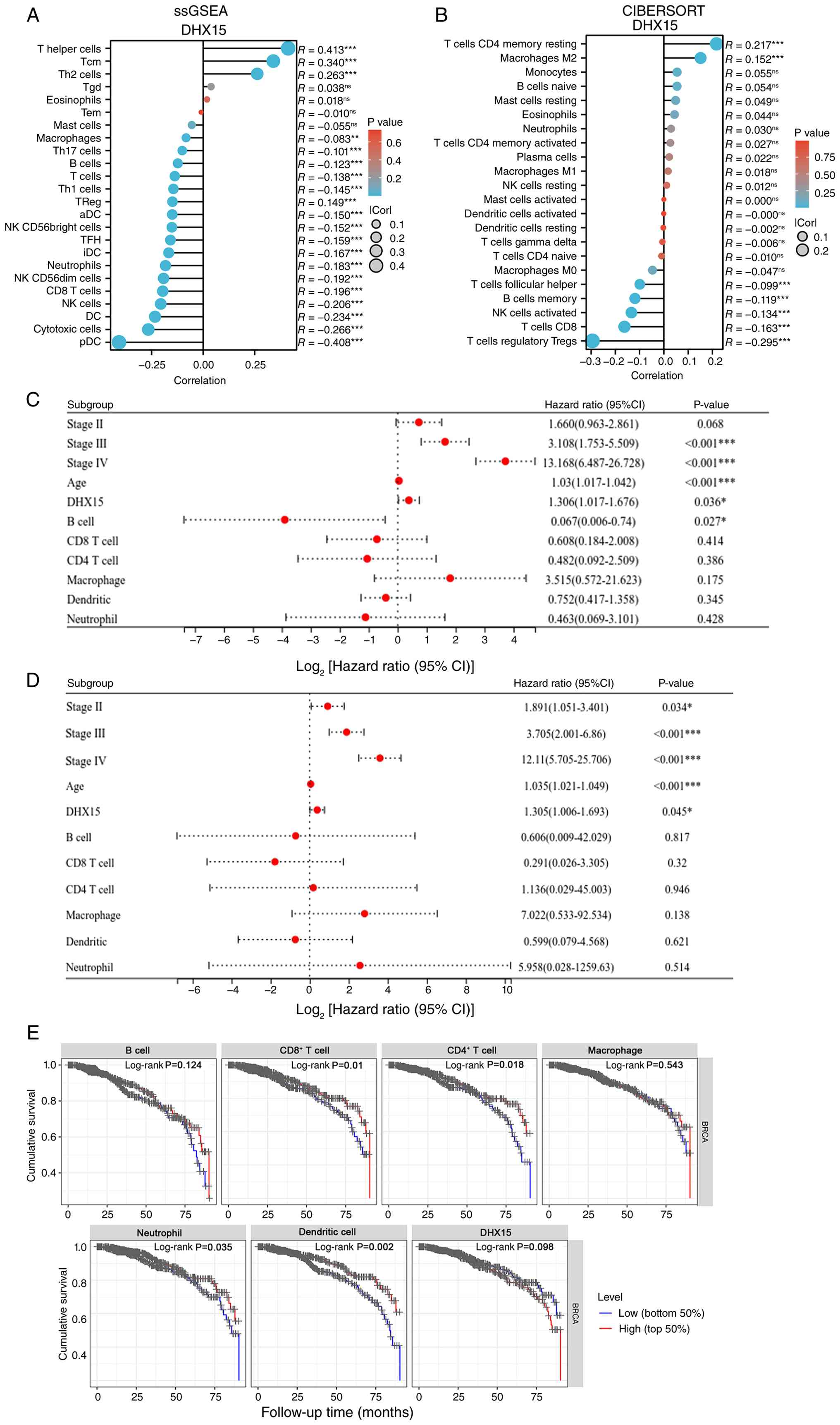 | Figure 6.Correlation between DHX15 and immune
infiltration in BC was analyzed using bioinformatics analysis. (A)
ssGSEA analysis demonstrates a comparison of infiltration and DHX15
expression across distinct immune cell types (cut-off value is the
median). (B) CIBERSORT analysis shows immune cell types
infiltration and DHX15 expression (cut-off value is the median).
(C) Univariate Cox screening of all candidate prognostic factors.
(D) Multivariable Cox proportional hazards models were used to
evaluate the impact of DHX15 expression on survival, adjusted for
the infiltration levels of multiple immune cells. DHX15 expression
was stratified by the median cutoff value, with low expression as
the reference. Age and immune cell infiltration levels were
analyzed as continuous covariates. Stages II, III, and IV were each
compared separately to Stage I. (E) Kaplan-Meier plots illustrating
the association between DHX15 expression and immune infiltration
status in BC. Each survival curve corresponds to a combined
subgroup defined by both DHX15 expression level and immune cell
infiltration status. *P<0.05, **P<0.01 and ***P<0.001. BC,
breast cancer; DHX15, DEAH-box helicase 15; ssGSEA, single-sample
gene set enrichment analysis; NK, natural killer; Treg, regulatory
T cell; Tem, Tcm, Tgd, TFH, aDC, iDC, pDC; ns, not significant;
Cor, correlation. Tem, effector memory T cell; Tcm, Ccentral memory
T cell; Tgd, γδ T cell; TFH, follicular helper T cell; aDC,
activated dendritic cell; iDC, immature dendritic cell; pDC,
plasmacytoid dendritic cell. |
Spearman's correlation analysis was employed to
evaluate the association between DHX15 expression and immune cell
markers in TCGA data, adjusting for tumor purity. As presented in
Table I, a marked positive
correlation was observed between DHX15 expression and monocyte
(CD86 and colony-stimulating factor 1 receptor), tumor associated
macrophage (CD68 and IL10), M1 macrophage (nitric oxide synthase 2
and cyclooxygenase-2), M2 macrophage (CD163, V-set and
immunoglobulin domain-containing 4 and membrane-spanning 4-domains
A4A), Th1 (STAT4, STAT1 and TNF), Th2 (GATA-binding protein 3,
STAT6 and STAT5A), Th17 (STAT3), T follicular helper cell (BCL6 and
IL21) and Treg (FOXP3, C-C motif chemokine receptor 8 and STAT5B)
markers. In addition, DHX15 was negatively correlated with
CD8+ T cell (CD8B), B cell (CD19A, CD79A) and T cell
exhaustion [programmed cell death 1 (PDCD1), lymphocyte-activation
gene 3 (LAG3) and granzyme B (GZMB)] markers.
 | Table I.Correlation analysis between DEAH-box
helicase 15 and immune cell markers in the Tumor Immune Estimation
Resource database. |
Table I.
Correlation analysis between DEAH-box
helicase 15 and immune cell markers in the Tumor Immune Estimation
Resource database.
|
|
| Breast cancer |
|---|
|
|
|
|
|---|
|
|
| Unadjusted |
Purity-adjusted |
|---|
|
|
|
|
|
|---|
| Description | Gene markers | Cor | P-value | partial.cor |
partial.P-value |
|---|
| CD8+ T
cell | CD8A | −0.006 |
8.53×10⁻¹ | 0.064 |
4.44×10⁻²a |
|
| CD8B | −0.110 |
2.50×10⁻⁴b | −0.055 |
8.55×10⁻² |
| T cell
(general) | CD3D | −0.129 |
1.84×10⁻⁵b | −0.077 |
1.52×10⁻²a |
|
| CD3E | −0.083 |
5.99×10⁻³a | −0.020 | 0.53 |
|
| CD2 | −0.037 |
1.50×10⁻¹ | 0.027 | 0.40 |
| B cell | CD19 | −0.094 |
1.84×10⁻³a | −0.056 |
7.60×10⁻²a |
|
| CD79A | −0.092 |
2.21×10⁻³a | −0.046 | 0.15 |
| Monocyte | CD86 | 0.115 |
1.40×10⁻⁴c | 0.176 |
2.32×10⁻⁸b |
|
| CD115 (CSF1R) | 0.076 |
1.19×10⁻² | 0.133 |
2.47×10⁻⁵b |
| TAM | CCL2 | −0.018 |
5.59×10⁻¹ | 0.040 | 0.21 |
|
| CD68 | 0.081 |
7.44×10⁻³a | 0.129 |
4.30×10⁻⁵b |
|
| IL10 | 0.160 |
1.03×10⁻⁷b | 0.216 |
5.75×10⁻¹²b |
| M1 macrophage | INOS (NOS2) | 0.061 |
4.29×10⁻²a | 0.073 |
2.10×10⁻²a |
|
| IRF5 | 0.031 | 0.3 | 0.048 | 0.13 |
|
| COX2 (PTGS2) | 0.085 |
4.95×10⁻³a | 0.146 |
3.99×10⁻⁶b |
| M2 macrophage | CD163 | 0.190 |
1.98×10⁻¹⁰b | 0.243 |
8.58×10⁻¹⁵b |
|
| VSIG4 | 0.112 |
1.99×10⁻⁴c | 0.159 |
4.89×10⁻⁷b |
|
| MS4A4A | 0.121 |
5.82×10⁻⁵b | 0.193 |
8.75×10⁻¹⁰b |
| Neutrophils | CD66b
(CEACAM8) | 0.016 |
6.05×10⁻¹ | 0.011 | 0.72 |
|
| CD11b (ITGAM) | 0.160 |
1.02×10⁻⁷b | 0.199 |
2.42×10⁻¹⁰b |
|
| CCR7 | −0.020 |
5.08×10⁻¹ | 0.048 | 0.13 |
| Natural killer
cell | KIR2DL1 | −0.016 |
5.87×10⁻¹ | 0.003 | 0.91 |
|
| KIR2DL3 | −0.006 |
8.45×10⁻¹ | 0.017 | 0.60 |
|
| KIR2DL4 | −0.055 |
6.66×10⁻² | −0.015 | 0.64 |
|
| KIR3DL1 | −0.008 |
7.89×10⁻¹ | 0.022 | 0.50 |
|
| KIR3DL2 | −0.044 |
1.42×10⁻¹ | −0.012 | 0.71 |
|
| KIR3DL3 | −0.012 |
7.02×10⁻¹ | −0.004 | 0.90 |
|
| KIR2DS4 | −0.042 |
1.63×10⁻¹ | −0.009 | 0.78 |
| Dendritic cell | HLA-DPB1 | −0.138 |
4.43×10⁻⁶b | −0.098 |
1.90×10⁻³a |
|
| HLA-DQB1 | −0.102 |
6.66×10⁻⁴b | −0.062 |
5.25×10⁻² |
|
| HLA-DRA | 0.037 |
2.16×10⁻¹ | 0.101 |
1.44×10⁻³a |
|
| HLA-DPA1 | 0.032 |
2.93×10⁻¹ | 0.099 |
1.84×10⁻³a |
|
| BCDA-1 (CD1C) | −0.022 |
4.61×10⁻¹a | 0.045 | 0.15 |
|
| BCDA-4 (NRP1) | 0.244 |
2.29×10⁻¹⁶b | 0.300 |
4.30×10⁻²²b |
|
| CD11c (ITGAX) | 0.068 |
2.42×10⁻²a | 0.135 |
1.91×10⁻⁵b |
| Th1 | T-bet (TBX21) | −0.078 |
1.00×10⁻²a | −0.023 | 0.46 |
|
| STAT4 | 0.056 |
6.28×10⁻² | 0.134 |
2.23×10⁻⁵b |
|
| STAT1 | 0.285 |
6.24×10⁻²²b | 0.305 |
8.23×10⁻²³b |
|
| IFN-γ (IFNG) | −0.039 |
1.91×10⁻¹ | 0.002 | 0.94 |
|
| TNF-α (TNF) | 0.053 |
8.02×10⁻² | 0.076 |
1.62×10⁻²a |
| Th2 | GATA3 | 0.266 |
3.18×10⁻¹⁹b | 0.240 |
1.64×10⁻¹⁴b |
|
| STAT6 | 0.236 |
2.39×10⁻¹⁵b | 0.249 |
1.83×10⁻¹⁵b |
|
| STAT5A | 0.057 |
5.84×10⁻² | 0.088 |
5.32×10⁻³a |
|
| IL13 | −0.021 |
4.87×10⁻¹ | 0.010 | 0.74 |
| Tfh | BCL6 | 0. 200 |
2.01×10⁻¹¹b | 0.228 |
3.74×10⁻¹³b |
|
| IL21 | 0.049 |
1.05×10⁻¹ | 0.078 |
1.35×10⁻²a |
| Th17 | STAT3 | 0.451 |
4.19×10−56b | 0.454 |
1.39×10−51b |
|
| IL17A | −0.029 |
3.33×10⁻¹ | −0.021 | 0.50 |
| Treg | FOXP3 | 0.033 |
2.72×10⁻¹ | 0.098 |
2.05×10⁻³a |
|
| CCR8 | 0.236 |
2.05×10⁻¹⁵ | 0.288 |
2.07×10⁻²⁰ |
|
| STAT5B | 0.328 |
5.62×10⁻²⁹ | 0.341 |
1.77×10⁻²⁸ |
|
| TGFβ (TGFB1) | −0.034 |
2.55×10⁻¹ | 0.007 | 0.816 |
| T cell
exhaustion | PD-1 (PDCD1) | −0.134 |
7.99×10⁻⁶ | −0.091 |
4.02×10⁻³ |
|
| CTLA4 | −0.033 |
2.75×10⁻¹ | 0.021 | 0.51 |
|
| LAG3 | −0.160 |
1.03×10⁻⁷ | −0.131 |
3.23×10⁻⁵ |
|
| TIM-3 (HAVCR2) | 0.140 |
3.39×10⁻⁶ | 0.193 |
7.78×10⁻¹⁰ |
|
| GZMB | −0.110 |
2.64×10⁻⁴ | −0.074 |
1.96×10⁻² |
DHX15 enhances proliferation,
migration and invasion in BC cells
The aforementioned preliminary bioinformatics
analysis revealed the biological function of DHX15 in BC. Given
that DHX15 is highly expressed in both BC tissues and cells, and
bioinformatics analysis indicates that patients with high
expression associate with worse outcomes, it was hypothesized that
DHX15 serves a key role in BC progression. The endogenous RNA and
protein expression levels of DHX15 in BC cells was analyzed using
data from the HPA database (Fig.
S1). The results suggested that DHX15 exhibited notably higher
endogenous expression at both the mRNA and protein levels in
MDA-MB-231 cells compared with MCF-7 cells. To validate the present
hypothesis, stably transfected cell lines were constructed by
downregulating DHX15 in MDA-MB-231 cells and upregulating DHX15 in
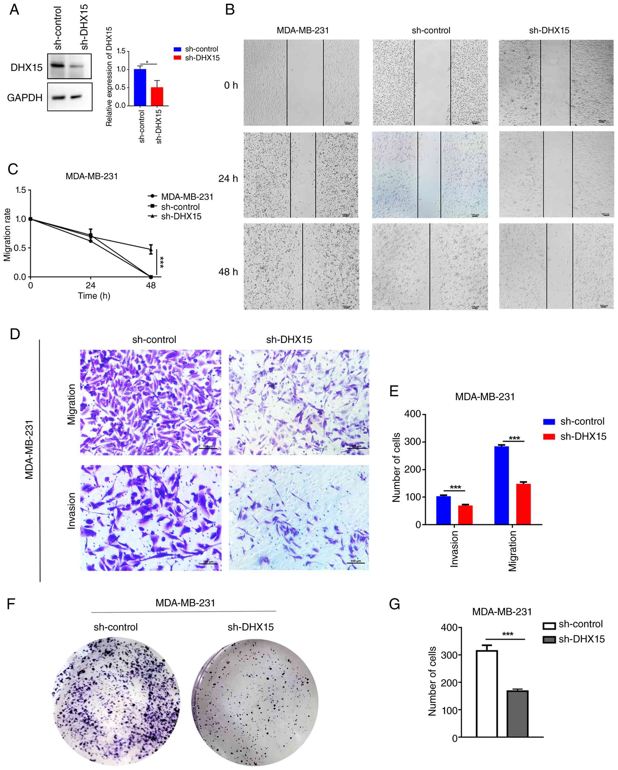
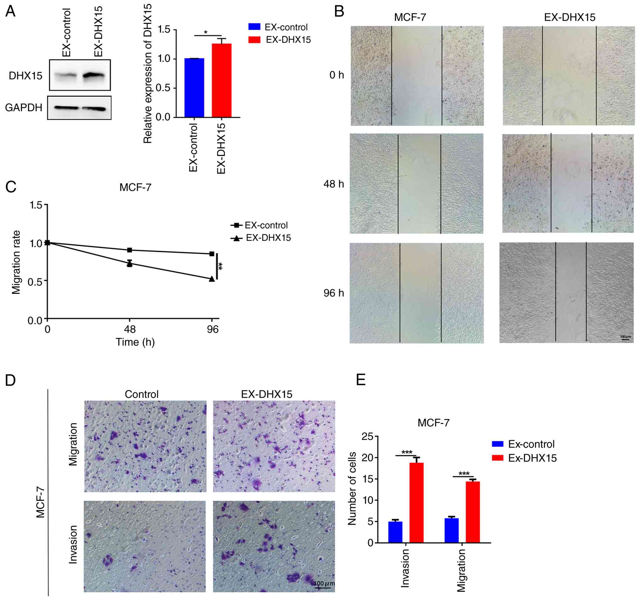
MCF-7 cells. Western blotting revealed that DHX15 expression levels
were reduced in sh-DHX15-transfected MDA-MB-231 cells (Fig. 7A) and were increased in EX-DHX15-
transfected MCF-7 cells (Fig. 8A).
Wound healing experiments indicated that knockdown of the DHX15
gene resulted in delayed healing (P<0.001; Fig. 7B and C). By contrast, overexpression
of the DHX15 gene resulted in accelerated healing (P<0.01;
Fig. 8B and C). Transwell assays
demonstrated that knocking down DHX15 expression resulted in
reduced migration and invasion capabilities of MDA-MB-231 cells
(P<0.001; Fig. 7D and E), but
overexpression of DHX15 resulted in increased migration and
invasion capabilities of MCF-7 cells (P<0.001; Fig. 8D and E). Furthermore, DHX15
knockdown led to a decrease in the number of colonies formed
(P<0.001; Fig. 7F and G). Colony
formation assay was performed only in DHX15-knockdown MDA-MB-231
cells, as MCF-7 cells display slow proliferation and poor
colony-forming capacity for this assay under conventional culture
conditions. Therefore, the knockdown of DHX15 may inhibit the
proliferation, migration and invasion of MDA-MB-231 cells.
Discussion
BC remains a key issue in the realm of global health
challenges. The complex pathogenesis and varied clinical
manifestations of BC pose notable obstacles to effective treatment
and prevention (1). Therefore, it
is key to characterize novel biomarkers for BC to detect and treat
the disease. As a member of the DEAD-box RNA-unwinding subfamily,
DHX15 is a key regulator of mRNA maturation and ribosome assembly
(25). Previous studies through
experiments have demonstrated that DXH15 is notably overexpressed
in hepatocellular carcinoma and serves a key role in controlling
hepatocellular carcinoma tumor growth and expansion (12,13,26,27).
This indicates that DHX15 serves a key role in the invasive
behavior of hepatocellular carcinoma and may serve as a biomarker
for migratory potential. Furthermore, high expression of DHX15 can
promote the progression of prostate cancer by stimulating siah
2-mediated ubiquitination of the androgen receptor (26), whereas upregulated DHX15 in BC is
associated with worse prognosis in patients (28). Nonetheless, the biological function
and molecular regulatory mechanisms in BC development remain
incompletely elucidated. Building on this foundation, the present
study utilized TCGA, the Kaplan-Meier database, Metascape,
wound-healing assays, Transwell assays and colony formation
experiments to investigate DHX15 as a potential therapeutic target
and biomarker for BC. The present study focused on gene expression
levels, predictive abilities, immune cell infiltration, protein
interaction network construction, single-cell functional analysis,
pathway analysis and molecular mechanisms.
From the analysis of GO, KEGG and PPI networks, the
present study demonstrated that DHX15 and its co-expressed genes
were enriched in certain pathways, such as ‘microtubule
cytoskeleton organization involved in mitosis’, SRP-dependent
cotranslational protein targeting to membrane’ and ‘DNA repair’.
The CancerSEA database indicated that the biological function of
DHX15 in BC single-cell data was primarily associated with DNA
damage, cell cycle, DNA repair, stemness and invasion. The present
results indicated that DHX15 may enhance tumor malignancy by
promoting transcriptional regulation and cell cycle control in BC
cells. Consistent research indicates that DHX15 enhances androgen
receptor transcriptional activity and contributes to prostate
cancer progression through Siah2 (26).
The tumor immune microenvironment (TIME) is
considered the environment surrounding the tumor. The composition
of the TIME is chiefly constituted by immune cells and cytokines,
which are mainly produced by tumor cells (29). The degree of immune cell
infiltration in tumor tissue is associated with the malignancy of
the tumor (30). Regulatory cells
of the TIME, including Th2, tumor-associated macrophage (TAMs),
Tregs and myeloid-derived suppressor cells, have been shown to be
associated with an immunosuppressive microenvironment and worse
outcomes (31). The M2 subtypes of
macrophages have been observed to be stimulated by Th2 cytokines,
including IL-4, IL-10 and IL-13. These cells have also been
revealed to express CD206 (mannose receptor), arginase 1 and
scavenger receptors (32,33). TAMs resemble M2 macrophages by
secreting pro-tumor cytokines (such as IL-10, CCL22, VEGF and MMP9)
(34), thereby facilitating tumor
progression. Kundu et al (31) reported a positive association
between M2 macrophage infiltration and the malignant progression of
BC. Patients with high infiltration levels of M2 macrophages in BC
typically have a worse prognosis and have decreased
immunotherapeutic sensitivity (35). The present findings indicated that
DHX15 expression was positively significant associated with T
helper cells, Tcm, and weak positive correlation Th2 cell and M2
macrophage infiltration through ssGSEA and CIBERSORT analyses.
Therefore, it is hypothesized that increased DHX15 expression in BC
cells may stimulate M2 macrophages by activating Th2 cell
infiltration, resulting in worse prognosis and immuno-resistance.
The single-cell transcriptomic analysis revealed that DHX15 was
highly expressed not only in malignant breast glandular cells but
also in T cells and macrophages within the TIME. DHX15 in
endometrial carcinoma may promote the secretion of chemotactic
factors that recruit immunosuppressive macrophages and T cells
(15), which is consistent with the
present correlation analysis showing a positive association between
DHX15 expression and the infiltration of M2 macrophages and Th2
cells. By contrast, DHX15 is also known to function as a cytosolic
nucleic acid sensor in innate immune cells, where it can modulate
NF-κB and IFN regulatory factor 3 signaling to influence cytokine
production and immune cell polarization (36,37).
Therefore, DHX15 expression in T cells and macrophages could
directly regulate their functional states, such as promoting M2
macrophage polarization or Th2 cell differentiation, independent of
tumor cell-derived signals.
The proportional hazards model analysis indicated
that DHX15 functions as an independent prognostic factor in cases
where multiple infiltrating immune cell types are present in BC.
Kaplan-Meier survival analysis combined with immune infiltration
data indicated that the infiltration of CD8+ T cells,
CD4+ T cells, neutrophils and dendritic cells was
significantly associated with the prognosis of BC, where the group
with high DHX15 expression exhibited a worse prognosis. This
finding indicates an association between DHX15, immune response and
clinical outcomes. Immune checkpoints are crucial for tumor
progression, where inhibiting these checkpoints can block cancer
cell immune escape (38). In the
present study, DHX15 expression was positively associated with Treg
markers (FOXP3, CCR8 and STAT5B) and negatively associated with the
immune checkpoints PDCD1, LAG3 and GZMB. Collectively, the
prognostic effect of DHX15 is tightly intertwined with the TIME.
DHX15 correlates positively with Treg signature genes to promote
immunosuppression, while it negatively associates with effector
immune checkpoint and cytotoxic markers, thereby limiting the
antitumor activity of CD8+ T cells and resulting in
worse clinical prognosis. Next, DHX15 expression in BC and its
biological functions through in vitro experiments were
determined. Following shRNA-mediated interference of DHX15
expression, BC cells exhibited reduced migration capacity, invasive
ability and proliferation levels, whilst overexpressing DHX15
expression increased these cellular functions, further validating
the accuracy of prior predictions. This is consistent with the
in vitro functional data showing that in non-small cell lung
cancer, DHX15 serves as an essential mediator of lncRNA-induced
invasive phenotypes, while DHX15 upregulation also facilitates
metastatic progression in endometrial, colorectal and liver cancers
through activation of STAT3/NF-κB axes and metabolic reprogramming
(27,39,40).
These findings suggest that DHX15 exerts a key biological role in
the progression of BC.
The present study also revealed that DHX15
expression was associated with immune cell infiltration in BC
tumors through bioinformatics analysis, demonstrating that DHX15
expression may be associated with the TIME and could serve as a
basis for future investigation into its role in immunotherapy
response.
There were, however, limitations to the present
study due to the lack of in vitro and in vivo assays
to validate the effects of DHX15 on BC cell immunity. For future
research, the role of DHX15 in regulating BC cell immunity and drug
sensitivity through both in vitro and in vivo
experiments should be validated. Whilst the present bioinformatics
analysis suggests that DHX15 may be involved in the p53/Rb, WNT and
mTOR signaling pathways, the present study did not experimentally
validate these predictions. Future studies employing
pathway-specific inhibitors, western blot analysis of key signaling
components and gene knockdown/rescue experiments are warranted to
elucidate the precise molecular mechanisms through which DHX15
promotes BC cell proliferation and migration. Notably, some
statistically significant correlations between DHX15 and immune
signatures from ssGSEA and CIBERSORT analyses presented correlation
coefficients below 0.3, which are defined as weak correlations, and
subsequent experimental validation is essential to validate these
preliminary bioinformatic observations. The present study served as
foundational preclinical work requiring subsequent in vivo
validation. Future studies should plan to conduct functional
co-culture assays or in vivo immune profiling following
DHX15 modulation and experimentally validate whether these
immune-related associations functionally contribute to the
malignant phenotype or to resistance against immunotherapy.
To conclude, the present study employed an
integrated approach combining bioinformatics analysis and
experiments to investigate the expression of DHX15, its biological
function and altered immune cell infiltration patterns in BC. The
findings suggest that high expression of DHX15 in BC is associated
with a worse prognosis and altered immune cell infiltration
patterns. Furthermore, functional assays revealed that DHX15 can
promote BC cell proliferation, migration and invasion in
vitro. Collectively, DHX15 serves as a novel prognostic
biomarker and a promoter of malignancy phenotypes in BC; however,
the immune-related findings in the present study are purely
bioinformatics predictions and await experimental verification in
subsequent research.
Supplementary Material
Supporting Data
Acknowledgements
Not applicable.
Funding
The present study was funded by the Tianjin Municipal Education
Commission's Scientific Research Plan Project (grant no.
2023KJ043).
Availability of data and materials
The data generated in the present study may be
requested from the corresponding author.
Authors' contributions
FL and QY designed and performed the experiments,
compiled and analyzed the data and assisted in writing the
manuscript. FL also performed the bioinformatics analysis. JL
designed the experiments and provided the funding. FL and JL
confirm the authenticity of all the raw data. All authors read and
approved the final version of the manuscript.
Ethics approval and consent to
participate
Not applicable.
Patient consent for publication
Not applicable.
Competing interests
The authors declare that they have no competing
interests.
References
|
1
|
Siegel RL, Kratzer TB, Giaquinto AN, Sung
H and Jemal A: Cancer statistics, 2025. CA J Clin. 75:10–45. 2025.
View Article : Google Scholar
|
|
2
|
Sung H, Ferlay J, Siegel RL, Laversanne M,
Soerjomataram I, Jemal A and Bray F: Global cancer statistics 2020:
GLOBOCAN Estimates of incidence and mortality worldwide for 36
cancers in 185 countries. CA Cancer J Clin. 71:209–249.
2021.PubMed/NCBI
|
|
3
|
Bergin ART and Loi S: Triple-negative
breast cancer: Recent treatment advances. F1000Res. 8:F10002019.
View Article : Google Scholar : PubMed/NCBI
|
|
4
|
Lei S, Zheng R, Zhang S, Chen R, Wang S,
Sun K, Zeng H, Wei W and He J: Breast cancer incidence and
mortality in women in China: Temporal trends and projections to
2030. Cancer Biol Med. 18:900–909. 2021. View Article : Google Scholar : PubMed/NCBI
|
|
5
|
Siegel RL, Miller KD and Jemal A: Cancer
statistics, 2020. CA J Clin. 70:7–30. 2020. View Article : Google Scholar
|
|
6
|
Chen XM, Liang YB, Zuo JX, Yang ZS, Zhang
LY, Zhang XY, Wan P and Ke Y: ZG16B: A key regulator of tumor
progression and immune microenvironment modulation in cancer
(Review). Int J Mol Med. 57:582026. View Article : Google Scholar : PubMed/NCBI
|
|
7
|
Li Q, Guo H, Xu J, Li X, Wang D, Guo Y,
Qing G, Van Vlierberghe P and Liu H: A helicase-independent role of
DHX15 promotes MYC stability and acute leukemia cell survival.
iScience. 27:1085712024. View Article : Google Scholar : PubMed/NCBI
|
|
8
|
Fan L, Guo X, Zhang J, Wang Y, Wang J and
Li Y: Relationship between DHX15 expression and survival in
colorectal cancer. Rev Esp Enferm Dig. 115:234–240. 2023.PubMed/NCBI
|
|
9
|
Detanico T, Virgen-Slane R, Steen-Fuentes
S, Lin WW, Rhode-Kurnow A, Chappell E, Correa RG, DiCandido MJ,
Mbow ML, Li J and Ware CF: Co-expression networks identify DHX15
RNA helicase as a B cell regulatory factor. Front Immunol.
10:29032019. View Article : Google Scholar : PubMed/NCBI
|
|
10
|
Wang G, Xiao X, Wang Y, Chu X, Dou Y,
Minze LJ, Ghobrial RM, Zhang Z and Li XC: The RNA helicase DHX15 is
a critical regulator of natural killer-cell homeostasis and
functions. Cell Mol Immunol. 19:687–701. 2022. View Article : Google Scholar : PubMed/NCBI
|
|
11
|
Xu Y, Song Q, Pascal LE, Zhong M, Zhou Y,
Zhou J, Deng FM, Huang J and Wang Z: DHX15 is up-regulated in
castration-resistant prostate cancer and required for androgen
receptor sensitivity to low DHT concentrations. Prostate.
79:657–666. 2019. View Article : Google Scholar : PubMed/NCBI
|
|
12
|
Xie C, Liao H, Zhang C and Zhang S:
Overexpression and clinical relevance of the RNA helicase DHX15 in
hepatocellular carcinoma. Hum Pathol. 84:213–220. 2019. View Article : Google Scholar : PubMed/NCBI
|
|
13
|
Pan L, Li Y, Zhang HY, Zheng Y, Liu XL, Hu
Z, Wang Y, Wang J, Cai YH, Liu Q, et al: DHX15 is associated with
poor prognosis in acute myeloid leukemia (AML) and regulates cell
apoptosis via the NF-kB signaling pathway. Oncotarget.
8:89643–89654. 2017. View Article : Google Scholar : PubMed/NCBI
|
|
14
|
Ito S, Koso H, Sakamoto K and Watanabe S:
RNA helicase DHX15 acts as a tumour suppressor in glioma. B J
Cancer. 117:1349–1359. 2017. View Article : Google Scholar : PubMed/NCBI
|
|
15
|
Balaya RD, Kanekar S, Kumar S and
Kandasamy RK: Role of DEAD/DEAH-box helicases in immunity,
infection and cancers. Cell Commun Signal. 23:2922025. View Article : Google Scholar : PubMed/NCBI
|
|
16
|
Hashemi V, Masjedi A, Hazhir-Karzar B,
Tanomand A, Shotorbani SS, Hojjat-Farsangi M, Ghalamfarsa G, Azizi
G, Anvari E, Baradaran B and Jadidi-Niaragh F: The role of DEAD-box
RNA helicase p68 (DDX5) in the development and treatment of breast
cancer. J Cell Physiol. 234:5478–5487. 2019. View Article : Google Scholar : PubMed/NCBI
|
|
17
|
Li T, Fan J, Wang B, Traugh N, Chen Q, Liu
JS, Li B and Liu XS: TIMER: A web server for comprehensive analysis
of tumor-infiltrating immune cells. Cancer Res. 77:e108–e110. 2017.
View Article : Google Scholar : PubMed/NCBI
|
|
18
|
Chandrashekar DS, Bashel B, Balasubramanya
SAH, Creighton CJ, Ponce-Rodriguez I, Chakravarthi BVSK and
Varambally S: UALCAN: A portal for facilitating tumor subgroup gene
expression and survival analyses. Neoplasia. 19:649–658. 2017.
View Article : Google Scholar : PubMed/NCBI
|
|
19
|
Gyorffy B, Lanczky A, Eklund AC, Denkert
C, Budczies J, Li Q and ZSzallasi Z: An online survival analysis
tool to rapidly assess the effect of 22,277 genes on breast cancer
prognosis using microarray data of 1,809 patients. Breast Cancer
Res Treat. 123:725–731. 2010. View Article : Google Scholar : PubMed/NCBI
|
|
20
|
Fekete JT and Gyorffy B: ROCplot.org:
Validating predictive biomarkers of chemotherapy/hormonal
therapy/anti-HER2 therapy using transcriptomic data of 3,104 breast
cancer patients. Int J Cancer. 145:3140–3151. 2019. View Article : Google Scholar : PubMed/NCBI
|
|
21
|
Vasaikar SV, Straub P, Wang J and Zhang B:
LinkedOmics: Analyzing multi-omics data within and across 32 cancer
types. Nucleic Acids Res. 46:D956–D963. 2018. View Article : Google Scholar : PubMed/NCBI
|
|
22
|
Zhou Y, Zhou B, Pache L, Chang M,
Khodabakhshi AH, Tanaseichuk O, Benner C and Chanda SK: Metascape
provides a biologist-oriented resource for the analysis of
systems-level datasets. Nat Commun. 10:15232019. View Article : Google Scholar : PubMed/NCBI
|
|
23
|
Uhlen M, Fagerberg L, Hallstrom BM,
Lindskog C, Oksvold P, Mardinoglu A, Sivertsson A, Kampf C,
Sjöstedt E, Asplund A, et al: Proteomics. Tissue-based map of the
human proteome. Science. 347:12604192015. View Article : Google Scholar : PubMed/NCBI
|
|
24
|
Yuan H, Yan M, Zhang G, Liu W, Deng C,
Liao G, Xu L, Luo T, Yan H, Long Z, et al: CancerSEA: A cancer
single-cell state atlas. Nucleic Acids Res. 47:D900–D908. 2019.
View Article : Google Scholar : PubMed/NCBI
|
|
25
|
Ren T, Wei G, Yi J, Zhang Y, Zhao H, Wu N,
Zhang H, Guo Z, Wang Y, Kuang J, et al: GPATCH3, a splicing
regulator that facilitates tumor immune evasion via the modulation
of ATPase activity of DHX15. Front Immunol. 16:16124612025.
View Article : Google Scholar : PubMed/NCBI
|
|
26
|
Jing Y, Nguyen MM, Wang D, Pascal LE, Guo
W, Xu Y, Ai J, Deng FM, Masoodi KZ, Yu X, et al: DHX15 promotes
prostate cancer progression by stimulating Siah2-mediated
ubiquitination of androgen receptor. Oncogene. 37:638–650. 2018.
View Article : Google Scholar : PubMed/NCBI
|
|
27
|
Portoles I, Ribera J, Fernandez-Galan E,
Lecue E, Casals G, Melgar-Lesmes P, Fernández-Varo G, Boix L,
Sanduzzi M, Aishwarya V, et al: Identification of Dhx15 as a major
regulator of liver development, regeneration, and tumor growth in
zebrafish and mice. Int J Mol Sci. 25:37162024. View Article : Google Scholar : PubMed/NCBI
|
|
28
|
Zheng W, Wang X, Yu Y, Ji C and Fang L:
CircRNF10-DHX15 interaction suppressed breast cancer progression by
antagonizing DHX15-NF-kappaB p65 positive feedback loop. Cell Mol
Biol Lett. 28:342023. View Article : Google Scholar : PubMed/NCBI
|
|
29
|
Lo YL, Lin HC, Lee Y, Chuang HY and Chou
TF: TME-responsive nanoparticles co-targeting VCP, NETs, and dual
immune checkpoints for immune revitalization in
EGFR/PD-L1/CTLA-4-driven colorectal cancer. Biomed Pharmacother.
192:1185652025. View Article : Google Scholar : PubMed/NCBI
|
|
30
|
Dieci MV, Miglietta F and Guarneri V:
Immune infiltrates in breast cancer: Recent updates and clinical
implications. Cells. 10:2232021. View Article : Google Scholar : PubMed/NCBI
|
|
31
|
Kundu M, Butti R, Panda VK, Malhotra D,
Das S, Mitra T, Kapse P, Gosavi SW and Kundu GC: Modulation of the
tumor microenvironment and mechanism of immunotherapy-based drug
resistance in breast cancer. Mol Cancer. 23:922024. View Article : Google Scholar : PubMed/NCBI
|
|
32
|
Sadeghalvad M, Mohammadi-Motlagh HR and
Rezaei N: Immune microenvironment in different molecular subtypes
of ductal breast carcinoma. Breast Cancer Res Treat. 185:261–279.
2021. View Article : Google Scholar : PubMed/NCBI
|
|
33
|
Del Alcazar CR, Huh SJ, Ekram MB, Trinh A,
Liu LL, Beca F, Zi X, Kwak M, Bergholtz H, Su Y, et al: Immune
escape in breast cancer during in situ to invasive carcinoma
transition. Cancer Discov. 7:1098–1115. 2017. View Article : Google Scholar : PubMed/NCBI
|
|
34
|
Wang Z, Zhang J, Chen H, Zhang X, Zhang K,
Zhang F, Xie Y, Ma H, Pan L, Zhang Q, et al: Molecular
characterization and prognostic modeling associated with M2-like
tumor-associated macrophages in breast cancer: Revealing the
immunosuppressive role of DLG3. Front Immunol. 16:16507262025.
View Article : Google Scholar : PubMed/NCBI
|
|
35
|
Zhang L, Gu S, Wang L, Zhao L, Li T, Zhao
X and Zhang L: M2 macrophages promote PD-L1 expression in
triple-negative breast cancer via secreting CXCL1. Pathol Res
Pract. 260:1554582024. View Article : Google Scholar : PubMed/NCBI
|
|
36
|
Ramnani B, Devale T, Manivannan P, Haridas
A and Malathi K: DHX15 and Rig-I coordinate apoptosis and innate
immune signaling by antiviral RNase L. Viruses. 16:19132024.
View Article : Google Scholar : PubMed/NCBI
|
|
37
|
Mosallanejad K, Sekine Y,
Ishikura-Kinoshita S, Kumagai K, Nagano T, Matsuzawa A, Takeda K,
Naguro I and Ichijo H: The DEAH-box RNA helicase DHX15 activates
NF-kappaB and MAPK signaling downstream of MAVS during antiviral
responses. Sci Signal. 7:ra402014. View Article : Google Scholar : PubMed/NCBI
|
|
38
|
Thomas R, Al-Khadairi G and Decock J:
Immune checkpoint inhibitors in triple negative breast cancer
treatment: Promising future prospects. Front Oncol. 10:6005732020.
View Article : Google Scholar : PubMed/NCBI
|
|
39
|
Yao G, Chen K, Qin Y, Niu Y, Zhang X, Xu
S, Zhang C, Feng M and Wang K: Long non-coding RNA JHDM1D-AS1
interacts with DHX15 protein to enhance non-small-cell lung cancer
growth and metastasis. Mol Ther Nucl Acids. 18:831–840. 2019.
View Article : Google Scholar : PubMed/NCBI
|
|
40
|
Ye L, Jiang G, Sun Y and Li B:
ARNTL-mediated INO80-DHX15 axis reprograms the glycolytic
metabolism and augments the progression of endometrial carcinoma.
Cell Death Dis. 16:4632025. View Article : Google Scholar : PubMed/NCBI
|