Introduction
Leiomyosarcoma (LMS) is a malignant soft-tissue
sarcoma (STS) originating from smooth muscle cells, which accounts
for 10–20% of all STS cases (1).
LMS most commonly arises in the uterus, retroperitoneum and soft
tissues of the extremities; however, primary LMS occurring in the
deep soft tissue of the lumbar region is rare, with only a few
isolated case reports in nearby or similar regions documented in
the literature (2,3). Due to its rarity, the clinical course,
optimal treatment strategies and long-term outcomes for this
specific anatomical presentation remain poorly defined.
For localized LMS, wide surgical resection with
negative margins is the cornerstone of curative treatment (4). Adjuvant radiotherapy may reduce local
recurrence, particularly in high-grade tumors, while adjuvant
chemotherapy remains controversial but is often considered for
patients with high-risk features, such as tumor size >5 cm, deep
fascial involvement and high-grade (grade 3) histology (5). In metastatic or recurrent LMS,
systemic therapy options include anthracycline-based regimens,
trabectedin and pazopanib (6).
Recently, the multitarget tyrosine kinase inhibitor anlotinib has
shown promising activity in advanced LMS, with a phase III trial
demonstrating a median overall survival (OS) time of 17.45 months
in heavily pretreated patients (7).
However, acquired resistance inevitably develops, limiting
long-term disease control.
The present report describes a case of primary LMS
arising in the deep soft tissue of the lumbar region with invasion
into the skeletal muscle and a Ki-67 proliferation index of 70%,
indicating high proliferative activity. The patient underwent three
surgical resections combined with chemoradiotherapy, followed by
anlotinib therapy. This case report provides a complete clinical
trajectory of a rare, aggressive LMS managed with sequential
multimodal therapy, and offers insights into the efficacy and
limitations of anlotinib in this challenging clinical scenario.
Case report
In February 2018, a 48-year-old man with a right
lumbar mass but no notable pain, with only mild local discomfort
when lying on his right side, presented to Guangshan County
People's Hospital (Henan, China) and underwent a mass resection 1
week later; the operation consisted of a wide local excision of the
right lumbar mass (Fig. 1).
Intraoperatively, the tumor was found to be adherent to the deep
fascia and involved the superficial layer of the erector spinae
aponeurosis, but there was no gross invasion of the underlying
muscle bellies. Subsequently, the patient was referred to The
General Hospital of the Central Theater Command, (China) for
further management. The pathological slides from the initial
resection were subsequently reviewed and confirmed at Central
Theater Command General Hospital of the People's Liberation Army
(Wuhan, China). Postoperative pathological examination confirmed a
diagnosis of right lumbar LMS. Microscopically, the tumor consisted
of intersecting fascicles of spindle cells with moderate atypia, a
mitotic count of 10 per 10 high-power fields (HPFs) and no
necrosis; the resection margin was microscopically positive (R1)
(Fig. S1A). The final diagnosis
was LMS, French Federation of Cancer Centers Sarcoma Group (FNCLCC)
grade 2 (8). The
immunohistochemistry results were as follows: Vimentin(+),
desmin(−), smooth muscle actin (SMA)(+), h-Caldesmon(+),
pan-cytokeratin (PCK)(−), CD34(−), S-100(−), CD68(−), anaplastic
lymphoma kinase 1(−), E3 ubiquitin-protein ligase Mdm2 (MDM2)(−),
cyclin-dependent kinase 4 (CDK4)(−), β-catenin(−), Bcl-2(−),
integrase interactor 1(+) and Ki-67(+; 15%) (Fig. S2). Although desmin expression was
negative, the diffuse strong SMA positivity, the fascicular growth
pattern and the lack of other lineage markers detected by
immunohistochemistry supported this diagnosis after exclusion of
other spindle cell malignancies. The patient received six cycles of
intravenous chemotherapy with ifosfamide (IFO) at 2 g/m2
per day on days 1–3 and pirarubicin (THP) at 50 mg/m2 on
day 1, repeated every 21 days. The chemotherapy was generally well
tolerated; the main toxicities were grade 2 nausea, grade 2
alopecia and grade 1 fatigue, without febrile neutropenia or
cardiotoxicity as per the National Cancer Institute Common
Terminology Criteria for Adverse Events version 5.0 (9). Subsequently, local radiotherapy was
administered to the right lumbar region using a clinical target
volume (CTV) of 40 Gy in 20 fractions, followed by a CTV boost of
20 Gy in eight fractions. A total of 3 months after radiotherapy,
computed tomography (CT) showed no evidence of recurrence (Fig. S1B).
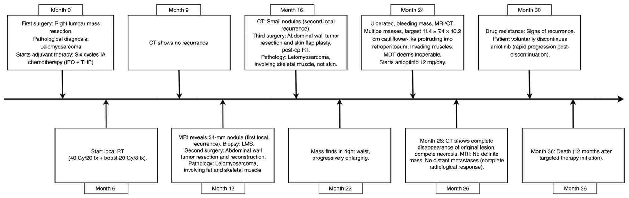 | Figure 1.A detailed timeline for each event:
First surgery, chemotherapy, radiotherapy, recurrences, subsequent
surgeries, initiation of anlotinib treatment, key imaging
assessments, progression and death. IFO, ifosfamide; THP,
pirarubicin; RT, radiation therapy; CT, computed tomography; LMS,
leiomyosarcoma; MRI, magnetic resonance imaging; fx, fractions;
MDT, multidisciplinary team. |
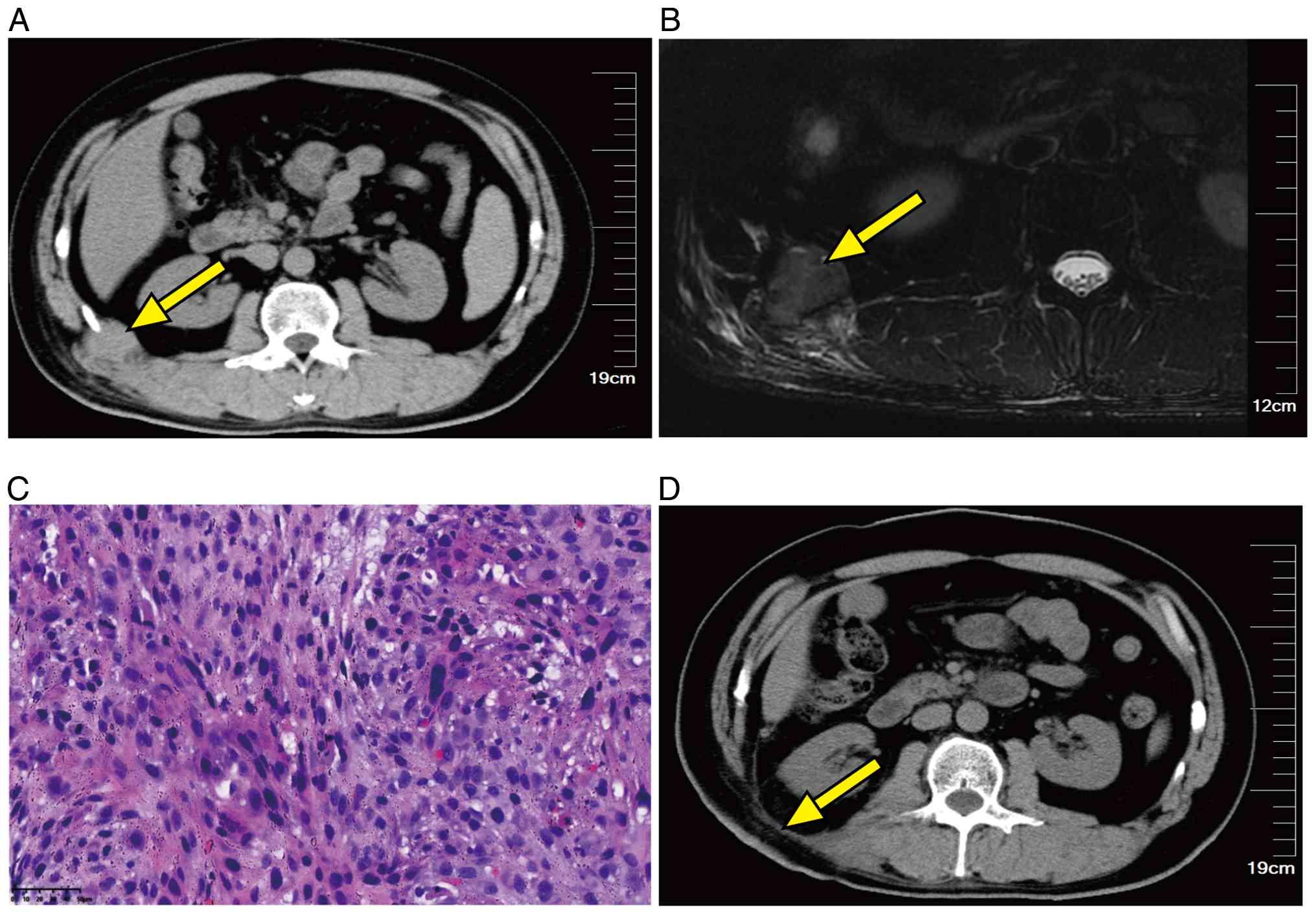
Notably, at the 1-year follow-up after the first
surgery, magnetic resonance imaging (MRI) revealed a new nodule in
the subcutaneous soft tissue of the right waist, measuring 34 mm at
the largest diameter, which was suggestive of tumor recurrence. An
ultrasound-guided biopsy of the right lumbar mass was immediately
performed and pathology confirmed recurrent LMS (data not shown).
The recurrent tumor was found to involve the full thickness of the
abdominal wall musculature, including the external oblique,
internal oblique and transversus abdominis, with the deep aspect
extending to the extraperitoneal fat. A fusiform incision measuring
~15×6 cm was made along the long axis of the tumor. The 12th rib
and a portion of the diaphragm adherent to the tumor were resected
en bloc to ensure clear margins, and the tumor together with the
surrounding soft tissues was completely excised. The resulting
full-thickness abdominal wall defect was repaired using a 15×15-cm
synthetic mesh placed in the preperitoneal space and secured to the
posterior rectus sheath. The abdominal wall incision was closed by
full-thickness suturing of the skin, subcutaneous tissue and
residual muscle layers (Fig. 2).
The tumor exhibited a mitotic count of 22/10 HPFs, 30% necrosis and
was classified as FNCLCC grade 3. The surgical margins were
positive (R1). The immunohistochemistry results of the second
specimen were as follows: Vimentin(+), desmin(−), SMA(+),
h-Caldesmon(−), MyoD1(−), PCK(+), epithelial membrane antigen
(EMA)(−), CD31(−), CD34(−), factor VIII (FVIII)(+), S-100(−),
HMB45(−), CD68(−), CDK4(−), MDM2(±) and Ki-67(+; 70%) (Fig. S3 and S4). The equivocal MDM2 staining, in
combination with negative CDK4 staining, indicated that
dedifferentiated liposarcoma was unlikely; however, MDM2
amplification testing was not performed. Despite negative staining
for h-Caldesmon and desmin, the diagnosis of LMS was favored based
on diffuse SMA positivity, characteristic fascicular architecture
and the exclusion of other types of sarcoma (including a malignant
peripheral nerve sheath tumor, dedifferentiated liposarcoma and
synovial sarcoma) by a dedicated sarcoma pathologist.
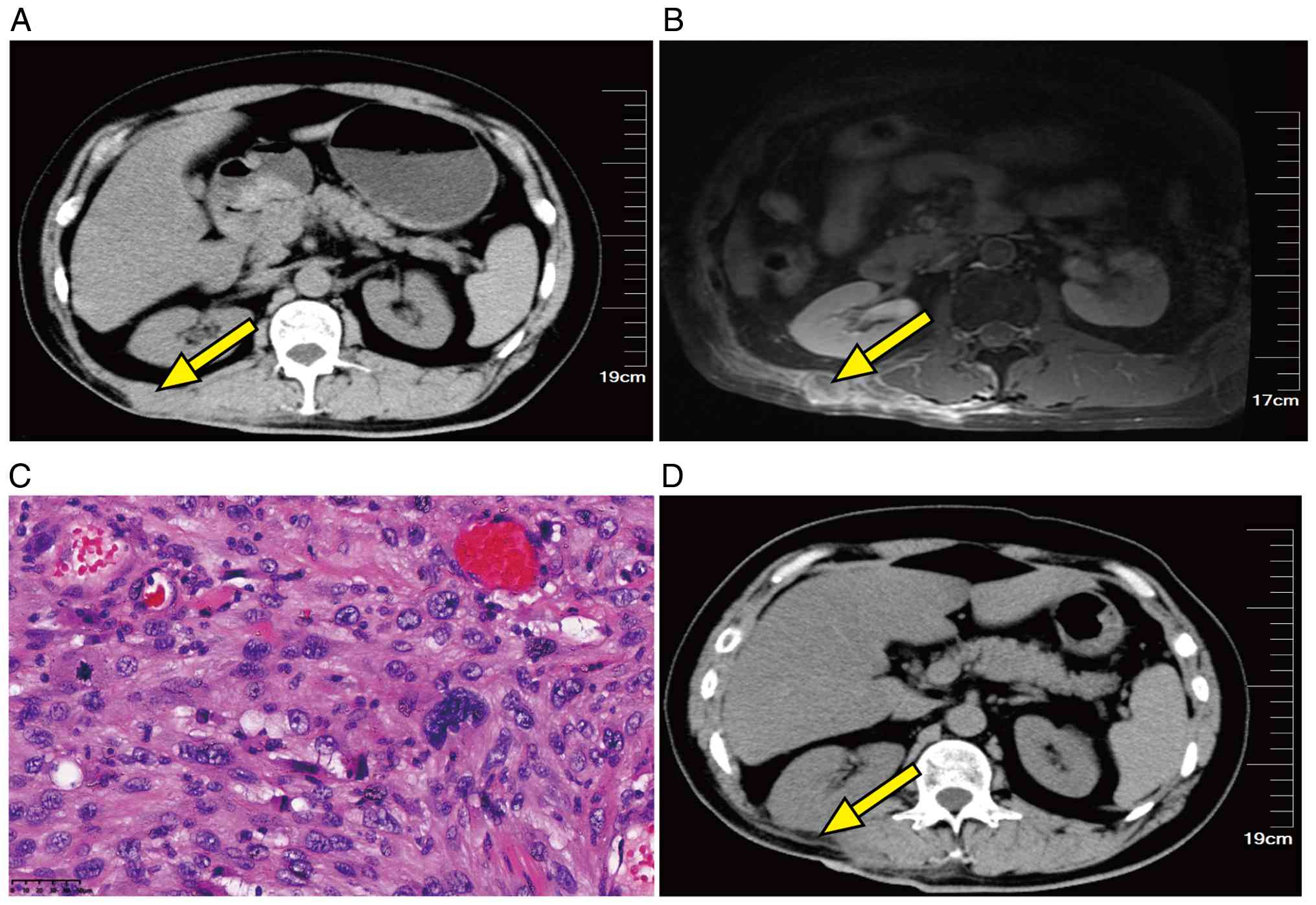
A total of 4 months after the second surgery, CT
revealed small nodules on the right lumbar back, indicating a
second recurrence. The third operation consisted of abdominal wall
tumor resection combined with skin flap surgery. The tumor
extensively involved the erector spinae, iliocostalis and
longissimus muscles. A resection margin of ~3 cm from the tumor
border was delineated, and the dissection proceeded layer by layer
through the skin, subcutaneous tissue and affected muscles down to
the extraperitoneal fat. The tumor and the involved segments of the
right lumbar back muscles were completely removed. The resultant
soft tissue defect was reconstructed using a skin flap based on the
Limberg technique: A rhomboid flap was designed, elevated and
mobilized over the right lumbar back defect, and the flap was
sutured in place. Pathological evaluation again confirmed recurrent
LMS involving skeletal muscle, without skin involvement (Fig. 3). The resection was R1, with a
mitotic count of 25/10 HPFs and 30% necrosis, consistent with
FNCLCC grade 3. The immunohistochemistry results of the specimen
were as follows: Vimentin(+), desmin(+), SMA(+), h-Caldesmon(+),
MyoD1(−), PCK(+), EMA(−), CD31(−), CD34(−), FVIII(−), S-100(−),
HMB45(−), CD68(−), MDM2(−), CDK4(−) and Ki-67(+; 60%) (Fig. S5). Postoperatively, a second course
of radiotherapy was administered to the right lumbar recurrent
region; a total dose of 30 Gy was delivered in 10 fractions of 3 Gy
over 2 weeks.
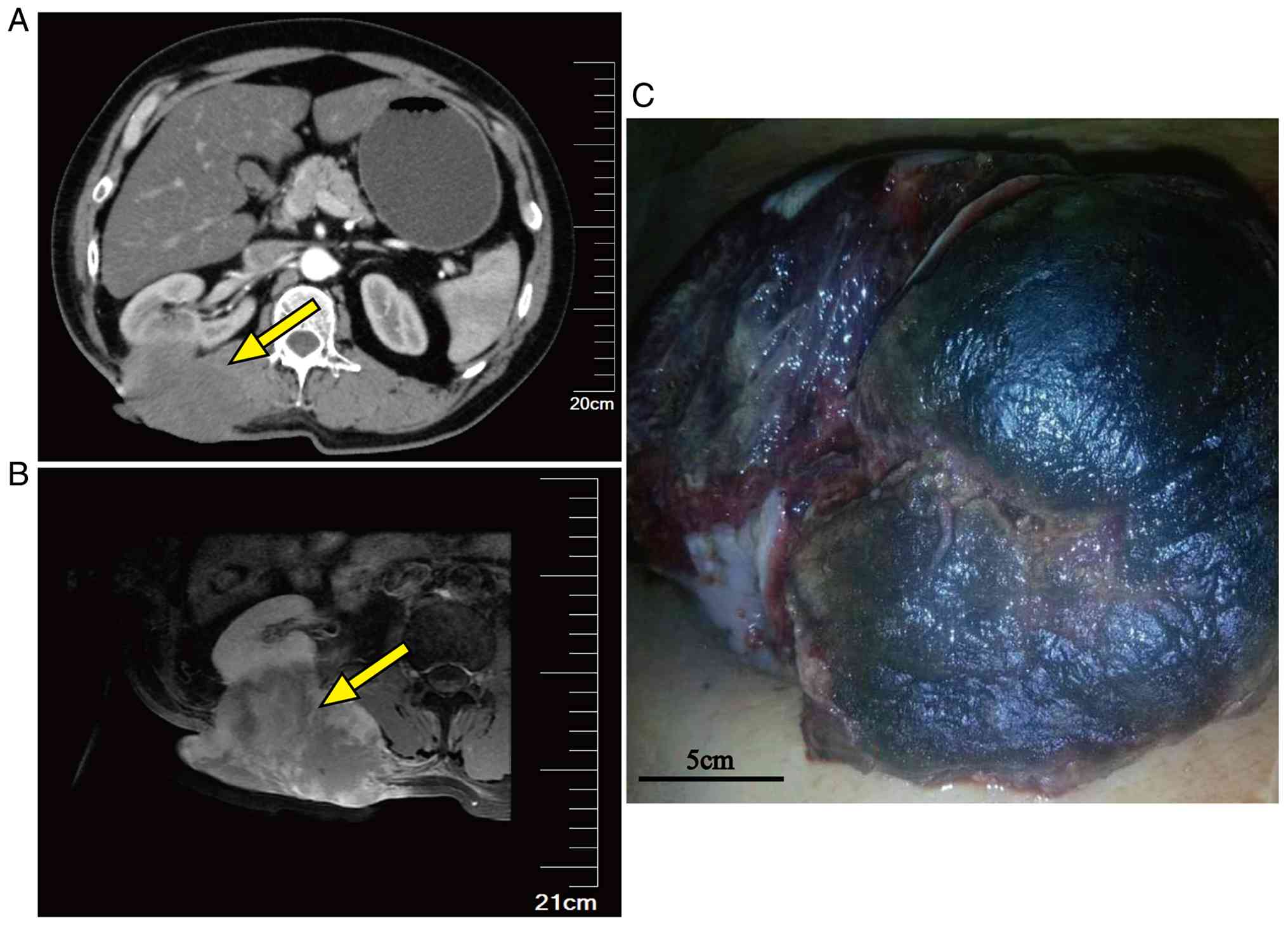
A total of 6 months after the third surgery, a right
lumbar mass was detected that was progressively enlarging. By 8
months, the mass surface had ulcerated and exhibited bleeding. MRI
confirmed tumor recurrence in the right lumbar back soft tissues
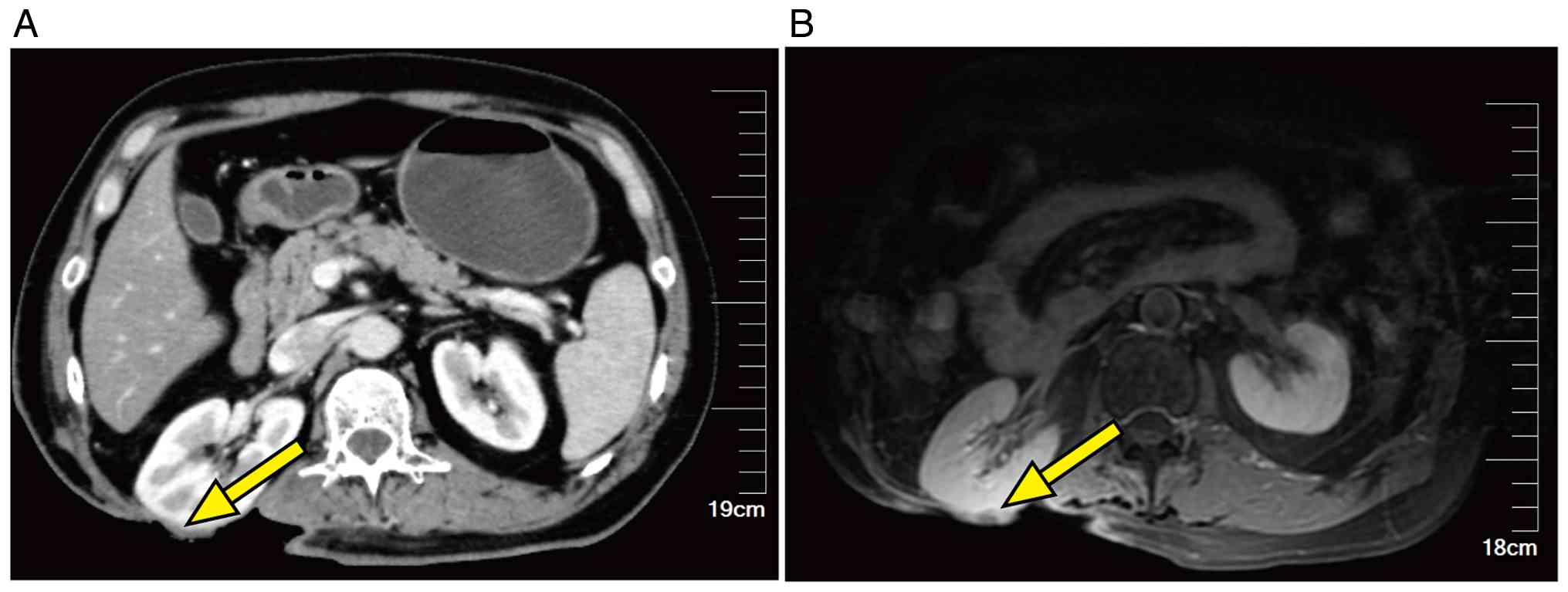
(Figs. 4 and 5). Contrast-enhanced CT with
three-dimensional reconstruction showed multiple irregular
mass-like and nodular lesions with mild heterogeneous enhancement
and ill-defined borders. The largest lesion was a
cauliflower-shaped mass protruding into the retroperitoneum,
measuring ~11.4×7.4×10.2 cm, with invasion of the right
iliocostalis, longissimus and erector spinae muscles. New pulmonary
nodules were also detected. Following a multidisciplinary sarcoma
tumor board discussion, surgery, radiotherapy and chemotherapy were
deemed inappropriate. The tumor was rapidly growing, making
resection no longer feasible. Genetic testing was recommended but
was declined by the family of the patient due to financial
constraints.
The patient was subsequently administered 12 mg
anlotinib orally once daily on a 2-weeks-on, 1-week-off schedule
(21-day cycles). The treatment was well tolerated, with the only
adverse events being grade 1 hand-foot syndrome and medically
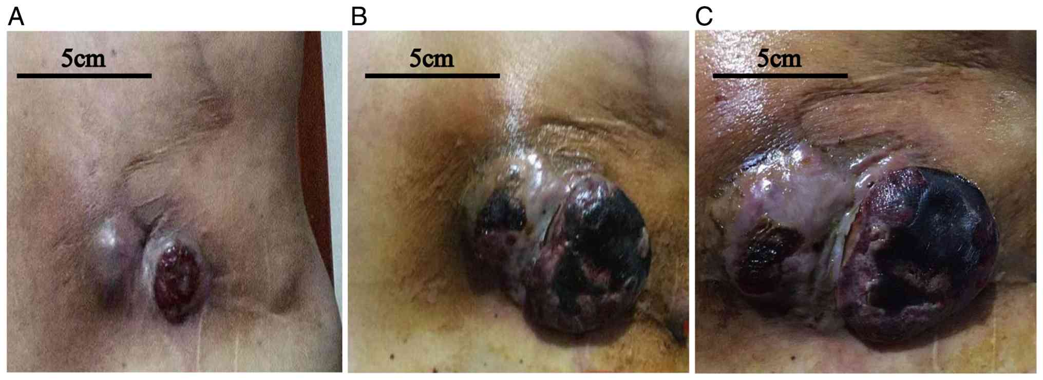
controlled hypertension. Tumor response to anlotinib was assessed
according to the Response Evaluation Criteria in Solid Tumors
(RECIST) version 1.1 (10),
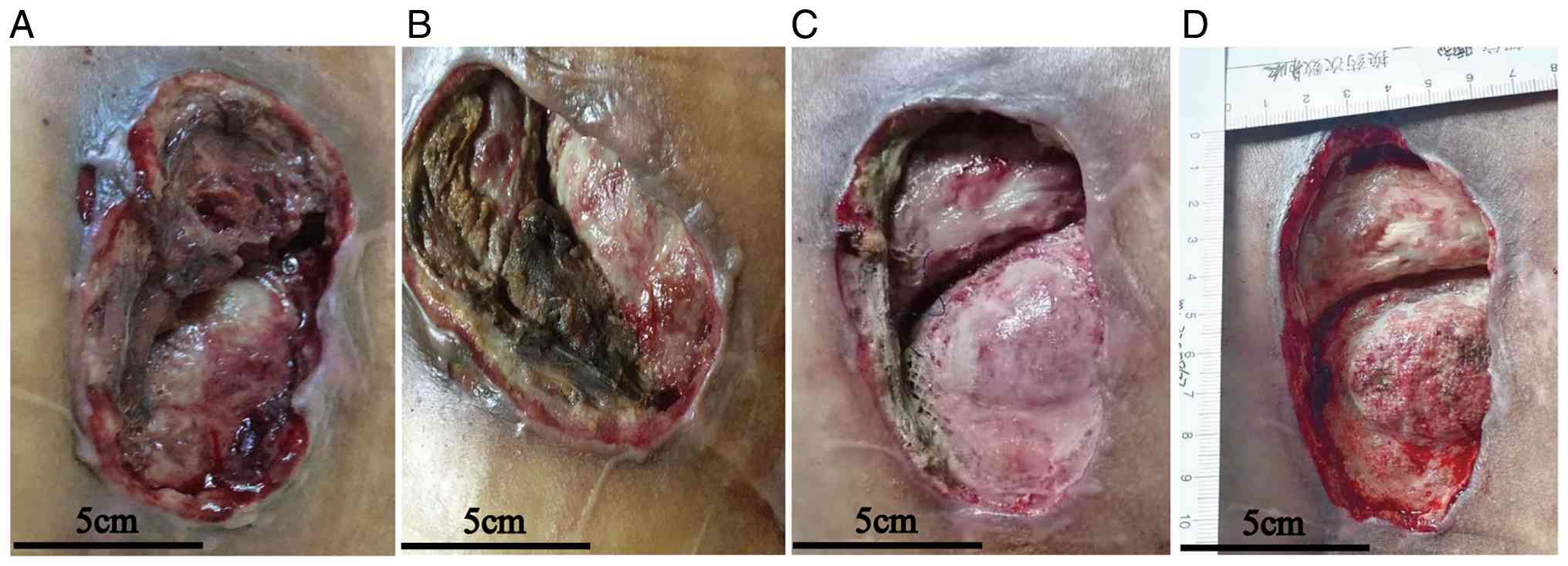
supplemented by serial clinical images documenting progressive
tumor necrosis and sloughing (Fig.
6). At 8 weeks after anlotinib initiation, the primary tumor
was no longer clinically detectable, no enlarged lymph nodes were
observed in the retroperitoneum or pelvic wall, and no distant
metastases were detected. Radiographic evaluation at ~2 months
demonstrated complete disappearance of the primary target lesion
(Fig. 7), consistent with a
complete response (CR) according to the RECIST 1.1 criteria, and no
new lesions were identified at that time. After 6 months of
targeted therapy, the patient developed drug resistance, with signs
of recurrence at the primary tumor site; thus, the patient
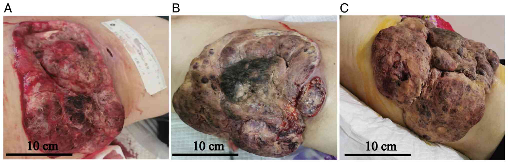
voluntarily discontinued anlotinib. Following drug withdrawal, the
disease progressed rapidly, with a marked increase in tumor volume
(Fig. 8). The patient succumbed 12
months after initiation of the targeted therapy.
Discussion
STS encompasses a heterogeneous group of >60
histological subtypes, together accounting for <1% of all adult
malignancies. LMS is a subtype defined by smooth muscle
differentiation and represents 10–20% of all STS cases (1). While LMS most commonly arises in the
uterus, retroperitoneum or extremities, primary occurrence in the
deep soft tissue of the lumbar region is rare (6,11),
with detailed descriptions of LMS confined to the lumbar paraspinal
or deep lumbar soft tissue remaining scarce. A 2025 report
documented the case of a 49-year-old woman with primary paraspinal
LMS who presented with lower back pain and lower extremity
weakness, and was successfully managed with complete surgical
excision; however, the patient was lost to follow-up after the
second visit to the clinic (3). An
earlier report described the case of a patient with unresectable
mismatch repair-deficient LMS harboring biallelic PTEN loss, in
whom the combination of antiangiogenic agents and pembrolizumab
rendered the tumor resectable, and CR was achieved after surgery
(2).
Unlike cutaneous or subcutaneous LMS, which
generally carries a lower metastatic potential, deep-seated LMS is
associated with a substantially higher risk of local recurrence
(40–60%) and distant metastasis (20–60%) (12). In the current patient, the tumor
invaded the skeletal muscle but spared the skin and bone,
consistent with a deep soft-tissue origin. This anatomical location
likely contributed to the aggressive clinical course, which was
characterized by multiple local recurrences despite repeated
resections. Pathological evaluation showed a spindle cell neoplasm
with marked cellular atypia, and immunohistochemistry revealed a
Ki-67 proliferation index of 70%, which is considerably higher than
the typical range of 20–40% reported for conventional LMS (13,14).
Such an elevated proliferative index is associated with aggressive
tumor behavior and has been related to shortened recurrence-free
survival times (15). This
pathological feature provides a biological explanation for the
rapid recurrences observed in the current patient and may also have
facilitated the emergence of resistant subclones under therapeutic
pressure.
For localized STS, the cornerstone of management is
wide surgical resection, frequently combined with perioperative
radiotherapy and chemotherapy, an approach that has improved
outcomes (16–18). The 5-year survival rate for
localized disease can reach 70–80%, whereas for advanced and
metastatic sarcoma, it remains at <20% (19). Over a 36-month disease course, the
patient described in the present study underwent three surgical
resections combined with adjuvant IFO-THP chemotherapy and
radiotherapy. Despite the high proliferative index and repeated
recurrences, the patient achieved an OS time of 36 months. This
prolonged survival may reflect the cumulative benefit of aggressive
local control through repeated surgeries and sequential systemic
therapies, consistent with current guidelines that underscore the
importance of multidisciplinary management for localized LMS, even
in cases of recurrence (4,5).
Anlotinib is an oral multitarget tyrosine kinase
inhibitor that targets VEGF receptors 1–3, platelet-derived growth
factor receptor-α, c-Kit and fibroblast growth factor receptors
1–3, thereby inhibiting tumor angiogenesis and proliferation
(20–22). Anlotinib is approved in China for
the treatment of advanced non-small cell lung cancer, small cell
lung cancer, medullary thyroid carcinoma and STS. Anlotinib has
been proven effective in a variety of STSs, such as liposarcoma and
synovial sarcoma (23,24). In the present patient, the early
clinical course following anlotinib initiation was characterized by
marked radiographic tumor regression, accompanied by clinically
evident necrosis and tumoral sloughing. Follow-up imaging performed
2 months after the start of anlotinib treatment demonstrated
complete disappearance of the primary target lesion according to
RECIST 1.1 criteria, with no residual tumor mass identifiable at
the original site. Mild, non-specific enhancement in the right
psoas muscle and adjacent subcutaneous tissue was interpreted as
post-treatment change, rather than evidence of active disease.
However, disease progression developed after ~6 months of therapy
and the patient survived for 12 months from the initiation of
anlotinib. Notably, the marked necrosis and sloughing observed at
the initiation of anlotinib treatment may reflect not only drug
effect but also contributions from tumor biology, ulceration,
vascular compromise, infection, prior treatment effects, or a
combination of these factors. Nevertheless, the temporal
association between anlotinib exposure and tumor regression,
followed by subsequent progression consistent with acquired
resistance, suggests a potential therapeutic effect during the
initial treatment period.
In the context of published data, a recent phase III
trial evaluating anlotinib in advanced LMS reported a median
progression-free survival time of 3.42 months (7). In the present patient, the
progression-free survival interval of ~6 months appears clinically
meaningful when viewed alongside this finding, although no
definitive comparative conclusion can be drawn from a single
uncontrolled observation. The development of progression after 6
months highlights the challenge of acquired resistance to tyrosine
kinase inhibitors in sarcoma and emphasizes the need for
combination strategies that may prolong durable disease control.
Recent studies have investigated the addition of anlotinib to
chemotherapy in the neoadjuvant conversion setting for unresectable
STS (25,26). In a study of 28 patients, the
combination of doxorubicin, IFO and anlotinib markedly improved
tumor regression, surgical conversion rates and R0 resection rates,
particularly in synovial sarcoma and liposarcoma, although with an
expected increase in manageable toxicity (27). The marked initial response observed
in the present case suggests that anlotinib may offer benefit in
selected patients with advanced LMS and provides a clinical
rationale for investigating anlotinib-based combination regimens in
this specific subtype.
Several limitations must be acknowledged when
interpreting the present case. First, as a single case report, the
findings cannot be generalized to broader populations. Second,
since post-progression tissue was not available for molecular
analysis, the exact mechanisms underlying the acquired resistance
to anlotinib remain undetermined; notably, this limitation
constrains any discussion of resistance pathways to the level of
hypothesis generation. Potential mechanisms, while speculative, may
include clonal selection of pre-existing resistant subclones,
activation of bypass signaling pathways such as MET or AXL, and
alterations in the tumor microenvironment (28–30).
In the current patient, the notably high Ki-67 index (70%) may have
accelerated the expansion of resistant clones once selective
pressure was applied. Future cases would benefit from paired pre-
and post-progression biopsies and genomic profiling to elucidate
resistance mechanisms.
In summary, the present case illustrates that a
sequential multimodal treatment approach, incorporating repeated
surgical resections, chemoradiotherapy and subsequent targeted
therapy, was able to achieve prolonged OS time in a patient with a
rare, highly aggressive primary LMS of the deep soft tissue in the
lumbar region. The rapid and profound response to anlotinib,
followed by the emergence of resistance, underscores the potential
activity of this agent in recurrent LMS, and highlights the
critical need for molecular profiling and the development of
rational combination strategies to delay or overcome drug
resistance.
Supplementary Material
Supporting Data
Acknowledgements
Not applicable.
Funding
Funding: No funding was received.
Availability of data and materials
The data generated in the present study are included
in the figures and/or tables of this article.
Authors' contributions
JHL and TF confirm the authenticity of all the raw
data. JHL and TF performed case data collection and manuscript
drafting, and conceived the study. DXL, ZH and DDM were in charge
of the literature search and review, acquiring the patient's
pathological images and interpreting the reports. TF, ZYZ and WDJ
revised the manuscript and advised on patient treatment. In
addition, all authors agreed on the journal to which the article
has been submitted and agree to be accountable for all aspects of
the work. All authors have read and approved the final
manuscript.
Ethics approval and consent to
participate
Not applicable.
Patient consent for publication
The patient described in this case report is
deceased. Written informed consent for the publication of this case
report and all accompanying clinical images, including identifiable
images, was obtained from the patient's son, who is the legal next
of kin.
Competing interests
The authors declare that they have no competing
interests.
References
|
1
|
WHO Classification of Tumours Editorial
Board, . WHO Classification of Tumours: Soft Tissue and Bone
Tumours. 5th Edition. International Agency for Research on Cancer
(IARC); Lyon: 2020
|
|
2
|
Guo X, Li S, Tong H, Zhang Y, Ji Y, Zhuang
R, Zhang C, You Y, Lu W and Zhou Y: Case report: Complete response
to antiangiogenesis and immune checkpoint blockade in an
unresectable MMR-deficient leiomyosarcoma harboring biallelic loss
of PTEN. Front Oncol. 12:8020742022. View Article : Google Scholar : PubMed/NCBI
|
|
3
|
Qadri HM, Batool S, Bashir R, Ali S and
Khizar A: Primary paraspinal leiomyosarcoma in a 49-year-old Woman:
Index case from Pakistan. Pak J Med Sci. 41 (13PINS-NNO
Suppl):S231–S234. 2025. View Article : Google Scholar : PubMed/NCBI
|
|
4
|
von Mehren M, Kane JM, Agulnik M, Bui MM,
Carr-Ascher J, Choy E, Connelly M, Dry S, Ganjoo KN, Gonzalez RJ,
et al: Soft tissue sarcoma, version 2.2022, NCCN clinical practice
guidelines in oncology. J Natl Compr Canc Netw. 20:815–833. 2022.
View Article : Google Scholar : PubMed/NCBI
|
|
5
|
Gronchi A, Miah AB, Dei Tos AP, Abecassis
N, Bajpai J, Bauer S, Biagini R, Bielack S, Blay JY, Bolle S, et
al: Soft tissue and visceral sarcomas: ESMO-EURACAN-GENTURIS
Clinical Practice Guidelines for diagnosis, treatment and
follow-up. Ann Oncol. 32:1348–1365. 2021. View Article : Google Scholar : PubMed/NCBI
|
|
6
|
Kerrison WGJ, Thway K, Jones RL and Huang
PH: The biology and treatment of leiomyosarcomas. Crit Rev Oncol
Hematol. 184:1039552023. View Article : Google Scholar : PubMed/NCBI
|
|
7
|
Jones RL, Chawla NS, Attia S, Pollack S,
Cranmer LD, Lopez-Pousa A, Seetharam M, Burgess MA, Chmielowski B,
Siontis BL, et al: A randomized phase III trial of catequentinib
hydrochloride (AL3818) versus placebo in subjects with metastatic
or advanced leiomyosarcoma (LMS). J Clin Oncol. 43:115062025.
View Article : Google Scholar
|
|
8
|
Neuville A, Chibon F and Coindre JM:
Grading of soft tissue sarcomas: From histological to molecular
assessment. Pathology. 46:113–120. 2014. View Article : Google Scholar : PubMed/NCBI
|
|
9
|
Freites-Martinez A, Santana N,
Arias-Santiago S and Viera A: Using the common terminology criteria
for adverse events (CTCAE-Version 5.0) to evaluate the severity of
adverse events of anticancer therapies. Actas Dermosifiliogr (Engl
Ed). 112:90–92. 2021.(In English, Spanish). View Article : Google Scholar : PubMed/NCBI
|
|
10
|
Eisenhauer EA, Therasse P, Bogaerts J,
Schwartz LH, Sargent D, Ford R, Dancey J, Arbuck S, Gwyther S,
Mooney M, et al: New response evaluation criteria in solid tumours:
Revised RECIST guideline (version 1.1). Eur J Cancer. 45:228–247.
2009. View Article : Google Scholar : PubMed/NCBI
|
|
11
|
Choi JH and Ro JY: The 2020 WHO
classification of tumors of bone: An updated review. Adv Anat
Pathol. 28:119–138. 2021. View Article : Google Scholar : PubMed/NCBI
|
|
12
|
Fields JP and Helwig EB: Leiomyosarcoma of
the skin and subcutaneous tissue. Cancer. 47:156–169. 1981.
View Article : Google Scholar : PubMed/NCBI
|
|
13
|
Svarvar C, Böhling T, Berlin Ö, Gustafson
P, Follerås G, Bjerkehagen B, Domanski HA, Sundby Hall K, Tukiainen
E and Blomqvist C: Clinical course of nonvisceral soft tissue
leiomyosarcoma in 225 patients from the Scandinavian Sarcoma Group.
Cancer. 109:282–291. 2007. View Article : Google Scholar : PubMed/NCBI
|
|
14
|
Massi D, Franchi A, Alos L, Cook M, Di
Palma S, Enguita AB, Ferrara G, Kazakov DV, Mentzel T, Michal M, et
al: Primary cutaneous leiomyosarcoma: Clinicopathological analysis
of 36 cases. Histopathology. 56:251–262. 2010. View Article : Google Scholar : PubMed/NCBI
|
|
15
|
Pisters PW, Leung DH, Woodruff J, Shi W
and Brennan MF: Analysis of prognostic factors in 1,041 patients
with localized soft tissue sarcomas of the extremities. J Clin
Oncol. 14:1679–1689. 1996. View Article : Google Scholar : PubMed/NCBI
|
|
16
|
Tanaka K and Ozaki T: Adjuvant and
neoadjuvant chemotherapy for soft tissue sarcomas: JCOG bone and
soft tissue tumor study group. Jpn J Clin Oncol. 51:180–184. 2021.
View Article : Google Scholar : PubMed/NCBI
|
|
17
|
Nakata E, Fujiwara T, Kunisada T, Ito T,
Takihira S and Ozaki T: Immunotherapy for sarcomas. Jpn J Clin
Oncol. 51:523–537. 2021. View Article : Google Scholar : PubMed/NCBI
|
|
18
|
Brownstein JM and Delaney TF: Malignant
Soft-tissue sarcomas. Hematol Oncol Clin North Am. 34:161–175.
2020. View Article : Google Scholar : PubMed/NCBI
|
|
19
|
Haduong JH, Martin AA, Skapek SX and
Mascarenhas L: Sarcomas. Pediatr Clin North Am. 62:179–200. 2015.
View Article : Google Scholar : PubMed/NCBI
|
|
20
|
Qin T, Liu Z, Wang J, Xia J, Liu S, Jia Y,
Liu H and Li K: Anlotinib suppresses lymphangiogenesis and
lymphatic metastasis in lung adenocarcinoma through a process
potentially involving VEGFR-3 signaling. Cancer Biol Med.
17:753–767. 2020. View Article : Google Scholar : PubMed/NCBI
|
|
21
|
Wang Y, Liang D, Chen J, Chen H, Fan R,
Gao Y, Gao Y, Tao R and Zhang H: Targeted therapy with Anlotinib
for a patient with an OncogenicFGFR3-TACC3 fusion and recurrent
Glioblastoma. Oncologist. 26:173–177. 2021. View Article : Google Scholar : PubMed/NCBI
|
|
22
|
Yang L, Zhou X, Sun J, Lei Q, Wang Q, Pan
D, Ding M and Ding Y: Reactive oxygen species mediate
anlotinib-induced apoptosis via activation of endoplasmic reticulum
stress in pancreatic cancer. Cell Death Dis. 11:7662020. View Article : Google Scholar : PubMed/NCBI
|
|
23
|
Yen C and Chen TW: Next frontiers in
systemic therapy for soft tissue sarcoma. Chin Clin Oncol.
7:432018. View Article : Google Scholar : PubMed/NCBI
|
|
24
|
Li S: Anlotinib: A novel targeted drug for
bone and soft tissue sarcoma. Front Oncol. 11:6648532021.
View Article : Google Scholar : PubMed/NCBI
|
|
25
|
Zhang K, Wang Y and Li Y: Delayed
neoadjuvant therapeutic response in rare giant pulmonary tumor: A
case report. Respir Med Case Rep. 57:1022702025.PubMed/NCBI
|
|
26
|
Liu J, Wang J, Dai H and Feng Q:
Neoadjuvant chemotherapy, DEB-TACE, and 3D-printed prosthesis for
primary pelvic pleomorphic undifferentiated sarcoma: A case report.
Front Oncol. 15:16410582025. View Article : Google Scholar : PubMed/NCBI
|
|
27
|
Long Z, Lu Y, Li M, Fu Z, Akbar Y, Li J,
Chen G, Zhang H, Wang Q, Xiang L and Wang Z: Evaluation of
anlotinib combined with adriamycin and ifosfamide as conversion
therapy for unresectable soft tissue sarcomas. Cancers (Basel).
15:7002023. View Article : Google Scholar : PubMed/NCBI
|
|
28
|
Italiano A, Mathoulin Pelissier S, Cesne
AL, Terrier P, Bonvalot S, Collin F, Michels JJ, Blay JY, Coindre
JM and Bui B: Trends in survival for patients with metastatic
soft-tissue sarcoma. Cancer. 117:1049–1054. 2011. View Article : Google Scholar : PubMed/NCBI
|
|
29
|
Haibe Y, Kreidieh M, El Hajj H, Khalifeh
I, Mukherji D, Temraz S and Shamseddine A: Resistance mechanisms to
Anti-angiogenic therapies in cancer. Front Oncol. 10:2212020.
View Article : Google Scholar : PubMed/NCBI
|
|
30
|
Pezzella F: Mechanisms of resistance to
anti-angiogenic treatments. Cancer Drug Resist. 2:595–607.
2019.PubMed/NCBI
|