Introduction
Angiosarcoma (AS) is a rare malignant neoplasm of
vascular endothelial origin, accounting for ~2% of all soft tissue
sarcomas (1). According to
anatomical site and etiology, AS may be classified into cutaneous,
lymphedema-associated, radiation-induced, breast and deep soft
tissue subtypes (1,2). Among these, cutaneous angiosarcoma
(cAS) is the most common form and predominantly affects the scalp
and face of elderly individuals, with an estimated annual incidence
of 0.26–3.0 cases per million population (3,4).
Despite its rarity, cAS is clinically important because of its
aggressive biological behavior, high local recurrence rate and
propensity for early distant metastasis, resulting in a poor
overall prognosis, with reported 5-year survival rates ranging from
11–50% (4,5).
Primary cAS is generally considered sporadic,
whereas secondary angiosarcoma may develop in association with
chronic lymphedema or previous radiotherapy. Although the precise
pathogenesis of primary cAS remains incompletely understood,
cumulative ultraviolet exposure, advanced age, genetic alterations
affecting endothelial cells and dysregulation of angiogenic
signaling pathways have been proposed as contributing factors
(1–4). Diagnosis requires careful
clinicopathological consideration. Histopathological examination
typically demonstrates irregular, anastomosing vascular channels
lined by atypical endothelial cells, while immunohistochemical
markers such as CD31, CD34 and erythroblast transformation-specific
related gene (ERG) are useful for confirming endothelial
differentiation and excluding histological mimics. Once the
diagnosis has been established, appropriate imaging is required to
evaluate regional lymph node involvement, local extension and
distant metastasis. Prognosis is influenced by the infiltrative and
occasionally multifocal growth pattern of the tumor, the
feasibility of achieving complete surgical excision and the
occurrence of local recurrence or metastatic spread (2–5).
One of the major clinical challenges associated with
cAS is that its early manifestations are frequently subtle and
nonspecific. Initial lesions may appear as bruise-like patches,
erythematous plaques or nodules, and may mimic benign inflammatory
or infectious conditions, including cellulitis, dermatitis, rosacea
and herpes zoster (6–9). This overlap often leads to delayed
recognition and delayed biopsy, which may in turn postpone
definitive treatment. Early histopathological confirmation is
therefore key to avoid diagnostic delay and facilitate timely
intervention.
For localized and technically resectable cAS,
complete surgical excision with histologically negative margins
remains the principal treatment. However, because the tumor
frequently exhibits poorly defined and infiltrative growth,
adequate local clearance may be difficult to achieve. Adjuvant
radiotherapy may be considered to improve local control,
particularly in patients with large, infiltrative or incompletely
excised tumors. For unresectable, recurrent or metastatic disease,
systemic treatment options include taxane-based chemotherapy and
selected anti-angiogenic agents, although treatment responses and
long-term outcomes remain variable. Consequently, therapeutic
decisions should be individualized according to tumor extent,
resectability, patient age and comorbidities, preferably through
multidisciplinary assessment (2,4,5).
The present study reports a case of primary cAS of
the scalp in a 95-year-old Asian man that was initially
misdiagnosed as cellulitis. The clinicopathological features,
diagnostic process and therapeutic management are described, and
selected previously reported cases are reviewed to highlight the
diagnostic pitfalls of this uncommon but aggressive malignancy.
Case report
A 95-year-old Asian man presented to the Department
of Dermatology, First People's Hospital of Horqin District
(Tongliao, China), in November 2025, with a 5-month history of a
progressively evolving scalp lesion and a 1-month history of
ulceration and crusting. Five months prior to presentation, a small
erythematous nodule had appeared on the vertex of the scalp without
pain or pruritus. Over time, the lesion gradually enlarged with
peripheral extension and became elevated and firm; 1 month before
presentation, minor trauma to the lesion resulted in ulceration and
bleeding, followed by crust formation. The surrounding skin
progressively developed a violaceous discoloration. The patient had
previously been diagnosed with cellulitis at another hospital and
treated with systemic antibiotics for 2 weeks; however, no notable
clinical improvement was observed. Their medical history included
diabetes mellitus and hypertension. There was no history of chronic
lymphedema, previous radiotherapy, immunosuppression or similar
tumors in the family.
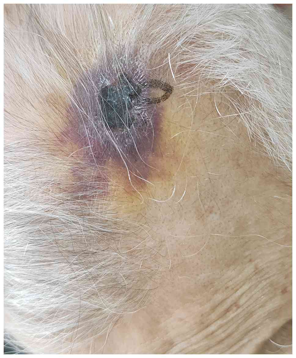
On physical examination, a 4×5 cm ill-defined
violaceous infiltrative plaque was observed on the vertex of the
scalp, with surrounding purpuric/ecchymotic discoloration, central
ulceration and dry crusting (Fig.
1). The lesion was firm on palpation and had poorly demarcated
borders. No palpable cervical lymphadenopathy was detected. No
additional suspicious cutaneous lesions were identified on routine
skin examination.
Routine laboratory investigations, including
complete blood count, liver and renal function tests, coagulation
profile and urinalysis, were all within normal limits. To evaluate
disease extent, computed tomography of the head and neck as well as
contrast-enhanced chest-abdomen imaging were performed, and no
evidence of regional lymph node involvement or distant metastasis
was detected.
Due to the atypical clinical appearance and poor
response to antibiotic therapy, a biopsy of the scalp lesion was
performed. The biopsy specimen was fixed in 10% neutral-buffered
formalin at room temperature for 24 h, routinely processed,
embedded in paraffin and sectioned at a thickness of 4 µm. The
sections were deparaffinized in xylene and rehydrated through a
graded ethanol series. Manual hematoxylin and eosin (H&E)
staining was performed at room temperature. Briefly, the sections
were stained with Harris hematoxylin for 5 min, rinsed in running
tap water, differentiated in 1% acid alcohol, blued in running tap
water and counterstained with eosin Y for 2 min. The stained
sections were subsequently dehydrated through graded ethanol,
cleared in xylene and mounted with neutral resin. Bright-field
images were acquired using a NanoZoomer digital slide scanner
(Hamamatsu Photonics K.K.), and representative images were reviewed
and exported using NDP.view2 software (version 2.9.29; Hamamatsu
Photonics K.K.). Histopathological examination of the specimen
revealed irregular, anastomosing vascular channels of varying size
within the dermis and subcutis. These slit-like and variably
dilated vascular spaces were lined by mildly atypical endothelial
cells exhibiting focal intraluminal protrusion and infiltrative
growth into the surrounding stroma (Fig. 2). The overall morphology was
consistent with a well-differentiated vasoformative malignant
vascular neoplasm.
For immunohistochemical analysis, 4 µm thick
sections were prepared from formalin-fixed, paraffin-embedded
tissue. The tissue was fixed in 10% neutral-buffered formalin at
room temperature for 24 h before routine processing and paraffin
embedding. Immunohistochemical staining was performed using an
Ultra 60 Plus automated immunohistochemistry stainer (OriGene
Technologies, Inc.). Heat-induced epitope retrieval for all nine
antibodies was performed within the instrument using an EDTA-based
antigen retrieval solution (pH 8.0; cat. no. ZLI-9067; OriGene
Technologies, Inc.) at 98°C for 25 min according to the
manufacturer's pre-optimized protocol. Endogenous peroxidase
activity was blocked with 3% hydrogen peroxide at room temperature
for 10 min.
All primary antibodies were supplied by OriGene
Technologies, Inc. in a ready-to-use format and required no further
dilution. The antibodies used were cytokeratin (CK; clone AE1/AE3;
cat. no. ZM-0069), CD31 (clone UMAB30; cat. no. ZM-0044), CD34
(clone QBEnd/10; cat. no. ZM-0046), ERG (clone UMAB78; cat. no.
ZM-0103), Ki-67 (clone UMAB107; cat. no. ZM-0166), C-MYC (clone
EP121; cat. no. ZA-0555), epithelial membrane antigen (EMA; clone
GP1.4; cat. no. ZM-0095), human herpesvirus 8 (HHV-8; clone 13B10;
cat. no. ZM-0404) and smooth muscle actin (SMA; clone UMAB237; cat.
no. ZM-0003). The sections were incubated with the ready-to-use
primary antibodies at 37°C for 60 min.
A universal two-step mouse/rabbit enhanced polymer
detection system (cat. no. PV-9000; OriGene Technologies, Inc.) was
used for immunodetection. The polymer detection reagent was
incubated at 37°C for 20 min, and immunoreactivity was visualized
using a 3,3′-diaminobenzidine (DAB) chromogenic detection kit (cat.
no. ZLI-9017; OriGene Technologies, Inc.). DAB color development
was performed at room temperature for 5 min, followed by
hematoxylin counterstaining at room temperature for 20 sec. All
staining procedures, including deparaffinization, antigen
retrieval, endogenous peroxidase blocking, primary antibody
incubation, polymer-based detection and chromogenic visualization,
were performed automatically according to the manufacturer's
standardized protocol. Appropriate positive controls were included
for each antibody. The immunohistochemically stained sections were
scanned using a NanoZoomer digital slide scanner (Hamamatsu
Photonics K.K.), and representative fields were reviewed and
exported using NDP.view2 software (version 2.9.29; Hamamatsu
Photonics K.K.).
Immunohistochemical staining demonstrated positivity
for CD31, CD34 and ERG, supporting endothelial differentiation. The
Ki-67 proliferation index was ~20% (Fig. 3), and C-MYC staining showed weak
focal positivity in ~10% of the lesional cells (Fig. S1A), whereas CK and EMA staining
were negative (Fig. S1B and C).
Additional immunohistochemical staining for HHV-8 and SMA was
subsequently performed on serial sections obtained from the same
representative paraffin block used for the original
immunohistochemical evaluation. HHV-8 staining was negative in the
lesional cells (Fig. 4A), whereas
SMA staining demonstrated the absence of a continuous SMA-positive
pericytic layer surrounding the irregular neoplastic vascular
channels (Fig. 4B). Taken together,
the histopathological and immunophenotypic findings supported the
diagnosis of well-differentiated primary cAS and argued against
Kaposi sarcoma.
Following clinicopathological confirmation of the
diagnosis and imaging exclusion of metastatic disease, the patient
underwent wide local excision in the Department of Surgery, First
People's Hospital of Horqin District (Tongliao, China), with a
gross peripheral margin of 2 cm from the clinically visible tumor
border. The lesion was excised en bloc to the level of the galea
aponeurotica. Intraoperative frozen-section examination showed no
tumor involvement of the circumferential peripheral margins or the
deep basal margin. Final permanent-section examination subsequently
confirmed that all submitted surgical margins were free of tumor;
however, the exact microscopic clearance distances were not
reported. The defect was reconstructed using an O-Z flap. The
postoperative course was uneventful. Adjuvant radiotherapy was not
administered because of the advanced age of the patient and
complete excision with negative surgical margins. At the most
recent outpatient follow-up in July 2026, ~8 months after surgery,
local examination showed no clinical evidence of recurrence.
Discussion
cAS most commonly affects elderly individuals and
often develops on the scalp and face (3,4). A
major reason for delayed diagnosis is that early lesions frequently
present as inconspicuous erythematous or violaceous patches,
bruise-like plaques or edematous infiltrative lesions, which can
resemble inflammatory or infectious processes rather than a
malignant vascular tumor (6–9). In
routine clinical practice, especially when scalp erythema, swelling
or crusting is present, benign conditions such as cellulitis or
dermatitis may be considered first, thereby postponing biopsy.
Several previously published case reports have
documented similar diagnostic pitfalls (Table I). Reported initial misdiagnoses
include cellulitis (6), diabetic
foot ulcer (7), refractory wound
(8), herpes zoster (9) and abscess (10). In those reports, the interval
between symptom onset and definitive diagnosis ranged from several
weeks to months. Such delays are clinically meaningful, as they may
allow continued local progression, reduce the likelihood of
complete surgical resection and adversely affect prognosis. The
cases summarized in Table I suggest
a recurring clinical pattern characterized by advanced age,
involvement of the scalp or face, violaceous or bruise-like
lesions, progressive enlargement and poor response to empirical
anti-inflammatory or antibiotic treatment.
 | Table I.Selected misdiagnosed cases of
cutaneous angiosarcoma in the literature. |
Table I.
Selected misdiagnosed cases of
cutaneous angiosarcoma in the literature.
| First author,
year | Age/sex | Location | Initial
misdiagnosis | Diagnostic delay | Outcome | (Refs.) |
|---|
| AlNodali et
al, 2024 | 87/F | Neck, face and
scalp | Cellulitis and
dermatitis | 2 months | Unresectable | (6) |
| Almheirat et
al, 2024 | 48/M | Foot | Diabetic foot
ulcer | 3 months | Deceased at 6 months
after amputation | (7) |
| Yan et al,
2023 | 78/M | Scalp | Refractory wound | 4 months | Surgical
resection | (8) |
| Yıldırım et
al, 2021 | 50/M | Scalp | Herpes zoster | 2 months | Deceased at 6
months | (9) |
| Lara-Martinez et
al, 2021 | 53/M | Face | Abscess | 4-6 months | Metastatic
progression | (10) |
The present case closely reflects this pattern. The
lesion was initially managed as cellulitis because of erythema,
swelling and subsequent ulceration. However, classic cellulitis
usually presents with warmth, tenderness and at least some degree
of systemic inflammatory response, and most cases improve with
appropriate antibiotic treatment. In the present patient, the
absence of systemic manifestations and the lack of clinical
improvement after antibiotic therapy should have prompted
reconsideration of the initial diagnosis. The present case
therefore emphasizes an important practical point: In elderly
patients, a progressive violaceous scalp lesion that does not
respond to conventional antibiotic therapy should raise suspicion
for malignancy and prompt early biopsy.
Histopathological examination remains the
cornerstone of diagnosis. Well-differentiated cAS typically
demonstrates irregular anastomosing vascular channels lined by
atypical endothelial cells infiltrating the dermis and subcutis,
whereas poorly differentiated lesions may show reduced
vasoformation and may mimic poorly differentiated carcinoma or
melanoma (2,4). Although the endothelial atypia in the
present case was relatively mild, the diagnosis of
well-differentiated cAS was supported by the irregular and
anastomosing vascular channels, infiltrative dissection of the
surrounding stroma, extension into the subcutis and focal
intraluminal endothelial protrusion. For this reason,
immunohistochemistry is of considerable diagnostic value.
Endothelial markers such as CD31, CD34, ERG and friend leukemia
integration 1 are commonly expressed, with CD31 and ERG considered
particularly useful markers of endothelial differentiation
(2,4). In the present case, the diagnosis was
supported by the characteristic vasoformative morphology together
with positivity for CD31, CD34 and ERG, and negativity for CK and
EMA, which helped exclude an epithelial neoplasm.
The principal histopathological differential
diagnoses included radiation-associated atypical vascular lesions,
benign vasoformative lesions and Kaposi sarcoma. A
radiation-associated atypical vascular lesion was considered
unlikely because the patient had no history of radiotherapy and
because the lesion demonstrated infiltrative involvement of both
the dermis and subcutis. The absence of chronic lymphedema also
supported classification of the tumor as primary rather than
lymphedema-associated cAS. Furthermore, SMA staining showed that
the irregular neoplastic vascular channels lacked a continuous
SMA-positive pericytic layer, supporting a malignant vasoformative
proliferation rather than a benign vascular lesion. HHV-8 staining
was negative in the lesional cells, arguing against Kaposi sarcoma.
C-MYC staining showed only weak focal positivity in ~10% of the
lesional cells, rather than diffuse strong expression; MYC gene
amplification was not evaluated. Taken together, these
morphological and immunophenotypic findings supported the diagnosis
of well-differentiated primary cAS.
Complete surgical excision with histologically
negative margins remains the cornerstone of treatment for localized
cAS (4,11). However, because cAS often exhibits
infiltrative and occasionally multifocal growth, achieving adequate
local control may be challenging. Adjuvant radiotherapy may improve
local control in selected cases, whereas systemic treatments,
including taxanes and anti-angiogenic agents, may be considered in
unresectable or metastatic disease (2,11–13).
Despite this, prognosis remains guarded, and multidisciplinary
management is generally recommended (5,14). In
the present patient, no distant metastasis was detected on
preoperative imaging, and wide local excision was performed with a
gross peripheral margin of 2 cm from the clinically visible tumor
border, extending to the level of the galea aponeurotica. Both
intraoperative frozen-section examination and final
permanent-section examination confirmed that the submitted
peripheral and deep margins were free of tumor, although the exact
microscopic clearance distances were not reported. Adjuvant
radiotherapy was not pursued because of the advanced age of the
patient and complete excision with negative surgical margins. At
the most recent outpatient follow-up in July 2026, ~8 months after
surgery, local examination showed no clinical evidence of
recurrence. Nevertheless, recurrence is common; in a retrospective
cohort of 143 patients with cAS of the face and scalp, 61 patients
(43%) developed local recurrence and 83 patients (58%) experienced
disease recurrence overall (15).
Therefore, long-term surveillance remains necessary.
The present report has certain limitations. First,
as a single case report, the conclusions that may be drawn are
limited. Second, the postoperative follow-up period was limited to
8 months and was based on clinical examination without repeat
staging imaging; therefore, longer follow-up is required to assess
long-term local control and metastatic risk. Third, although all
submitted surgical margins were tumor-free on permanent-section
examination, the exact microscopic clearance distances were not
documented in the original pathology report. In addition, C-MYC
expression was assessed using immunohistochemistry, but MYC gene
amplification was not evaluated. Despite these limitations, the
present case adds to the existing literature on the varied
presentation of cAS and reinforces the importance of early tissue
diagnosis in atypical scalp lesions. Reports of other rare sarcomas
have similarly emphasized the value of integrating detailed
morphological and immunophenotypic characterization with a review
of previously reported cases to improve diagnostic recognition
(16).
In summary, primary cAS of the scalp may clinically
mimic cellulitis, particularly in elderly patients. Failure to
respond to antibiotic therapy, especially in the setting of a
progressive violaceous scalp lesion, should prompt early biopsy and
histopathological evaluation. Early diagnosis and timely definitive
treatment are important for reducing diagnostic delay and
facilitating appropriate management.
Supplementary Material
Supporting Data
Acknowledgements
Not applicable.
Funding
Funding: No funding was received.
Availability of data and materials
The data generated in the present study are included
in the figures and/or tables of this article.
Authors' contributions
BQ conceived and designed the case report,
participated in the clinical diagnosis and management of the
patient, acquired and interpreted the clinical data, and drafted
the manuscript. QL and YG contributed to the acquisition, analysis
and interpretation of the clinical data, conducted the literature
review and critically revised the manuscript for important
intellectual content. YT collected the clinical data. YY
contributed to the conception and design of the case report,
participated in the clinicopathological diagnosis and therapeutic
decision-making, interpreted the clinical and pathological
findings, and critically revised the manuscript for important
intellectual content. BQ and YY confirm the authenticity of all the
raw data. All authors read and approved the final version of the
manuscript.
Ethics approval and consent to
participate
The present study was approved by the Ethics
Committee of the First People's Hospital of Horqin District
(Tongliao, China; approval no. 2026001). Written informed consent
was obtained from the patient.
Patient consent for publication
Written informed consent was obtained from the
patient for publication of the present case report and any
accompanying images.
Competing interests
The authors declare that they have no competing
interests.
References
|
1
|
Young RJ, Brown NJ, Reed MW, Hughes D and
Woll PJ: Angiosarcoma. Lancet Oncol. 11:983–991. 2010. View Article : Google Scholar : PubMed/NCBI
|
|
2
|
Cao J, Wang J, He C and Fang M:
Angiosarcoma: A review of diagnosis and current treatment. Am J
Cancer Res. 9:2303–2313. 2019.PubMed/NCBI
|
|
3
|
Shustef E, Kazlouskaya V, Prieto VG, Ivan
D and Aung PP: Cutaneous angiosarcoma: A current update. J Clin
Pathol. 70:917–925. 2017. View Article : Google Scholar : PubMed/NCBI
|
|
4
|
Yamakawa K, Ogata D, Namikawa K, Nakano E,
Yamaguchi Y and Yamazaki N: A review of cutaneous angiosarcoma:
Epidemiology, diagnosis, prognosis, and treatment options. Jpn J
Clin Oncol. 55:707–719. 2025. View Article : Google Scholar : PubMed/NCBI
|
|
5
|
Houpe JE, Seger EW, Neill BC, Kang BY,
Hocker TLH, Alam M and Tolkachjov SN: Treatment of angiosarcoma of
the head and neck: A systematic review. Cutis. 111:247–251. 2023.
View Article : Google Scholar : PubMed/NCBI
|
|
6
|
AlNodali N, Alqahtani N, Sharafuddin A,
Atef R, AlNodali H, Dhulaimi M, Alshomrani B and Alwagdani Y:
Cutaneous angiosarcoma masquerading as photodermatitis: A case
report. Acta Dermatovenerol Alp Pannonica Adriat. 33:213–215.
2024.PubMed/NCBI
|
|
7
|
Almheirat Y, Elyamani L, Hormi O, Zerrouki
N, Zizi N and Dikhaye S: Misdiagnosis of cutaneous epithelioid
angiosarcoma as diabetic foot ulcer: A case study. Wounds.
36:43–46. 2024. View Article : Google Scholar : PubMed/NCBI
|
|
8
|
Yan ZH, Li ZL, Chen XW, Lian YW, Liu LX
and Duan HY: Misdiagnosis of scalp angiosarcoma: A case report.
World J Clin Cases. 11:3099–3104. 2023. View Article : Google Scholar : PubMed/NCBI
|
|
9
|
Yıldırım S, Erdemoğlu Y, Durmaz EO, Erfan
G, Duman D and Dikicioğlu Çetin E: Primary cutaneous angiosarcoma
of the scalp with cranial invasion in a patient with metastatic
breast cancer. J Clin Aesthet Dermatol. 14:22–24. 2021.PubMed/NCBI
|
|
10
|
Lara-Martinez H, Weinberg M, Baratam P,
Horn J, Ward K and Styler M: Angiosarcoma of the face: A case study
and literature review of local and metastatic angiosarcoma. Case
Rep Oncol Med. 2021:88235852021.PubMed/NCBI
|
|
11
|
Florou V and Wilky BA: Current and future
directions for angiosarcoma therapy. Curr Treat Options Oncol.
19:142018. View Article : Google Scholar : PubMed/NCBI
|
|
12
|
Richards E, Schimmel J, Renzi M and
Lawrence N: An update on cutaneous angiosarcoma diagnosis and
treatment. Cutis. 113:218–223. 2024. View Article : Google Scholar : PubMed/NCBI
|
|
13
|
Klein JC and Dominguez AR: Cutaneous
angiosarcoma. JAMA Dermatol. 159:3322023. View Article : Google Scholar : PubMed/NCBI
|
|
14
|
Sturm EC, Marasco IS and Katz SC:
Multidisciplinary management of angiosarcoma: A review. J Surg Res.
257:213–220. 2021. View Article : Google Scholar : PubMed/NCBI
|
|
15
|
Yoder AK, Farooqi AS, Wernz C, Subramaniam
A, Ravi V, Goepfert R, Sturgis EM, Mitra D, Bishop AJ and
Guadagnolo BA: Outcomes after definitive treatment for cutaneous
angiosarcomas of the face and scalp: Reevaluating the role of
surgery and radiation therapy. Head Neck. 45:1943–1951. 2023.
View Article : Google Scholar : PubMed/NCBI
|
|
16
|
Bai Q, Lin Y, Fu W, Ning Y, Niu Q, Su W,
Wang X, Lan Y, Wang Y, Zhang Y, et al: Characteristics of
intracranial sclerosing epithelioid fibrosarcoma and review of
related literature. Oncol Transl Med. 11:187–194. 2025. View Article : Google Scholar
|