Introduction
Esophageal cancer is the most common malignant tumor
of the digestive system. Globally, the number of new cases of
esophageal cancer ranks 7th and 13th among malignant tumors in male
and female patients, respectively. China accounts for 53.7 and
55.3% of the global incidence and mortality, rates respectively
(1). The mortality rate of
esophageal cancer in China is second only to gastric cancer among
malignant tumors of the digestive system. There are two main
histological types of esophageal cancer: Esophageal squamous cell
carcinoma (ESCC), which is the predominant type (accounting for
~90% of cases) present in China, and esophageal adenocarcinoma.
Despite the effectiveness of surgical treatment, radiotherapy,
chemotherapy and immunotherapy for esophageal cancer having
improved, the majority of patients are diagnosed at an advanced
stage. In addition, the high rates of postoperative recurrence and
metastasis contribute to poor prognosis, with a 5-year overall
survival rate of ~25% (2).
Therefore, identifying specific molecular markers and investigating
their molecular mechanisms in the occurrence and progression of
esophageal cancer are of great importance for improving diagnosis,
treatment outcomes and long-term survival rates.
Zic family members (ZIC) are transcriptional
regulatory factors containing highly conserved C2H2 zinc finger
motifs. A total of five ZIC genes (ZIC1-5) have been identified and
studied during embryonic development in vertebrates (3,4). These
identified genes are vertebrate homologs of the Drosophila
odd-paired genes, structurally similar to each other and partially
functionally identical (5). As a
member of the Zic family, ZIC5 has been reported to be involved in
the malignant progression of numerous types of human cancer, such
as cervical cancer (6), prostate
cancer (7), melanoma (8), liver cancer (9), colon cancer (10) and gastric cancer (11). However, there have been a limited
number of studies regarding the expression and underlying mechanism
of ZIC5 in ESCC.
Therefore, the present study used bioinformatics
methods to analyze the expression levels of ZIC5 in ESCC and normal
esophageal tissue, as well as analyze its association with the
survival of patients with ESCC. The possible functional mechanism
underlying this was also explored. Immunohistochemistry was used to
determine the expression of ZIC5 protein in ESCC and further
analysis was conducted regarding its association with clinical
characteristics and prognosis. The present findings therefore
provide a theoretical basis for the diagnosis and prognosis of ESCC
and lay the foundation for improving the survival rate of
patients.
Materials and methods
Data collection and analysis
Firstly, the expression levels of the ZIC5 gene in
various pan-cancer samples from The Cancer Genome Atlas (TCGA;
portal.gdc.cancer.gov/) on the University of California, Santa Cruz
Xena platform (xenabrowser.net) were extracted. An unpaired
Student's t-test was used to compare the expression of the ZIC5
gene between tumor and normal tissues in various cancer types and
box plots were drawn to observe the differential expression of the
ZIC5 gene in different cancer types. To observe the expression of
the ZIC5 gene in ESCC samples, samples with the pathological type
of ESCC from TCGA esophageal cancer dataset (96 squamous cell
carcinomas and 11 adjacent tissues) were extracted. Box plots were
plotted showing the expression of the ZIC5 gene in ESCC and normal
tissues. The GSE53625 dataset for ESCC was downloaded from National
Center for Biotechnology Information Gene Expression Omnibus (GEO;
ncbi.nlm.nih.gov/geo), which contains gene expression profiles of
cancer tissues and paired adjacent tissues from 179 ESCC samples
(12). Further box plots were
plotted comparing the expression of the ZIC5 gene between ESCC and
control groups in GSE53625. A paired Student's t-test was used to
calculate the significance P-value.
Prognostic value analysis of ZIC5 in
ESCC
Overall survival (OS) of the samples in the
TCGA-ESCC and GSE53625 datasets and the expression values of the
ZIC5 gene in the corresponding samples were extracted. Survival
analysis was performed using the R ‘survival’ package (version
3.8–9, CRAN.R-project.org/package=survival) on TCGA-ESCC and
GSE53625 ESCC samples. A log-rank P<0.05 was considered to
indicate a statistically significant difference and survival curves
were subsequently plotted using the same R package.
Transcriptional regulation analysis of
ZIC5
miRWalk (version 3.0; http://mirwalk.umm.uni-heidelberg.de) was used to
predict microRNA (miRNA/miR) binding sites on ZIC5. Default
parameters were applied, specifically a binding probability ≥0.95
and binding site localization within the 3′-untranslated region, to
identify miRNA-mRNA interactions. The online tool miRNA Target
Prediction Database (miRDB; http://mirdb.org/) was used for miRNA and target gene
prediction, selecting an miRDB score ≥50 as the threshold.
Similarly, the TargetScan 8.0 database (https://www.targetscan.org/vert_80/) was accessed for
miRNA-target gene prediction, applying a total context++ score
<0 as the cut-off criterion. At least two of the three databases
were identified as having miRNAs predicted to target ZIC5 as
potential regulators of ZIC5 expression.
Analysis of downstream pathways
regulated by ZIC5 gene
Molecular Signatures Database (version 7.1;
http://software.broadinstitute.org/gsea/msigdb/index.jsp)
was used for pathway analysis. Kyoto Encyclopedia of Genes and
Genomes (KEGG; genome.jp/kegg/) pathway gene sets, specifically the
'c2.cp.kegg.v7.4.symbols.gmt' was used as the enrichment
background. Based on the expression values of all genes in all
tumor samples of TCGA-ESCC and GSE53625, using the Pearson
correlation coefficient ranking between each gene and ZIC5, Gene
Set Enrichment Analysis (GSEA) was conducted using local GSEA
software (version 4.3.1, Broad Institute; http://www.gsea-msigdb.org/gsea/index.jsp) to identify
the positive and negative pathways associated with ZIC5 expression.
Significant enrichment was defined as normalized enrichment score
>1.5 with enrichment P<0.05. Subsequently, the ‘ggplot2’
package (version 4.0.3, ggplot2.tidyverse.org/) within R software
was used to construct a GSEA enrichment analysis bubble plot.
Clinical data collection and
follow-up
A total of 20 paired fresh ESCC and adjacent
non-cancerous tissue specimens from patients presented at Tianjin
Medical University Cancer Institute and Hospital (Tianjin, China)
from January 2025 to June 2025 were collected prospectively for
western blotting analysis. Among the 20 patients, 16 were male and
four were female, with a median age of 61 years (range: 55–74
years). Furthermore, 176 specimens of ESCC and normal esophageal
tissue >5 cm away from the cancer periphery were collected from
patients that underwent surgical treatment in Tianjin Medical
University Cancer Institute and Hospital from January 2009 to
December 2010 for immunohistochemistry analysis. The inclusion
criteria were as follows: i) Histological diagnosis of ESCC; ii) no
history of other malignant tumors and; iii) specimens had not
received radiotherapy, chemotherapy or immunotherapy before
surgery. The exclusion criteria were as follows: i) Specimens with
a histological diagnosis other than ESCC or with an uncertain
diagnosis; ii) cases with incomplete clinical or follow-up data;
and iii) specimens with poor quality or adjacent non-cancerous
tissues located less than 5 cm from the tumor margin. Among the 176
patients, 144 were male and 32 were female, with a median age of 68
years (range, 39–85 years). Clinical data of patients were
collected and followed up on patient recurrence and survival status
via telephone. The samples were accessed from the institutional
biobank between January 2023 and March 2023. The collection of both
the 20 paired samples and the 176 tissue specimens in the present
study was approved by the Tianjin Medical University Cancer
Institute and Hospital ethics committee (approval no. bc2021340)
and written informed consent was obtained from all patients.
Western blotting
Total protein was extracted from ESCC tumor tissue
using RIPA lysis buffer (Beyotime Biotechnology) supplemented with
protease inhibitor cocktail and 1 mM PMSF. Subsequently, a BCA
protein assay kit (Pierce; Thermo Fisher Scientific, Inc.) was
utilized for protein quantitation. Equal amounts of protein (20 µg
per lane) were separated by 10% SDS-PAGE, transferred onto PVDF
membranes and blocked with TBS-Tween containing 5% skimmed milk for
1 h at ambient temperature. Subsequently, the membrane was
incubated with primary antibodies overnight at 4°C, followed by
incubation with secondary antibody for 2 h at room temperature. The
primary antibodies were as following: ZIC5 (1:500; cat. no.
bs-12147R; BIOSS), and GAPDH (1:5,000; cat. no. 10494-1-AP;
Proteintech Group, Inc.). The secondary antibody was HRP-conjugated
goat anti-rabbit IgG (1:1,000; cat. no. 7074S; Cell Signaling
Technology, Inc.). Enhanced chemiluminescence reagent (Merck
Millipore) was used to visualize the immunoreactive bands. ImageJ
(version 1.53e; National Institutes of Health) was used to measure
and analyze the optical density.
Immunohistochemical detection
ZIC5 protein levels were assessed through
immunohistochemistry. Briefly, tissue samples were fixed in 10%
neutral-buffered formalin for 24 h at room temperature and embedded
in paraffin. Sections were cut at a thickness of 4 µm,
deparaffinized in xylene and rehydrated through a descending
ethanol series. Antigen retrieval was performed by heating the
sections in 10 mM sodium citrate buffer at 95–100°C for 15 min.
Endogenous peroxidase activity was quenched using 0.3% hydrogen
peroxide and the sections were blocked in 5% goat serum (Vector
Laboratories) at room temperature for 1 h. Sections were incubated
overnight at 4°C with an anti-ZIC5 polyclonal antibody (1:100; cat.
no. bs-12147R; BIOSS), then exposed to a biotinylated goat
anti-rabbit IgG secondary antibody (cat. no. BA-1000; Vector
Laboratories; 1:200) for 20 min at room temperature.
Diaminobenzidine served as the chromogenic substrate and slides
were initially examined under a light microscope (Olympus BX53,
Japan) and digitized using an automated slide scanning system.
Results were independently evaluated in a
double-blind manner by two experienced pathologists who were
blinded to the clinicopathological information.
Immunohistochemistry scoring was performed as previously described
(13). Staining intensity was
graded as 0 (negative), 1 (weak, light yellow), 2 (moderate, yellow
brown) or 3 (strong, brown). The extent of staining was scored as
follows: 0, <10%; 1, 10–25%; 2, 26–50%; 3, 51–75%; and 4,
>75%. The immunoreactivity score was calculated by multiplying
the intensity score by the extent score. Patients with a total
score <4 were considered to have low expression, and those with
a total score of ≥4 were considered to have high expression.
Statistical analysis
Statistical analysis was performed using SPSS
(version 25.0; IBM Corp.) and R software (version 4.2.1; R
Foundation for Statistical Computing). The ‘limma’ package (v.3.21)
was used to identify differentially expressed genes between
ZIC5-high and ZIC5-low groups, and the resulting gene ranking was
subsequently used for GSEA. Categorical data are presented as
numbers and percentages. Parametric continuous data are presented
as the mean ± SD). Independent sample Student's t-tests were used
for comparisons between two groups, while multiple-group
comparisons were performed using one-way ANOVA. The associations
between ZIC5 expression and clinicopathological factors were
assessed using the χ2 test or Fisher's exact test.
Survival rates were calculated using the Kaplan-Meier method and
differences in survival curves were assessed using log-rank tests.
Cox regression analysis was conducted in both univariate and
multivariate models to determine independent factors affecting
prognosis. All statistical tests were two-sided and P<0.05 was
considered to indicate a statistically significant difference.
Results
Expression of ZIC5 gene in ESCC
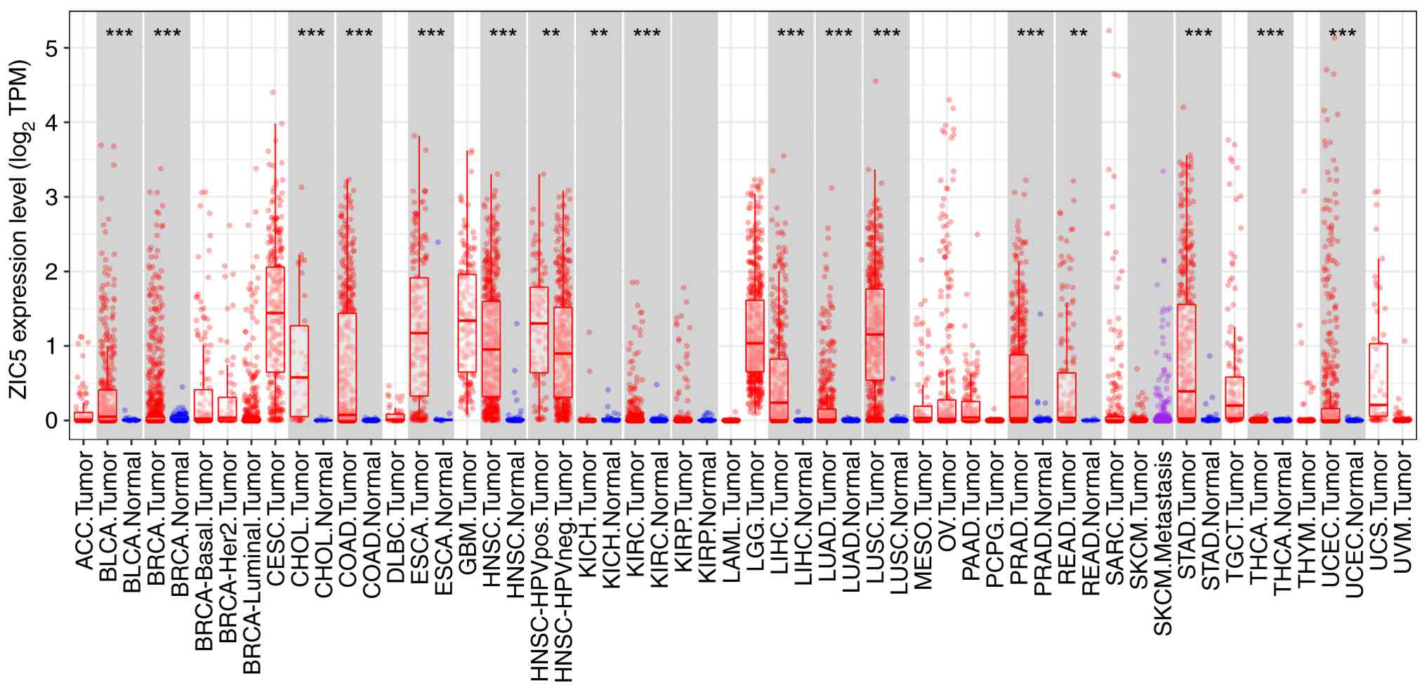
ZIC5 expression was significantly increased in head
and neck squamous cell carcinoma, breast cancer, esophageal cancer,
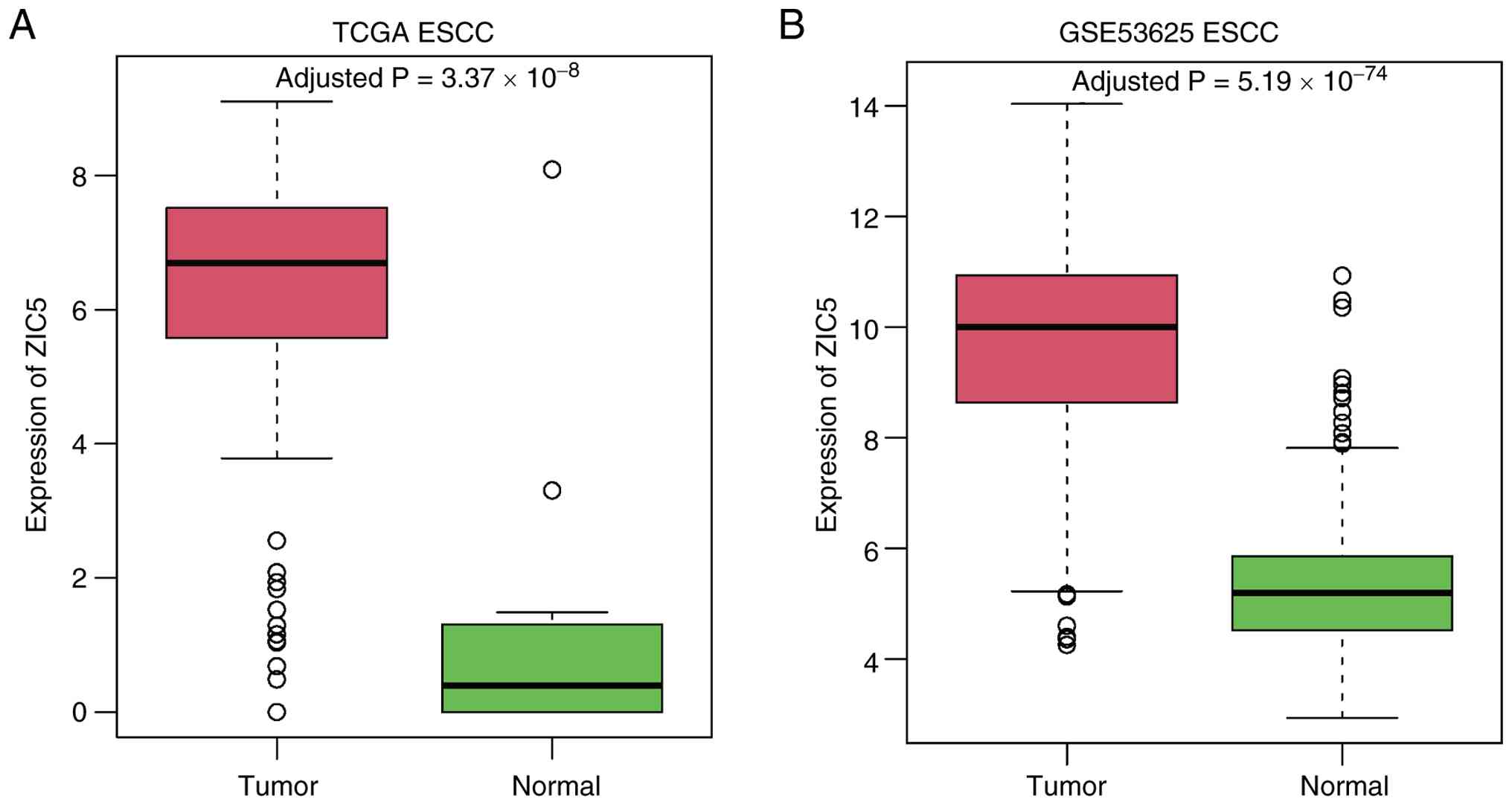
gastric cancer and bladder cancer, amongst others (Fig. 1). To examine ZIC5 expression in
ESCC, box plots of ZIC5 expression were generated based on the
TCGA-ESCC and GSE53625-ESCC datasets to compare ESCC samples with
controls. ZIC5 expression was significantly upregulated in ESCC
samples when compared with the control group, with statistically
significant differences (Fig.
2).
Impact of ZIC5 mRNA on the prognosis
of ESCC
Survival analysis was performed on TCGA-ESCC and
GSE53625-ESCC samples using the optimal cut-off value determined by
X-tile for grouping. As shown in Fig.
3, ZIC5 expression was associated with the prognosis of
patients with ESCC. Patients with high ZIC5 expression exhibited
significantly shorter survival times when compared with those with
low expression.
Transcriptional regulation analysis of
ZIC5
As aforementioned, miRNA target prediction for ZIC5
was performed. The miRNAs predicted by at least two of the three
databases were identified, as summarized in Table SI. Analysis revealed that 40 miRNAs
potentially regulate ZIC5, among which hsa-miR-212-5p was predicted
by all three databases (Fig.
4).
Analysis of downstream pathways
regulated by ZIC5
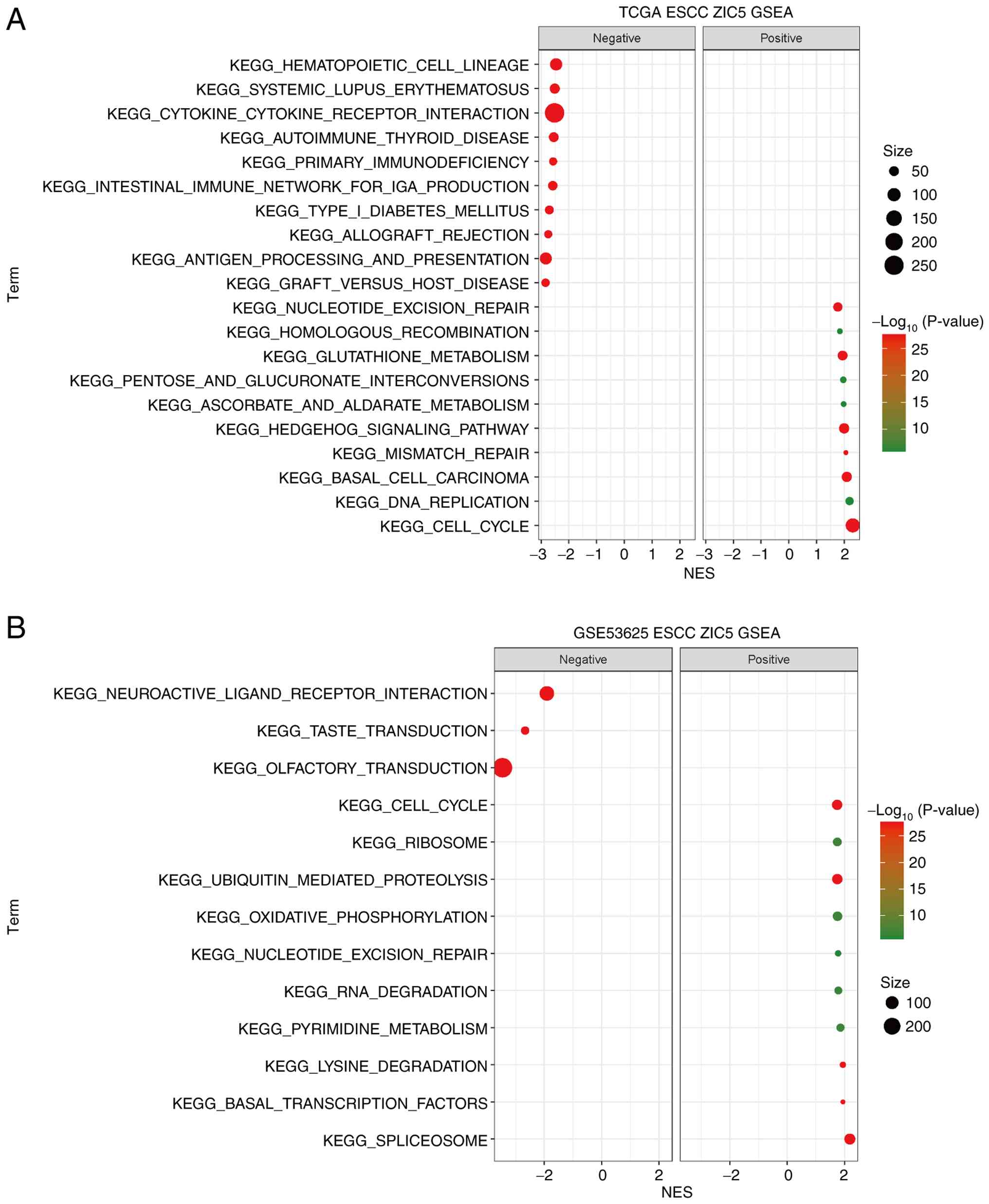
KEGG pathway enrichment analysis for ZIC5 was
conducted by GSEA on TCGA-ESCC and GSE53625-ESCC datasets. From
each dataset, the top pathways exhibiting positive and negative
association with ZIC5 were selected and visualized as a bubble plot
(Fig. 5).
Enrichment score profiles revealed that
KEGG_CELL_CYCLE and KEGG_NUCLEOTIDE_EXCISION_REPAIR were
consistently enriched in ZIC5-high tumors in both datasets
(Fig. 6). In both cohorts, the
maximum enrichment was reached near the leading edge of the ranked
gene list, indicating that the enrichment was driven by a
coordinated set of core pathway genes rather than by a few
outliers. Functionally, the enrichment of the cell-cycle pathway
suggests that ZIC5 may promote tumor proliferation by accelerating
cell division, whereas the enrichment of the nucleotide excision
repair pathway implies an enhanced DNA damage repair capacity that
helps maintain genomic stability and evade apoptosis.
Expression of ZIC5 protein in ESCC
tissue and its association with prognosis
First, the significant upregulation of ZIC5 in 20
paired fresh ESCC tissues compared with adjacent paracancerous
tissues was validated through western blotting (Fig. 7A). To further explore the expression
of ZIC5 protein in ESCC tissues and its association with
clinicopathological characteristics and prognosis, 176
treatment-naïve ESCC tissue specimens and normal esophageal tissue
specimens were collected from the Tianjin Medical University Cancer
Institute and Hospital, and ZIC5 protein expression was detected.
Immunohistochemical results showed that ZIC5 protein expression was
primarily localized in the nucleus of cancer cells, and the
expression rate was significantly higher in ESCC tissues than in
normal tissues (68.2% vs. 44.3%). Based on ZIC5 expression levels,
all ESCC patients were divided into a low-expression group (n=56)
and a high-expression group (n=120). Representative images are
shown in Fig. 7B-E).
The association between ZIC5 expression and
different clinicopathological characteristics is shown in Table I. High ZIC5 expression was
associated with tumor size, invasion depth, lymph node metastasis
and TNM staging. By contrast, ZIC5 expression displayed no
significant association with sex, age, smoking history, alcohol
intake, tumor location or histological grade.
 | Table I.Association between ZIC5 expression
and clinical pathological characteristics in patients with
esophageal squamous cell carcinoma. |
Table I.
Association between ZIC5 expression
and clinical pathological characteristics in patients with
esophageal squamous cell carcinoma.
|
|
| ZIC5
expression |
|
|
|---|
|
|
|
|
|
|
|---|
| Variable | n | Low expression | High
expression | χ2 | P-value |
|---|
| Total | 176 | 56 (31.8%) | 120 (68.2%) |
|
|
| Sex |
|
|
| 0.838 | 0.360 |
|
Male | 144 | 48 (33.3%) | 96 (66.7%) |
|
|
|
Female | 32 | 8 (25.0%) | 24 (75.0%) |
|
|
| Age, years |
|
|
| 2.728 | 0.099 |
|
≤68 | 82 | 21 (25.6%) | 61 (74.4%) |
|
|
|
>68 | 94 | 35 (37.2%) | 59 (62.8%) |
|
|
| Smoking
history |
|
|
| 0.274 | 0.600 |
|
Yes | 121 | 37 (30.6%) | 84 (69.4%) |
|
|
| No | 55 | 19 (34.5%) | 36 (65.5%) |
|
|
| Alcohol intake |
|
|
| 1.265 | 0.261 |
|
Yes | 105 | 30 (28.6%) | 75 (71.4%) |
|
|
| No | 71 | 26 (36.6%) | 45 (63.4%) |
|
|
| Tumor location |
|
|
| 5.497 | 0.064 |
|
Upper | 7 | 0 (0%) | 7 (100.0%) |
|
|
|
Middle | 148 | 49 (33.1%) | 99 (66.9%) |
|
|
|
Lower | 21 | 7 (33.3%) | 14 (66.7%) |
|
|
| Tumor size, cm |
|
|
| 4.949 | 0.026a |
|
<3.5 | 73 | 30 (41.1%) | 43 (58.9%) |
|
|
|
≥3.5 | 103 | 26 (25.2%) | 77 (74.8%) |
|
|
| Histological
grade |
|
|
| 2.023 | 0.364 |
| G1 | 9 | 3 (33.3%) | 6 (66.7%) |
|
|
| G2 | 131 | 45 (34.4%) | 86 (65.6%) |
|
|
| G3 | 36 | 8 (22.2%) | 28 (77.8%) |
|
|
| Invasion depth |
|
|
| 6.706 | 0.035a |
|
pT1-2 | 30 | 14 (46.7%) | 16 (53.3%) |
|
|
|
pT3 | 95 | 32 (33.7%) | 63 (66.3%) |
|
|
|
pT4 | 51 | 10 (19.6%) | 41 (80.4%) |
|
|
| Lymph node
metastasis |
|
|
| 5.315 | 0.021a |
|
Yes | 107 | 41 (38.3%) | 66 (61.7%) |
|
|
| No | 69 | 15 (21.7%) | 54 (78.3%) |
|
|
| TNM staging |
|
|
| 11.865 | 0.001a |
|
I–II | 68 | 32 (47.1%) | 36 (52.9%) |
|
|
|
III | 108 | 24 (22.2%) | 84 (77.8%) |
|
|
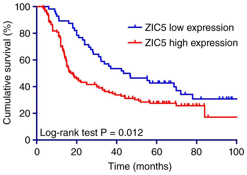
In addition, Kaplan-Meier survival analysis revealed
that patients with high ZIC5 levels exhibited a significantly worse
5-year OS rate compared with those with low ZIC5 levels (27.5 vs.
42.7%; Fig. 8). Cox univariate
analysis showed that age, tumor size, invasion depth, lymph node
metastasis and ZIC5 protein expression were all associated with
patient survival. Multivariate analysis indicated that age, lymph
node metastasis and ZIC5 protein expression levels (hazard
ratio=1.519; 95% CI: 1.017–2.269; Table II) were independent prognostic
indicators affecting the survival of patients with ESCC.
| Table II.Impact of ZIC5 and clinical
pathological features on the prognosis of patients with esophageal
squamous cell carcinoma. |
Table II.
Impact of ZIC5 and clinical
pathological features on the prognosis of patients with esophageal
squamous cell carcinoma.
|
| Univariate
analysis | Multivariate
analysis |
|---|
|
|
|
|
|---|
| Variable | HR | 95% CI | P-value | HR | 95% CI | P-value |
|---|
| Sex (Female vs.
Male) | 0.966 | 0.767–1.217 | 0.772 |
|
|
|
| Age, years (>68
vs. ≤68) | 1.459 | 1.022–2.083 | 0.038a | 1.518 | 1.056–2.183 | 0.024a |
| Smoking history
(Yes vs. No) | 1.107 | 0.753–1.627 | 0.604 |
|
|
|
| Alcohol intake (Yes
vs. No) | 1.304 | 0.907–1.874 | 0.152 |
|
|
|
| Tumor location
(Lower vs. Middle vs. Upper) | 0.835 | 0.543–1.284 | 0.411 |
|
|
|
| Tumor size, cm
(≥3.5 vs. <3.5) | 1.497 | 1.040–2.156 | 0.030a | 1.288 | 0.887–1.869 | 0.184 |
| Histological grade
(G3 vs. G2 vs. G1) | 1.301 | 0.908–1.864 | 0.152 |
|
|
|
| Invasion depth (pT4
vs. pT3 vs. pT1-2) | 1.438 | 1.100–1.880 | 0.008a | 1.300 | 0.980–1.725 | 0.069 |
| Lymph node
metastasis (Yes vs. No) | 2.049 | 1.438–2.919 | 0.000a | 1.733 | 1.207–2.490 | 0.003a |
| ZIC5 expression
(High vs. Low) | 1.645 | 1.117–2.421 | 0.012a | 1.519 | 1.017–2.269 | 0.041a |
Discussion
Esophageal cancer is a common malignant tumor of the
upper gastrointestinal tract. Due to the atypical symptoms of early
esophageal cancer, the majority of patients seek medical attention
at middle to late stages, with a 5-year survival rate of only
15–25% (14–16). Therefore, identifying effective
biomarkers is of great clinical importance for the early diagnosis
of esophageal cancer. In the present study, the expression of the
ZIC5 gene in pan-cancer was first analyzed, revealing that ZIC5
expression was significantly increased in head and neck squamous
cell carcinoma, breast cancer, esophageal cancer, gastric cancer
and bladder cancer. To investigate ZIC5 expression in ESCC, ZIC5
mRNA expression levels in TCGA and GEO databases were analyzed,
revealing that ZIC5 was highly expressed in ESCC. In addition,
elevated ZIC5 protein expression level was significantly associated
with poor prognosis in patients with ESCC.
Members of the Zic family serve important roles in
neural development and tumorigenesis. As a member of the cerebellar
zinc finger family, ZIC5 has been reported as a transcription
factor involved in regulating the expression of target genes
(5). A number of studies have found
that ZIC5 is highly expressed in certain malignant tumors,
including lung cancer, prostate cancer and glioma (17–19).
Zeng et al (20) found that
ZIC5 is highly expressed in lung adenocarcinoma tissue. In A549 and
H1299 cells, silencing ZIC5 suppressed its expression, inhibited
cell proliferation and attenuated metabolic activity as evidenced
by reduced glucose uptake, lactate production and ATP levels.
Associated research has also been conducted regarding ZIC5 in
digestive system tumors. Satow et al (21) found that ZIC5 was upregulated in
colon cancer and promoted the proliferation and primary drug
resistance of colon cancer cells. The OS rate of patients with
pancreatic cancer with high ZIC5 expression was markedly lower
compared with that of patients with low ZIC5 expression. Targeting
ZIC5 therefore enhances the efficacy of gemcitabine treatment by
effectively reducing tumor volume in pancreatic cancer in
vivo (22). ZIC5 can markedly
inhibit the proliferation of liver cancer cells (9) and induce apoptosis of colon cancer,
pancreatic cancer and cholangiocarcinoma cells after being silenced
(21,23).
ZIC5 may serve a role in the occurrence and
development of tumors through the following pathways: i) ZIC5 may
act as a proto-oncogene to affect the expression of cyclin B1 and
CDK1 complexes and participate in regulating cell proliferation
(17); ii) ZIC5 promotes tumor cell
proliferation and invasion by regulating the activity of certain
miRNAs (19,24); iii) ZIC5 may promote tumor growth,
invasion and migration by regulating the Wnt/β-catenin signaling
pathway (10); iv) ZIC5 promotes
malignant progression of tumor cells by regulating the CDK1/CDC25c
signaling pathway (25). However,
there are limited research reports on the expression and mechanism
of action of ZIC5 in ESCC.
miRNA serves an important regulatory role in
tumorigenesis. In recent years, numerous studies have found that
miRNAs are abnormally expressed in certain types of malignant
tumors and exert notable effects on cell proliferation, apoptosis,
invasion and migration, amongst other processes. mRNAs, as targets
of miRNAs, also serve important roles in tumor malignancy (26,27).
The present study was based on analysis results from three
databases. ZIC5 may be a target gene of hsa-miR-212-5p and also
serves an important role in the malignant progression of ESCC. A
number of studies have shown that hsa-miR-212-5p exhibits
carcinogenic properties in different tumor types (28,29).
Qu et al (30) found that
circZFR regulates the activity of superoxide dismutase 2 by
inhibiting hsa-miR-212-5p, thereby promoting the progression of
ovarian cancer cells. In addition, further analysis was carried out
of the downstream pathways regulated by the ZIC5 gene through GSEA,
with results showing that ZIC5 may promote the proliferation of
ESCC cells by participating in the cell cycle and repair processes.
However, these results need to be further determined through gain-
and loss-of-function experiments, transcriptomic profiling and
targeted molecular assays.
There are a number of limitations to the present
study. Firstly, the present study was a single-institute study with
a small sample size and there may be selection bias during patient
and data collection. In addition, the present study did not
incorporate functional experiments to validate the specific
mechanisms by which ZIC5 influences ESCC progression. This lack of
experimental validation may limit the robustness of the present
conclusions regarding the role of ZIC5 in ESCC. Furthermore, the
absence of multi-center validation may hinder the external
applicability of the present results. Lastly, inherent differences
between the datasets used (TCGA and GSE53625) could introduce batch
effects, potentially impacting the consistency of the present
findings across different cohorts.
In conclusion, bioinformatics was used to screen for
differentially expressed ZIC5 associated with the prognosis of
patients with ESCC through TCGA and GEO databases, with the
expression of ZIC5 protein in ESCC tissues further verified through
immunohistochemistry. The results showed that ZIC5 is highly
expressed in ESCC tissues and is notably associated with poor
prognosis in patients with ESCC. ZIC5 may be a target gene of
hsa-miR-212-5p that regulates the malignant progression of ESCC
cells. Further in vivo and in vitro experimental
studies are needed to validate these findings. The present study
highlighted that the ZIC5 gene has the potential to become a new
prognostic marker for ESCC and may provide a reference for targeted
therapy and prognosis of patients with ESCC.
Supplementary Material
Supporting Data
Acknowledgements
Not applicable.
Funding
The present study was supported by the National Natural Science
Foundation of China (grant no. 82002551).
Availability of data and materials
The data generated in the present study may be
requested from the corresponding author.
Authors' contributions
BY, BW, YZ and ZZ conceived and designed the present
workflow. BY, BW, HZ and ZZ performed the experiments and analyzed
the data. HD and YL conducted the bioinformatics analysis. SW and
HY contributed to the histological analysis. BY and BW wrote the
manuscript. BW, YZ and ZZ revised the manuscript. All authors read
and approved the final manuscript. YZ and ZZ confirm the
authenticity of all the raw data.
Ethics approval and consent to
participate
All methods were performed in accordance with the
relevant ethical regulations and were conducted under approval from
the Research Ethics Committee of Tianjin Medical University Cancer
Institute and Hospital (Tianjin, China; approval no. bc2021340).
Written informed consent was acquired from all patients of the
primary cohort for the acquisition of clinical and pathological
information and the use of surgical specimens.
Patient consent for publication
The patients provided written informed consent for
publication of their data.
Competing interests
The authors declare that they have no competing
interests.
Glossary
Abbreviations
Abbreviations:
|
ESCC
|
esophageal squamous cell carcinoma
|
|
ZIC5
|
Zic family member 5
|
|
TCGA
|
The Cancer Genome Atlas
|
|
GEO
|
Gene Expression Omnibus
|
|
OS
|
overall survival
|
|
GSEA
|
Gene Set Enrichment Analysis
|
|
KEGG
|
Kyoto Encyclopedia of Genes and
Genomes
|
References
|
1
|
Sung H, Ferlay J, Siegel RL, Laversanne M,
Soerjomataram I, Jemal A and Bray F: Global cancer statistics 2020:
GLOBOCAN estimates of incidence and mortality worldwide for 36
cancers in 185 countries. CA Cancer J Clin. 71:209–249.
2021.PubMed/NCBI
|
|
2
|
Meng P, Gu T, Xu J, Huang H, Jin H, Wang
Y, Zhang H and Ruan Z: Pretreatment CALLY index as promising novel
biomarker in the prediction of surgical and oncological outcomes in
esophageal cancer: A multi-center retrospective cohort study. Front
Immunol. 16:16050672025. View Article : Google Scholar : PubMed/NCBI
|
|
3
|
Grinberg I and Millen KJ: The ZIC gene
family in development and disease. Clin Genet. 67:290–296. 2005.
View Article : Google Scholar : PubMed/NCBI
|
|
4
|
Houtmeyers R, Souopgui J, Tejpar S and
Arkell R: The ZIC gene family encodes multi-functional proteins
essential for patterning and morphogenesis. Cell Mol Life Sci.
70:3791–3811. 2013. View Article : Google Scholar : PubMed/NCBI
|
|
5
|
Ali RG, Bellchambers HM and Arkell RM:
Zinc fingers of the cerebellum (Zic): Transcription factors and
co-factors. Int J Biochem Cell Biol. 44:2065–2068. 2012. View Article : Google Scholar : PubMed/NCBI
|
|
6
|
Jia Q, Song J, Xu T, Liu J, Chai J, Yang
Y, Li L, Li M and Yang X: ZIC5 promotes aggressiveness and cancer
stemness in cervical squamous cell carcinoma. Pathol Res Pract.
241:1542682023. View Article : Google Scholar : PubMed/NCBI
|
|
7
|
Tan YF, Zhang Y, Ge SY, Zhong F, Sun CY
and Xia GW: AR-regulated ZIC5 contributes to the aggressiveness of
prostate cancer. Cell Death Discov. 8:3932022. View Article : Google Scholar : PubMed/NCBI
|
|
8
|
Satow R, Nakamura T, Kato C, Endo M,
Tamura M, Batori R, Tomura S, Murayama Y and Fukami K: ZIC5 drives
melanoma aggressiveness by PDGFD-mediated activation of FAK and
STAT3. Cancer Res. 77:366–377. 2017. View Article : Google Scholar : PubMed/NCBI
|
|
9
|
Liu L, Hu X, Sun D, Wu Y and Zhao Z: ZIC5
facilitates the growth of hepatocellular carcinoma through
activating Wnt/β-catenin pathway. Biochem Biophys Res Commun.
503:2173–2179. 2018. View Article : Google Scholar : PubMed/NCBI
|
|
10
|
Zhao Z, Wang L, Bartom E, Marshall S,
Rendleman E, Ryan C, Shilati A, Savas J, Chandel N and Shilatifard
A: β-Catenin/Tcf7l2-dependent transcriptional regulation of GLUT1
gene expression by Zic family proteins in colon cancer. Sci Adv.
5:eaax06982019. View Article : Google Scholar : PubMed/NCBI
|
|
11
|
Ma G, Dai W, Sang A, Yang X and Li Q:
Roles of ZIC family genes in human gastric cancer. Int J Mol Med.
38:259–266. 2016. View Article : Google Scholar : PubMed/NCBI
|
|
12
|
Li J, Chen Z, Tian L, Zhou C, He MY, Gao
Y, Wang S, Zhou F, Shi S, Feng X, et al: LncRNA profile study
reveals a three-lncRNA signature associated with the survival of
patients with oesophageal squamous cell carcinoma. Gut.
63:1700–1710. 2014. View Article : Google Scholar : PubMed/NCBI
|
|
13
|
Zhang H, Han Y, Xiao W, Gao Y, Sui Z, Ren
P, Meng F, Tang P and Yu Z: USP4 promotes the proliferation,
migration, and invasion of esophageal squamous cell carcinoma by
targeting TAK1. Cell Death Dis. 14:7302023. View Article : Google Scholar : PubMed/NCBI
|
|
14
|
He S, Xu J, Liu X and Zhen Y: Advances and
challenges in the treatment of esophageal cancer. Acta Pharm Sin B.
11:3379–3392. 2021. View Article : Google Scholar : PubMed/NCBI
|
|
15
|
Yang J, Liu X, Cao S, Dong X, Rao S and
Cai K: Understanding esophageal cancer: The challenges and
opportunities for the next decade. Front Oncol. 10:17272020.
View Article : Google Scholar : PubMed/NCBI
|
|
16
|
Domper Arnal MJ, Ferrández Arenas Á and
Lanas Arbeloa Á: Esophageal cancer: Risk factors, screening and
endoscopic treatment in Western and Eastern countries. World J
Gastroenterol. 21:7933–7943. 2015. View Article : Google Scholar : PubMed/NCBI
|
|
17
|
Sun Q, Shi R, Wang X, Li D, Wu H and Ren
B: Overexpression of ZIC5 promotes proliferation in non-small cell
lung cancer. Biochem Biophys Res Commun. 479:502–509. 2016.
View Article : Google Scholar : PubMed/NCBI
|
|
18
|
Wang ZY, Duan Y and Wang P: SP1-mediated
upregulation of lncRNA SNHG4 functions as a ceRNA for miR-377 to
facilitate prostate cancer progression through regulation of ZIC5.
J Cell Physiol. 235:3916–3927. 2020. View Article : Google Scholar : PubMed/NCBI
|
|
19
|
Li GF, Li L, Yao ZQ and Zhuang SJ:
Hsa_circ_0007534/miR-761/ZIC5 regulatory loop modulates the
proliferation and migration of glioma cells. Biochem Biophys Res
Commun. 499:765–771. 2018. View Article : Google Scholar : PubMed/NCBI
|
|
20
|
Zeng C, Huang D, Wang L, Liang H and Ma X:
Silencing ZIC5 suppresses glycolysis and promotes disulfidptosis in
lung adenocarcinoma cells. Cancer Biol Ther. 26:25017802025.
View Article : Google Scholar : PubMed/NCBI
|
|
21
|
Satow R, Inagaki S, Kato C, Shimozawa M
and Fukami K: Identification of zinc finger protein of the
cerebellum 5 as a survival factor of prostate and colorectal cancer
cells. Cancer Sci. 108:2405–2412. 2017. View Article : Google Scholar : PubMed/NCBI
|
|
22
|
Satow R, Kashiwaba Y, Okao M, Takano S,
Aiga Y, Yoneda A, Hosomichi K and Fukami K: Zic family member 5
promotes RIO kinase 3 expression to enhance pancreatic cancer
survival. FEBS J. 292:4057–4068. 2025. View Article : Google Scholar : PubMed/NCBI
|
|
23
|
Satow R, Aiga Y, Watanabe T, Ishizuka N,
Yoneda A and Fukami K: Zic family member 5 promotes survival in
human pancreatic cancer and cholangiocarcinoma cells. Biochem
Biophys Rep. 31:1012892022.PubMed/NCBI
|
|
24
|
Yang X, Wu P, Wang Z, Su X, Wu Z, Ma X, Wu
F and Zhang D: Constructed the ceRNA network and predicted a
FEZF1-AS1/miR-92b-3p/ZIC5 axis in colon cancer. Mol Cell Biochem.
478:1083–1097. 2023. View Article : Google Scholar : PubMed/NCBI
|
|
25
|
Maimaiti A, Aizezi A, Anniwaer J, Ayitula
Ali B and Dilixiati M: Zinc finger of the cerebellum 5 promotes
colorectal cancer cell proliferation and cell cycle progression
through enhanced CDK1/CDC25c signaling. Arch Med Sci. 17:449–461.
2019. View Article : Google Scholar : PubMed/NCBI
|
|
26
|
Fu Z, Zhou Y, Zhang Y, Zhou Z, Yu Y, Yuan
C, Dong J and Duan S: MicroRNA-325: A comprehensive exploration of
its multifaceted roles in cancer pathogenesis and therapeutic
implications (Review). Oncol Lett. 28:4592024. View Article : Google Scholar : PubMed/NCBI
|
|
27
|
Zhang N, Chi M, Pan W, Zhang C, Wang Y,
Gao X, Bai C and Liu X: miR-576-3p/M-phase phosphoprotein 8 axis
regulates the malignant progression of hepatocellular carcinoma
cells via the PI3K/Akt signaling pathway. Oncol Lett. 28:3272024.
View Article : Google Scholar : PubMed/NCBI
|
|
28
|
Chen FF, Sun N, Wang Y, Xi HY, Yang Y, Yu
BZ and Li XJ: miR-212-5p exerts tumor promoter function by
regulating the Id3/PI3K/Akt axis in lung adenocarcinoma cells. J
Cell Physiol. 235:7273–7282. 2020. View Article : Google Scholar : PubMed/NCBI
|
|
29
|
Yuan M, Zhang X, Yue F, Zhang F, Jiang S,
Zhou X, Lv J, Zhang Z, Sun Y, Chen Z, et al: CircNOLC1 promotes
colorectal cancer liver metastasis by interacting with AZGP1 and
sponging miR-212-5p to regulate reprogramming of the oxidative
pentose phosphate pathway. Adv Sci (Weinh). 10:e22052292023.
View Article : Google Scholar : PubMed/NCBI
|
|
30
|
Qu D, Zou X and Liu Z: Propofol modulates
glycolysis reprogramming of ovarian tumor via restraining circular
RNA-zinc finger RNA-binding protein/microRNA-212-5p/superoxide
dismutase 2 axis. Bioengineered. 13:11881–11892. 2022. View Article : Google Scholar : PubMed/NCBI
|