Introduction
Lung cancer is the most frequently diagnosed cancer
and the leading cause of cancer-related mortality worldwide, with
~2.6 million new cases and 1.9 million mortalities estimated
globally in 2024, while lung adenocarcinoma (LUAD) is the most
prevalent histological subtype of non-small cell lung cancer
(NSCLC) (1–4). Despite notable advances in early
diagnosis and the development of targeted therapies and immune
checkpoint inhibitors, platinum-based chemotherapy remains an
established treatment for patients with advanced or metastatic LUAD
who lack actionable driver mutations (5–7).
Cisplatin, one of the most widely used platinum agents, exerts its
antitumor effects primarily through the induction of DNA
cross-linking and subsequent apoptosis (8,9).
However, the clinical benefit of cisplatin is frequently
compromised by the rapid emergence of acquired resistance, which
leads to treatment failure and disease progression (10,11).
Cisplatin resistance therefore represents a major clinical
challenge and a key barrier to improving long-term survival
outcomes in patients with LUAD.
To overcome cisplatin resistance, a number of
therapeutic strategies have been explored in recent years.
Combination regimens integrating cisplatin with targeted agents,
immune checkpoint inhibitors or modulators have shown partial
efficacy in delaying resistance and improving response rates
(12–16). In addition, previous studies have
demonstrated that the inhibition of DNA damage repair pathways,
modulation of oxidative stress responses and targeting of cell
cycle regulators may sensitize tumor cells to cisplatin (17–22).
Despite this, these approaches often yield heterogeneous clinical
responses and may be accompanied by increased toxicity or limited
applicability to specific patient subgroups. These limitations
highlight the urgent need to identify robust molecular determinants
of cisplatin resistance and to develop more precise,
mechanism-based therapeutic strategies.
At the molecular level, cisplatin resistance is a
multifactorial process that involves alterations in DNA repair
capacity, cell cycle regulation, metabolic reprogramming, apoptosis
evasion, autophagy and tumor microenvironment remodeling (23–26).
Recently, posttranslational modifications have emerged as important
regulators of drug resistance (27–29).
Among them, protein palmitoylation is a reversible lipid
modification that serves a key role in controlling protein
stability, subcellular localization and signal transduction
(30–32). Accumulating evidence has further
suggested that dysregulated palmitoylation contributes to tumor
progression and therapeutic resistance by affecting oncogenic
signaling pathways (such as the ZDHHC Palmitoyltransferase 20-YTH
domain-containing family protein 3-MYC and claudin 4-Notch axes),
cellular metabolism and stress responses (33–38).
However, the contribution of palmitoylation-associated genes to
cisplatin resistance in LUAD, as well as their interactions with
tumor heterogeneity and the immune microenvironment, remain largely
unexplored.
Recent studies have demonstrated that integrated
bioinformatics and machine learning approaches can effectively
identify disease-specific biomarkers and characterize immune
landscapes across diverse pathological conditions. For example,
mitochondrial dysfunction-associated biomarkers identified in liver
ischemia-reperfusion injury have been associated with increased
neutrophil and activated mast cell infiltration, along with reduced
M2 macrophages (39). In addition,
in clear cell renal cell carcinoma, an immunogenic cell
death-associated prognostic signature effectively predicted overall
survival, disease progression, distinct immune infiltration
patterns and improved immunotherapy responses in patients at
high-risk (40).
Similarly, in hepatocellular carcinoma, a polyamine
metabolism-associated prognostic signature showed strong prognostic
value and revealed notable differences in immune cell infiltration
and immune treatment sensitivity between risk groups (41). These findings further highlight the
translational potential of such strategies in precision medicine.
The present study systematically investigated cisplatin resistance
in LUAD by defining resistance-associated molecular subtypes and
identifying key regulatory genes with prognostic and functional
relevance. In this study, we aimed to systematically characterize
cisplatin resistance heterogeneity in LUAD by integrating
multilevel transcriptomic analyses, including bulk and single-cell
RNA sequencing approaches. We sought to identify
resistance-associated molecular subtypes, discover key molecular
determinants underlying cisplatin resistance, and investigate their
potential biological relevance through comprehensive bioinformatic
analyses and experimental validation. This integrated framework may
provide new insights into the molecular mechanisms of cisplatin
resistance and facilitate the development of more precise
therapeutic strategies for patients with LUAD.
Materials and methods
Data collection
Transcriptomic data of LUAD were obtained from The
Cancer Genome Atlas (TCGA) database (https://portal.gdc.cancer.gov), including gene
expression profiles and corresponding clinical information. An
independent cohort from the International Cancer Genome Consortium
(ICGC) database (https://dcc.icgc.org), specifically
the ICGC-LUAD-US cohort, was used for external validation of the
prognostic model. Cisplatin-resistant transcriptomic data were
retrieved from the Gene Expression Omnibus (GEO) database
(https://www.ncbi.nlm.nih.gov/geo),
specifically dataset GSE213102 (42). Single-cell RNA sequencing (RNA-seq)
data were obtained from the GEO dataset GSE229253 (43). All datasets were processed and
normalized according to their respective preprocessing pipelines
prior to downstream analyses.
Collection of cisplatin resistance
gene sets
Cisplatin resistance-associated genes were retrieved
from the GeneCards database (https://www.genecards.org/). The keyword
‘cisplatin-resistant’ was used to perform a systematic search and
genes with relevance scores provided by GeneCards were extracted.
The resulting gene list was downloaded and curated to obtain a
cisplatin resistance gene set for subsequent analyses.
Unsupervised clustering analysis
Unsupervised clustering was performed using the
‘ConsensusClusterPlus’ R package (44) (version 1.68.0) based on the
expression profiles of treatment group samples from the TCGA-LUAD
cohort. The expression matrix was subjected to consensus clustering
with the k-means algorithm and Euclidean distance. Clustering
stability was evaluated across 50 resampling iterations, with 80%
of the samples and all the features randomly selected in each
iteration. The optimal number of clusters was determined by
inspecting the cumulative distribution function curves and
consensus heatmaps, supplemented by item-consensus and
cluster-consensus scores derived from the integrated consensus
likelihood analysis (44). The
samples were subsequently assigned to their respective consensus
clusters and the resulting subtype information was exported for
downstream analyses.
Single-sample Gene Set Enrichment
Analysis (ssGSEA)
To quantify pathway activity at the individual
sample level, ssGSEA was performed using gene expression data from
the TCGA-LUAD cohort. Gene expression matrices were preprocessed by
converting expression values to numeric formats, removing
duplicated genes through average expression consolidation and genes
with low overall expression were filtered. Curated gene sets were
imported from a gene matrix transposed (GMT) file using the
GSEABase R package (version 1.70.0; DOI,
10.18129/B9.bioc.GSEABase). ssGSEA scores for each gene set were
then computed using the ‘ssgsea’ method and Gaussian kernel
estimation and the genes were ranked by absolute expression. To
facilitate cross-sample comparison, raw ssGSEA enrichment scores
were normalized using a min-max scaling transformation. The
resulting normalized enrichment matrix was used for subsequent
statistical analyses.
GSEA
GSEA was performed using gene expression data from
the TCGA-LUAD cohort to identify pathway differences between the
two molecular subgroups. The analysis was conducted using GSEA
software (https://www.gsea-msigdb.org/gsea/index.jsp) with the
Kyoto Encyclopedia of Genes and Genomes (KEGG) and REACTOME gene
sets downloaded from the Molecular Signatures Database (MSigDB;
http://www.gsea-msigdb.org/gsea/msigdb/). Genes were
ranked according to the signal-to-noise ratio between the two
subgroups and enrichment was assessed using 1,000 permutations.
Pathways with a nominal P<0.05 were considered to be
significantly enriched.
Cell-type identification by estimating
relative subsets of RNA transcripts (CIBERSORT) immune infiltration
analysis
Immune cell infiltration was quantified using the
CIBERSORT algorithm (45) based on
bulk RNA sequencing data from the TCGA-LUAD cohort, which applied
ν-support vector regression (ν-SVR) to deconvolute the relative
proportions of 22 immune cell subsets. The LM22 leukocyte signature
matrix was used as the reference gene expression profile and the
mixed expression matrix was quantile-normalized prior to
deconvolution. To improve robustness, CIBERSORT fitted multiple
ν-SVR models with different ν parameters and selected the model
with the minimal root-mean-square error. For each sample, CIBERSORT
returned the estimated immune cell fractions along with correlation
coefficients and root-mean-square error values reflecting
deconvolution accuracy. Permutation testing was performed when
needed to compute empirical P-values and samples with P<0.05
were considered to have reliable immune infiltration estimates.
Tumor mutational burden (TMB)
analysis
Tumor mutation burden analysis was performed using
somatic mutation data downloaded from TCGA-LUAD cohort. Masked
somatic mutation (MAF) files were retrieved from the National
Cancer Institute Genomic Data Commons database (https://portal.gdc.cancer.gov/) and merged to
generate a unified mutation profile. The MAF data were processed
and annotated using the ‘maftools’ R package (version 2.18.0)
(46) and TMB was calculated as the
total number of non-synonymous somatic mutations per megabase for
each sample. To compare mutational landscapes between molecular
subgroups, sample identifiers were matched to cluster assignments
and subgroup-specific MAF objects (for example, C1 and C2) were
generated. Mutation profiles were visualized using the oncoplots
function implemented in maftools, which displays the most
frequently mutated genes across molecular subgroups.
Differential gene expression
analysis
Differential expression analysis was performed on
TCGA-LUAD transcriptomic data using the ‘limma’ R package (version
3.58.1) (47). Expression matrices
were log2-transformed prior to analysis. A design matrix
was constructed based on control and treatment groups, and linear
models were fitted using the lmFit function implemented in limma,
followed by empirical Bayes moderation. Differentially expressed
genes were identified using a contrast comparison between the C1
and C2 groups. In parallel, the GSE213102 dataset, which includes
cisplatin-resistant and cisplatin-sensitive cell lines, was
analyzed using the same ‘limma’-based differential expression
pipeline to identify resistance-associated genes.
Weighted gene coexpression network
analysis (WGCNA)
WGCNA was performed using the ‘WGCNA’ R package
(version 1.73) (48) based on gene
expression data from the TCGA-LUAD cohort. After low-variance genes
were removed, the top 25% of the most variable genes were selected
for network construction. The sample quality was assessed by
hierarchical clustering and outlier samples were excluded.
Soft-thresholding power was determined using the scale-free
topology criterion (48) and an
adjacency matrix was constructed and transformed into a topological
overlap matrix (TOM). Genes were clustered on the basis of TOM
dissimilarity and coexpression modules were identified using
dynamic tree cutting. Module eigengenes were calculated and
correlated with cluster membership (C1 and C2 subgroups) to
identify trait-associated modules. Gene significance and module
membership were computed to screen for hub genes within key
modules.
Functional enrichment analysis
Gene Ontology (GO) (49) and KEGG (50) enrichment analyses were performed
using the ‘clusterProfiler’ R package (version 4.10.1) (51). Gene sets were first converted from
gene symbols to Entrez IDs using the org.Hs.eg.db annotation
database (https://bioconductor.org/packages/org.Hs.eg.db/) and
genes without valid Entrez IDs were removed. GO enrichment analysis
[biological processes (BPs), cellular components and molecular
functions] and KEGG pathway enrichment analysis were subsequently
performed, with the organism set to ‘hsa’. Enriched terms were
filtered using P<0.05 as the significance threshold. The results
were visualized using the ‘enrichplot’ (version 1.22.0) (https://bioconductor.org/packages/enrichplot/) and
‘ggplot2’ R packages (version 3.5.0) (https://cran.r-project.org/package=ggplot2).
Protein-protein interaction (PPI)
network analysis
Gene set were imported into the Search Tool for the
Retrieval of Interacting Genes/Proteins (STRING) database
(https://string-db.org/) to construct a
protein-protein interaction network using a confidence score
threshold of 0.4. The resulting interaction network was downloaded
and further visualized in Cytoscape (https://cytoscape.org/). Topological parameters were
calculated and key hub genes were identified using the built-in
‘NetworkAnalyzer’ tool or the ‘CytoHubba’ plugin.
Machine learning-based feature
selection and modeling
To construct a robust prognostic signature, an
integrative machine learning framework encompassing 101 different
algorithmic combinations was employed. Gene expression data from
the TCGA served as the training set, while ICGC data were used for
independent validation. Candidate genes were preselected based on
univariate Cox regression analysis (P<0.05). The machine
learning framework incorporated 10 algorithms, including Random
Survival Forest (RSF), Elastic Net (Enet), Stepwise Cox regression
(StepCox), CoxBoost, partial least squares regression for Cox
(plsRcox), supervised principal components (superpc), gradient
boosting machine (GBM), survival support vector machine
(survival-SVM), Ridge regression and Lasso regression, as well as
their specified pairwise combinations, resulting in 101 candidate
models. Model development and selection were performed using
ML.Dev.Prog. The Sig pipeline in R (with the following packages:
‘GSEABase’ (version 1.70.0; DOI, 10.18129/B9.bioc.GSEABase), Gene
Set Variation Analysis (‘GSVA’; version 1.50.5) (52), ‘cancerclass’ (version 1.46.0)
(53), ‘mixOmics’ (version 6.26.0)
(54), ‘sparrow’ (version 1.8.5;
DOI, 10.18129/B9.bioc.sparrow), ‘sva’ (version 3.50.0) (55), ‘ComplexHeatmap’ (version 2.18.0)
(56), ‘CoxBoost’ (version 1.5)
(57), ‘fastAdaboost’ [version
1.0.0; http://github.com/souravc83/fastAdaboost) and ‘Mime1’
(version 0.0.0.9) (58)] was used.
Model performance was evaluated by the concordance index (C-index)
in both the training and validation cohorts. Visualization of
C-index distributions across all the models was performed using the
‘cindex_dis_all’ function, with the top-performing models further
selected for downstream analyses.
Single-cell RNA-seq analysis
Single-cell RNA-seq data from the GSE229253 dataset
were processed using the ‘Seurat’ R package (version 5.3) (59). Quality control was performed by
calculating the mitochondrial gene ratio (percent.mt) and
hemoglobin gene ratio (percent.HB), followed by filtering cells
with nFeature_RNA >200, nFeature_RNA <7,500, nCount_RNA
>1,000 and percent.mt <20%. After normalization, highly
variable genes were identified and scaling and dimensionality
reduction were conducted using principal component analysis. Batch
effects were corrected using the Harmony algorithm. Clustering was
carried out with ‘FindNeighbors’ and ‘FindClusters’ and the results
were visualized using Uniform Manifold Approximation and
Projection. Cluster-specific marker genes were identified using
‘FindAllMarkers’ with the Wilcoxon test. Cell-type annotation was
performed with ‘SingleR’ (version 2.4.1) (60) using the Human Primary Cell Atlas as
the reference and further validated by cross-referencing
significant marker genes with the CellMarker database (http://bio-bigdata.hrbmu.edu.cn/CellMarker/).
Cell-cell communication analysis
Cell-cell communication was analyzed using the
‘CellChat’ R package (version 1.6.1) (61). Normalized count matrices and
cell-type annotations were used to construct the CellChat object
and the human ligand-receptor database was used to identify
overexpressed signaling genes and ligand-receptor pairs. The
communication probabilities and pathway-level interactions were
computed, filtered and visualized to characterize the intercellular
signaling networks.
Single-cell GSEA
To evaluate the activity of a custom cisplatin
resistance-associated gene set at the single-cell level, tumor
epithelial cells were extracted from the GSE229253 single-cell
RNA-sequencing dataset and analyzed using the immune-related GSEA
(‘irGSEA’) R package (version 3.3.2; http://github.com/chuiqin/irGSEA). The custom gene set
was generated from cisplatin resistance-associated genes identified
in the present study and converted into GMT format for enrichment
analysis. The expression matrix was normalized; highly variable
genes were identified and data were scaled using the ‘Seurat’ R
package. Gene set activity in individual cells was calculated using
six scoring methods implemented in ‘irGSEA’, including AUCell,
UCell, Variance-adjusted Mahalanobis, Single-Cell Signature
Explorer, ssGSEA and singscore. The resulting enrichment scores are
visualized as violin plots to compare the activity of the custom
cisplatin resistance-associated gene set across different tumor
epithelial cell subpopulations.
Copy number variation (CNV)
analysis
Single-cell CNVs were inferred using the ‘inferCNV’
R package (version 1.18.1) (62).
Raw unique molecular identifier count matrices were extracted from
the Seurat object and gene genomic coordinates were annotated and
ordered using the AnnoProbe R package (version 0.1.7; http://github.com/shengqh/AnnoProbe),
excluding genes on chromosome (chr)-M, chrX and chrY. Normal cells
were used as the reference group. The inferCNV object was
constructed from the expression matrix, cell annotations and gene
order file and CNV inference was performed with a cut-off of 0.1,
denoising enabled and hierarchical clustering using the ward.D2
method. The resulting CNV profiles were used to identify malignant
epithelial cell populations.
Cell culture
Human LUAD cell line A549 and NSCLC cell line H1299
were obtained from the American Type Culture Collection. A549 cells
are derived from human LUAD epithelial cells, while H1299 cells are
derived from human non-small cell lung carcinoma lacking p53
expression. A549 cells were cultured in F-12K medium (cat. no.
PM150910; Procell Life Science & Technology Co., Ltd.) and
H1299 cells were maintained in RPMI-1640 medium (cat. no. PM150110;
Procell Life Science & Technology Co., Ltd.), both supplemented
with 10% FBS (cat. no. FSD500; Shanghai ExCell Biology, Inc.) and
1% penicillin-streptomycin. All cells were cultured at 37°C in a
humidified incubator with 5% CO2.
Gene knockdown assays
Gene knockdown was performed using short hairpin
(sh)-RNA expression vectors targeting human CCT2. A total of two
shRNA sequences targeting human CCT2 were newly designed and cloned
into the pGPU6-GFP-Neo vector backbone by Shanghai GenePharma Co.,
Ltd. A non-targeting shRNA vector was used as the negative control.
Cells were seeded in 6-well plates at 50–60% confluence and
transfected with 2.5 µg shRNA plasmid per well using Hieff Trans™
Booster DNA/RNA Transfection Reagent (Lipo3000 alternative; cat.
no. 40801ES04; Shanghai Yeasen Biotechnology Co., Ltd.) according
to the manufacturer's instructions. After 48–72 h, the knockdown
efficiency was assessed by reverse transcription-quantitative PCR
(RT-qPCR) and western blotting. For RT-qPCR, total RNA was
extracted using TRNzol Universal Reagent (cat. no. DP424; Tiangen
Biotech Co., Ltd.), reverse transcribed according to the
manufacturer's instructions with a Hifair® III 1st
Strand cDNA Synthesis SuperMix for qPCR (gDNA digester plus; cat.
no. 11141ES10; Shanghai Yeasen Biotechnology Co., Ltd.) and
amplified using Hieff UNICON™ Universal Blue qPCR Master Mix (SYBR;
cat. no. 11184ES03; Shanghai Yeasen Biotechnology Co., Ltd.). qPCR
amplification was performed on a QuantStudio™ 3 Real-Time PCR
System with an initial denaturation at 95°C for 30 sec, followed by
40 cycles of 95°C for 3 sec and 60°C for 20 sec. Relative gene
expression was normalized to GAPDH and calculated using the
2−ΔΔCq method (63). All
shRNA and primer sequences are listed in the Table SI.
Western blotting
Total protein was extracted from A549 and H1299
cells using RIPA lysis buffer (Yeasen Biotechnology Co., Ltd.; cat.
no. 20115ES60) and quantified using a BCA assay. Equal amounts of
protein (20 µg per lane) were separated by 12% SDS-PAGE and
transferred onto PVDF membranes. After blocking with 5% skim milk
at room temperature for 2 h, membranes were incubated overnight at
4°C with primary antibodies against CCT2 (cat. no. HA722240;
1:1,000; HUABIO) and GAPDH (cat. no. ET1601-4; 1:50,000; HUABIO),
diluted in 5% BSA. After washing, membranes were incubated with
HRP-conjugated secondary antibodies (cat. no. HA1001; 1:50,000;
HUABIO) at room temperature for 1 h. Protein bands were visualized
using an enhanced chemiluminescence system (Enhanced
Chemiluminescence Detection Kit; Yeasen Biotechnology Co., Ltd.;
cat. no. 36208ES) and quantified using ImageJ software (version
1.8.0; National Institutes of Health).
Cell proliferation assay
Cell proliferation was evaluated using a Cell
Counting Kit-8 (CCK-8; cat. no. AC11L054; Heyuan Liji (Shanghai)
Biotechnology Co., Ltd.) assay. Cells were seeded in 96-well plates
at 2×10³ cells per well and incubated at 37°C for the
indicated durations. At each time point, 10 µl CCK-8 solution was
added to each well and incubated for 2 h. The absorbance at 450 nm
was measured using a microplate reader. All experiments were
performed in triplicate.
Cisplatin treatment and cell viability
assay
A549 and H1299 cells were first transfected with
shRNA expression vectors targeting CCT2 or control shRNA vectors.
After transfection, cells were treated with cisplatin (cat. no.
P4394; Sigma-Aldrich; Merck KGaA) at final concentrations of 20 µM
for A549 cells and 15 µM for H1299 cells at 37°C for 48 h. Cells
were then incubated for 48 h at 37°C in a humidified atmosphere
with 5% CO2. A total of four experimental groups were
included: i) Control; ii) sh-CCT2; iii) cisplatin alone and; iv)
cisplatin combined with sh-CCT2. After treatment, cell viability
was assessed using the CCK-8 assay according to the manufacturer's
instructions.
Molecular docking and molecular
dynamics simulation
Molecular docking of CCT2 was performed using the
Molecular Operating Environment (MOE; http://www.chemcomp.com). The crystal structure of
human CCT2 was prepared by removing water molecules, adding
hydrogens, optimizing protonation states and minimizing the protein
with the AMBER10:EHT force field. Natural-product ligands were
energy-minimized and docked into the predicted binding pocket using
the Triangle Matcher placement method (64) and London dG scoring, followed by
refinement with Generalized Born Volume Integral/Weighted Surface
Area (GBVI/WSA) dG scoring function to obtain the optimal binding
poses. The top docking complexes were subsequently subjected to
molecular dynamics simulations using GROMACS (https://www.gromacs.org). Protein topologies were
generated with the AMBER ff14SB force field and ligand parameters
with General Amber Force Field (GAFF). Each complex was placed in a
Transferable Intermolecular Potential 3-Point (TIP3P) water box
with 0.15 M sodium chloride, energy-minimized, equilibrated under
NVT and NPT conditions and simulated for 100 ns at 310 K and 1 bar.
Trajectories were analyzed for RMSD, radius of gyration and solvent
accessible surface area to evaluate the dynamic behavior and
binding stability of each ligand-CCT2 complex.
Statistical analysis
Statistical analyses were performed using GraphPad
Prism (version 10.0; Dotmatics). Data are presented as the mean ±
SD from at least three independent biological replicates.
Comparisons among multiple groups were performed using one-way
ANOVA tests followed by Dunnett's or Tukey's multiple comparisons
test, as appropriate. For experiments involving two independent
variables, statistical significance was determined using two-way
ANOVA tests followed by Šidák's multiple comparisons test.
P<0.05 was considered to indicate a statistically significant
difference.
Results
Identification of cisplatin
resistance-associated gene signatures in LUAD
First, the transcriptome data of LUAD from the
TCGA-LUAD dataset was downloaded, which included 541 tumor samples
and 59 adjacent normal lung tissue samples. Differential expression
analysis [log2 fold change (FC) >1; P<0.05]
revealed 3,234 differentially expressed genes (DEGs), among which
1,229 were upregulated and 2,005 were downregulated in tumor
tissues (Fig. 1A). To further
identify cisplatin resistance-associated genes, the GSE213102
dataset was retrieved from the GEO database, which contains the
transcriptomic profiles of cisplatin-resistant cell lines (A549,
H1299, 3B1A and H1573). GEO2R analysis with the same thresholds
yielded 1,976 shared DEGs across all the resistant cell lines
(Fig. 1B). Univariate Cox
regression was then performed to screen prognostic genes among
these candidates and obtained cisplatin resistance-associated genes
from the GeneCards database. The intersection of upregulated genes,
prognostic risk genes, GeneCards-derived cisplatin resistance genes
and cisplatin resistance-associated DEGs in LUAD resulted in the
identification of a final set of nine cisplatin
resistance-associated genes (Fig.
1C). Gene correlation analysis using GeneMANIA (https://genemania.org/) revealed extensive
coexpression, physical interactions and colocalization among these
genes, which were enriched primarily in BPs such as ‘cell cycle
G2/M phase transition’, ‘chromosome separation’ and
‘regulation of nuclear division’ (Fig.
1D). Unsupervised consensus clustering based on these nine
genes revealed two robust molecular subtypes, designated C1 and C2
(Fig. 1E). ssGSEA demonstrated that
the cisplatin resistance score was significantly greater in the C1
subtype compared with the C2 subtype (Fig. 1F). Survival analysis further
revealed that patients in the C1 cluster exhibited significantly
worse overall survival compared with those in the C2 cluster
(log-rank test, P<0.001; Fig.
1G).
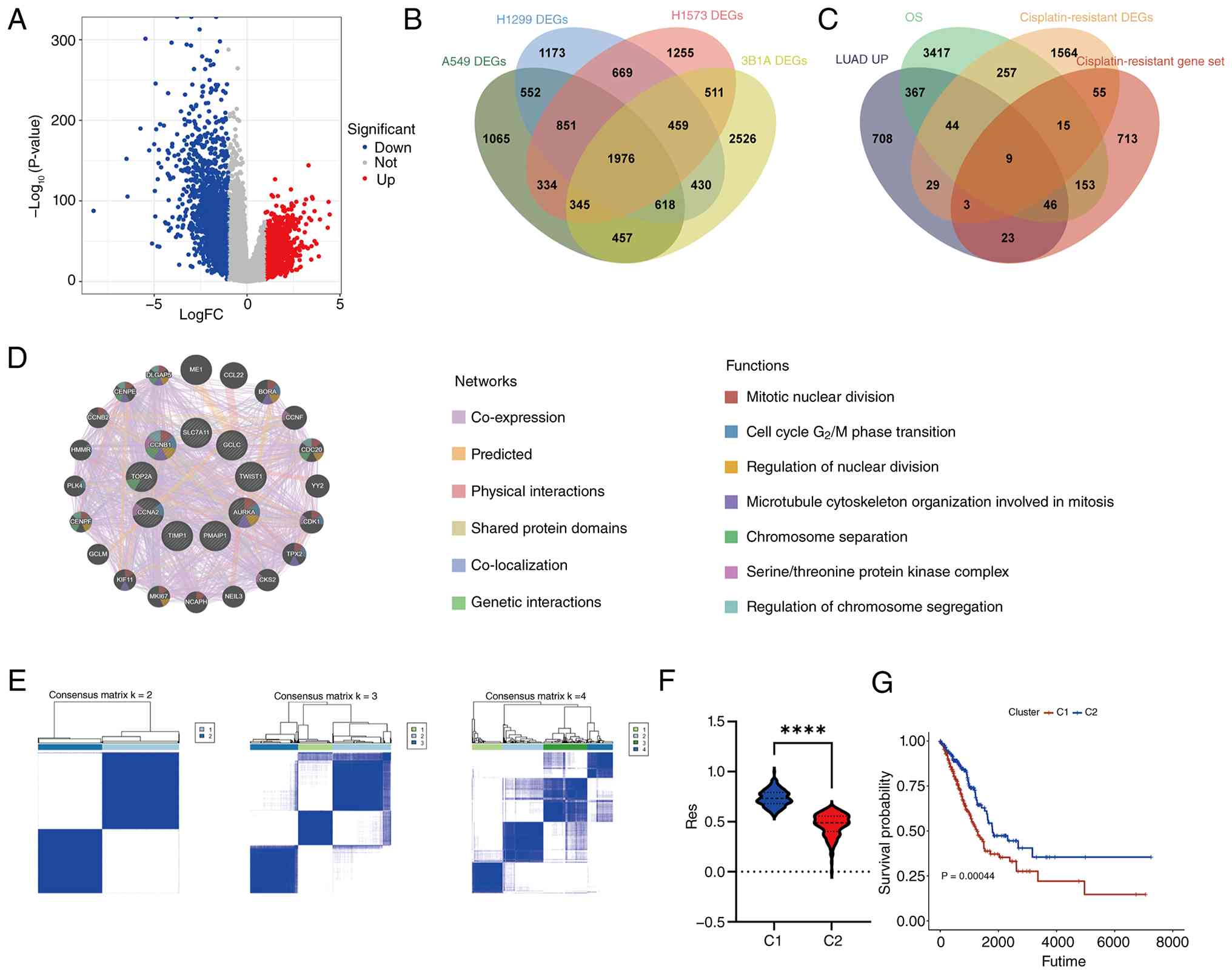 | Figure 1.Identification of cisplatin
resistance-associated gene signatures and molecular subtypes in
LUAD. (A) DEGs between LUAD tumor and normal tissues in The Cancer
Genome Atlas cohort. (B) Venn diagram showing the overlap of DEGs
among cisplatin-resistant cell lines from the GSE213102 dataset.
(C) Venn diagram illustrating the intersection of upregulated genes
in LUAD, prognostic risk genes, GeneCards-derived cisplatin
resistance genes and cisplatin resistance-associated DEGs, yielding
nine candidate genes. (D) GeneMANIA network depicting coexpression
and functional interactions among the nine cisplatin
resistance-associated genes. (E) Consensus clustering of LUAD
samples based on the nine-gene signature, defining two molecular
subtypes (C1 and C2). (F) Comparison of single sample Gene Set
Enrichment Analysis-derived cisplatin resistance scores between the
two subtypes. ****P<0.0001. (G) Kaplan-Meier OS analysis of
patients stratified by C1 and C2 subtypes. LUAD, lung
adenocarcinoma; DEGs, differentially expressed genes; OS, overall
survival; FC, fold change; Up, upregulated; Down, downregulated;
Res, resistance. |
Genomic and pathway characteristics of
cisplatin-resistance subtypes
To elucidate the differences in gene expression
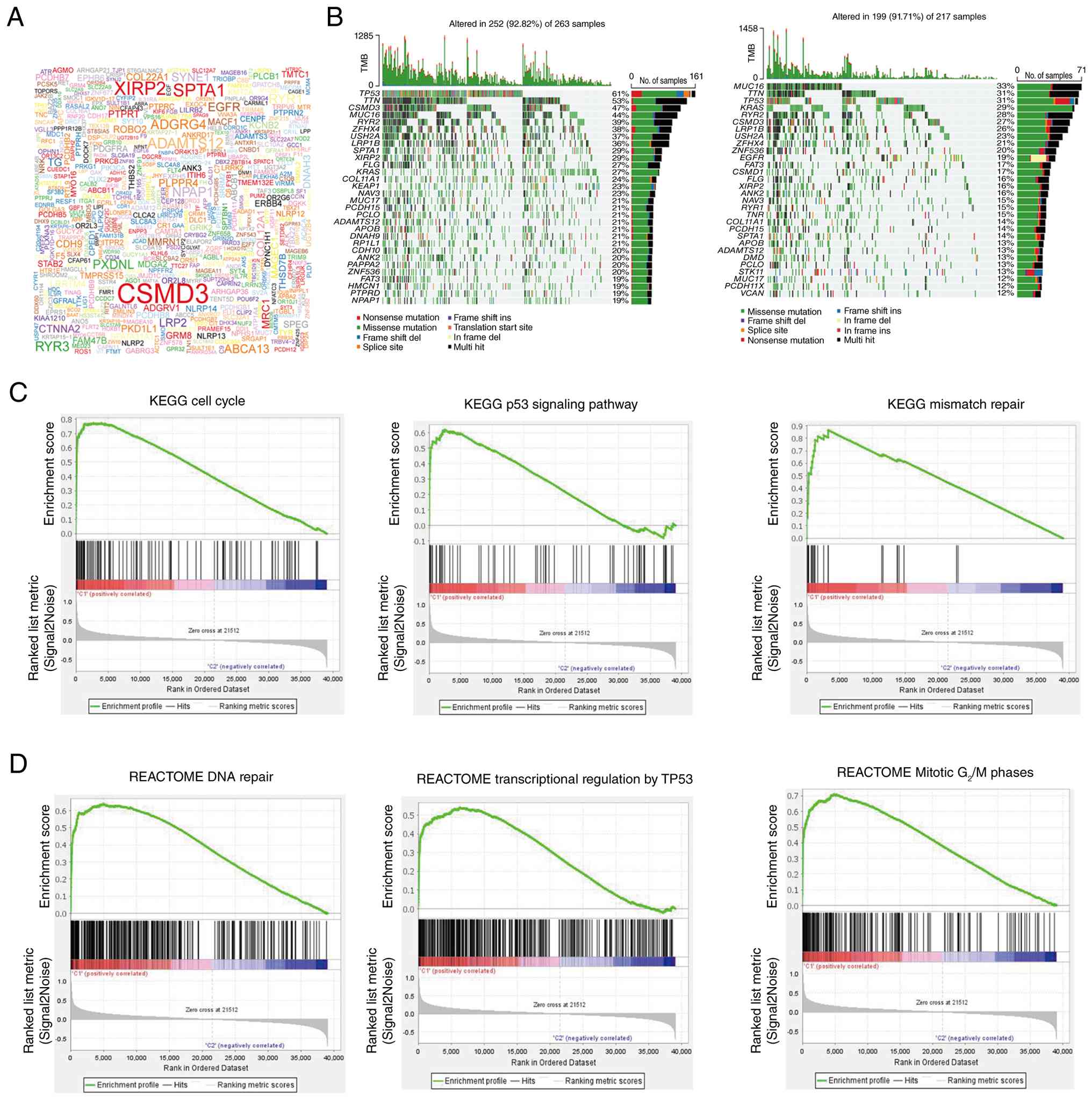
between the two subtypes, TMB analysis was first performed. The
highly mutated genes included CUB and sushi multiple domains 3
(CSMD3), erythrocytic spectrin α 1 and xin actin binding repeat
containing 2 (Fig. 2A). Notably,
compared with the C2 subtype, the C1 subtype exhibited
significantly higher mutation frequencies for TP53, titin and CSMD3
(Fig. 2B). The present study
subsequently investigated differences in pathway activity between
subtypes using GSEA. KEGG-based GSEA revealed that ‘KEGG cell
cycle’, ‘KEGG p53 signaling pathway’ and ‘KEGG mismatch repair’
were markedly enriched in the C1 subtype (Fig. 2C). Consistent with these findings, a
reactome GSEA revealed significant activation of ‘REACTOME DNA
repair’, ‘REACTOME transcriptional regulation by TP53’ and
‘REACTOME Mitotic G2/M phases’ in the C1 subtype
(Fig. 2D). These findings suggested
that enhanced genomic instability and checkpoint activation were
key molecular hallmarks of the high-resistance C1 cluster.
Immune landscape differences between
cisplatin-resistance subtypes
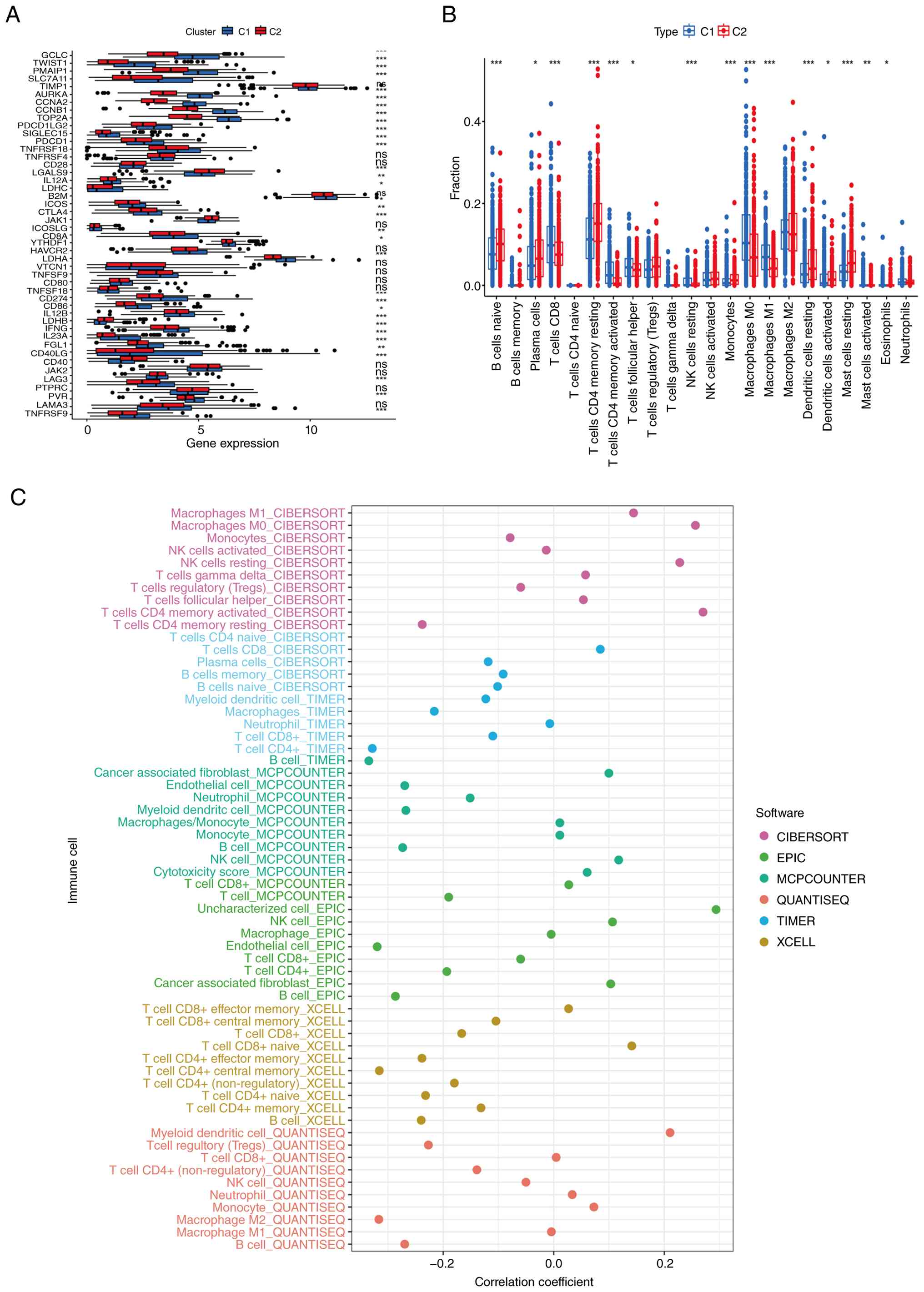
Immunological heterogeneity between the two
cisplatin resistance subtypes was first characterized by comparing
the expression levels of immune checkpoint-associated genes. The
majority of immune checkpoint-associated genes, including
glutamate-cysteine ligase catalytic subunit, twist family BHLH
transcription factor 1 and phorbol-12-myristate-13-acetate-induced
protein 1, were significantly upregulated in the C1 subtype
relative to the C2 subtype (Fig.
3A). Immune cell infiltration was then assessed using the
CIBERSORT algorithm. A number of immune cell populations, such as
naïve B cells, plasma cells and resting CD4 memory T cells, were
found to be more abundant in the C2 subtype, whereas CD8 T cells
were significantly more abundant in the C1 subtype (Fig. 3B). Numerous computational algorithms
were subsequently applied to evaluate the correlation between
immune infiltration patterns and cisplatin resistance scores. The
resistance score was found to be positively correlated with that of
resting natural killer cells and follicular helper T cells,
indicating an association between the immunological activation
state and the cisplatin resistance phenotype (Fig. 3C).
Integrated molecular profiling
identifies key pathways and hub genes associated with cisplatin
resistance
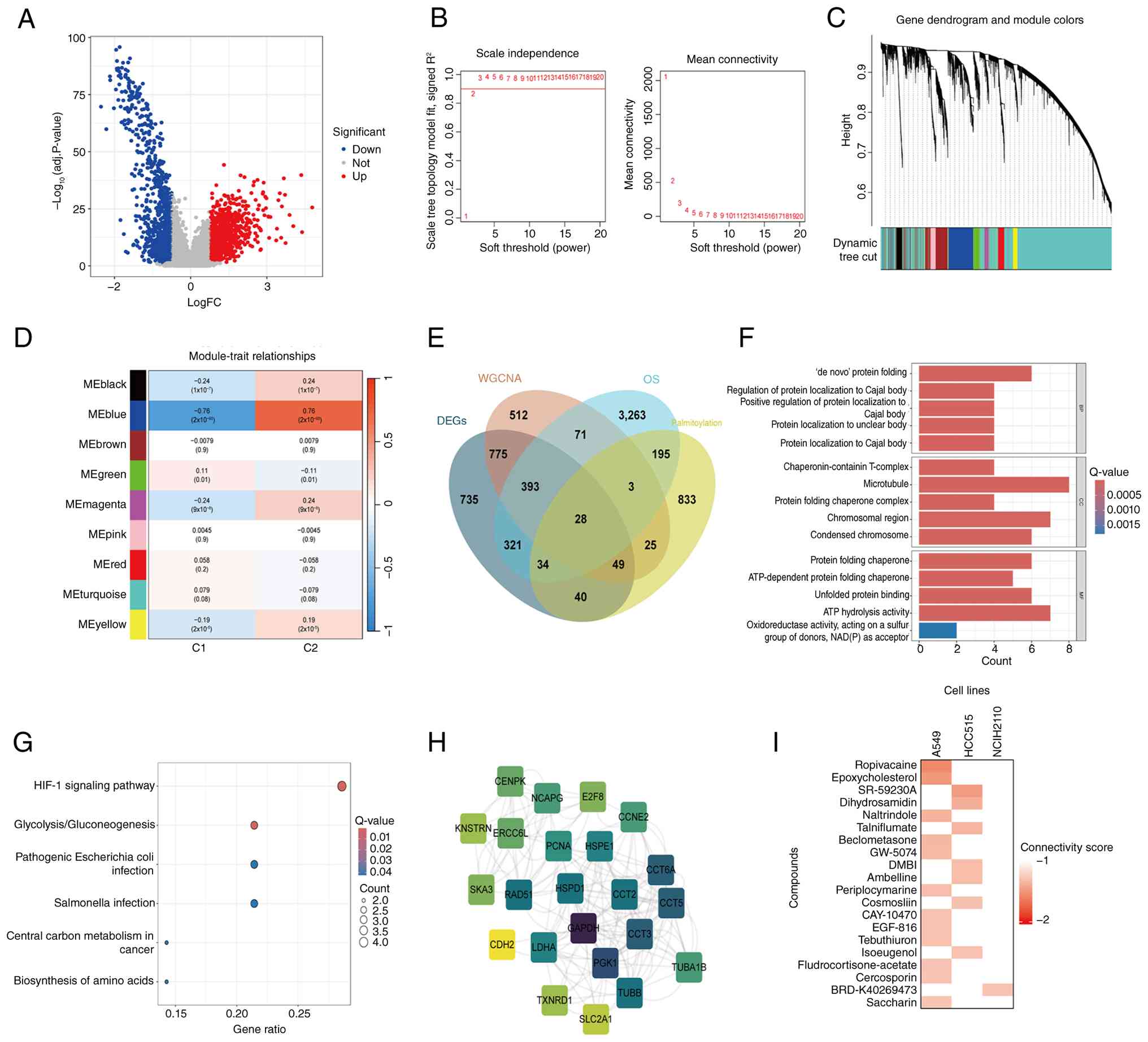
To investigate the molecular disparities between the
two subtypes, differential expression analysis was performed
(log2 FC >0.5; P<0.05) to identify 2,375 DEGs,
including 1,224 upregulated genes and 1,151 downregulated genes, in
the C2 relative to C1 subtype (Fig.
4A). WGCNA was then conducted, with a soft-thresholding power
of 3 (Fig. 4B), resulting in a
number of coexpression modules (Fig.
4C). Module-trait correlation analysis revealed numerous
clinically relevant modules (Fig.
4D), which were selected for downstream analyses. Given the key
role of protein palmitoylation in chemoresistance,
palmitoylation-associated genes were incorporated into the
analysis. An intersection of DEGs, WGCNA-derived module genes,
prognostic risk genes and palmitoylation-associated genes yielded
28 shared genes (Fig. 4E). To
elucidate the biological functions associated with these genes, GO
and KEGG enrichment analyses were performed. GO enrichment revealed
involvement in ‘de novo protein folding’, ‘regulation of
protein localization to Cajal body’, ‘chaperonin-containing
T-complex’ assembly, ‘microtubule’ and ‘protein folding chaperone’
activity (Fig. 4F). KEGG analysis
revealed significant enrichment of the ‘HIF-1 signaling pathway’
and ‘glycolysis/gluconeogenesis’ (Fig.
4G). PPI analysis using the STRING database revealed GAPDH,
CCT2 and CCT3 as central hub genes within the network (Fig. 4H). Finally, CMAP (https://clue.io/cmap) analysis identified several
compounds with negative or low connectivity scores, including
ropivacaine, epoxycholesterol, naltrindole and beclometasone in
A549 cells; SR-59230A, dihydrosamidin, talniflumate, GW-5074, DMBI,
cosmosiin and isoeugenol in HCC515 cells; and BRD-K40269473 in
NCI-H2110 cells (Fig. 4I).
Identification of key
cisplatin-resistance genes using an integrated 101-algorithm
machine learning framework
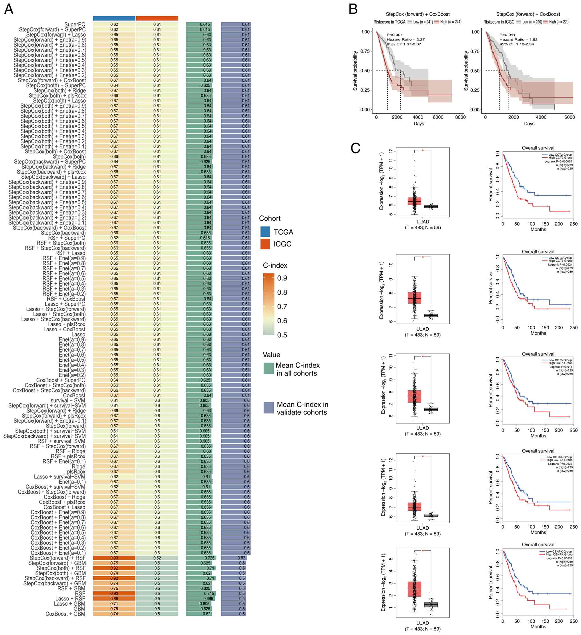
A total of 101 machine learning algorithms were
integrated to refine the expression of cisplatin
resistance-associated core genes. The TCGA cohort was used for
model training and the ICGC cohort was used for external
validation. The performance heatmaps of all the algorithmic models
are presented in Fig. 5A. Among all
combinations, the StepCox (forward) + CoxBoost model demonstrated
the highest predictive accuracy. Survival curves generated from
both the training and validation datasets, on the basis of the
model-derived risk scores, consistently revealed significant
prognostic stratification (Fig.
5B). This optimized model identified five key genes, CCT2,
CCT3, CCT5, CCT6A and centromere protein K (CENPK), as the most
robust predictors. All five genes were markedly upregulated in LUAD
tissues compared with normal lung tissues and patients with high
expression of these genes exhibited significantly poorer overall
survival compared with those with low expression levels (Fig. 5C). These findings suggested that the
five-gene signature served a key role in mediating cisplatin
resistance and may serve as a potent biomarker panel for risk
assessment. Notably, numerous members of the CCT family (CCT2,
CCT3, CCT5 and CCT6A) were consistently identified, suggesting that
the CCT family may serve a broader and coordinated role in
cisplatin resistance. Among these, CCT2 exhibited the most
significant prognostic association, as it exhibited the smallest
log-rank P-value in Kaplan-Meier survival analysis (Fig. 5C) and patients with high CCT2
expression demonstrated significantly worse overall survival, which
led it to be prioritized for further investigation.
Single-cell transcriptomic profiling
reveals the cell type-specific distribution and cisplatin
resistance enrichment of core genes
Single-cell RNA-seq data from lung cancer samples
retrieved from the GEO database (GSE229253) were used to explore
the cellular origin and functional heterogeneity of the core genes
at single-cell resolution. Dimensional reduction and clustering
revealed key cell populations, including epithelial cells,
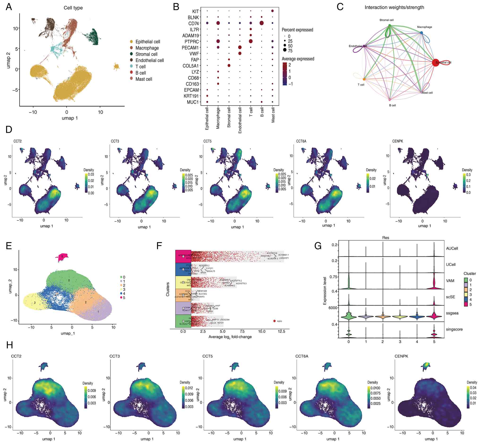
macrophages, endothelial cells, T cells and B cells (Fig. 6A), with representative marker genes
for each lineage displayed in Fig.
6B. Cell-cell communication networks inferred using CellChat
revealed extensive intercellular interactions across these cell
types (Fig. 6C). Visualization of
core gene expression across cell populations demonstrated that
CCT2, CCT3, CCT5 and CCT6A were predominantly enriched in
epithelial cells (Fig. 6D). Further
reclustering of epithelial cells revealed six distinct epithelial
cell subpopulations, each characterized by unique marker gene
signatures (Fig. 6F). According to
the results of the IRGSEA for resistance gene-set enrichment,
subclusters 0 and 5 exhibited strong enrichment for cisplatin
resistance signatures across numerous algorithms (Fig. 6G). Finally, mapping the five core
genes onto epithelial subclusters demonstrated that CCT2, CCT3,
CCT5 and CCT6A were significantly enriched in both
cisplatin-resistant subclusters 0 and 5, whereas CENPK expression
was predominantly enriched in subcluster 5 (Fig. 6H). These findings highlighted
specific epithelial cell subpopulations as the principal cellular
reservoirs of cisplatin resistance-associated gene activation.
Single-cell functional
characterization of CCT2 in epithelial subclusters
GSVA across six epithelial subclusters revealed
distinct biological functions of CCT2, with subcluster 0 exhibiting
significant enrichment of genes associated with oncogenic and
proliferative pathways, including genes associated with ‘KRAS
signaling up’, ‘Wnt beta-catenin signaling’ and ‘cell cycle’
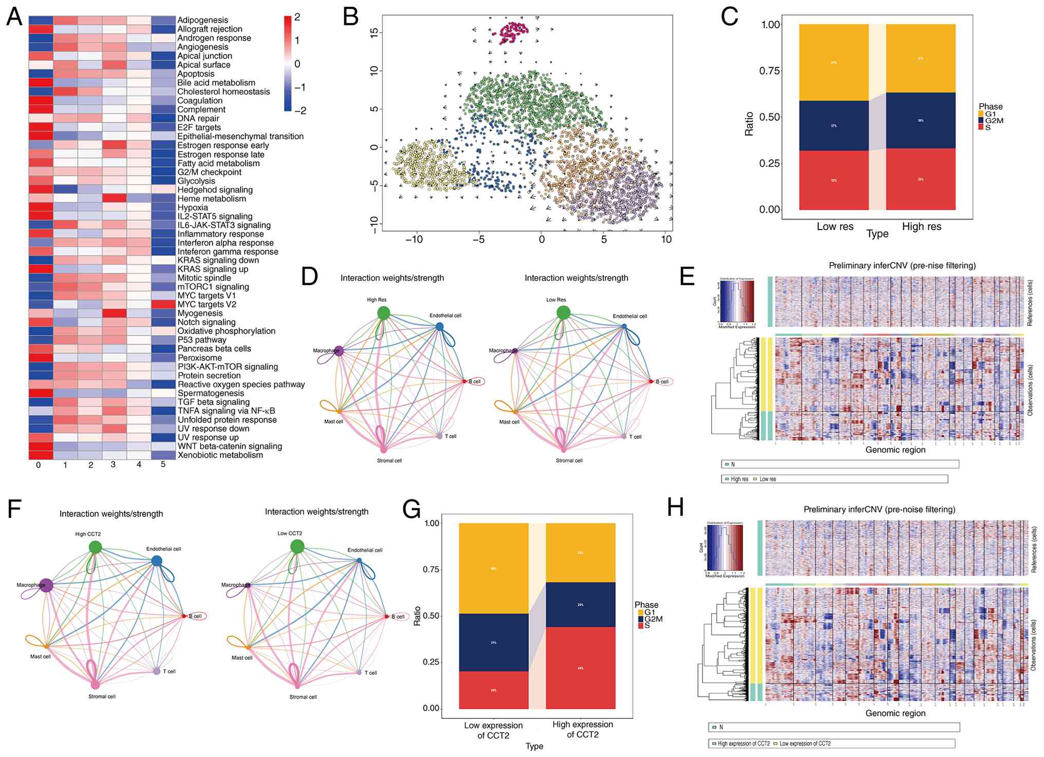
(Figs. 7A and S1). RNA velocity analysis further
demonstrated a directional flow from subcluster 0 toward other
epithelial subclusters, suggesting that subcluster 0 may represent
a transcriptionally active and developmental origin state (Fig. 7B). On the basis of the
IRGSEA-derived drug resistance signatures, the epithelial cells
were stratified into high- and low-resistance groups. Cell cycle
phase scoring indicated that, compared with the low-resistance
group, the high-resistance group exhibited a markedly higher
proportion of proliferating cells (S and G2/M phases;
Fig. 7C). Cell-cell communication
analysis revealed that the high-resistance epithelial cell
population exhibited stronger interactions with endothelial cells
and mast cells compared with the low-resistance population, as
indicated by the increased interaction strength in the
communication network (Fig. 7D).
Consistently, ligand-receptor analysis identified a greater number
of significant interactions between these cell populations,
including representative ligand-receptor pairs such as
semaphorin-3C (SEMA3C)-[neuropilin (NRP)-2 + plexin-A2 (PLXNA2)],
SEMA3C-(NRP1 + PLXNA2), SEMA3C-(NRP1 + NRP2), cell adhesion
molecule 1 (CADM1)-CADM1 and amphiregulin (AREG)-EGFR, among others
(Fig. S2). CNV profiling further
demonstrated increased chromosomal instability in the resistant
epithelial population (Fig. 7E). To
clarify the contribution of CCT2 expression, epithelial cells were
separately analyzed according to their CCT2 expression level.
High-CCT2 epithelial cells exhibited stronger interactions with
endothelial cells and mast cells compared with low-CCT2 epithelial
cells, as indicated by increased interaction strength in the
communication network (Fig. 7F).
Consistently, ligand-receptor analysis revealed a greater number of
significant interactions between high-CCT2 epithelial cells and
these cell populations, including representative ligand-receptor
pairs such as SEMA3C-(NRP1 + PLXNA2), SEMA3C-(NRP1 + NRP2),
secretoglobin family 3A member 2-macrophage receptor with
collagenous structure, CADM1-CADM1 and AREG-EGFR, among others
(Fig. S3). Consistent with the
resistance-associated cell population, CCT2-high epithelial cells
also exhibited a higher proportion of proliferating cells (S and
G2/M phases; Fig. 7G).
In addition, CNV analysis showed greater genomic alterations in the
high-CCT2 subgroup (Fig. 7H),
collectively indicating that CCT2 upregulation was associated with
proliferative activity, enhanced cellular communication and genomic
instability within epithelial tumor cells.
Experimental validation of CCT2
function in cisplatin resistance
Functional roles of CCT2 were next validated in the
cell lines A549 and NCI-H1299, respectively. A total of two shRNA
expression constructs targeting CCT2 were transfected into both
cell lines and qPCR determined the efficient knockdown by sh-CCT2-1
and sh-CCT2-2 (Fig. 8A; n=3;
P<0.05). Western blotting analysis demonstrated that
CCT2-targeting shRNA vectors effectively reduced CCT2 protein
expression in A549 and NCI-H1299 cells (Fig. 8B; n=3; P<0.05). Cell
proliferation assessed by CCK-8 assays at 24 and 48 h revealed that
CCT2 depletion significantly reduced the proliferative capacity of
both A549 and NCI-H1299 cells (Fig.
8C; n=5; P<0.05). To further evaluate whether CCT2
influences the cellular response to cisplatin, cells were treated
with cisplatin following CCT2 knockdown. Cisplatin alone
significantly suppressed cell viability, whereas combined treatment
with cisplatin and CCT2 silencing resulted in a notably greater
reduction in cell viability (Fig.
8D; n=5; P<0.05). Compared with cisplatin treatment alone,
CCT2 knockdown further enhanced the inhibitory effect of cisplatin
on cell viability, indicating that depletion of CCT2 increased the
sensitivity of lung cancer cells to cisplatin. These findings
indicated that CCT2 promoted lung cancer cell proliferation and
modulated the sensitivity of lung cancer cells to cisplatin.
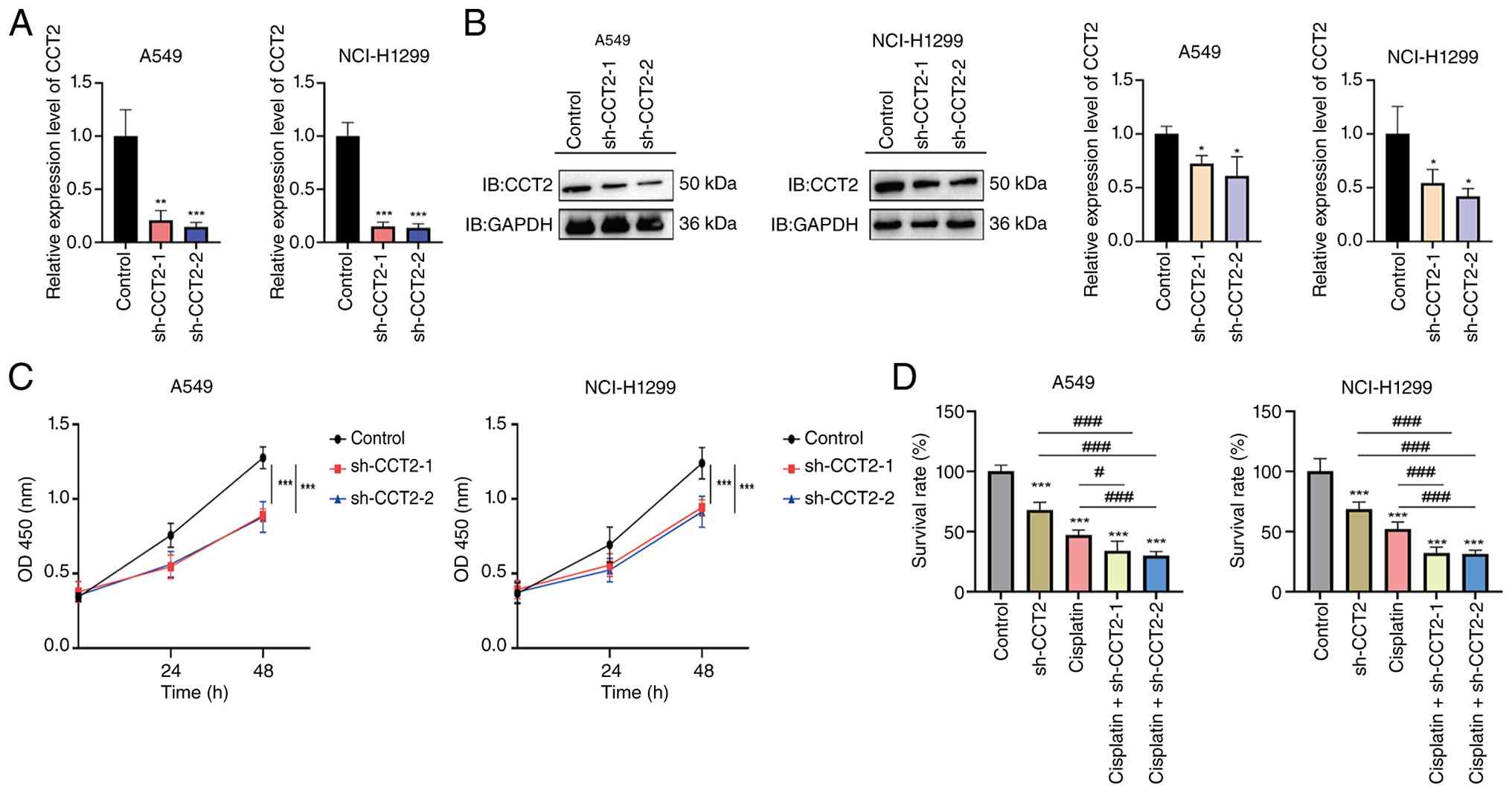 | Figure 8.Experimental validation of the
function of CCT2 in cisplatin resistance in lung cancer cells. (A)
Quantitative PCR validation of the knockdown efficiency of CCT2 in
A549 and H1299 cells transfected with shRNA expression vectors
targeting CCT2. (B) Western blotting determination of CCT2 protein
depletion following transfection with shRNA expression vectors
targeting CCT2. (C) Cell Counting Kit-8 assays showing reduced cell
proliferation after CCT2 silencing. (D) Cell viability under
cisplatin treatment with or without CCT2 knockdown, indicating
enhanced cisplatin sensitivity upon CCT2 depletion. Data are
presented as the mean ± SD from at least three independent
biological replicates. Statistical analysis for (A) and (B) were
performed using one-way ANOVAs followed by Dunnett's multiple
comparisons test, with all treatment groups compared to the control
group. For (C), statistical significance was determined using
two-way ANOVAs followed by Šidák's multiple comparisons test. For
(D), statistical analysis was performed using a one-way ANOVA
followed by Tukey's multiple comparisons test. *P<0.05,
**P<0.01, ***P<0.001 vs. the control group;
#P<0.05 and ###P<0.001 indicate the
comparisons between the indicated groups, as shown by the
horizontal lines. ns, not significant; IB, immunoblotting; sh-,
short hairpin; CCT, chaperonin-containing TCP-1; OD, optical
density. |
Identification of potential
therapeutic compounds targeting CCT2
Molecular docking was performed using a
natural-product compound library within the MOE platform to
identify potential inhibitors of CCT2. A number of compounds,
including deslanoside (−11.9 kcal/mol; Fig. 9A), digitoxin (−10.7 kcal/mol;
Fig. 9B), lanatoside C (−11.8
kcal/mol; Fig. 9C), lecithin (−11.7
kcal/mol; Fig. 9D) and thymopentin
(−10.9 kcal/mol; Fig. 9E),
demonstrated strong binding affinities. Subsequent molecular
dynamics simulations revealed stable RMSD, radius of gyration and
solvent-accessible surface area profiles for each ligand-protein
complex, indicating stable binding conformations. These results
suggested that the identified compounds may serve as promising
candidates for targeting CCT2 in future therapeutic
development.
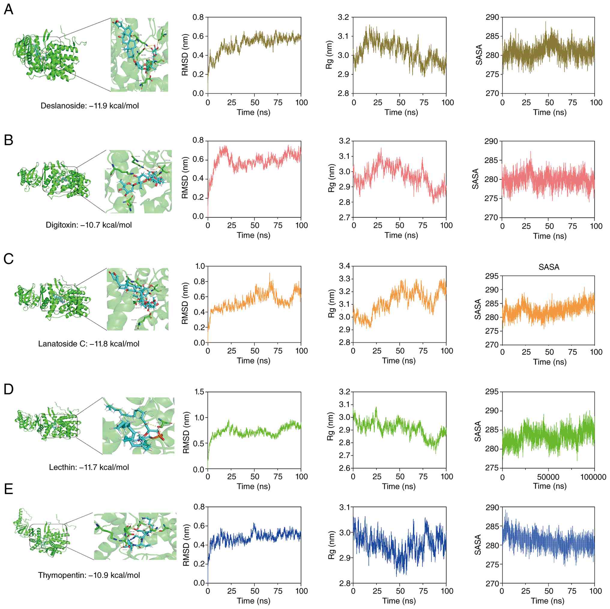 | Figure 9.Identification of potential
therapeutic compounds targeting CCT2. (A) Deslanoside, (B)
digitoxin, (C) lanatoside C, (D) lecithin and (E) thymopentin.
Molecular docking conformations and corresponding molecular
dynamics simulation results with CCT2, including RMSD, Rg and SASA,
indicating stable ligand-CCT2 interactions. CCT,
chaperonin-containing TCP-1; RMSD, root mean square deviation; Rg,
radius of gyration; SASA, solvent-accessible surface area. |
Discussion
Despite marked advances in the treatment of LUAD,
including targeted therapies, immunotherapy and emerging
metabolism-oriented strategies, cisplatin-based chemotherapy
remains a cornerstone of clinical management, particularly for
patients without actionable driver mutations or those who are
refractory to targeted and immunotherapeutic approaches (65–69).
Although cisplatin-based chemotherapy provides substantial clinical
benefit, its efficacy is often constrained by the development of
adaptive resistance, including the emergence of resistant tumor
cell populations with enhanced DNA damage repair capacity and
altered apoptotic responses following cisplatin exposure (70,71).
In particular, the efficacy of cisplatin is frequently undermined
by the rapid development of acquired resistance, which is
associated with alterations in DNA damage responses, cell cycle
control, metabolic reprogramming and the tumor microenvironment
(72–74). In routine clinical practice, the
management of cisplatin resistance still relies largely on
empirical combination therapies or treatment switching rather than
mechanism-driven interventions, reflecting the lack of robust
molecular markers and validated therapeutic targets specific to
cisplatin resistance in LUAD. Collectively, these difficulties
underscore the need to systematically elucidate the molecular and
cellular basis of cisplatin resistance to develop more precise and
durable therapeutic strategies.
In the context of the clinical challenge of
overcoming cisplatin resistance in LUAD, previous efforts have
focused on identifying molecular determinants that underlie
differential treatment responses. A number of genes and pathways,
including TP53 signaling, cell cycle regulators and glycolytic
pathways, have been implicated, yet their prognostic and
therapeutic relevance remains unclear across cohorts (75–77).
The present study systematically integrated bulk and single-cell
transcriptomic data to define cisplatin resistance-associated
molecular subtypes in LUAD. The present findings not only
corroborate the enrichment of cell cycle- and DNA repair-associated
pathways in resistant tumors but also reveal distinct immune
infiltration patterns and notable cellular heterogeneity associated
with resistance phenotypes. Notably, through machine learning-based
feature selection, a robust core gene signature with both
prognostic relevance and biological relevance was refined, offering
a complementary approach to previous studies and enabling more
comprehensive candidate gene identification and prognostic
evaluation (78–80).
Notably, the present study highlights the CCT gene
family, particularly CCT2, as key regulators of cisplatin
resistance. The chaperonin-containing TCP-1 (CCT/TRiC) complex is
essential for protein folding, cytoskeletal organization and cell
cycle control, processes that are closely associated with tumor
cell proliferation and stress adaptation (81–83).
Emerging evidence has suggested that posttranslational
modifications, including palmitoylation, modulate protein stability
and signaling dynamics in cancer cells (84). By integrating
palmitoylation-associated genes obtained from the GeneCards
database with the present cisplatin resistance-associated gene
sets, CCT2 was identified as a candidate gene of interest. In the
present study, CCT2 was predominantly enriched in
cisplatin-resistant epithelial cell subpopulation. In addition,
CCT2-high epithelial cells exhibited a higher proportion of
proliferating cells (S and G2/M phases) and stronger
interactions with endothelial cells and mast cells compared with
CCT2-low epithelial cells, as indicated by increased communication
strength in the network analysis and a greater number of
ligand-receptor interactions/Functional validation further
demonstrated that CCT2 knockdown enhanced the inhibitory effect of
cisplatin on cell viability, suggesting that CCT2 depletion
sensitizes lung cancer cells to cisplatin. Based on previous
studies demonstrating the involvement of CCT2 in
chaperonin-mediated protein folding and cell cycle regulation
(85,86), it was speculated that CCT2 may
contribute to cisplatin resistance by promoting tumor cell
adaptation to chemotherapeutic stress. Previous studies have
demonstrated that dysregulated G2/M checkpoint
progression and chaperonin-mediated proteostasis contribute notably
to cisplatin resistance and tumor stress adaptation (87,88).
In ovarian cancer, anlotinib reversed cisplatin resistance by
inducing G2/M arrest and apoptosis through the aurora
kinase A/p53 pathway, highlighting the importance of cell cycle
dysregulation in platinum resistance (87). In addition, CCT3 was upregulated in
cisplatin-resistant LUAD cells, whereas CCT3 knockdown induced
G2/M arrest and apoptosis and restored cisplatin
sensitivity through inhibition of the JAK2/STAT3 pathway (88).
In summary, the present study provides a
comprehensive framework for understanding cisplatin resistance in
LUAD through the integration of molecular subtyping, immune
profiling, machine learning and experimental validation. The
identification of CCT2 as a key resistance-associated gene offers
new insights into the role of chaperonin-mediated regulation in
chemotherapy resistance. Despite this, a number of limitations
should be acknowledged. First, although the present integrative
analyses and experimental validation suggest that CCT2 is
associated with cisplatin resistance, the precise molecular
mechanisms underlying its function, particularly its roles in
protein folding and cell cycle regulation, require further in-depth
investigation. Notably, future studies should include
pathway-specific functional assays to directly determine whether
CCT2 regulates these BPs. In addition, future studies should
include CCT2 overexpression and rescue experiments (such as CCT2
re-expression following knockdown) to further establish the causal
role of CCT2 in cisplatin resistance and determine whether CCT2
overexpression attenuates cisplatin-induced growth inhibition.
Second, the present study was primarily based on public datasets
and in vitro experiments and thus lacks validation in
large-scale clinical cohorts and in vivo models. Therefore,
the clinical predictive value and therapeutic potential of CCT2
need to be further determined in prospective studies. Third,
although potential compounds targeting CCT2 have been identified
through molecular docking, their biological efficacy and safety
remain to be experimentally validated. Collectively, the present
study provides a foundation for future mechanistic and
translational studies aimed at overcoming cisplatin resistance in
LUAD.
In conclusion, the present study systematically
elucidated the molecular landscape of cisplatin resistance in LUAD
through integrated multiomics analyses, machine learning-based
modeling and experimental validation. A total of two distinct
cisplatin resistance-associated molecular subtypes with
significantly different prognoses, immune infiltration patterns and
genomic alteration profiles were identified. By combining
differential expression analysis, WGCNA, palmitoylation-associated
gene sets and large-scale machine learning screening, the present
study established a robust five-gene signature and highlighted CCT2
as a key regulator of cisplatin resistance. Single-cell analyses
further revealed that CCT2 is preferentially enriched in resistant
epithelial cell subpopulations and is associated with enhanced cell
cycle activity, increased intercellular communication and genomic
instability. Functional assays demonstrated that CCT2 knockdown
significantly suppressed tumor cell proliferation and sensitized
LUAD cells to cisplatin treatment. In addition, molecular docking
and molecular dynamics simulations revealed a number of candidate
compounds with stable binding to CCT2, suggesting potential
therapeutic avenues. Collectively, the present findings provide
novel insights into the mechanisms underlying cisplatin resistance
in LUAD and identified CCT2 as a promising biomarker and
therapeutic target, thereby offering a potential strategy for
improving treatment efficacy and patient outcomes.
Supplementary Material
Supporting Data
Supporting Data
Acknowledgements
Not applicable.
Funding
Funding: No funding was received.
Availability of data and materials
The data generated in the present study may be
requested from the corresponding author.
Authors' contributions
HM was responsible for study conceptualization,
methodology, bioinformatics analysis, data curation, figure
preparation and visualization and writing the original manuscript
draft. CS was responsible for software implementation,
computational analysis and interpretation of the data, validation
of analytical results, investigation, and writing, reviewing and
editing the manuscript. XH contributed to study conceptualization,
interpretation of the data, scientific supervision, and writing,
reviewing and editing the manuscript. GW was responsible for study
conceptualization, funding acquisition, providing supervision and
writing, reviewing and editing the manuscript. HM and GW confirm
the authenticity of all the raw data. All authors read and approved
the final version of the manuscript.
Ethics approval and consent to
participate
Not applicable.
Patient consent for publication
Not applicable.
Competing interests
The authors declare that they have no competing
interests.
Glossary
Abbreviations
Abbreviations:
|
LUAD
|
lung adenocarcinoma
|
|
NSCLC
|
non-small cell lung cancer
|
|
TCGA
|
The Cancer Genome Atlas
|
|
ICGC
|
International Cancer Genome
Consortium
|
|
GEO
|
Gene Expression Omnibus
|
|
DEGs
|
differentially expressed genes
|
|
GSEA
|
Gene Set Enrichment Analysis
|
|
ssGSEA
|
single-sample GSEA
|
|
GSVA
|
Gene Set Variation Analysis
|
|
IRGSEA
|
immune-related GSEA
|
|
WGCNA
|
weighted gene co-expression network
analysis
|
|
PPI
|
protein-protein interaction
|
|
CNV
|
copy number variation
|
|
KEGG
|
Kyoto Encyclopedia of Genes and
Genomes
|
|
BP
|
biological process
|
|
TMB
|
tumor mutational burden
|
|
CCT
|
chaperonin-containing TCP-1
|
|
CMAP
|
connectivity map
|
|
MOE
|
Molecular Operating Environment
|
|
RMSD
|
root mean square deviation
|
References
|
1
|
Mangang KN and Rao G: Lung cancer
biomarkers. Clin Chim Acta. 580:1207072026. View Article : Google Scholar : PubMed/NCBI
|
|
2
|
Ma J, Li S, Zhang X, Zhao P, Zhao B, Yue
J, Han L, Bai J, Zhao Z and Zhang Y: Lung cancer organoids in
translational research: Bridging basic science, drug development,
and clinical therapeutics. Crit Rev Oncol Hematol. 214:1048402025.
View Article : Google Scholar : PubMed/NCBI
|
|
3
|
Wang Y, Xu M, Wei X, Huang H, Chen Q, Chen
B, Bao X and Li J: Prospective proteomics for discovering
biomarkers in lung adenocarcinoma: A literature review. Transl
Cancer Res. 14:6102–6117. 2025. View Article : Google Scholar : PubMed/NCBI
|
|
4
|
Sung H, Filho AM, Laversanne M, Ferlay J,
Siegel RL, Soerjomataram I, Jemal A and Bray F: Global cancer
statistics 2024: GLOBOCAN estimates of incidence and mortality
worldwide for 34 cancers in 186 countries. CA Cancer J Clin.
76:e700902026.PubMed/NCBI
|
|
5
|
Vokes NI, Pan K and Le X: Efficacy of
immunotherapy in oncogene-driven non-small-cell lung cancer. Ther
Adv Med Oncol. 15:175883592311614092023. View Article : Google Scholar : PubMed/NCBI
|
|
6
|
Turcott JG, Cárdenas-Fernández D,
Sánchez-Lara K, Palomares-Palomares CB and Arrieta O: Nutritional
approach on management of diarrhea induced by EGFR-TKI's in
advanced Non-Small cell lung cancer patients. Nutr Cancer.
77:567–574. 2025. View Article : Google Scholar : PubMed/NCBI
|
|
7
|
Ricco G, Seminerio R, Andrini E, Malvi D,
Gruppioni E, Altimari A, Zagnoni S, Campana D and Lamberti G: BRAF
V600E-mutated large cell neuroendocrine carcinoma responding to
targeted therapy: A case report and review of the literature.
Anticancer Drugs. 34:1076–1084. 2023. View Article : Google Scholar : PubMed/NCBI
|
|
8
|
Groehler A IV, Maratova A, Dao NM, Mahkmut
A and Schärer OD: Development of comprehensive ultraperformance
liquid Chromatography-High-Resolution mass spectrometry assays to
quantitate Cisplatin-Induced DNA-DNA Cross-links. Chem Res Toxicol.
36:822–837. 2023. View Article : Google Scholar : PubMed/NCBI
|
|
9
|
Chen YF, Pang YC, Wang HC, Wu PE, Chen ZJ,
Huang D, Peng DL, Yan YM, Liu C, Wu LC, et al: Identification of
arnicolide C as a novel chemosensitizer to suppress
mTOR/E2F1/FANCD2 axis in non-small cell lung cancer. Br J
Pharmacol. 181:1221–1237. 2024. View Article : Google Scholar : PubMed/NCBI
|
|
10
|
Kuang L, Wang P, Zhou L and Li Y:
Transformation of lung adenocarcinoma to small cell lung cancer
following osimertinib treatment: A case report and literature
review. Anticancer Drugs. 36:253–259. 2025. View Article : Google Scholar : PubMed/NCBI
|
|
11
|
Fennell DA, Summers Y, Cadranel J, Benepal
T, Christoph DC, Lal R, Das M, Maxwell F, Visseren-Grul C and Ferry
D: Cisplatin in the modern era: The backbone of first-line
chemotherapy for non-small cell lung cancer. Cancer Treat Rev.
44:42–50. 2016. View Article : Google Scholar : PubMed/NCBI
|
|
12
|
Zhao Y, Zhang L, Xia L, E H, Wang T, Lu H,
Chen H, She Y, Tang H, Wu J, et al: A METTL3-NFE2L3 axis mediates
tumor stemness and progression in lung adenocarcinoma. Sci Adv.
11:eadt76822025. View Article : Google Scholar : PubMed/NCBI
|
|
13
|
Park JH, You GL, Ahn MJ, Kim SW, Hong MH,
Han JY, Ock CY, Lee JS, Oh IJ, Lee SY, et al: Real-world outcomes
of anti-PD1 antibodies in platinum-refractory, PD-L1-positive
recurrent and/or metastatic non-small cell lung cancer, and its
potential practical predictors: First report from Korean cancer
study group LU19-05. J Cancer Res Clin Oncol. 147:2459–2469. 2021.
View Article : Google Scholar : PubMed/NCBI
|
|
14
|
Liang J, Bi G, Huang Y, Zhao G, Sui Q,
Zhang H, Bian Y, Yin J, Wang Q, Chen Z and Zhan C: MAFF confers
vulnerability to cisplatin-based and ionizing radiation treatments
by modulating ferroptosis and cell cycle progression in lung
adenocarcinoma. Drug Resist Updat. 73:1010572024. View Article : Google Scholar : PubMed/NCBI
|
|
15
|
Jiang F, Shen Q, Zhang F, Fu J, Hu L, Wang
J, Zhou H, Chen J and Wang Y: ADH1C facilitates cisplatin
resistance of lung adenocarcinoma cells. DNA Cell Biol. 41:631–640.
2022. View Article : Google Scholar : PubMed/NCBI
|
|
16
|
Huang L, Lou K, Wang K, Liang L, Chen Y
and Zhang J: Let-7c-5p represses cisplatin resistance of lung
adenocarcinoma cells by targeting CDC25A. Appl Biochem Biotechnol.
195:1644–1655. 2023. View Article : Google Scholar : PubMed/NCBI
|
|
17
|
Kiss RC, Xia F and Acklin S: Targeting DNA
damage response and repair to enhance therapeutic index in
Cisplatin-Based cancer treatment. Int J Mol Sci. 22:81992021.
View Article : Google Scholar : PubMed/NCBI
|
|
18
|
Qiao X, Li W, Zheng Z, Liu C, Zhao L, He Y
and Li H: Inhibition of the HMGB1/RAGE axis protects against
cisplatin-induced ototoxicity via suppression of inflammation and
oxidative stress. Int J Biol Sci. 20:784–800. 2024. View Article : Google Scholar : PubMed/NCBI
|
|
19
|
Cai L, Zhang Q, Du L and Zheng F:
Silencing of miR-1246 induces cell cycle arrest and apoptosis in
Cisplatin-Resistant ovarian cancer cells by promoting ZNF23
transcription. Cytogenet Genome Res. 161:488–500. 2021. View Article : Google Scholar : PubMed/NCBI
|
|
20
|
Haroon M and Kang SC: Kaempferol
synergistically enhances Cisplatin-induced apoptosis and cell cycle
arrest in colon cancer cells. J Cancer Prev. 29:69–87. 2024.
View Article : Google Scholar : PubMed/NCBI
|
|
21
|
Rao L, Guo D and Wu JP:
Cisplatin-resistance induces lung squamous carcinoma cell growth by
nicotine-mediated α7nAchR/HDAC1/Cyclin D1/pRb cell cycle
activation. Cell Biochem Funct. 42:e39902024. View Article : Google Scholar : PubMed/NCBI
|
|
22
|
Xie R, Cheng L, Huang M, Huang L, Chen Z,
Zhang Q, Li H, Lu J, Wang H, Zhou Q, et al: NAT10 drives cisplatin
chemoresistance by enhancing ac4C-Associated DNA repair in bladder
cancer. Cancer Res. 83:1666–1683. 2023. View Article : Google Scholar : PubMed/NCBI
|
|
23
|
Ye W, Fan C, Fu K, Wang X, Lin J, Nian S,
Liu C and Zhou W: The SAR and action mechanisms of autophagy
inhibitors that eliminate drug resistance. Eur J Med Chem.
244:1148462022. View Article : Google Scholar : PubMed/NCBI
|
|
24
|
Yang L, Xie HJ, Li YY, Wang X, Liu XX and
Mai J: Molecular mechanisms of platinum-based chemotherapy
resistance in ovarian cancer (Review). Oncol Rep. 47:822022.
View Article : Google Scholar : PubMed/NCBI
|
|
25
|
Laurino S, Mazzone P, Ruggieri V, Zoppoli
P, Calice G, Lapenta A, Ciuffi M, Ignomirelli O, Vita G, Sgambato
A, et al: Cationic channel TRPV2 overexpression promotes resistance
to Cisplatin-induced apoptosis in gastric cancer cells. Front
Pharmacol. 12:7466282021. View Article : Google Scholar : PubMed/NCBI
|
|
26
|
Wang D, Zhao C, Xu F, Zhang A, Jin M,
Zhang K, Liu L, Hua Q, Zhao J, Liu J, et al: Cisplatin-resistant
NSCLC cells induced by hypoxia transmit resistance to sensitive
cells through exosomal PKM2. Theranostics. 11:2860–2875. 2021.
View Article : Google Scholar : PubMed/NCBI
|
|
27
|
Miao C, Huang Y, Zhang C, Wang X, Wang B,
Zhou X, Song Y, Wu P, Chen ZS and Feng Y: Post-translational
modifications in drug resistance. Drug Resist Updat. 78:1011732025.
View Article : Google Scholar : PubMed/NCBI
|
|
28
|
Arora G, Bothra A, Prosser G, Arora K and
Sajid A: Role of post-translational modifications in the
acquisition of drug resistance in Mycobacterium tuberculosis. FEBS
J. 288:3375–3393. 2021. View Article : Google Scholar : PubMed/NCBI
|
|
29
|
Hu S, Xu J, Cui W, Jin H, Wang X and
Maimaitiyiming Y: Post-translational modifications in multiple
myeloma: Mechanisms of drug resistance and therapeutic
opportunities. Biomolecules. 15:7022025. View Article : Google Scholar : PubMed/NCBI
|
|
30
|
Jin J, Zhi X, Wang X and Meng D: Protein
palmitoylation and its pathophysiological relevance. J Cell
Physiol. 236:3220–3233. 2021. View Article : Google Scholar : PubMed/NCBI
|
|
31
|
He Q, Qu M, Shen T, Su J, Xu Y, Xu C,
Barkat MQ, Cai J, Zhu H, Zeng LH and Wu X: Control of
mitochondria-associated endoplasmic reticulum membranes by protein
S-palmitoylation: Novel therapeutic targets for neurodegenerative
diseases. Ageing Res Rev. 87:1019202023. View Article : Google Scholar : PubMed/NCBI
|
|
32
|
He Y, Li S, Jiang L, Wu K, Chen S, Su L,
Liu C, Liu P, Luo W, Zhong S and Li Z: Palmitic acid accelerates
endothelial cell injury and cardiovascular dysfunction via
palmitoylation of PKM2. Adv Sci (Weinh). 12:e24128952025.
View Article : Google Scholar : PubMed/NCBI
|
|
33
|
Zhang H, Sun Y, Wang Z, Huang X, Tang L,
Jiang K and Jin X: ZDHHC20-mediated S-palmitoylation of YTHDF3
stabilizes MYC mRNA to promote pancreatic cancer progression. Nat
Commun. 15:46422024. View Article : Google Scholar : PubMed/NCBI
|
|
34
|
Liu Z, Xiao M, Mo Y, Wang H, Han Y, Zhao
X, Yang X, Liu Z and Xu B: Emerging roles of protein palmitoylation
and its modifying enzymes in cancer cell signal transduction and
cancer therapy. Int J Biol Sci. 18:3447–3457. 2022. View Article : Google Scholar : PubMed/NCBI
|
|
35
|
Xu M, Zheng Y, Chen J, Gao C, Zhu M, Ma A,
Liang B, Xu W, Fan J, Zhou H, et al: CLDN4 palmitoylation promotes
hepatic-to-biliary lineage transition and lenvatinib resistance in
hepatocellular carcinoma. Cell Rep Med. 6:1022082025. View Article : Google Scholar : PubMed/NCBI
|
|
36
|
Jeong DW, Park JW, Kim KS, Kim J, Huh J,
Seo J, Kim YL, Cho JY, Lee KW, Fukuda J and Chun YS:
Palmitoylation-driven PHF2 ubiquitination remodels lipid metabolism
through the SREBP1c axis in hepatocellular carcinoma. Nat Commun.
14:63702023. View Article : Google Scholar : PubMed/NCBI
|
|
37
|
Wang J, Li DL, Zheng LF, Ren S, Huang ZQ,
Tao Y, Liu Z, Shang Y, Pang D, Guo H, et al: Dynamic palmitoylation
of STX11 controls injury-induced fatty acid uptake to promote
muscle regeneration. Dev Cell. 59:384–399.e5. 2024. View Article : Google Scholar : PubMed/NCBI
|
|
38
|
Teuber JP, Scissors RE, Subramani A,
Madamanchi N and Brody MJ: Rac1 palmitoylation is required for
cardiac stress adaptation and regulation of protein kinase A
signaling. JCI Insight. 10:e1937332025. View Article : Google Scholar : PubMed/NCBI
|
|
39
|
Chen X, Zhou Y, Zhang Z, Yuan G, He S and
Xiao F: Identification of mitochondrial dysfunction-related
biomarkers and immune infiltration in liver ischemia-reperfusion
injury via integrated bioinformatics and machine learning. Biochem
Biophys Res Commun. 807:1534352026. View Article : Google Scholar : PubMed/NCBI
|
|
40
|
Liu J, Shi Y and Zhang Y: Multi-omics
identification of an immunogenic cell death-related signature for
clear cell renal cell carcinoma in the context of 3P medicine and
based on a 101-combination machine learning computational
framework. EPMA J. 14:275–305. 2023. View Article : Google Scholar : PubMed/NCBI
|
|
41
|
Yu Y, Liu H, Liu K, Zhao M, Zhang Y, Jiang
R and Wang F: Multi-omics identification of a polyamine metabolism
related signature for hepatocellular carcinoma and revealing tumor
microenvironment characteristics. Front Immunol. 16:15703782025.
View Article : Google Scholar : PubMed/NCBI
|
|
42
|
Chavez-Dominguez R, Aguilar-Cazares D,
Perez-Medina M, Avila-Rios S, Soto-Nava M, Mendez-Tenorio A,
Islas-Vazquez L, Benito-Lopez JJ, Galicia-Velasco M and
Lopez-Gonzalez JS: Transcriptional signature of early cisplatin
drug-tolerant persister cells in lung adenocarcinoma. Front Oncol.
13:12084032023. View Article : Google Scholar : PubMed/NCBI
|
|
43
|
Dolgalev I, Zhou H, Murrell N, Le H,
Sakellaropoulos T, Coudray N, Zhu K, Vasudevaraja V, Yeaton A,
Goparaju C, et al: Inflammation in the tumor-adjacent lung as a
predictor of clinical outcome in lung adenocarcinoma. Nat Commun.
14:67642023. View Article : Google Scholar : PubMed/NCBI
|
|
44
|
Wilkerson MD and Hayes DN:
ConsensusClusterPlus: A class discovery tool with confidence
assessments and item tracking. Bioinformatics. 26:1572–1573. 2010.
View Article : Google Scholar : PubMed/NCBI
|
|
45
|
Newman AM, Liu CL, Green MR, Gentles AJ,
Feng W, Xu Y, Hoang CD, Diehn M and Alizadeh AA: Robust enumeration
of cell subsets from tissue expression profiles. Nat Methods.
12:453–457. 2015. View Article : Google Scholar : PubMed/NCBI
|
|
46
|
Mayakonda A, Lin DC, Assenov Y, Plass C
and Koeffler HP: Maftools: Efficient and comprehensive analysis of
somatic variants in cancer. Genome Res. 28:1747–1756. 2018.
View Article : Google Scholar : PubMed/NCBI
|
|
47
|
Ritchie ME, Phipson B, Wu D, Hu Y, Law CW,
Shi W and Smyth GK: Limma powers differential expression analyses
for RNA-sequencing and microarray studies. Nucleic Acids Res.
43:e472015. View Article : Google Scholar : PubMed/NCBI
|
|
48
|
Langfelder P and Horvath S: WGCNA: An R
package for weighted correlation network analysis. BMC
Bioinformatics. 9:5592008. View Article : Google Scholar : PubMed/NCBI
|
|
49
|
Gene Ontology Consortium, . The Gene
Ontology resource: Enriching a GOld mine. Nucleic Acids Res.
49:D325–D334. 2021. View Article : Google Scholar : PubMed/NCBI
|
|
50
|
Kanehisa M, Furumichi M, Sato Y, Kawashima
M and Ishiguro-Watanabe M: KEGG for taxonomy-based analysis of
pathways and genomes. Nucleic Acids Res. 51:D587–D592. 2023.
View Article : Google Scholar : PubMed/NCBI
|
|
51
|
Wu T, Hu E, Xu S, Chen M, Guo P, Dai Z,
Feng T, Zhou L, Tang W, Zhan L, et al: clusterProfiler 4.0: A
universal enrichment tool for interpreting omics data. Innovation
(Camb). 2:1001412021.PubMed/NCBI
|
|
52
|
Hänzelmann S, Castelo R and Guinney J:
GSVA: Gene set variation analysis for microarray and RNA-seq data.
BMC Bioinformatics. 14:72013. View Article : Google Scholar : PubMed/NCBI
|
|
53
|
Jan B, Kosztyla D, von Törne C, et al:
cancerclass: An R Package for development and validation of
diagnostic tests from high-dimensional molecular data. J Statist
Software. 59:1–19. 2014.
|
|
54
|
Rohart F, Gautier B, Singh A and Lê Cao
KA: mixOmics: An R package for ‘omics feature selection and
multiple data integration. PLoS Comput Biol. 13:e10057522017.
View Article : Google Scholar : PubMed/NCBI
|
|
55
|
Leek JT and Storey JD: Capturing
heterogeneity in gene expression studies by surrogate variable
analysis. PLoS Genet. 3:1724–1735. 2007. View Article : Google Scholar : PubMed/NCBI
|
|
56
|
Gu Z, Eils R and Schlesner M: Complex
heatmaps reveal patterns and correlations in multidimensional
genomic data. Bioinformatics. 32:2847–2849. 2016. View Article : Google Scholar : PubMed/NCBI
|
|
57
|
Binder H and Schumacher M: Allowing for
mandatory covariates in boosting estimation of sparse
high-dimensional survival models. BMC Bioinformatics. 9:142008.
View Article : Google Scholar : PubMed/NCBI
|
|
58
|
Liu H, Zhang W, Zhang Y, Adegboro AA,
Fasoranti DO, Dai L, Pan Z, Liu H, Xiong Y, Li W, et al: Mime: A
flexible machine-learning framework to construct and visualize
models for clinical characteristics prediction and feature
selection. Comput Struct Biotechnol J. 23:2798–2810. 2024.
View Article : Google Scholar : PubMed/NCBI
|
|
59
|
Hao Y, Hao S, Andersen-Nissen E, Mauck WM
III, Zheng S, Butler A, Lee MJ, Wilk AJ, Darby C, Zager M, et al:
Integrated analysis of multimodal single-cell data. Cell.
184:3573–3587.e29. 2021. View Article : Google Scholar : PubMed/NCBI
|
|
60
|
Aran D, Looney AP, Liu L, Wu E, Fong V,
Hsu A, Chak S, Naikawadi RP, Wolters PJ, Abate AR, et al:
Reference-based analysis of lung single-cell sequencing reveals a
transitional profibrotic macrophage. Nat Immunol. 20:163–172. 2019.
View Article : Google Scholar : PubMed/NCBI
|
|
61
|
Jin S, Guerrero-Juarez CF, Zhang L, Chang
I, Ramos R, Kuan CH, Myung P, Plikus MV and Nie Q: Inference and
analysis of cell-cell communication using CellChat. Nat Commun.
12:10882021. View Article : Google Scholar : PubMed/NCBI
|
|
62
|
Patel AP, Tirosh I, Trombetta JJ, Shalek
AK, Gillespie SM, Wakimoto H, Cahill DP, Nahed BV, Curry WT,
Martuza RL, et al: Single-cell RNA-seq highlights intratumoral
heterogeneity in primary glioblastoma. Science. 344:1396–1401.
2014. View Article : Google Scholar : PubMed/NCBI
|
|
63
|
Livak KJ and Schmittgen TD: Analysis of
relative gene expression data using real-time quantitative PCR and
the 2(−Delta Delta C(T)) method. Methods. 25:402–408. 2001.
View Article : Google Scholar : PubMed/NCBI
|
|
64
|
Corbeil CR, Williams CI and Labute P:
Variability in docking success rates due to dataset preparation. J
Comput Aided Mol Des. 26:775–786. 2012. View Article : Google Scholar : PubMed/NCBI
|
|
65
|
Qin L, Cheng X, Wang S, Gong G, Su H,
Huang H, Chen T, Damdinjav D, Dorjsuren B, Li Z, et al: Discovery
of novel aminobutanoic Acid-Based ASCT2 inhibitors for the
treatment of Non-Small-cell lung cancer. J Med Chem. 67:988–1007.
2024. View Article : Google Scholar : PubMed/NCBI
|
|
66
|
Tan AC and Tan DSW: Targeted therapies for
lung cancer patients with oncogenic driver molecular alterations. J
Clin Oncol. 40:611–625. 2022. View Article : Google Scholar : PubMed/NCBI
|
|
67
|
Lahiri A, Maji A, Potdar PD, Singh N,
Parikh P, Bisht B, Mukherjee A and Paul MK: Lung cancer
immunotherapy: Progress, pitfalls, and promises. Mol Cancer.
22:402023. View Article : Google Scholar : PubMed/NCBI
|
|
68
|
Meyer ML, Fitzgerald BG, Paz-Ares L,
Cappuzzo F, Jänne PA, Peters S and Hirsch FR: New promises and
challenges in the treatment of advanced non-small-cell lung cancer.
Lancet. 404:803–822. 2024. View Article : Google Scholar : PubMed/NCBI
|
|
69
|
Chaft JE, Shyr Y, Sepesi B and Forde PM:
Preoperative and postoperative systemic therapy for operable
Non-Small-Cell lung cancer. J Clin Oncol. 40:546–555. 2022.
View Article : Google Scholar : PubMed/NCBI
|
|
70
|
Galluzzi L, Senovilla L, Vitale I, Michels
J, Martins I, Kepp O, Castedo M and Kroemer G: Molecular mechanisms
of cisplatin resistance. Oncogene. 31:1869–1883. 2012. View Article : Google Scholar : PubMed/NCBI
|
|
71
|
Amable L: Cisplatin resistance and
opportunities for precision medicine. Pharmacol Res. 106:27–36.
2016. View Article : Google Scholar : PubMed/NCBI
|
|
72
|
Liu C, Pan S, Pan X, Yang J, Yao H, Yang
Z, Hao S, Liu Y, Liu P and Zhang S: High-throughput single-cell
metabolites profiling reveals metabolic reprogramming confers
cisplatin resistance in lung cancer. Talanta. 285:1273552025.
View Article : Google Scholar : PubMed/NCBI
|
|
73
|
Kryczka J, Kryczka J, Czarnecka-Chrebelska
KH and Brzeziańska-Lasota E: Molecular mechanisms of
chemoresistance induced by cisplatin in NSCLC cancer therapy. Int J
Mol Sci. 22:88852021. View Article : Google Scholar : PubMed/NCBI
|
|
74
|
Yu X, Jia L, Tang Q, Zhou Q, Wang G and
Wang S: Regulation of cisplatin resistance in lung cancer by
epigenetic mechanisms. Clin Epigenetics. 17:1452025. View Article : Google Scholar : PubMed/NCBI
|
|
75
|
Lin L, Zou Y and Zhang D: Silencing
ribosome biogenesis regulator 1 homolog (RRS1) inhibits
angiogenesis and cisplatin resistance of lung cancer cells by
activating ferroptosis mediated by p53 pathway. Tissue Cell.
94:1027962025. View Article : Google Scholar : PubMed/NCBI
|
|
76
|
Jang M, Park R, Park YI, Park Y, Lee JI,
Namkoong S, Lee EJ and Park J: LNX1 Contributes to cell cycle
progression and cisplatin resistance. Cancers (Basel). 13:40662021.
View Article : Google Scholar : PubMed/NCBI
|
|
77
|
Su P, Mao X, Ma J, Huang L, Yu L, Tang S,
Zhuang M, Lu Z, Osafo KS, Ren Y, et al: ERRα promotes glycolytic
metabolism and targets the NLRP3/caspase-1/GSDMD pathway to
regulate pyroptosis in endometrial cancer. J Exp Clin Cancer Res.
42:2742023. View Article : Google Scholar : PubMed/NCBI
|
|
78
|
Lv M, Tang L, Yang L, Yu J, Ji Q and Zhao
R: Identification and experimental verification of a novel
cisplatin resistance-related lncRNA signature for predicting
prognosis and immune response in lung adenocarcinoma. Clin Transl
Oncol. 28:1178–1197. 2026. View Article : Google Scholar : PubMed/NCBI
|
|
79
|
Mao X, Xu S, Wang H, Xiao P, Li S, Wu J,
Sun J, Jin C, Shen M, Shi Y, et al: Integrated analysis reveals
critical cisplatin-resistance regulators E2F7 contributed to tumor
progression and metastasis in lung adenocarcinoma. Cancer Cell Int.
24:1732024. View Article : Google Scholar : PubMed/NCBI
|
|
80
|
Yu H, Zhang W, Xu XR and Chen S: Drug
resistance related genes in lung adenocarcinoma predict patient
prognosis and influence the tumor microenvironment. Scientific
reports. 13:96822023. View Article : Google Scholar : PubMed/NCBI
|
|
81
|
Shen PS and Willardson BM: Protein folding
by the CCT/TRiC chaperone complex. Curr Opin Struct Biol.
91:1029992025. View Article : Google Scholar : PubMed/NCBI
|
|
82
|
Wang DY, Kamuda K, Montoya G and Mesa P:
The TRiC/CCT chaperonin and its role in uncontrolled proliferation.
Adv Exp Med Biol. 1243:21–40. 2020. View Article : Google Scholar : PubMed/NCBI
|
|
83
|
Vallin J and Grantham J: The role of the
molecular chaperone CCT in protein folding and mediation of
cytoskeleton-associated processes: Implications for cancer cell
biology. Cell Stress Chaperones. 24:17–27. 2019. View Article : Google Scholar : PubMed/NCBI
|
|
84
|
Ko PJ and Dixon SJ: Protein palmitoylation
and cancer. EMBO Rep. 19:e466662018. View Article : Google Scholar : PubMed/NCBI
|
|
85
|
Ghozlan H, Showalter A, Lee E, Zhu X and
Khaled AR: Chaperonin-containing TCP1 complex (CCT) promotes breast
cancer growth through correlations with key cell cycle regulators.
Front Oncol. 11:6638772021. View Article : Google Scholar : PubMed/NCBI
|
|
86
|
Zhao F, Yao Z, Li Y, Zhao W, Sun Y, Yang
X, Zhao Z, Huang B, Wang J, Li X and Chen A: Targeting the
molecular chaperone CCT2 inhibits GBM progression by influencing
KRAS stability. Cancer Lett. 590:2168442024. View Article : Google Scholar : PubMed/NCBI
|
|
87
|
Wang H and Wang Y: Anlotinib induces
apoptosis and second growth/mitosis phase block in
cisplatin-resistant ovarian cancer cells via the aurora kinase
A/p53 pathway. Hum Exp Toxicol. 42:96032712311857742023. View Article : Google Scholar : PubMed/NCBI
|
|
88
|
Danni X, Jiangzheng Z, Huamao S, Yinglian
P, Changcheng Y and Yanda L: Chaperonin containing TCP1 subunit 3
(CCT3) promotes cisplatin resistance of lung adenocarcinoma cells
through targeting the Janus kinase 2/signal transducers and
activators of transcription 3 (JAK2/STAT3) pathway. Bioengineered.
12:7335–7347. 2021. View Article : Google Scholar : PubMed/NCBI
|