Introduction
Cancer is one of the leading causes of mortality
worldwide, accounting for nearly 10 million deaths in 2020
(1), and its initiation and
progression are associated with lifestyle, environmental exposure
and genetic susceptibility. In recent years, increasing attention
has been directed toward the role of psychosocial factors in tumor
development with accumulating evidence demonstrating that chronic
stress promotes tumor progression through neuroendocrine-mediated
modulation of the tumor microenvironment, including effects on
tumor cell stemness, metastasis and immune evasion (2–4).
Following a cancer diagnosis, patients experiencing chronic
psychological stress typically exhibit shortened survival,
increased metastatic risk and accelerated disease progression
(5,6). Epidemiological studies (7,8) have
reported associations between persistent stress-related conditions,
including anxiety, depression and social isolation and the risk of
certain cancers; however, the strength and consistency of these
associations vary across cancer types and study populations
(9). For example, pretreatment
emotional distress has been associated with worse clinical outcomes
in patients with melanoma, including a lower response to immune
checkpoint blockade and an increased risk of disease progression
(10). Although behavioral and
treatment-associated factors may contribute to these associations,
experimental studies (11,12) support the biological plausibility
that stress-related neuroendocrine signaling may also influence
tumor progression.
The physiological stress response primarily involves
activation of the hypothalamic-pituitary-adrenal axis and the
sympathetic nervous system, leading to the release of
glucocorticoids and catecholamines, including norepinephrine (NE)
and epinephrine. These stress mediators subsequently activate
intracellular signaling cascades by binding to specific receptors,
such as β2-adrenergic and glucocorticoid receptors, across numerous
cell types (13). Early studies
(14,15) mainly focused on stress-induced
immune suppression, suggesting that impaired immune surveillance
represents a major mechanism underlying stress-mediated tumor
progression. For example, chronic stress decreases natural killer
(NK) cell cytotoxicity and impairs T-cell function, thereby
weakening antitumor immunity (16).
However, studies (9,14) using immunodeficient mouse models
have demonstrated that chronic stress can still accelerate tumor
growth, indicating that stress may also exert direct
tumor-promoting effects independent of immune regulation.
Over the past decade, accumulating evidence has
demonstrated that stress signaling directly regulates tumor cell
behavior through a number of molecular pathways. For example, NE
activates the adenosine monophosphate-activated protein kinase
(AMPK)-UNC-51 like autophagy activating kinase 1 (ULK1) pathway
through β2-adrenergic receptor (ADRB2) signaling, thereby inducing
protective autophagy and promoting gastric cancer cell survival
(17,18). In colorectal cancer, chronic stress
enhances glycolytic enzyme expression through the β2-AR/protein
kinase A (PKA)/cyclic adenosine monophosphate (cAMP) response
element-binding protein (CREB) signaling cascade, leading to
metabolic reprogramming and increased energy metabolism in tumor
cells (19). Similarly, in breast
cancer, epinephrine promotes cancer stem cell-like properties
through activation of the lactate dehydrogenase A
(LDHA)/ubiquitin-specific protease 28 (USP28)/MYC/snail family
transcriptional repressor 2 (SLUG) pathway (20). An et al (21) showed chronic psychological stress
notably influences the tumor microenvironment by promoting the
expansion of myeloid-derived suppressor cells (MDSCs) through the
β-AR/IL-6/STAT3 signaling pathway, thereby facilitating metastasis
in breast cancer. Adrenergic signaling also impairs antitumor
immunity by inducing metabolic dysfunction and exhaustion of CD8+ T
cells (22). Advances have also
been achieved in the emerging field of neuro-tumor interactions.
Magnon et al (23)
demonstrated that sympathetic denervation markedly suppresses
prostate cancer progression. Emerging evidence indicates that
sensory neurons communicate directly with pancreatic cancer cells
through glutamate-mediated pseudosynaptic interactions, thereby
promoting tumor progression (24).
Notwithstanding recent progress, key issues persist, such as the
tumor-specific variability in stress signaling pathways, the
temporal and spatial regulation of neuro-immune-tumor interactions
and the combined influence of numerous stress-associated
mechanisms. In addition, clinical translation remains challenging,
particularly regarding patient stratification and the development
of effective combination therapeutic strategies. Therefore,
systematic investigation of stress-mediated tumor progression from
the perspectives of tumor cell-intrinsic signaling, tumor
microenvironment remodeling and neuro-tumor interactions is
required. A deeper understanding of these mechanisms may provide a
theoretical basis for integrating stress management into
comprehensive cancer treatment and facilitating the clinical
translation of novel therapeutic strategies.
The present review summarizes stress-mediated tumor
progression from five interconnected biological perspectives,
including tumor cell-intrinsic adaptation, metabolic reprogramming,
immune suppression, neuro-tumor communication and
angiogenic/metastatic remodeling, while discussing current
evidence, limitations and future translational opportunities. A
summary of representative cancer types, stress models, experimental
systems and clinical relevance is provided in Table I.
 | Table I.Representative evidence supporting
stress-associated mechanisms in cancer progression. |
Table I.
Representative evidence supporting
stress-associated mechanisms in cancer progression.
| Cancer type | Stress model/stress
measurement | Major pathway | Experimental
system | Biological
outcome | Clinical
relevance |
|---|
| Gastric | Chronic
stress/norepinephrine stimulation |
ADRB2-AMPK-ULK1 | Cell line + mouse
model | Protective
autophagy and survival | Potential
stress-targeted therapy |
| Breast cancer | Chronic
unpredictable stress |
β-AR/IL-6/STAT3 | Mouse model | MDSC expansion and
metastasis | Immune
suppression |
| Melanoma | Adrenergic
stress | β-AR signaling | Mouse model +
clinical samples | CD8+ T
cell exhaustion | Immunotherapy
response |
| Glioblastoma | Chronic stress |
DRD2/ERK/β-catenin | Cell + animal
model | Tumor
progression | Potential DRD2
targeting |
| Pancreatic ductal
adenocarcinoma | Neuron-derived
glutamate | NMDAR2D/GRIN2D | Mouse model +
molecular analysis | Neural
invasion | Neuro-tumor
therapeutic target |
| Prostate
cancer | Sympathetic
innervation | ADRB2/ADRB3 | Mouse model | Tumor
progression | Clinical
translation challenges |
Tumor cell-intrinsic adaptation under
psychological stress
Adrenergic signaling promotes tumor
survival
Cell survival and protective autophagy
The ADRB2/cAMP/PKA/CREB signaling pathway is widely
recognized (18,25) as a central mechanism mediating the
transmission of stress-related signals in tumor cells. In a number
of malignancies, including gastric, colorectal and breast cancer,
this pathway promotes tumor cell proliferation, inhibits apoptosis
and enhances protective autophagy. Stress hormones directly bind to
ADRB2, a G protein-coupled receptor, resulting in increased
intracellular cAMP levels and subsequent activation of PKA.
Activated PKA further phosphorylates downstream transcription
factors, particularly cAMP response element-binding protein. Upon
phosphorylation, CREB moves into the nucleus, where it modulates
the transcription of genes involved in cell survival, such as
Bcl-2, cyclin D1 and autophagy-related genes such as Bcl-2, cyclin
D1, Beclin-1 and BNIP3, thereby promoting tumor cell viability
(26).
Previous studies have demonstrated that induction of
autophagy represents an important downstream consequence of β2-AR
activation in gastric cancer cells. Specifically, NE activates the
AMPK-ULK1 signaling pathway through the ADRB2/cAMP/PKA/CREB axis
(27). Studies (28,29)
have indicated that, during specific forms of regulated mitophagy,
AMPK modulates autophagic activity through phosphorylation of ULK1
at serine residues 556 and 694). Collectively, the findings suggest
that the ADRB2/cAMP/PKA/CREB signaling pathway promotes protective
autophagy through activation of the AMPK-ULK1 axis, thereby
supporting tumor cell survival under stress conditions. This
adaptive mechanism enables malignant cells to tolerate nutrient
deprivation and hypoxia, ultimately facilitating tumor progression
(Fig. 1).
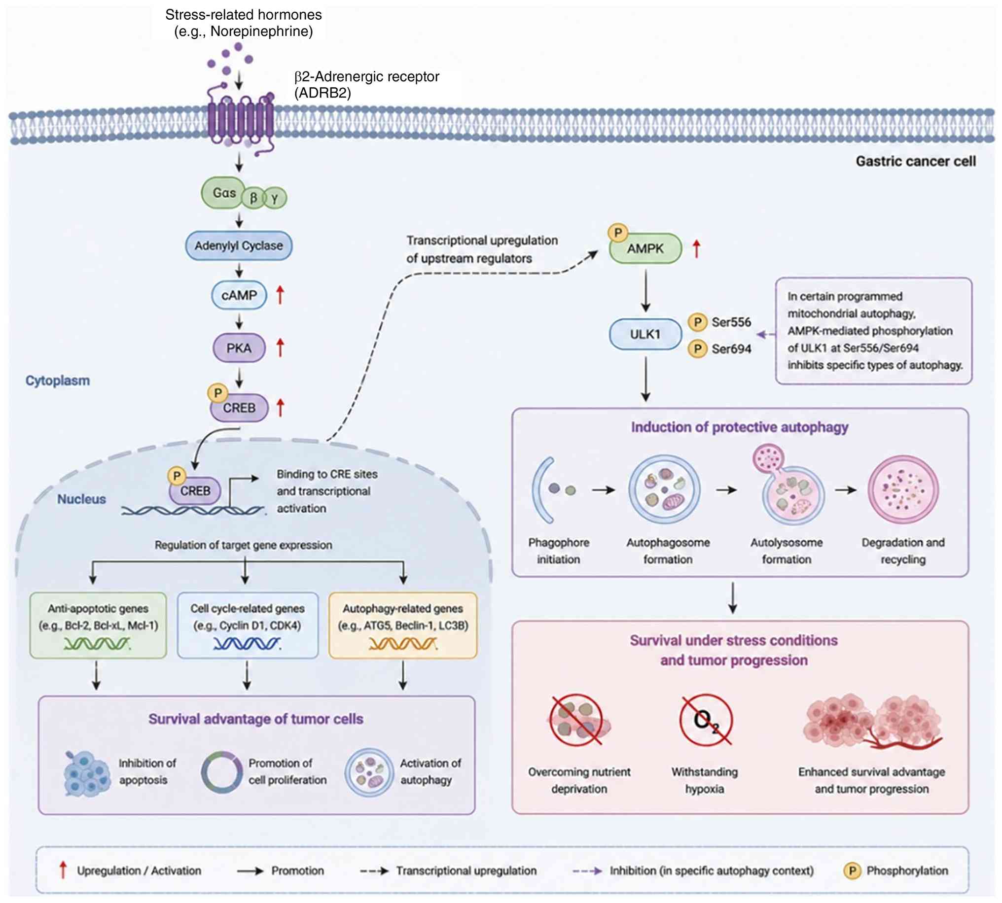 | Figure 1.Schematic representation of the
ADRB2/cAMP/PKA/CREB signaling cascade. Norepinephrine, a
stress-associated hormone, triggers the activation of the ADRB2,
which then initiates the adenylate cyclase-mediated cAMP/PKA
signaling pathway. Downstream, CREB is phosphorylated and
translocates into the nucleus. This drives higher expression of
genes associated to anti-apoptotic effects (Bcl-2), cell cycle
advancement (Cyclin D1) and autophagy regulation (ATG5, Beclin-1
and LC3B). In parallel, this signaling pathway enhances protective
autophagy through stimulation of the AMPK-ULK1 axis, allowing tumor
cells to survive under nutrient deprivation and hypoxic conditions,
thereby providing a net survival benefit. ADRB2, β2-adrenergic
receptor; cAMP, cyclic adenosine monophosphate; PKA, protein kinase
A; CREB, cAMP response element-binding protein; AMPK, AMP-activated
protein kinase; ULK1, UNC-51-like kinase 1; CRE, cAMP response
element; ATG5, autophagy protein 5; LC3B, microtubule-associated
protein 1 light chain 3β. |
Another important downstream consequence of ADRB2
activation is the regulation of CREB-dependent transcription. In
colorectal cancer, catecholamine stimulation activates the cAMP/PKA
signaling pathway through β2-ARs, resulting in CREB1
phosphorylation and transcriptional activation of glycolytic
enzymes (30).
ADRB2-mediated calcium signaling and
insulin-like growth factor 2 (IGF2) secretion
The ADRB2/PKA/voltage-dependent calcium channel
(VDCC)/Ca2+/IGF2 signaling pathway functions as an
important signal amplification mechanism in stress-mediated tumor
progression. Through calcium-dependent exocytosis, the pathway
rapidly promotes the release of IGF2, which subsequently activate
downstream signaling pathways associated with tumor cell survival.
In lung cancer, activation of this signaling cascade enhances the
survival of lung epithelial cells and contributes to tumorigenesis
(31).
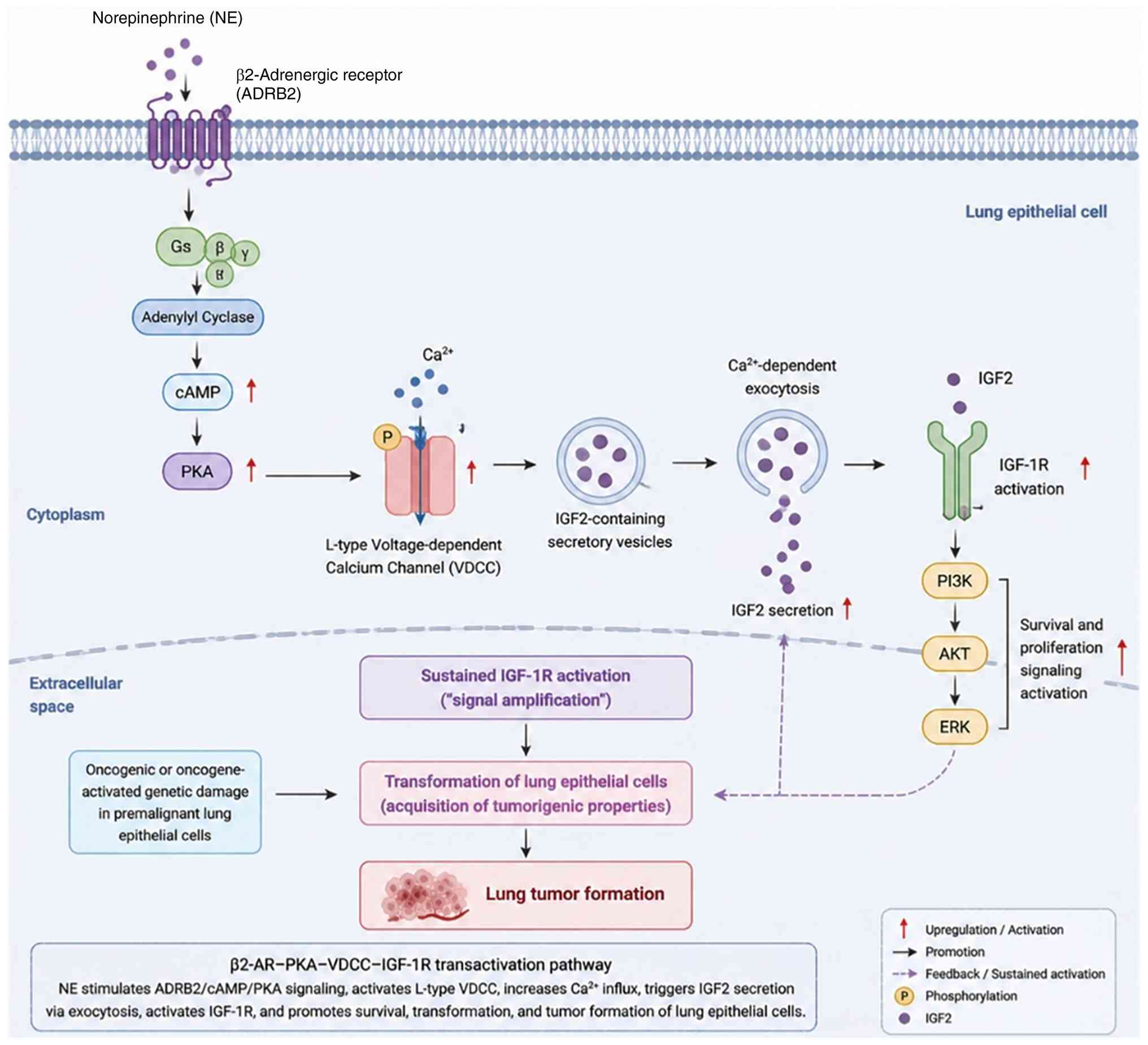
NE activates the ADRB2/cAMP/PKA signaling pathway,
which subsequently stimulates L-type VDCCs and induces
Ca2+ influx. The resulting calcium-dependent exocytosis
promotes the secretion of IGF2, leading to sustained activation of
IGF-1 receptor (IGF-1R) signaling. Consequently, this signaling
cascade facilitates the acquisition of tumor-associated phenotypes
by lung epithelial cells and supports the survival of precancerous
cells despite carcinogen-induced genetic damage, thereby promoting
lung tumorigenesis (Fig. 2)
(31).
Collectively, these observations suggest that VDCCs
represent a promising therapeutic target for interrupting
stress-induced calcium signaling (32).
Mechanistically, the
ADRB2/PKA/VDCC/Ca2+/IGF2 axis illustrates that
adrenergic signaling amplifies tumor progression not only through
intracellular signaling but also by promoting calcium-dependent
growth factor secretion.
Angiogenic signaling
The Notch signaling pathway serves as an important
mediator associating stress-related signaling to tumor angiogenesis
and primarily contributes to the pro-angiogenic effects induced by
catecholamines. Catecholamines upregulate the expression of
Jagged1, a signaling molecule targeting the Notch receptor through
activation of the β2-AR/PKA/mTOR signaling pathway. Increased
Jagged1 expression subsequently activates Notch signaling in
endothelial cells of blood vessels, thereby promoting the
proliferation of tumor-associated vascular endothelium.
In breast cancer, NE stimulation increases the
expression of the Notch ligand Jagged1 in tumor cells, which
subsequently activates Notch signaling in adjacent endothelial
cells. Pharmacological inhibition of β2-AR, PKA or mTOR signaling
markedly attenuates Jagged1 induction, highlighting the key role of
the β2-AR/PKA/mTOR axis in stress-induced angiogenic signaling
(33). Activation of endothelial
Notch signaling further enhances vascular endothelial activity and
promotes tumor angiogenesis, as demonstrated in both in
vitro co-culture systems and mouse xenograft models (34,35).
Beyond Jagged1-mediated signaling, other Notch
ligands also contribute to tumor vascular remodeling. Delta-like
ligand 4, a key regulator of vascular development, has been shown
to modulate tumor angiogenesis and represents a potential
therapeutic target for anti-angiogenic strategies (35). Collectively, these findings suggest
that stress-associated adrenergic signaling promotes tumor
angiogenesis not only through direct regulation of pro-angiogenic
factors but also through tumor-endothelial communication mediated
by Notch signaling.
Dopamine (DA) signaling promotes tumor
plasticity
DA receptor D2 (DRD2)/ERK/β-catenin
signaling
The DRD2/ERK/β-catenin signaling pathway represents
an important mechanism through which DA-mediated signaling promotes
tumor progression, particularly in glioblastoma (GBM).
Chronic psychological stress increases DA production
in GBM, leading to activation of DRD2 signaling. Sustained DRD2
activation stimulates the ERK signaling pathway, which suppresses
glycogen synthase kinase-3β (GSK3β) activity and promotes β-catenin
stabilization and nuclear translocation. Activated β-catenin
subsequently drives transcriptional programs associated with tumor
proliferation, stemness and malignant progression. In parallel,
ERK1/2 signaling upregulates tyrosine hydroxylase (TH), the
rate-limiting enzyme in DA biosynthesis, thereby increasing DA
production and establishing an autocrine positive-feedback loop
that further amplifies DRD2 signaling. Collectively, the
DRD2/ERK/β-catenin signaling axis and the DA/ERK/TH regulatory
circuit reinforce stress-induced glioblastoma progression through
sustained activation of oncogenic signaling (36).
DRD2-mediated stabilization of
hypoxia-inducible factor-1α (HIF-1α) promotes
epithelial-mesenchymal transition (EMT)
Beyond activation of the ERK/β-catenin pathway, DRD2
also promotes tumor progression by stabilizing HIF-1α. Within the
nucleus, DRD2 interacts with the tumor suppressor von Hippel-Lindau
(VHL) protein and competes with HIF-1α for VHL binding. This
interaction reduces the ubiquitin-mediated degradation of HIF-1α,
leading to its stabilization and subsequent induction of EMT.
During EMT, epithelial cells lose polarity and intercellular
adhesion while acquiring mesenchymal characteristics, ultimately
enhancing migratory and invasive capacities and facilitating tumor
metastasis (37).
Collectively, these findings indicate that DRD2
contributes to tumor progression through a number of mechanisms. In
addition to promoting proliferative signaling, DRD2 enhances tumor
cell plasticity by stabilizing HIF-1α and inducing EMT, thereby
expanding the spectrum of stress-induced malignant phenotypes.
Stress-induced metabolic
reprogramming
Unlike genetic alterations that constitutively
rewire cancer metabolism, psychological stress induces a dynamic
metabolic adaptation through neuroendocrine signaling. Recent
studies (38,39) indicate that stress hormones and
neuron-derived neurotransmitters converge on glycolytic pathways to
support tumor cell survival, stemness and metastatic
dissemination.
In breast cancer, chronic stress-induced adrenergic
signaling promotes metabolic reprogramming through activation of
the LDHA/USP28/MYC/SLUG axis. Stress-released adrenaline enhances
LDHA activity, resulting in increased lactate production and
intracellular acidification. The altered intracellular pH
environment facilitates USP28-mediated deubiquitination and
stabilization of the oncogenic transcription factor MYC. Stabilized
MYC subsequently activates SLUG transcription, promoting the
acquisition of breast cancer stem cell-like properties and
enhancing tumor cell plasticity (40).
In pancreatic ductal adenocarcinoma (PDAC),
stress-associated metabolic reprogramming can also be mediated
through direct neuron-tumor communication. Neuron-derived glutamate
promotes glycolytic adaptation in pancreatic ductal adenocarcinoma
cells by inducing Ca2+ influx and activating the
calcium/calmodulin-dependent protein kinase (CaMK)II/ERK-MAPK
signaling cascade. This pathway enhances hexokinase 2 expression
through m6A RNA modification, resulting in increased glycolytic
activity that supports tumor growth and metastatic dissemination.
In addition, glutamate release from sensory neurons is regulated by
the IGF-1/IGF-1R/transient receptor potential vanilloid 1/PI3K/Akt
signaling axis, establishing a reciprocal regulatory interaction
between pancreatic cancer cells and surrounding neural components
(41).
Collectively, these findings suggest that metabolic
reprogramming is a common downstream consequence of stress
signaling in cancer. Although different tumor types utilize
distinct upstream mediators, both adrenergic hormones and
neuron-derived neurotransmitters ultimately enhance glycolytic
activity to support tumor cell survival, stemness and metastatic
progression. However, current evidence is largely based on
experimental models, and further studies are required to determine
whether these mechanisms are broadly applicable across different
human cancers (Table II).
| Table II.Major stress-induced signaling
pathways underlying tumor cell-intrinsic adaptation. |
Table II.
Major stress-induced signaling
pathways underlying tumor cell-intrinsic adaptation.
| Biological
theme | Stress
mediator | Major signaling
pathway | Representative
tumor | Major biological
effect |
|---|
| Cell survival and
protective autophagy | NE | ADRB2/cAMP/PKA/CREB
→ AMPK/ULK1 | Gastric cancer | Cell survival and
protective autophagy |
| Calcium-dependent
growth factor secretion | NE |
ADRB2/PKA/VDCC/Ca2+/IGF2 | Lung cancer | Growth factor
secretion and tumor initiation |
| Angiogenic
signaling | Catecholamines |
ADRB2/PKA/mTOR/Jagged1/Notch | Breast cancer | Angiogenesis |
| Tumor
plasticity | DA |
DRD2/ERK/β-catenin | Glioblastoma | Proliferation and
stemness |
| EMT | DA |
DRD2/VHL/HIF-1α | Numerous
tumors | EMT and
invasion |
| Metabolic
reprogramming | Adrenaline |
LDHA/USP28/MYC/SLUG | Breast cancer | Glycolysis and
stemness |
| Metabolic
reprogramming | Glutamate | CaMKII/ERK/HK2 | Pancreatic ductal
adenocarcinoma | Glycolysis and
metastasis |
Pressure-induced reshaping of the tumor
microenvironment
In addition to directly reshaping tumor cell
metabolism, chronic stress profoundly alters the composition and
function of immune cells within the tumor microenvironment.
Stress-induced expansion of MDSCs
Chronic psychological stress promotes the expansion
of MDSCs, a heterogeneous population of immature myeloid cells that
serve a central role in tumor immune evasion. In a chronic
unpredictable stress model, activation of β1- and β2-ARs stimulated
the IL-6/STAT3 signaling pathway, leading to MDSC expansion and
enhanced pulmonary metastasis. Pharmacological inhibition of
IL-6/STAT3 attenuated MDSC accumulation and reduced metastatic
dissemination, highlighting the importance of β-adrenergic
signaling in stress-induced immunosuppression (42).
These findings indicate that chronic stress promotes
tumor immune evasion by expanding immunosuppressive myeloid
populations, thereby creating a microenvironment permissive for
metastatic progression.
CD8+ T-cell dysfunction and
exhaustion
Chronic stress-induced adrenergic signaling impairs
the antitumor activity of CD8+ T cells by promoting an
exhausted phenotype. In melanoma, activation of AR signaling
upregulates inhibitory receptors, including programmed cell death
protein 1 (PD-1) and T-cell immunoglobulin and mucin-domain
containing-3 (Tim-3), thereby driving CD8+ T-cell
exhaustion. This exhausted state is further characterized by
metabolic dysfunction and impaired effector function, ultimately
contributing to immunosuppression within the tumor microenvironment
(43).
Accumulating evidence suggests that metabolic
fitness is a key determinant of CD8+ T-cell antitumor
activity. Within the tumor microenvironment, metabolic competition
between tumor cells and immune cells may further aggravate
stress-induced T-cell dysfunction by limiting nutrient availability
and impairing cellular bioenergetics. Consequently,
stress-associated immune suppression may involve not only immune
checkpoint activation but also immune metabolic reprogramming,
providing a potential mechanistic association between chronic
stress and resistance to cancer immunotherapy. Together, these
findings indicate that immune checkpoint signaling, and immune
metabolic dysfunction may cooperatively contribute to
stress-associated immunosuppression and reduced responsiveness to
immunotherapy (44,45).
Stress-induced macrophage
polarization
Chronic stress promotes macrophage polarization
toward an immunosuppressive M2-like phenotype within the lung tumor
microenvironment. Elevated expression of the long non-coding RNA,
HIF1A-antisense RNA 3 (AS3), activates HIF-1α signaling through
interaction with Y-box binding protein 1, establishing a positive
feedback loop that sustains HIF1A-AS3/HIF-1α activation. This
signaling axis induces M2-like macrophage polarization, enhances
macrophage motility and impairs macrophage phagocytic activity,
thereby promoting immune evasion and lung cancer progression
(Fig. 3) (46).
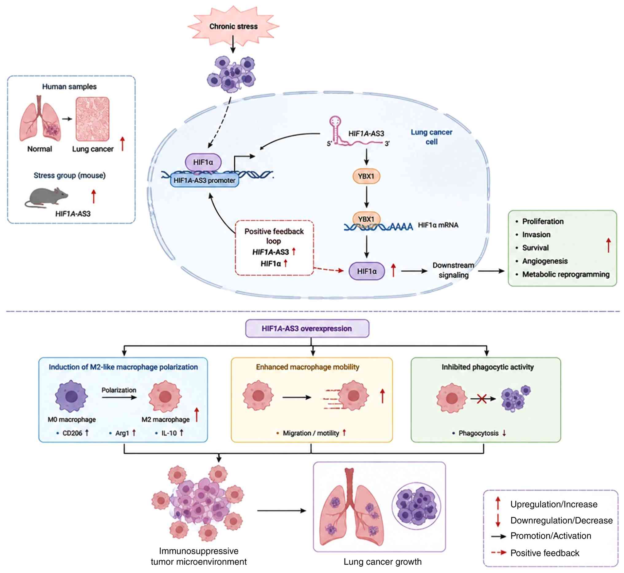 | Figure 3.Schematic representation of the
HIF1A-AS3/HIF-1α positive feedback loop in lung cancer under
chronic stress. Chronic stress increases HIF1A-AS3 expression,
which directly interacts with YBX1 to activate HIF-1α signaling,
thereby initiating downstream oncogenic pathways. HIF-1α, in turn,
transcriptionally upregulates HIF1A-AS3 via its promoter,
establishing a reciprocal positive feedback loop. This loop
promotes tumor cell proliferation, invasion and metabolic
reprogramming, while simultaneously inducing M2-like macrophage
polarization and suppressing phagocytosis, ultimately creating an
immunosuppressive microenvironment that accelerates lung cancer
progression. HIF1A-AS3, hypoxia-inducible factor 1α antisense RNA
3; YBX1, Y-box-binding protein 1; GCs, glucocorticoids; NETs,
neutrophil extracellular traps; ECM, extracellular
matrix. |
Chronic stress reshapes the immune microenvironment
not only by altering immune cell composition but also by
reprogramming macrophage phenotype toward an immunosuppressive
state.
Stress-induced NK-cell
dysfunction
Chronic stress suppresses NK cell-mediated antitumor
immunity through neuroimmune interactions within the pancreatic
tumor microenvironment.
In pancreatic cancer, elevated integrin α-5 (ITGA5)
expression enhances interactions between tumor cells and Schwann
cells (SCs), thereby promoting perineural invasion (PNI). SCs
subsequently undergo a transition toward a repair-like phenotype
and secrete nerve growth factor (NGF), which stimulates
neurogenesis while simultaneously suppressing IFN-γ-mediated
NK-cell cytotoxicity. The resulting impairment of NK-cell function
contributes to immune evasion and pancreatic cancer progression
(47). These findings suggest that
stress-associated neuroimmune interactions impair NK-cell-mediated
tumor surveillance, thereby associating PNI with immune suppression
in pancreatic cancer.
Stress-induced neutrophil activation
and neutrophil extracellular traps (NET) formation
Neutrophils represent another important immune
component affected by chronic stress within the tumor
microenvironment. Stress-induced glucocorticoid release promotes
neutrophil activation and the formation of NETs, thereby creating
an immunologically permissive environment that favors tumor
progression. NET formation contributes to metastatic dissemination
by altering the surrounding stromal environment and promoting
interactions between immune cells, fibroblasts and tumor cells
(48). Following NET induction,
activated fibroblasts increase fibronectin production and release
MMPs, resulting in extracellular matrix (ECM) remodeling. These
changes enhance tumor cell migration and invasion, associating
stress-induced immune alterations with metastatic progression
(Fig. 4; Table III) (48).
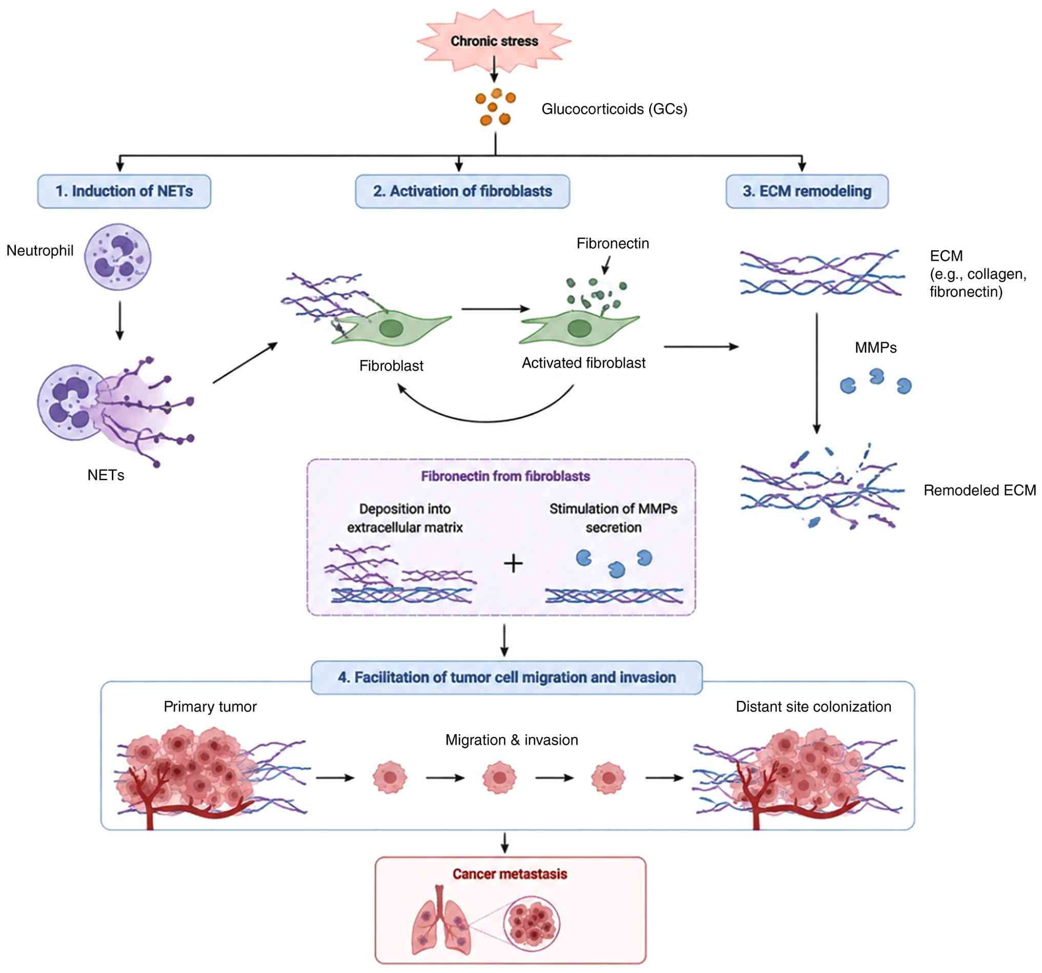 | Figure 4.Schematic overview of how chronic
stress promotes metastasis by triggering NET-dependent ECM
remodeling. Under chronic stress, the body releases
glucocorticoids, which then stimulate neutrophils to form
neutrophil NETs. After NETs are generated, they activate
fibroblasts and thereby increase the production and deposition of
fibronectin in the ECM. In parallel, NETs also promote the
secretion of MMPs, leading to further changes in the ECM.
Collectively, these events support tumor cell migration, invasion
and eventual distant metastatic spread. GCs, glucocorticoids; NETs,
neutrophil extracellular traps; ECM, extracellular matrix; MMPs,
matrix metalloproteinases. |
| Table III.Stress-induced alterations in the
tumor immune microenvironment. |
Table III.
Stress-induced alterations in the
tumor immune microenvironment.
| Immune cell | Stress
mediator | Major pathway | Functional
consequence | Tumor type |
|---|
| MDSCs | Catecholamines |
β-AR/IL-6/STAT3 | Expansion of
MDSCs | Breast cancer |
| CD8+ T
cells | Adrenergic
signaling | PD-1/Tim-3 | T-cell
exhaustion | Melanoma |
| Macrophages | HIF1A-AS3 | HIF-1α/YBX1 | M2
polarization | Lung cancer |
| Natural killer
cells | ITGA5/nerve growth
factor | IFN-γ
inhibition | Reduced
cytotoxicity | Pancreatic
cancer |
| Neutrophils |
Glucocorticoids | Neutrophil
extracellular traps | Extracellular
matrix remodeling | Numerous
tumors |
Neuro-tumor communication and neuroimmune
regulation
Sympathetic nerve-tumor
communication
Sympathetic nerve fibers are actively present within
the tumor microenvironment and represent a notable source of
catecholamine signals. NE released from sympathetic terminals
directly regulates tumor cell behavior through AR, particularly
ADRB2. In gastric cancer, sympathetic-derived NE activates
ADRB2-mediated AMPK-ULK1 signaling, promoting protective autophagy
and enabling tumor cells to survive under stress conditions
(27). In prostate cancer,
sympathetic denervation or genetic disruption of ADRB2/ADRB3
signaling markedly suppresses tumor progression, highlighting the
functional importance of sympathetic innervation in tumor
development (49). In genitourinary
malignancy, the effects of stress-related signaling may also be
shaped by sex hormone receptor activity and the tissue-specific
immune microenvironment. In prostate cancer, androgen receptor
signaling may interact with adrenergic and glucocorticoid pathways
to regulate tumor cell plasticity, inflammatory signaling and
immune-cell recruitment. In bladder cancer, sex hormone receptor
expression and sex-associated differences in immune composition may
likewise influence the biological consequences of chronic stress.
These observations suggest that adrenergic and glucocorticoid
signaling should not be considered in isolation, but rather within
the broader context of hormone-dependent and immune-regulatory
networks in genitourinary tumors (50).
Beyond direct regulation of tumor cells, sympathetic
signaling also reshapes the surrounding tumor ecosystem. NE
released from sympathetic nerves activates cAMP/PKA-dependent
transcriptional programs, including CREB, NF-κB and STAT3, which
promote angiogenic and metastatic responses through regulation of
VEGF and MMP (51). In addition,
chronic psychological stress enhances sympathetic activation,
increasing NE levels in circulation and tumor tissues. This
sustained adrenergic stimulation promotes tumor growth and immune
remodeling, including β-adrenergic-mediated recruitment of myeloid
cells through the C-X-C motif chemokine receptor type 2/C-X-C motif
chemokine ligand 2/C-X-C motif chemokine ligand 2 pathway in
hepatocellular carcinoma (52,53).
Collectively, these findings establish sympathetic
innervation as an important regulator of tumor biology by
integrating neural signals with tumor cell adaptation, immune
remodeling, angiogenesis and metastatic progression. However, the
biological consequences of sympathetic signaling are likely to vary
according to tumor lineage, hormone receptor status and the
composition of the local immune microenvironment. Further studies
are therefore needed to elucidate the spatial organization,
temporal dynamics and clinical importance of sympathetic
innervation across different tumor types.
Parasympathetic nerve-tumor
communication
The parasympathetic nervous system exerts
context-dependent effects on tumor progression, with both
tumor-promoting and tumor-suppressive functions observed across
different cancer types. Unlike sympathetic signaling, which is
generally associated with stress-induced tumor progression,
parasympathetic signaling appears to regulate tumor behavior
through tissue-specific neurotransmitter pathways and immune
modulation (54).
In gastric cancer, parasympathetic signaling
promotes tumor progression primarily through acetylcholine
(ACh)-mediated activation of muscarinic acetylcholine receptor M3
(CHRM3). Activation of CHRM3 stimulates downstream
Wnt/yes-associated protein, ERK1/2 and PI3K/AKT signaling pathways,
thereby enhancing tumor cell proliferation, survival and migration.
In addition, ACh signaling promotes EGFR activation and induces
MMP-7 expression, further facilitating malignant progression
(54). These findings indicate that
cholinergic signaling can directly regulate tumor cell behavior
through receptor-dependent mechanisms.
Conversely, parasympathetic activity may exert
antitumor effects in certain contexts. In breast cancer models,
modulation of parasympathetic nerve activity reduced tumor growth
and metastatic dissemination, which was associated with decreased
PD-1/programmed death-ligand 1 expression and enhanced antitumor
immune responses. Notably, parasympathetic activation did not
markedly alter tumor-infiltrating lymphocyte numbers but instead
influenced immune function by regulating immune checkpoint
expression and promoting IFN-γ production (50).
Beyond direct neural regulation, ACh-mediated
signaling also contributes to tumor-associated neurogenesis. ACh
released from tuft cells promotes neural development within and
around tumor tissues through interactions with neurotrophic growth
factors, establishing a feedback loop that facilitates neural
remodeling in the tumor microenvironment (55).
Collectively, parasympathetic signaling represents a
context-dependent regulator of tumor progression. The divergent
effects between tumor types may depend on receptor expression
patterns, cellular targets and the surrounding immune
microenvironment.
Sensory neuron-tumor communication
through glutamatergic pseudosynapse
Sensory neurons can directly communicate with tumor
cells through glutamatergic pseudosynapses. In PDAC, sensory nerve
terminals form synapse-like contacts with tumor cells that express
high levels of the N-methyl-D-aspartate receptor subunit, NMDAR2D,
also known as GRIN2D. Neuron-derived glutamate activates these
receptors and promotes Ca2+-dependent signaling in PDAC
cells. Glutamate stimulation also enhances GRIN2D expression
through the CaMKIV/CREB signaling pathway, thereby reinforcing
tumor responsiveness to neural inputs. Functional studies (56,57) in
mouse models further indicate that GRIN2D is required for tumor
growth and neural innervation. Collectively, these findings
identify glutamatergic pseudosynaptic communication as a direct
mechanism through which sensory neurons promote pancreatic cancer
progression (Fig. 5) (58).
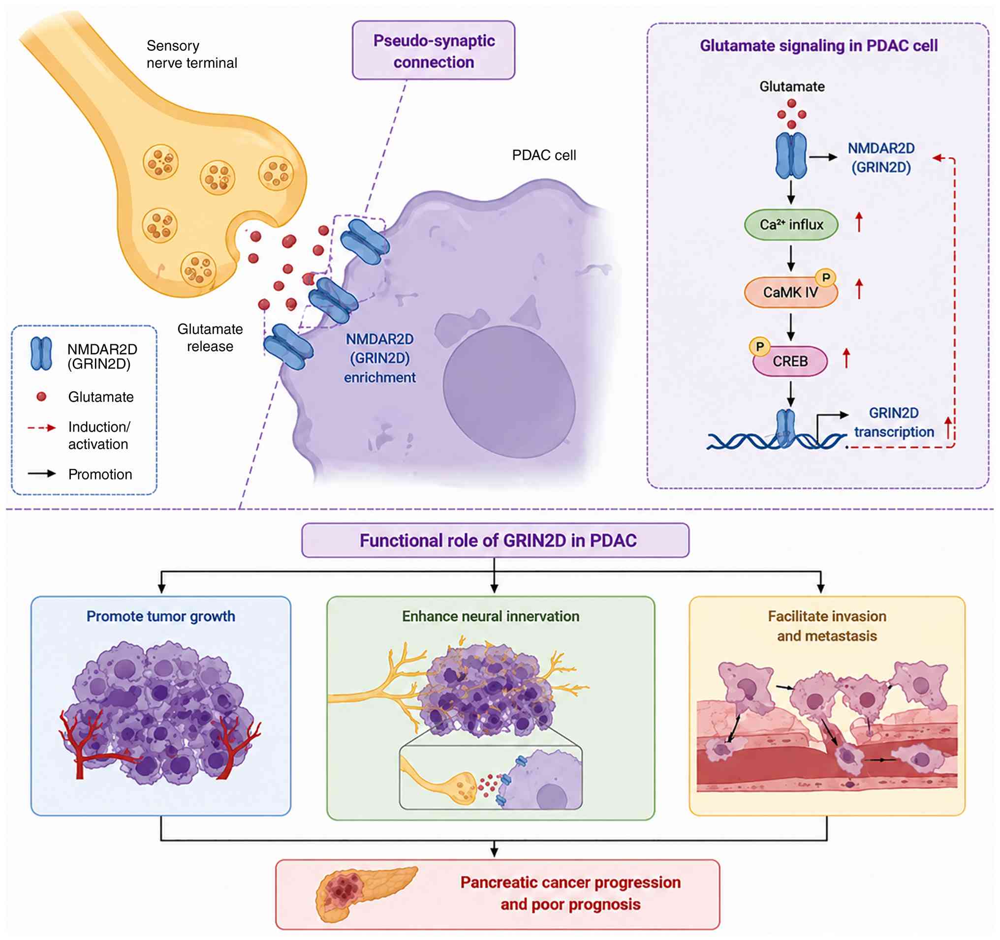 | Figure 5.Schematic representation of sensory
neuron-driven pancreatic cancer progression through glutamatergic
pseudosynapses. Sensory nerve terminals form pseudosynaptic
contacts with PDAC cells and release glutamate. The released
glutamate binds to NMDAR2D (GRIN2D) receptors, which are expressed
on the surface of the cancer cells. This binding leads to
Ca2+ influx and, in turn, activates the CaMK IV-CREB
signaling pathway. As a result, tumor proliferation, neural
innervation, cellular invasion and metastatic spread are promoted.
PDAC, pancreatic ductal adenocarcinoma; NMDAR2D/GRIN2D,
N-methyl-D-aspartate receptor subunit 2D; CaMK IV,
calcium/calmodulin-dependent protein kinase IV; cAMP, cyclic
adenosine monophosphate; CREB, cAMP response element-binding
protein. |
Context-dependent effects of
neurotransmitters
Neurotransmitter-mediated tumor regulation is not
restricted to nerve-derived signals. In glioblastoma, tumor-derived
DA can establish an autocrine DRD2/ERK/TH feedback loop that
sustains oncogenic signaling. This finding illustrates that
neurotransmitter signaling within tumors may involve both neural
and tumor-cell-derived sources.
Overall, neuro-tumor communication is mediated by
diverse neurotransmitters whose biological effects depend on their
cellular source, receptor distribution and tumor context. NE and
glutamate generally promote tumor progression through sympathetic
and sensory neural inputs, whereas ACh may exert either
tumor-promoting or tumor-suppressive effects. DA further
illustrates that neurotransmitter signaling may also arise from
tumor-cell-autonomous production rather than exclusively from
neural innervation (Table IV).
| Table IV.Neuro-tumor interactions and
potential therapeutic strategies. |
Table IV.
Neuro-tumor interactions and
potential therapeutic strategies.
| Neural
component |
Neurotransmitter | Major
receptor/pathway | Biological
effect | Potential
therapeutic target |
|---|
| Sympathetic
nerve | Norepinephrine | ADRB2 | Growth and
invasion | β-blockers |
| Parasympathetic
nerve | Acetylcholine | CHRM3 |
Context-dependent | CHRM3
inhibitors |
| Sensory neuron | Glutamate | NMDAR2D | Pseudosynapse | NMDAR
inhibitors |
| Dopamine | Dopamine | DRD2 | Plasticity | DRD2
antagonists |
| Tumor-Schwann
cell | Nerve growth
factor | ITGA5 | Perineural
invasion. | ITGA5
inhibitors |
Stress-induced vascular remodeling and
metastatic dissemination
Stress-induced angiogenesis
Angiogenesis is an key component of tumor
progression, providing oxygen and nutrients while facilitating
local invasion and metastatic dissemination. Chronic stress
promotes angiogenic remodeling by shifting the balance between
pro-angiogenic and anti-angiogenic signals within the tumor
microenvironment. Although adrenergic signaling can increase the
expression of VEGF, its angiogenic effects also involve suppression
of endogenous anti-angiogenic regulators.
In prostate cancer, stress-associated cAMP/PKA/CREB
signaling promotes angiogenesis through two downstream regulatory
branches involving histone deacetylase 2 (HDAC2) and G
protein-coupled receptor kinase 3 (GRK3). HDAC2 contributes to the
epigenetic repression of thrombospondin-1 (TSP1), whereas GRK3
suppresses the expression of both TSP1 and plasminogen activator
inhibitor-2 (PAI2). As TSP1 and PAI2 normally restrain vascular
growth, their downregulation creates a microenvironment favorable
for endothelial activation and neovascularization. Together with
increased VEGF signaling, these alterations enhance tumor
vascularization and support prostate cancer progression (Fig. 6) (59).
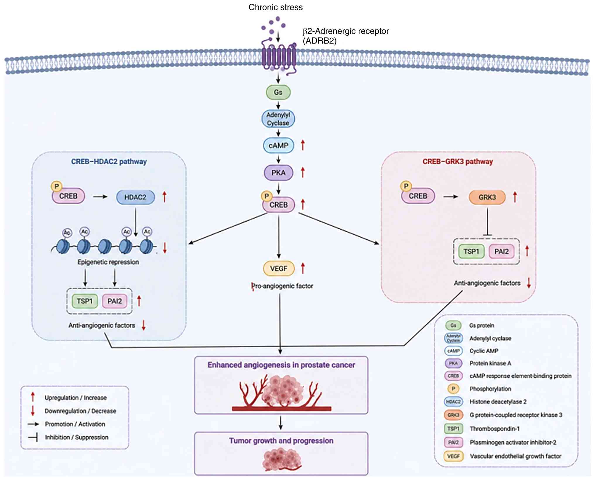 | Figure 6.Schematic representation of
stress-induced angiogenic signaling in prostate cancer. Chronic
stress activates ADRB2 on tumor cells, triggering the
Gs/cAMP/PKA/CREB signaling axis. CREB regulates angiogenesis via
two downstream pathways: the CREB-HDAC2 pathway, in which
HDAC2-mediated epigenetic repression decreases anti-angiogenic
factors TSP1 and PAI2; and the CREB-GRK3 pathway, in which GRK3
suppresses TSP1 and PAI2. Both pathways converge to upregulate
VEGF, a major pro-angiogenic factor, resulting in enhanced
angiogenesis and subsequent tumor growth and progression. Red
arrows indicate upregulation, down arrows indicate downregulation
and black arrows represent promotion or inhibition as indicated in
the diagram. ADRB2, β2-adrenergic receptor; Gs, G protein; cAMP,
cyclic adenosine monophosphate; PKA, protein kinase A; CREB, cAMP
response element-binding protein; HDAC2, histone deacetylase 2;
GRK3, G protein-coupled receptor kinase 3; TSP1, thrombospondin-1;
PAI2, plasminogen activator inhibitor-2; VEGF, vascular endothelial
growth factor. |
Collectively, stress-induced angiogenesis is driven
not only by increased production of pro-angiogenic mediators but
also by the loss of endogenous angiogenic restraints. This
coordinated remodeling of vascular signaling may facilitate tumor
growth and metastatic dissemination; however, the clinical
relevance of these mechanisms remains to be established.
Stress-induced lymphangiogenesis and
lymphatic dissemination
Chronic psychological stress promotes lymphatic
remodeling within and surrounding the tumor microenvironment,
thereby facilitating lymphatic metastasis. Activation of
β-adrenergic signaling enhances the remodeling of tumor-associated
lymphatic vessels, while sympathetic nervous system activity
regulates lymphatic flow dynamics. These alterations are
characterized by increased lymphatic vessel density, lymphatic
vessel dilation and enhanced lymphatic drainage, creating favorable
conditions for the dissemination of tumor cells through the
lymphatic circulation (60).
Collectively, these findings suggest that stress-induced
lymphangiogenesis not only provides structural routes for
metastatic spread but also promotes the efficient transport of
tumor cells to regional lymph nodes and distant metastatic
sites.
Together, stress-induced remodeling of both blood
and lymphatic vasculature provides complementary routes that
support tumor growth and metastatic dissemination.
Treatment and prevention
β-blockers
β-AR antagonists, particularly the non-selective
β-blocker propranolol, have demonstrated potential in alleviating
immunosuppression and enhancing the efficacy of immune checkpoint
inhibitors in both preclinical and retrospective clinical studies.
In a melanoma mouse model, combined treatment with propranolol and
anti-PD-1 therapy markedly enhanced antitumor activity, resulting
in delayed tumor progression, increased infiltration of
CD8+ T lymphocytes into the tumor microenvironment, and
restoration of T-cell effector function (61). This synergistic effect is considered
to result from the ability of propranolol to attenuate chronic
stress-induced T-cell exhaustion.
In mouse models subjected to chronic adrenergic
stress, treatment with propranolol markedly decreased the
proportion of tumor-infiltrating lymphocytes expressing exhaustion
markers, including PD-1, Tim-3 and lymphocyte activation gene 3.
Concurrently, propranolol expands the population of pre-exhausted T
cells, which exhibit greater self-renewal potential and enhanced
antitumor effector activity (62).
In breast cancer models, β-AR antagonists were also found to
improve the efficacy of anti-PD-1 therapy by reducing MDSC
accumulation and suppressing their immunosuppressive functions
(63). Furthermore, β-blockers have
been reported to enhance the effectiveness of both chemotherapy and
radiotherapy, underscoring their broad immunomodulatory properties
(64).
Retrospective clinical studies further support these
preclinical findings. In patients with melanoma, treatment with
β-blockers, particularly β2-selective antagonists administered for
non-oncological indications, was associated with notably improved
responses to immunotherapy compared with patients who were not
receiving these agents (65). In a
separate phase II biomarker-driven clinical study, patients with
breast cancer who received propranolol for one week before surgical
intervention displayed elevated infiltration of CD68+
macrophages and CD8+ T cells within the tumor
microenvironment. This increase in immune cell presence was
associated with downregulation of genes associated to metastatic
progression and inflammatory signaling pathways (63). Additionally, a phase I clinical
trial evaluating the combination of propranolol with the anti-PD-1
antibody pembrolizumab in patients with advanced melanoma reported
encouraging therapeutic results (66).
Collectively, the favorable results observed in
epidemiological studies, clinical investigations and mechanistic
research provide a strong theoretical rationale for combining
β-blockers with immunotherapy. At present, numerous clinical trials
are ongoing to further evaluate the efficacy and safety of this
combined therapeutic strategy.
Targeting key nodes
Because stress-induced carcinogenesis involves
numerous complex signaling pathways, therapeutic strategies
targeting key molecular mediators may provide substantial clinical
benefits. Among these approaches, autophagy inhibition has been
extensively investigated. Since tumor cells typically depend on
protective autophagy to adapt to stress and resist chemotherapy or
radiotherapy, inhibition of autophagy may enhance the therapeutic
efficacy of conventional anticancer treatments (67). In gastric cancer, chronic stress
activates the AMPK-ULK1 signaling pathway through ADRB2, thereby
inducing protective autophagy and promoting tumor cell
proliferation and survival. These findings suggest that
pharmacological inhibition of autophagy may attenuate the
tumor-promoting effects of chronic stress (27).
In addition, HDAC inhibitors exert antitumor
activity by inducing endoplasmic reticulum stress. Previous studies
(67,68) have indicated that HDAC inhibition
activates the protein kinase R-like endoplasmic reticulum
kinase-eukaryotic translation initiation factor 2α pathway, thereby
initiating the unfolded protein response and ultimately promoting
apoptosis of tumor cells . In prostate cancer, HDAC2 is a key
mediator of stress-induced angiogenesis. During tumor progression,
CREB facilitates tumor growth by epigenetically repressing the
anti-angiogenic factor TSP1 via upregulation of HDAC2.
Collectively, these observations suggest that HDAC2-targeted
inhibitors could serve as a potential therapeutic strategy for
aggressive prostate cancer. Additional mechanistic insights are
provided in the section on prostate cancer.
GRK3, an important downstream component of the ADRB
signaling pathway, has also emerged as a potential therapeutic
target in prostate cancer. Previous studies have demonstrated that
GRK3 is important for metastatic tumor cell survival and
proliferation. Furthermore, elevated GRK3 expression promotes
angiogenesis through downregulation of TSP1 and PAI-2, thereby
facilitating prostate cancer progression and metastasis (69). Importantly, GRK3 is
transcriptionally regulated by CREB and functions as a key mediator
within the chronic stress-activated cAMP/PKA/CREB signaling
pathway. Suppression of GRK3 expression inhibits neuroendocrine
differentiation while simultaneously increasing the proliferation
of neuroendocrine prostate cancer cells. Therefore, the development
of selective GRK3 kinase inhibitors may provide a promising
therapeutic strategy for aggressive prostate cancer subtypes.
DRD2 antagonists have also demonstrated antitumor
potential in a number of cancer models. In particular, the
classical DRD2 antagonist trifluoperazine (TFP) has shown
inhibitory effects on glioblastoma growth through suppression of
tumor cell proliferation and survival. The therapeutic efficacy of
TFP may depend on the specific DA receptor expression profile
within tumors (70). In
stress-induced tumor models, tissue factor pathway inhibitor (TFPI)
promotes degradation of HIF-1α through disruption of the
interaction between DRD2 and VHL, thereby inhibiting stress-induced
tumor growth. TFP has been shown to suppress tumor cell
proliferation and migration in breast cancer models, primarily
through antagonism of DRD2 (71).
LDHA represent another important strategy for
reversing tumor-associated metabolic reprogramming. In breast
cancer, chronic stress-induced epinephrine exposure promotes
aerobic glycolysis through activation of LDHA. Elevated lactate
production subsequently induces USP28-mediated deubiquitination and
stabilization of MYC, ultimately activating the SLUG promoter and
promoting cancer stem cell-like phenotypes (72). Drug screening studies (72,73)
further demonstrated that vitamin C suppresses LDHA activity and
attenuates chronic stress-induced cancer stem cell-like
characteristics, suggesting its potential therapeutic value in
stress-associated breast cancer
Neuromodulation strategies
Chemical sympathectomy represents an important
experimental strategy for validating the role of the
sympathetic-tumor axis in tumor progression. In a murine prostate
cancer model, selective ablation of peripheral sympathetic nerves
using 6-hydroxydopamine notably suppressed tumor growth (74). Similarly, in gastric cancer models,
administration of 6-hydroxydopamine attenuated the tumor-promoting
effects of chronic stress and restored serum catecholamine levels
to baseline values. Relevant signaling mechanisms are discussed in
the ADRB2-related section of gastric cancer. As an important
approach for regulating parasympathetic nervous system activity,
vagus nerve stimulation has also attracted attention because of its
potential antitumor effects. Experimental evidence (75) demonstrated that vagus nerve
stimulation suppresses tumor growth and metastasis through
reduction of PD-1 and forkhead box (FOX) P3 expression in
tumor-infiltrating lymphocytes.
Targeting NMDARs represents another important
strategy for modulating glutamatergic signaling during neuro-tumor
interactions. In pancreatic cancer, sensory neurons establish
functional pseudosynaptic connections with NMDAR2D receptors
expressed on tumor cells through glutamate release, thereby
promoting tumor progression and neural invasion (53). In addition, targeting ITGA5 disrupts
interactions between tumor cells and SCs. Tumor cells expressing
ITGA5 adhere to fibronectin within the SC-associated ECM,
reprogramming SCs and promoting NGF production. Preclinical studies
(42,47) further suggest that combination
treatment with the ITGA5 inhibitor cilengitide and anti-PD-1
immunotherapy enhances therapeutic efficacy, highlighting the
potential of targeting neuroimmune interactions in pancreatic
cancer.
Suppression of NETs is an important strategy for
inhibiting stress-induced metastasis through modulation of
neutrophil activity. Chronic stress increases glucocorticoid
production, thereby promoting NET formation and establishing a
microenvironment favorable for metastatic progression (76).
Psychological interventions and
exosomes
Imipramine, a tricyclic antidepressant, has
potential therapeutic benefits in cancer treatment through
suppression of metastasis-associated exosome production. Imipramine
not only alleviates depressive symptoms in patients with cancer but
also inhibits communication between tumor cells and stromal cells
through blockade of exosome release. Collectively, these effects
contribute to marked tumor regression in breast cancer models
(77). In addition, imipramine
derivatives have been reported to suppress breast cancer
progression and metastasis through inhibition of the FOXM1-mediated
DNA repair pathway. This therapeutic effect selectively targets
tumor cells while exerting minimal effects on normal cells
(77).
In breast cancer, patients participating in stress
management interventions maintained or enhanced T cell
proliferative activity, whereas patients in control groups
exhibited progressive declines in immune function (78). Similarly, among patients with
melanoma receiving immunotherapy, individuals experiencing high
levels of emotional distress demonstrated notably lower objective
response rates compared with emotionally stable patients (37 vs.
69%) (79). Stress-induced
glucocorticoid release has been shown to impair
treatment-associated antitumor immune surveillance, at least
partially through upregulation of TSC22 domain family protein 3
expression (80).
Collectively, these findings suggest that
incorporation of psychological stress management into
immunotherapeutic regimens may represent a promising adjunctive
strategy for improving the efficacy of cancer immunotherapy.
Conclusions and future perspectives
Recent evidence suggests that psychological stress
promotes tumor progression through a number of interconnected
mechanisms. Within tumor cells, stress hormones activate signaling
pathways such as ADRB2/cAMP/PKA/CREB and DRD2/ERK/β-catenin,
thereby regulating tumor cell proliferation, protective autophagy
and metabolic reprogramming. Within the tumor microenvironment,
chronic stress promotes the accumulation of MDSCs, induces T-cell
exhaustion, facilitates M2 macrophage polarization and enhances
angiogenesis, collectively contributing to an immunosuppressive
microenvironment. In addition, at the neuro-tumor interface,
sympathetic, parasympathetic and sensory nerves directly regulate
tumor behavior through neurotransmitter release and pseudosynaptic
communication. Based on these mechanisms, therapeutic strategies
including β-blockers, inhibitors targeting DRD2, LDHA and GRK3, as
well as psychological interventions, have demonstrated promising
therapeutic potential.
Despite substantial progress, several important
limitations remain in current research. First, the tumor-specific
heterogeneity of stress-related signaling pathways remains
incompletely understood. In addition, systematic investigation of
the spatiotemporal dynamics underlying neuro-immune-tumor
interactions is still lacking. Studies examining synergistic
interactions among numerous stress-associated mechanisms also
remain limited. Furthermore, clinical translation remains
challenging due to the lack of validated stress-related biomarkers,
uncertainty regarding patient selection, limited prospective
clinical trials and difficulties in translating findings from
experimental stress models to heterogeneous human cancer
populations.
Single-cell and spatial multi-omics technologies may
provide deeper insight into the cellular heterogeneity of
stress-associated tumor microenvironments. Combination strategies
integrating β-adrenergic blockade with immune checkpoint inhibitors
represent another promising direction for future research and
warrant further evaluation in prospective clinical trials. In
addition, stress-associated biomarkers for patient stratification
are required to identify patient populations most likely to benefit
from stress-targeted interventions. Through multidisciplinary
collaboration and continued mechanistic investigation, stress
management strategies may ultimately be integrated more effectively
into comprehensive cancer treatment paradigms.
Acknowledgements
Not applicable.
Funding
Funding: No funding was received.
Availability of data and materials
Not applicable.
Authors' contributions
YZ wrote the manuscript. YK contributed to the
literature search. SZ generated the figures, ensured visual data
accuracy and contributed to drafting the manuscript. YZ and YK
reviewed and edited the manuscript. All authors have read and
approved the final version of the manuscript. Data authentication
is not applicable.
Ethics approval and consent to
participate
Not applicable.
Patient consent for publication
Not applicable.
Competing interests
The authors declare that they have no competing
interests.
Use of artificial intelligence tools
During the preparation of this work, artificial
intelligence tools were used to improve the readability and
language of the manuscript or to generate images, and subsequently,
the authors revised and edited the content produced by the
artificial intelligence tools as necessary, taking full
responsibility for the ultimate content of the present
manuscript.
References
|
1
|
Sung H, Ferlay J, Siegel RL, Laversanne M,
Soerjomataram I, Jemal A and Bray F: Global cancer statistics 2020:
GLOBOCAN estimates of incidence and mortality worldwide for 36
cancers in 185 countries. CA Cancer J Clin. 71:209–249.
2021.PubMed/NCBI
|
|
2
|
Yan J, Chen Y, Luo M, Hu X, Li H, Liu Q
and Zou Z: Chronic stress in solid tumor development: From
mechanisms to interventions. J Biomed Sci. 30:82023. View Article : Google Scholar : PubMed/NCBI
|
|
3
|
Wang Y, Wang X, Wang K, Qi J, Zhang Y,
Wang X, Zhang L, Zhou Y, Gu L, Yu R and Zhou X: Chronic stress
accelerates glioblastoma progression via DRD2/ERK/β-catenin axis
and Dopamine/ERK/TH positive feedback loop. J Exp Clin Cancer Res.
42:1612023. View Article : Google Scholar : PubMed/NCBI
|
|
4
|
He XY, Gao Y, Ng D, Michalopoulou E,
George S, Adrover JM, Sun L, Albrengues J, Daßler-Plenker J, Han X,
et al: Chronic stress increases metastasis via neutrophil-mediated
changes to the microenvironment. Cancer Cel. 42:474–486. 2024.
View Article : Google Scholar
|
|
5
|
Herbetko K, Kaczor J, Sołtyk A,
Kisielewska M, Opęchowski M, Sztuder A and Kulbacka J: The impact
of chronic stress on treatment outcomes of cancer patients with
divergent survival rates: A systematic review. Int J Mol Sci.
27:6862026. View Article : Google Scholar : PubMed/NCBI
|
|
6
|
He XY, Gao Y, Ng D, Michalopoulou E,
George S, Adrover JM, Sun L, Albrengues J, Daßler-Plenker J, Han X,
et al: Chronic stress increases metastasis via neutrophil-mediated
changes to the microenvironment. Cancer Cell. 42:474–486.e12. 2024.
View Article : Google Scholar : PubMed/NCBI
|
|
7
|
Cohen S, Janicki-Deverts D and Miller GE:
Psychological stress and disease. JAMA. 298:1685–1687. 2007.
View Article : Google Scholar : PubMed/NCBI
|
|
8
|
Chida Y, Hamer M, Wardle J and Steptoe A:
Do stress-related psychosocial factors contribute to cancer
incidence and survival? Nat Clin Pract Oncol. 5:466–475. 2008.
View Article : Google Scholar : PubMed/NCBI
|
|
9
|
Roberts AL, Huang T, Koenen KC, Kim Y,
Kubzansky LD and Tworoger SS: Posttraumatic stress disorder is
associated with increased risk of ovarian cancer: A Prospective and
retrospective longitudinal cohort study. Cancer Res. 79:5113–5120.
2019. View Article : Google Scholar : PubMed/NCBI
|
|
10
|
Fraterman I, Reijers ILM, Dimitriadis P,
Broeks A, Gonzalez M, Menzies AMM, Lopez-Yurda M, Kapiteijn E, van
der Veldt AAM, Suijkerbuijk KPM, et al: Association between
pretreatment emotional distress and neoadjuvant immune checkpoint
blockade response in melanoma. Nat Med. 29:3090–3099. 2023.
View Article : Google Scholar : PubMed/NCBI
|
|
11
|
He XY, Gao Y, Ng D, Michalopoulou E,
George S, Adrover JM, Sun L, Albrengues J, Daßler-Plenker J, Han X,
et al: Chronic stress increases metastasis via neutrophil-mediated
changes to the microenvironment. Cancer Cell. 42:474–486.e12. 2024.
View Article : Google Scholar : PubMed/NCBI
|
|
12
|
Thaker PH, Han LY, Kamat AA, Arevalo JM,
Takahashi R, Lu C, Ozols RF and Sood AK: Chronic stress promotes
tumor growth and angiogenesis in a mouse model of ovarian
carcinoma. Nat Med. 12:939–944. 2006. View
Article : Google Scholar : PubMed/NCBI
|
|
13
|
Zhang X, Zhang Y, He Z, Yin K, Li B, Zhang
L and Xu Z: Chronic stress promotes gastric cancer progression and
metastasis: An essential role for ADRB2. Cell Death Dis.
10:7882019. View Article : Google Scholar : PubMed/NCBI
|
|
14
|
Shakhar G and Ben-Eliyahu S: In vivo
beta-adrenergic stimulation suppresses natural killer activity and
compromises resistance to tumor metastasis in rats. J Immunol.
160:3251–3258. 1998. View Article : Google Scholar : PubMed/NCBI
|
|
15
|
Reiche EM, Nunes SO and Morimoto HK:
Stress, depression, the immune system, and cancer. Lancet Oncol.
5:617–625. 2004. View Article : Google Scholar : PubMed/NCBI
|
|
16
|
Qiao G, Chen M, Mohammadpour H, MacDonald
CR, Bucsek MJ, Hylander BL, Barbi JJ and Repasky EA: Chronic
adrenergic stress contributes to metabolic dysfunction and an
exhausted phenotype in T cells in the tumor microenvironment.
Cancer Immunol Res. 9:651–664. 2021. View Article : Google Scholar : PubMed/NCBI
|
|
17
|
Jiang W, Li Y, Li ZZ, Sun J, Li JW, Wei W,
Li L, Zhang C, Huang C, Yang SY, et al: Chronic restraint stress
promotes hepatocellular carcinoma growth by mobilizing splenic
myeloid cells through activating β-adrenergic signaling. Brain
Behav Immun. 80:825–838. 2019. View Article : Google Scholar : PubMed/NCBI
|
|
18
|
Zhi X, Li B, Li Z, Zhang J, Yu J, Zhang L
and Xu Z: Adrenergic modulation of AMPK-dependent autophagy by
chronic stress enhances cell proliferation and survival in gastric
cancer. Int J Oncol. 54:1625–1638. 2019.PubMed/NCBI
|
|
19
|
Guan Y, Yao W, Yu H, Feng Y, Zhao Y, Zhan
X and Wang Y: Chronic stress promotes colorectal cancer progression
by enhancing glycolysis through β2-AR/CREB1 signal pathway. Int J
Biol Sci. 19:2006–2019. 2023. View Article : Google Scholar : PubMed/NCBI
|
|
20
|
Cui B, Luo Y, Tian P, Peng F, Lu J, Yang
Y, Su Q, Liu B, Yu J, Luo X, et al: Stress-induced epinephrine
enhances lactate dehydrogenase A and promotes breast cancer
stem-like cells. J Clin Invest. 129:1030–1046. 2019. View Article : Google Scholar : PubMed/NCBI
|
|
21
|
An J, Feng L, Ren J, Li Y, Li G, Liu C,
Yao Y, Yao Y, Jiang Z, Gao Y, et al: Chronic stress promotes breast
carcinoma metastasis by accumulating myeloid-derived suppressor
cells through activating β-adrenergic signaling. Oncoimmunology.
10:20046592021. View Article : Google Scholar : PubMed/NCBI
|
|
22
|
Qiao G, Chen M, Mohammadpour H, MacDonald
CR, Bucsek MJ, Hylander BL, Barbi JJ and Repasky EA: Chronic
adrenergic stress contributes to metabolic dysfunction and an
exhausted phenotype in T cells in the tumor microenvironment.
Cancer Immunol Res. 9:651–664. 2021. View Article : Google Scholar : PubMed/NCBI
|
|
23
|
Magnon C, Hall SJ, Lin J, Xue X, Gerber L,
Freedland SJ and Frenette PS: Autonomic nerve development
contributes to prostate cancer progression. Science.
341:12363612013. View Article : Google Scholar : PubMed/NCBI
|
|
24
|
Ren L, Liu C, Çifcibaşı K, Ballmann M,
Rammes G, Mota Reyes C, Tokalov S, Klingl A, Grünert J, Goyal K, et
al: Sensory neurons drive pancreatic cancer progression through
glutamatergic neuron-cancer pseudo-synapses. Cancer Cell.
43:2241–2258. 2025. View Article : Google Scholar : PubMed/NCBI
|
|
25
|
Wang Y, Sun W, Li H, Xu F and Cui W:
Decoding the metastatic nexus: How chronic stress reprograms
neuroendocrine-metabolic-microbiome circuits to fuel tumor
metastasis. Am J Cancer Res. 15:5058–5083. 2025. View Article : Google Scholar : PubMed/NCBI
|
|
26
|
Zhang X, Zhang Y, He Z, Yin K, Li B, Zhang
L and Xu Z: Chronic stress promotes gastric cancer progression and
metastasis: An essential role for ADRB2. Cell Death Dis.
10:7882019. View Article : Google Scholar : PubMed/NCBI
|
|
27
|
Sastry KS, Karpova Y, Prokopovich S, Smith
AJ, Essau B, Gersappe A, Carson JP, Weber MJ, Register TC, Chen YQ,
et al: Epinephrine protects cancer cells from apoptosis via
activation of cAMP-dependent protein kinase and BAD
phosphorylation. J Biol Chem. 282:14094–14100. 2007. View Article : Google Scholar : PubMed/NCBI
|
|
28
|
Longo M, Bishnu A, Risiglione P,
Montava-Garriga L, Cuenco J, Sakamoto K, MacKintosh C and Ganley
IG: Opposing roles for AMPK in regulating distinct mitophagy
pathways. Mol Cell. 84:4350–4367.e9. 2024. View Article : Google Scholar : PubMed/NCBI
|
|
29
|
Egan DF, Shackelford DB, Mihaylova MM,
Gelino S, Kohnz RA, Mair W, Vasquez DS, Joshi A, Gwinn DM, Taylor
R, et al: Phosphorylation of ULK1 (hATG1) by AMP-activated protein
kinase connects energy sensing to mitophagy. Science. 331:456–461.
2011. View Article : Google Scholar : PubMed/NCBI
|
|
30
|
Guan Y, Yao W, Yu H, Feng Y, Zhao Y, Zhan
X and Wang Y: Chronic stress promotes colorectal cancer progression
by enhancing glycolysis through β2-AR/CREB1 signal pathway. Int J
Biol Sci. 19:2006–2019. 2023. View Article : Google Scholar : PubMed/NCBI
|
|
31
|
Jang HJ, Boo HJ, Lee HJ, Min HY and Lee
HY: Chronic stress facilitates lung tumorigenesis by promoting
exocytosis of IGF2 in lung epithelial cells. Cancer Res.
76:6607–6619. 2016. View Article : Google Scholar : PubMed/NCBI
|
|
32
|
Phan NN, Wang CY, Chen CF, Sun Z, Lai MD
and Lin YC: Voltage-gated calcium channels: Novel targets for
cancer therapy. Oncol Lett. 14:2059–2074. 2017. View Article : Google Scholar : PubMed/NCBI
|
|
33
|
Chen H, Liu D, Yang Z, Sun L, Deng Q, Yang
S, Qian L, Guo L, Yu M, Hu M, et al: Adrenergic signaling promotes
angiogenesis through endothelial cell-tumor cell crosstalk. Endocr
Relat Cancer. 21:783–95. 2014. View Article : Google Scholar : PubMed/NCBI
|
|
34
|
Li JL and Harris AL: Notch signaling from
tumor cells: A new mechanism of angiogenesis. Cancer Cell. 8:1–3.
2005. View Article : Google Scholar : PubMed/NCBI
|
|
35
|
Roca C and Adams RH: Regulation of
vascular morphogenesis by Notch signaling. Genes Dev. 21:2511–2524.
2007. View Article : Google Scholar : PubMed/NCBI
|
|
36
|
Wang Y, Wang X, Wang K, Qi J, Zhang Y,
Wang X, Zhang L, Zhou Y, Gu L, Yu R and Zhou X: Chronic stress
accelerates glioblastoma progression via DRD2/ERK/β-catenin axis
and Dopamine/ERK/TH positive feedback loop. J Exp Clin Cancer Res.
42:1612023. View Article : Google Scholar : PubMed/NCBI
|
|
37
|
Liu H, Yang J, Zhang Y, Han J, Yang Y,
Zhao Z, Dai X, Wang H, Ding X, Liu Y, et al: Psychological stress
drives progression of malignant tumors via DRD2/HIF1α signaling.
Cancer Res. 81:5353–5365. 2021. View Article : Google Scholar : PubMed/NCBI
|
|
38
|
Maccio L, Gkountelias K and Magnon C:
Neuronal regulation of cancer metabolism. Nat Rev Cancer.
24:515–531. 2024.PubMed/NCBI
|
|
39
|
Sun C, Shen Y, Wang F, et al: Sympathetic
nervous system in tumor progression and metabolic regulation:
mechanisms and clinical potential. J Transl Med. 23:8362025.
View Article : Google Scholar : PubMed/NCBI
|
|
40
|
Cui B, Luo Y, Tian P, Peng F, Lu J, Yang
Y, Su Q, Liu B, Yu J, Luo X, et al: Stress-induced epinephrine
enhances lactate dehydrogenase A and promotes breast cancer
stem-like cells. J Clin Invest. 129:1030–1046. 2019. View Article : Google Scholar : PubMed/NCBI
|
|
41
|
Li F, He C, Yao H, Zhao Y, Ye X, Zhou S,
Zou J, Li Y, Li J, Chen S, et al: Glutamate from nerve cells
promotes perineural invasion in pancreatic cancer by regulating
tumor glycolysis through HK2 mRNA-m6A modification. Pharmacol Res.
187:1065552023. View Article : Google Scholar : PubMed/NCBI
|
|
42
|
An J, Feng L, Ren J, Li Y, Li G, Liu C,
Yao Y, Yao Y, Jiang Z, Gao Y, et al: Chronic stress promotes breast
carcinoma metastasis by accumulating myeloid-derived suppressor
cells through activating β-adrenergic signaling. Oncoimmunology.
10:20046592021. View Article : Google Scholar : PubMed/NCBI
|
|
43
|
Qiao G, Chen M, Mohammadpour H, MacDonald
CR, Bucsek MJ, Hylander BL, Barbi JJ and Repasky EA: Chronic
adrenergic stress contributes to metabolic dysfunction and an
exhausted phenotype in T cells in the tumor microenvironment.
Cancer Immunol Res. 9:651–664. 2021. View Article : Google Scholar : PubMed/NCBI
|
|
44
|
Sun L, Zhao G, Wang S and Li N: Immune
cell metabolism in cancer drug resistance: Advances in target
discovery and clinical translation. Chin J Cancer Res. 37:432–445.
2025. View Article : Google Scholar : PubMed/NCBI
|
|
45
|
Liu H, Yang W and Jiang J: Targeting tumor
metabolism to augment CD8+ T cell anti-tumor immunity. J Pharm
Anal. 15:1011502025. View Article : Google Scholar : PubMed/NCBI
|
|
46
|
Liu C, Du H, Yu G, Qi J, Dong H, Hu R,
Wang F, Cui B, Chen W, Zhang Q, et al: Chronic stress stimulates
protumor macrophage polarization to propel lung cancer progression.
Cancer Res. 85:2429–2447. 2025. View Article : Google Scholar : PubMed/NCBI
|
|
47
|
Liu Y, Han G, Feng K, Lin X, Zhong W, Liu
Y, Wang C, Zhang C and Liu X: ITGA5-expressing tumor cells interact
with Schwann cells to drive nerve growth factor-mediated
immunosuppression of NK cells. Mol Ther. 33:5591–5610. 2025.
View Article : Google Scholar : PubMed/NCBI
|
|
48
|
He XY, Gao Y, Ng D, Michalopoulou E,
George S, Adrover JM, Sun L, Albrengues J, Daßler-Plenker J, Han X,
et al: Chronic stress increases metastasis via neutrophil-mediated
changes to the microenvironment. Cancer Cell. 42:474–486.e12. 2024.
View Article : Google Scholar : PubMed/NCBI
|
|
49
|
Zahalka AH, Arnal-Estapé A, Maryanovich M,
Nakahara F, Cruz CD, Finley LWS and Frenette PS: Adrenergic nerves
activate an angio-metabolic switch in prostate cancer. Science.
358:321–326. 2017. View Article : Google Scholar : PubMed/NCBI
|
|
50
|
Li D, Wang Z, Yu Q, Wang J, Wu R, Tuo Z,
Yoo KH, Wusiman D, Ye L, Guo Y, et al: Tracing the evolution of sex
hormones and receptor-Mediated immune microenvironmental
differences in prostate and bladder cancers: From embryonic
development to disease. Adv Sci (Weinh). 12:e24077152025.
View Article : Google Scholar : PubMed/NCBI
|
|
51
|
Zhang X, Zhang Y, He Z, Yin K, Li B, Zhang
L and Xu Z: Chronic stress promotes gastric cancer progression and
metastasis: An essential role for ADRB2. Cell Death Dis.
10:7882019. View Article : Google Scholar : PubMed/NCBI
|
|
52
|
Jiang W, Li Y, Li ZZ, Sun J, Li JW, Wei W,
Li L, Zhang C, Huang C, Yang SY, et al: Chronic restraint stress
promotes hepatocellular carcinoma growth by mobilizing splenic
myeloid cells through activating β-adrenergic signaling. Brain
Behav Immun. 80:825–838. 2019. View Article : Google Scholar : PubMed/NCBI
|
|
53
|
Kamiya A, Hayama Y, Kato S, Shimomura A,
Shimomura T, Irie K, Kaneko R, Yanagawa Y, Kobayashi K and Ochiya
T: Genetic manipulation of autonomic nerve fiber innervation and
activity and its effect on breast cancer progression. Nat Neurosci.
22:1289–1305. 2019. View Article : Google Scholar : PubMed/NCBI
|
|
54
|
Yaman I, Ağaç Çobanoğlu D, Xie T, Ye Y and
Amit M: Advances in understanding cancer-associated neurogenesis
and its implications on the neuroimmune axis in cancer. Pharmacol
Ther. 239:1081992022. View Article : Google Scholar : PubMed/NCBI
|
|
55
|
Renz BW, Takahashi R, Tanaka T, Macchini
M, Hayakawa Y, Dantes Z, Maurer HC, Chen X, Jiang Z, Westphalen CB,
et al: β2 Adrenergic-neurotrophin feedforward loop promotes
pancreatic cancer. Cancer Cell. 33:75–90. 2018. View Article : Google Scholar : PubMed/NCBI
|
|
56
|
Ferrarelli LK: Neuron-tumor synapses drive
PDAC. Sci Signal. 19:26502026. View Article : Google Scholar : PubMed/NCBI
|
|
57
|
Le M, Qing M, Zeng X and Cheng S:
m6A-YTHDF1 mediated regulation of GRIN2D in bladder cancer
progression and aerobic glycolysis. Biochem Genet. 63:3220–3239.
2025. View Article : Google Scholar : PubMed/NCBI
|
|
58
|
Ren L, Liu C, Çifcibaşı K, Ballmann M,
Rammes G, Mota Reyes C, Tokalov S, Klingl A, Grünert J, Goyal K, et
al: Sensory neurons drive pancreatic cancer progression through
glutamatergic neuron-cancer pseudo-synapses. Cancer Cell.
43:2241–2258.e8. 2025. View Article : Google Scholar : PubMed/NCBI
|
|
59
|
Zhao Y and Li W: Beta-adrenergic signaling
on neuroendocrine differentiation, angiogenesis, and metastasis in
prostate cancer progression. Asian J Androl. 21:253–259. 2019.
View Article : Google Scholar : PubMed/NCBI
|
|
60
|
Le CP, Nowell CJ, Kim-Fuchs C, Botteri E,
Hiller JG, Ismail H, Pimentel MA, Chai MG, Karnezis T, Rotmensz N,
et al: Chronic stress in mice remodels lymph vasculature to promote
tumour cell dissemination. Nat Commun. 7:106342016. View Article : Google Scholar : PubMed/NCBI
|
|
61
|
Bucsek MJ, Qiao G, MacDonald CR,
Giridharan T, Evans L, Niedzwecki B, Liu H, Kokolus KM, Eng JW,
Messmer MN, et al: β-Adrenergic signaling in mice housed at
standard temperatures suppresses an effector phenotype in CD8+ T
cells and undermines checkpoint inhibitor therapy. Cancer Res.
77:5639–5651. 2017. View Article : Google Scholar : PubMed/NCBI
|
|
62
|
Qiao G, Chen M, Mohammadpour H, MacDonald
CR, Bucsek MJ, Hylander BL, Barbi JJ and Repasky EA: Chronic
adrenergic stress contributes to metabolic dysfunction and an
exhausted phenotype in T cells in the tumor microenvironment.
Cancer Immunol Res. 9:651–664. 2021. View Article : Google Scholar : PubMed/NCBI
|
|
63
|
Mohammadpour H, MacDonald CR, Qiao G, Chen
M, Dong B, Hylander BL, McCarthy PL, Abrams SI and Repasky EA: β2
adrenergic receptor-mediated signaling regulates the
immunosuppressive potential of myeloid-derived suppressor cells. J
Clin Invest. 129:5537–5552. 2019. View Article : Google Scholar : PubMed/NCBI
|
|
64
|
Kokolus KM, Zhang Y, Sivik JM, Schmeck C,
Zhu J, Repasky EA, Drabick JJ and Schell TD: Beta blocker use
correlates with better overall survival in metastatic melanoma
patients and improves the efficacy of immunotherapies in mice.
Oncoimmunology. 7:e14052052017. View Article : Google Scholar : PubMed/NCBI
|
|
65
|
Elshazly AM, Hosseini N, Elzahed AA and
Gewirtz DA: An overview of autophagy inhibition as a potential
clinical strategy in cancer therapy. Mol Pharmacol. 107:1000562025.
View Article : Google Scholar : PubMed/NCBI
|
|
66
|
Hiller JG, Cole SW, Crone EM, Byrne DJ,
Shackleford DM, Pang JB, Henderson MA, Nightingale SS, Ho KM, Myles
PS, et al: Preoperative β-Blockade with propranolol reduces
biomarkers of metastasis in breast cancer: A Phase II randomized
trial. Clin Cancer Res. 26:1803–1811. 2020. View Article : Google Scholar : PubMed/NCBI
|
|
67
|
Bolden JE, Peart MJ and Johnstone RW:
Anticancer activities of histone deacetylase inhibitors. Nat Rev
Drug Discov. 5:769–784. 2006. View Article : Google Scholar : PubMed/NCBI
|
|
68
|
Wang X, Yang C, Yang Y, Xu F, Yuan D and
Gu Y: Inhibition of histone deacetylases induces cancer cell
apoptosis through the PERK pathway of ER stress response. J Cell
Mol Med. 29:e709282025. View Article : Google Scholar : PubMed/NCBI
|
|
69
|
Li W, Ai N, Wang S, Bhattacharya N,
Vrbanac V, Collins M, Signoretti S, Hu Y, Boyce FM, Gravdal K, et
al: GRK3 is essential for metastatic cells and promotes prostate
tumor progression. Proc Natl Acad Sci USA. 111:1521–1526. 2014.
View Article : Google Scholar : PubMed/NCBI
|
|
70
|
Pinheiro T, Otrocka M, Seashore-Ludlow B,
Rraklli V, Holmberg J, Forsberg-Nilsson K, Simon A and Kirkham M:
Reprint of: A chemical screen identifies trifluoperazine as an
inhibitor of glioblastoma growth. Biochem Biophys Res Commun.
499:136–142. 2018. View Article : Google Scholar : PubMed/NCBI
|
|
71
|
Liu S, Fan Y, Chen A, Jalali A, Minami K,
Ogawa K, Nakshatri H, Li BY and Yokota H: Osteocyte-driven
downregulation of snail restrains effects of Drd2 inhibitors on
mammary tumor cells. Cancer Res. 78:3865–3876. 2018. View Article : Google Scholar : PubMed/NCBI
|
|
72
|
Cui B, Luo Y, Tian P, Peng F, Lu J, Yang
Y, Su Q, Liu B, Yu J, Luo X, et al: Stress-induced epinephrine
enhances lactate dehydrogenase A and promotes breast cancer
stem-like cells. J Clin Invest. 129:1030–1046. 2019. View Article : Google Scholar : PubMed/NCBI
|
|
73
|
Zhang Y, Wang T, Li C, et al: Vitamin C
suppresses glycolysis and stemness in breast cancer via inhibition
of lactate dehydrogenase A. Cancer Res. 83:136–149. 2023.
|
|
74
|
Magnon C, Hall SJ, Lin J, Xue X, Gerber L,
Freedland SJ and Frenette PS: Autonomic nerve development
contributes to prostate cancer progression. Science.
341:12363612013. View Article : Google Scholar : PubMed/NCBI
|
|
75
|
Kamiya A, Hayama Y, Kato S, Shimomura A,
Shimomura T, Irie K, Kaneko R, Yanagawa Y, Kobayashi K and Ochiya
T: Genetic manipulation of autonomic nerve fiber innervation and
activity and its effect on breast cancer progression. Nat Neurosci.
22:1289–1305. 2019. View Article : Google Scholar : PubMed/NCBI
|
|
76
|
He XY, Gao Y, Ng D, Michalopoulou E,
George S, Adrover JM, Sun L, Albrengues J, Daßler-Plenker J, Han X,
et al: Chronic stress increases metastasis via neutrophil-mediated
changes to the microenvironment. Cancer Cell. 42:474–486.e12. 2024.
View Article : Google Scholar : PubMed/NCBI
|
|
77
|
Rajamanickam S, Panneerdoss S, Gorthi A,
Timilsina S, Onyeagucha B, Kovalskyy D, Ivanov D, Hanes MA,
Vadlamudi RK, Chen Y, et al: Inhibition of FoxM1-Mediated DNA
repair by imipramine blue suppresses breast cancer growth and
metastasis. Clin Cancer Res. 22:3524–36. 2016. View Article : Google Scholar : PubMed/NCBI
|
|
78
|
Andersen BL, Farrar WB, Golden-Kreutz DM,
Glaser R, Emery CF, Crespin TR, Shapiro CL and Carson WE III:
Psychological, behavioral, and immune changes after a psychological
intervention: A clinical trial. J Clin Oncol. 22:3570–80. 2004.
View Article : Google Scholar : PubMed/NCBI
|
|
79
|
Egeler MD, Klobuch S, Tak WS, Rohaan MW,
Borch TH, Noringriis IM, Tissier R, Lucas MW, van Houdt W, van
Akkooi ACJ, et al: The association between pretreatment emotional
distress and response to tumor-infiltrating lymphocyte therapy in
advanced melanoma. Eur J Cancer. 2271156402025.
|
|
80
|
Yang H, Xia L, Chen J, Zhang S, Martin V,
Li Q, Lin S, Chen J, Calmette J, Lu M, et al:
Stress-glucocorticoid-TSC22D3 axis compromises therapy-induced
antitumor immunity. Nat Med. 25:1428–1441. 2019. View Article : Google Scholar : PubMed/NCBI
|