Introduction
Cancer is a highly complex and intrinsically
heterogeneous disease. Despite substantial advances in conventional
therapeutic modalities, tumour recurrence, metastasis and acquired
resistance to anticancer agents remain major clinical obstacles.
These persistent challenges have driven intensive investigation
into the molecular and cellular mechanisms underlying
tumourigenesis and disease progression, with the aim of identifying
novel therapeutic targets and strategies. The tumour
microenvironment (TME), often conceptualised as the ‘soil’ that
supports tumour growth and evolution, has emerged as a critical
determinant of cancer biology and treatment response (1). Accumulating evidence indicates that
cancer-derived extracellular vesicles (EVs) actively remodel the
TME and facilitate key oncogenic processes (2), including immune evasion, angiogenesis
and pre-metastatic niche formation (3).
EVs are a heterogeneous population of lipid
bilayer-enclosed membrane vesicles that are actively secreted by
virtually all cell types and are ubiquitously present in biological
fluids, including blood, urine, saliva and cerebrospinal fluid
(4). As endogenous biological
nanocarriers, EVs selectively package and transport diverse
bioactive cargoes, including proteins, nucleic acids [such as mRNA,
microRNA (miR/miRNA) and long non-coding RNA] and lipids, enabling
intercellular communication through targeted delivery to recipient
cells (5). During tumourigenesis
and cancer progression, malignant cells secrete increased
quantities of EVs enriched with tumour-associated antigens,
oncoproteins and oncogenic nucleic acids. These EV-derived
molecular signatures closely mirror the pathological and phenotypic
features of their parental cells and collectively contribute to key
hallmarks of cancer, including sustained proliferative signalling,
invasion and metastasis, and evasion of immune surveillance
(6,7).
EVs derived from cancer cells serve as pivotal
mediators of TME remodelling and tumour progression. Proteins and
other bioactive molecules encapsulated within these EVs regulate
key oncogenic processes, including tumour cell proliferation,
invasion, distant metastasis, immune evasion and therapeutic
resistance, through diverse direct and indirect mechanisms
(8). For example, tumour-derived
EVs can impair immune cell function, thereby facilitating immune
evasion. They also contribute to the establishment of
pre-metastatic niches by preconditioning distant organ
microenvironments to support subsequent colonisation by cancer
cells (9,10).
Conventional nanocarrier systems, including
liposomes and polymeric nanoparticles, frequently encounter major
limitations, such as suboptimal biocompatibility, rapid
opsonisation and immune clearance, inadequate tumour-specific
targeting and insufficient intratumoural distribution. In recent
years, EVs, as endogenous and naturally evolved nanoscale carriers,
have emerged as a promising platform for overcoming these
challenges (11). Owing to their
intrinsic biocompatibility, low immunogenicity, and inherent
capacity for tissue penetration and cellular uptake, EVs are
increasingly recognised as a highly promising next-generation
system for tumour-targeted drug delivery (12). Consequently, EV-based delivery
strategies represent a rapidly advancing frontier in oncology
therapeutics research.
Therefore, systematically elucidating the specific
roles, underlying molecular mechanisms and key functional molecules
mediated by EVs in tumour malignancy has pivotal scientific and
translational importance. This line of research not only advances
the fundamental understanding of cancer biology, shifting the
conceptual framework from the classical ‘cancer cell-autonomous’
paradigm to a more integrative ‘tumour ecosystem’ perspective, but
also opens promising avenues for clinical translation.
Specifically, EVs have substantial potential as non-invasive liquid
biopsy biomarkers for early detection and prognostic
stratification, as biocompatible targeted drug delivery vehicles
with enhanced efficacy and reduced off-target toxicity, and as
novel therapeutic targets for intervention. Against this
background, the present review makes the following distinctive
contributions and presents innovative perspectives. First, by
focusing on the protein cargo of EVs, it systematically delineates
the molecular mechanisms governing five core malignant processes:
Tumour invasion and metastasis, immune evasion, angiogenesis,
metabolic reprogramming and therapeutic resistance. The specific
functional pathways mediated by distinct functional proteins are
also clarified. Second, the review establishes an integrated
‘mechanism-molecule-clinic’ framework that maps previously
fragmented research on liquid biopsy biomarkers and engineered
EV-based therapeutic strategies to the basic mechanisms elaborated
on, thereby elucidating the molecular basis underlying clinical
translation. Third, grounded in the functional molecular
heterogeneity of EVs, the present review provides an in-depth
analysis of the core bottlenecks currently limiting clinical
translation and proposes future development directions integrating
single-vesicle multi-omics, microfluidic technology and artificial
intelligence. The review aims to provide a systematic theoretical
reference for basic research in the field of tumour EVs and to
identify more targeted research pathways for clinical translation
and application (Fig. 1; Table I).
 | Table I.Classification of EV subpopulations
and summary of cancer-specific diagnostic markers. |
Table I.
Classification of EV subpopulations
and summary of cancer-specific diagnostic markers.
| EV
subpopulation | Biogenesis and
basic characteristics | General reference
markers | Cancer-specific
markers (corresponding cancer types) | Cell type-specific
markers (cellular origin) | Applicable cancer
types | Clinical diagnosis
and application scenarios |
|---|
| Exosomes (small
EVs) | Endosomal origin;
formed by inward budding of late endosomal membranes to generate
MVBs, and released upon fusion of MVBs with the plasma
membrane | Tetraspanins (CD9,
CD63, CD81), ESCRT complex proteins (TSG101, Alix), heat shock
proteins (HSP70, HSP90) and flotillin-1/2 | GPC1 (pancreatic
cancer), BARHL2 methylation (gastric cancer), miR-21/ miR-155
(pancreatic cancer), HER2 (breast cancer) and EGFR (lung
cancer) | EpCAM (epithelial
cell-derived), CD45 (immune cell-derived), ApoE/ApoB (hepatocyte-
derived), CD41 (platelet- derived) and CD31 (endothelial
cell-derived) | Pancreatic,
gastric, breast, lung, ovarian and colorectal, prostate cancer | Non-invasive early
cancer screening, minimal residual disease detection, recurrence
monitoring, prognostic evaluation and adjuvant treatment regimen
guidance |
| Microvesicles
(large EVs/ectosomes) | Plasma membrane
origin; formed by outward budding and fission of the plasma
membrane; size, 100 nm-1 µm | Annexin A1, CD40L,
PS, integrins and MMPs | MMP9 (melanoma),
VEGFR2 (colorectal cancer), TF (pancreatic cancer), miR-122 (liver
cancer) and CA125 (ovarian cancer) | CD44 (endothelial
cell-derived), CD62P (platelet-derived), CD11b (myeloid
cell-derived), CD14 (monocyte-derived) and cytokeratin 18 (CK18,
epithelial cell-derived) | Melanoma,
pancreatic, colorectal, lung, breast and ovarian cancer | Assessment of
tumour invasive potential, prediction of cancer- associated
thrombosis risk, metastasis risk assessment and evaluation of
tumour microenvironment status |
| Apoptotic
bodies | Apoptosis origin;
released as membrane blebs from cells undergoing programmed cell
death; size, 1–5 µm | Phosphatidylserine,
MHC I/II, thrombospondin, LAMP1, histones and nuclear DNA
fragments | Tumour-specific
nuclear DNA mutations, Annexin V positivity/high phosphatidylserine
expression, CK18 fragments (epithelial tumours) and M30 neoepitope
(apoptotic levels tumour cells) | CK8/CK18
(epithelial cell-derived), CD45 (immune cell-derived),
thrombospondin (endothelial cell-derived) and histones (nuclear
origin) | Pan-cancer
(especially solid tumours), haematological malignancies, lymphoma
and leukaemia | Tumour burden
monitoring, chemotherapy response evaluation, minimal residual
disease detection and evaluation of therapy-induced apoptosis |
| Large oncosomes
(tumour- specific large EVs) | Tumour cell plasma
membrane origin; large, irregularly shaped vesicles shed by highly
invasive tumour cells; size, 1–10 µm | Cytokeratins (CK18,
CK19), CD44v6, integrins, myosin and actin | Oncogenic
signalling proteins (AKT, Myc, Ras mutants), EGFRvIII
(glioblastoma), androgen receptor splice variant 7 (prostate
cancer) and miR-221 (aggressive breast cancer) | EpCAM/CK (tumour
epithelial cell-derived), CD44 (cancer stem cell- derived) and
vimentin (mesenchymal phenotype tumour cell-derived) | Prostate, breast
and pancreatic cancer, glioblastoma and melanoma | Evaluation of
tumour metastatic potential, assessment of circulating tumour cell
status, detection of cancer stem cell activity and prediction of
therapeutic efficacy |
Core mechanisms underlying the EV-mediated
regulation of malignant tumours
EV-mediated transport of
metastasis-associated proteins facilitates tumour invasion
Tumour metastasis is a highly orchestrated,
multistep process encompassing local invasion, intravasation,
survival in the circulation, extravasation and colonisation of
distant organs. A central mechanism underlying this process
involves tumour-derived EVs, which facilitate organotropic
metastasis and remodel the pre-metastatic niche by selectively
delivering bioactive proteins, thereby promoting tumour cell
invasion and dissemination (13).
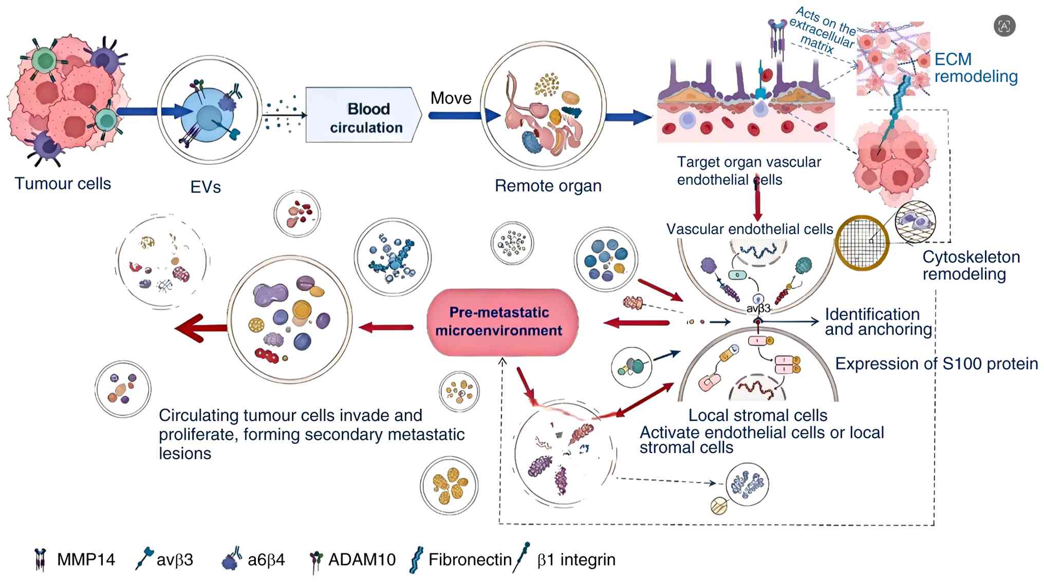
The specific targeting of EVs to distant organs
mainly depends on integrin family proteins, such as α6β4 and αvβ5,
on the vesicle membrane surface. Integrins are important molecules
that mediate cell-extracellular matrix adhesion and confer organ
tropism on vesicles. Different integrin heterodimers can be
recognised and taken up by vascular endothelial cells in specific
target organs. For example, EVs carrying integrin αvβ3 tend to
target lung tissue, whereas integrins α6β4 and αvβ5 are associated
with liver and brain metastasis, respectively (14). Clinical studies have also confirmed
that breast cancer-derived EVs can target and interact with lung
tissue endothelial cells by delivering integrin αvβ3, thereby
activating local S100 protein expression, promoting formation of a
pre-metastatic microenvironment in the lung and creating favourable
conditions for the colonisation of circulating tumour cells
(15,16).
Following the preliminary formation of the
pre-metastatic niche, EVs deliver hydrolases, including matrix
metalloproteinases (MMPs). Encapsulated within vesicles and
trafficked to the local microenvironment of target organs, these
hydrolases specifically degrade extracellular matrix (ECM)
structural proteins, disrupt tissue barriers and trigger matrix
remodelling, thereby clearing physical obstacles to tumour cell
invasion and the establishment of metastatic foci (17). For example, cervical cancer-derived
EVs deliver MMP-14 to recipient cells, markedly enhancing ECM
remodelling efficiency by activating the ERK signalling pathway and
accelerating remodelling of the tumour metastatic microenvironment
(18). Furthermore, EV-mediated ECM
remodelling itself forms a positive feedback loop that augments the
invasive potential of tumour cells. Neuroblastoma-secreted EVs are
enriched with a disintegrin and metalloproteinase 10 (ADAM10);
after transport to the local microenvironment via vesicles, ADAM10
directly facilitates local invasion and directed migration of
tumour cells (19).
Moreover, integrins and the ECM are functionally
interdependent rather than independent entities. As pivotal
transmembrane receptors, integrins mediate bidirectional
communication between cells and the ECM. Integrins facilitate
cell-ECM adhesion and transduce extracellular cues into
intracellular signalling cascades, thereby critically regulating
tumour invasion and metastasis (19). For example, β1 integrin is
consistently endogenously upregulated in multiple human
malignancies. β1 integrin promotes tumour cell adhesion to specific
ECM components (20), such as
fibronectin and collagen, and activates downstream effectors,
including FAK, Src and PI3K/Akt pathways. Concurrently, it
orchestrates cytoskeletal reorganisation through Rho
GTPase-mediated actomyosin contractility, collectively enhancing
metastatic capacity (21).
Furthermore, integrins serve as mechanosensors that detect ECM
stiffness, topography and ligand density. Integrins on the surface
of EVs also possess mechanosensory functions. This mechanical input
is integrated with biochemical signals, particularly through
crosstalk with Rho family GTPases, to coordinate polarised
cytoskeletal dynamics and directional tumour cell migration
(22).
In terms of clinical translation of the
aforementioned mechanisms, applications can be categorised into
three major dimensions. First, integrin subtypes present on the EV
membrane surface can serve as predictive biomarkers for
organ-specific metastasis. Supported by the experimental and
literature evidence outlined as aforementioned, the endogenous
upregulation of integrin αvβ3 indicates an elevated risk of
pulmonary metastasis in breast cancer, whereas the natural high
expression of integrins α6β4 and αvβ5 is associated with liver and
brain metastasis, respectively. In addition, ADAM10 and MMP-14
encapsulated in EVs are closely associated with local tumour
invasiveness (23). We propose that
this molecular signature can be applied to evaluate preoperative
tumour staging and define the extent of surgical resection. Second,
targeting metastasis-associated proteins carried by EVs to block
formation of the pre-metastatic niche provides a viable direction
for therapeutic target selection. Breast cancer-derived EVs drive
metastatic progression by delivering integrin αvβ3 (24). We posit that clinical intervention
targeting integrin αvβ3 can attenuate metastatic dissemination in
breast cancer. Meanwhile, inhibiting MMP-14 secretion and enzymatic
activity in EVs can effectively reduce ECM remodelling and local
infiltration in cervical cancer. Third, engineered EVs can be
rationally modified to achieve lesion-specific drug delivery in
clinical settings. Integrin αvβ3-functionalised EVs enable precise
delivery of therapeutic agents to pulmonary metastatic lesions,
enhancing the pulmonary accumulation efficiency of chemotherapeutic
drugs while reducing systemic toxic and adverse effects.
Tumour-derived EVs systemically drive tumour
invasion and distant metastasis through a three-tiered mechanistic
cascade: Integrin-mediated organ targeting, matrix
protease-dependent barrier degradation and integrin-ECM
synergistically regulated cell migration. Within this regulatory
network, integrin subtypes determine the organ specificity of
metastatic dissemination. At present, the differential
contributions of distinct EV subpopulations to pre-metastatic niche
formation remain poorly defined. Meanwhile, whether
integrin-mediated EV targeting is modulated by the physical
properties of the TME remains under debate. Detection of specific
integrin subtypes in circulating EVs can serve as a predictive
biomarker for assessing the risk of tumour metastasis. Small EVs
(for example, exosomes) primarily reach distant organs via the
systemic circulation, mediate organ-specific targeting via
membrane-bound integrins and induce pre-metastatic niche
establishment. Disrupting the interaction between EV integrins and
endothelial cells of target organs holds promise as a novel
strategy to inhibit distant tumour metastasis (Fig. 2).
EVs carrying immunosuppressive
proteins facilitate tumour immune evasion
Tumour immune escape is a multifaceted biological
process in which malignant cells evade immune recognition and
elimination through a range of molecular and cellular mechanisms,
thereby supporting their survival, proliferation and metastatic
dissemination. As a hallmark of tumour progression and systemic
spread, immune escape involves complex interactions between tumour
cells and host immune components. Among the most extensively
characterised mechanisms is the secretion of EVs by tumour cells,
which carry immunosuppressive cargo, including cytokines,
checkpoint ligands and regulatory miRNAs, that actively remodels
the TME into an immunosuppressive niche, impairing antitumour
immunity and enabling immune evasion (25,26).
One of the best-established mechanisms by which
tumour cells evade immune surveillance is the activation of immune
checkpoint pathways. Among these, programmed death-ligand 1 (PD-L1)
is the most extensively characterised immune checkpoint molecule.
PD-L1 is frequently endogenously upregulated at the cell surface in
multiple human malignancies, such as non-small-cell lung carcinoma,
triple-negative breast cancer, gastric adenocarcinoma and cutaneous
melanoma. Upon binding to its cognate receptor programmed cell
death protein 1 (PD-1) on activated T lymphocytes, PD-L1 triggers
intracellular signalling through the immunoreceptor tyrosine-based
inhibitory motif and the immunoreceptor tyrosine-based switch motif
(ITSM), leading to recruitment and activation of the inhibitory
phosphatase Src homology region 2 domain-containing phosphatase-2.
This cascade ultimately suppresses T-cell proliferation, reduces
cytokine production and induces functional exhaustion (27). Notably, ITSM phosphorylation is
enhanced upon PD-1/PD-L1 engagement, thereby amplifying the
inhibitory signal.
EVs secreted by tumour cells serve as critical
carriers, enabling PD-L1 to exert systemic immunosuppressive
effects. Accumulating evidence indicates that EVs derived from
multiple malignant tumours, including melanoma and non-small cell
lung cancer, can functionally express PD-L1 on their surface. These
PD-L1-bearing EVs bind to PD-1 on the surface of T cells, markedly
inhibiting CD8+ T-cell activation and proliferation (25). Meanwhile, PD-L1 signalling induces
T-cell apoptosis and blocks T-cell receptor signal transduction. In
addition to suppressing T-cell activation, PD-L1 inhibits the
PI3K/Akt/mTOR signalling pathway and enhances the expression and
function of phosphatase and tensin homologue. It also promotes the
differentiation of inducible regulatory T cells (Tregs), which
secrete TGF-β to further repress effector T-cell function. These
cascading regulatory effects collectively establish systemic
immunosuppression, thereby facilitating tumour immune evasion
(28).
In the indirect pathway, members of the TGF-β
protein family predominantly drive the differentiation of CD4+ T
cells into Tregs, which exert potent immunosuppressive effects,
mainly through the canonical SMAD signalling cascade (29). This pathway is tightly regulated at
multiple hierarchical levels, including ubiquitin-mediated
proteasomal degradation of SMAD proteins, phosphatase-catalysed
dephosphorylation, and competitive inhibition of receptor-regulated
SMADs by inhibitory SMADs (30).
Notably, TGF-β has a context-dependent dual role in tumourigenesis:
it functions predominantly as a tumour suppressor during early
carcinogenesis but frequently acquires pro-tumourigenic properties
in advanced disease (31).
EVs modulate immune responses through both direct
intercellular communication and indirect signalling pathways,
primarily by transferring immunoregulatory molecules such as PD-L1
and TGF-β. Advances in clinical detection methods, such as PD-L1
immunohistochemistry, circulating tumour-DNA sequencing and
immune-cell profiling (32), have
supported the development of a multilayered immunotherapeutic
framework and a comprehensive regulatory network governing immune
evasion. Collectively, these findings provide mechanistic insights
into tumour-induced immunosuppression and immune escape, as well as
a translational basis for the clinical implementation of related
diagnostic tools and therapeutic interventions. Specifically,
tumour-derived EVs synergistically shape local and systemic
immunosuppressive microenvironments through two mechanisms: Direct
delivery of PD-L1 to suppress effector T-cell function and indirect
delivery of TGF-β to induce Treg differentiation. Furthermore, the
intrinsic vesicular carrier properties of EVs markedly prolong the
duration and expand the spatial range of action of these
immunosuppressive molecules.
Nevertheless, the relative contributions of
EV-associated PD-L1 and tumour cell surface PD-L1 to immune evasion
remain controversial. The active forms of TGF-β and the specific
target molecules in EVs from different tumour origins have not been
fully elucidated. Meanwhile, circulating EV-associated PD-L1 levels
may serve as predictive biomarkers of the therapeutic efficacy of
immune checkpoint inhibitors. Bispecific inhibitors simultaneously
targeting EV-associated PD-L1 and EV-associated TGF-β may help
overcome resistance to single-agent immunotherapy in the future
(33) (Fig. 3).
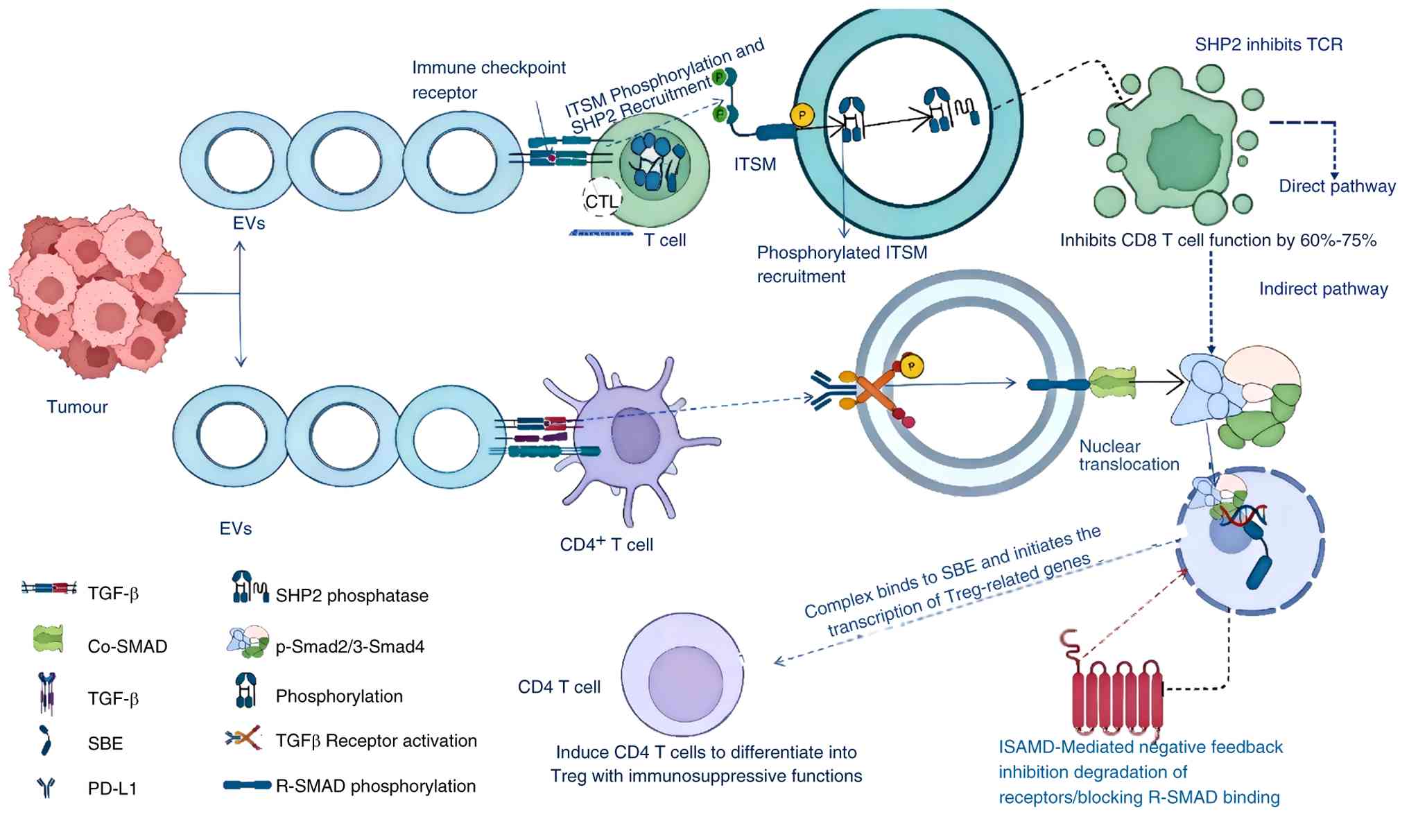 | Figure 3.EVs facilitate tumour immune evasion.
Schematic illustration of the dual pathways of EV-mediated tumour
immune evasion. EVs suppress antitumour immunity through two core
pathways. The direct pathway relies on PD-L1 on the EV surface to
inhibit the effector functions of CD8+ T cells. The indirect
pathway involves EV-derived TGF-β driving the differentiation of
CD4+ T cells into immunosuppressive Tregs. EVs, extracellular
vesicles; PD-L1, programmed cell death ligand 1; TCR, T-cell
receptor; TGF-β, transforming growth factor-β; Tregs, regulatory T
cells; SHP2, Src homology 2 domain-containing phosphatase 2; ITSM,
immunoreceptor tyrosine-based switch motif; TCR, T-cell receptor;
SBE, Smad-binding element; TGFβ receptor, TGF-β receptor; ISAMD,
immunoreceptor tyrosine-based switch motif-associated degradation.
Created using BioRender. |
Angiogenic factors modulate the TME to
promote neovascularization and support tumour progression
EV-associated pro-angiogenic factors promote tumour
angiogenesis. Tumour growth requires the formation of new blood
vessels to supply oxygen and nutrients, a process known as tumour
angiogenesis (34). Recent studies
have shown that tumour-derived EVs are critical mediators of
angiogenesis (35). By delivering
multiple pro-angiogenic bioactive factors, EVs help establish a
tumour endothelial microenvironment that supports angiogenesis
(36).
EVs directly initiate angiogenic programmes by
enriching and delivering core factors, including VEGF and basic
fibroblast growth factor (bFGF) (37). Among the signalling pathways
implicated in this process, the VEGF-VEGFR axis is a central
regulatory mechanism governing both angiogenesis and tumour
progression (38). Tumour-derived
EVs selectively enrich and stabilise VEGF, facilitating its
targeted delivery to vascular endothelial cells. This interaction
activates pro-angiogenic signalling pathways, thereby promoting
endothelial cell proliferation, migration and tubule formation,
which are key steps in tumour angiogenesis. Clinical and
experimental evidence supports this mechanism. For example,
glioblastoma-derived EVs markedly increase VEGF levels in
cerebrospinal fluid and enhance the in vitro tubulogenesis
capacity of human vascular endothelial cells (39).
In addition to VEGF, EVs carry a range of bioactive
molecules, including bFGF, platelet-derived growth factor and IL-8
(32). Notably, bFGF binds to its
cognate receptor, fibroblast growth factor receptor, to stimulate
tumour endothelial cell migration and acts synergistically with
VEGF to enhance angiogenesis (40).
Furthermore, MMPs packaged within EVs facilitate extracellular
matrix degradation, creating permissive microenvironments for
neovessel sprouting and stabilisation (36).
EVs enriched in pro-angiogenic factors show an
enhanced capacity to stimulate angiogenesis (41). EVs can respond to microenvironmental
changes and dynamically modulate the magnitude of angiogenesis. In
tumour cells, stress conditions such as hypoxia and oncogene
activation upregulate transcription factors, including
hypoxia-inducible factor 1α (HIF-1α), leading to increased
expression of pro-angiogenic genes such as VEGF and bFGF, thereby
altering EV cargo composition (42). Notably, EVs carrying miR-210
suppress key anti-angiogenic regulators, including ephrin-A3,
effectively initiating and sustaining the tumour angiogenic switch
(43). These molecular mechanisms
not only supply tumour cells with essential oxygen and nutrients,
but also facilitate local invasion and dissemination into adjacent
tissues (44).
This mechanism has substantial translational
potential in clinical practice. Clinical evidence indicates that
serum EV-associated VEGF levels in patients with colorectal cancer
are markedly and positively associated with intratumoural micro
vessel density (MVD) scores (45,46).
These findings indicate that EV-associated VEGF may serve as a
promising non-invasive biomarker for assessing tumour angiogenic
activity and predicting patient prognosis, highlighting its
potential clinical utility.
From a liquid biopsy perspective, the positive
correlation between VEGF levels in serum EVs and tumour tissue MVD
suggests that EV-carried VEGF may be useful for evaluating tumour
angiogenic status and patient prognosis (47). In therapeutic target screening, the
VEGF signalling pathway represents one of the core mechanisms
driving tumour neovascularisation (48). Clinical experiments have shown that
glioblastoma-derived EVs markedly increase VEGF concentrations in
cerebrospinal fluid, exceeding normal levels. Targeting EV-mediated
angiogenesis may therefore help overcome resistance induced by
conventional anti-VEGF agents. Finally, in the application of
engineered EVs, previous studies have shown that EVs loaded with
miR-210 can suppress key anti-angiogenic regulators such as
ephrin-A3 and effectively trigger the tumour angiogenic switch.
Accordingly, miRNAs may be used in engineered EV-based strategies
to specifically target tumour endothelial cells (49).
The VEGF signalling pathway constitutes a central
regulatory mechanism underlying tumour angiogenesis and is widely
recognised as a clinically relevant biomarker in oncology (50,51).
Given the pivotal role of VEGF in promoting neovascularisation,
therapeutic strategies targeting VEGF or its cognate receptor,
VEGFR, have become a major focus in anticancer drug development.
Nevertheless, clinical experience indicates that monotherapy based
solely on the inhibition of pro-angiogenic factors frequently has
limitations, including the development of resistance and suboptimal
therapeutic responses.
In conclusion, tumour-derived EVs trigger tumour
angiogenesis through a multidimensional cascade involving cytokine
activation, extracellular matrix remodelling and hypoxia-induced
miRNA regulation, with multiple pro-angiogenic factors encapsulated
within EVs exerting synergistic effects. However, the relative
contributions of EVs and free pro-angiogenic factors to tumour
angiogenesis remain unclear. Moreover, the angiogenic potency of
EVs secreted by distinct cell populations requires further
systematic investigation. Circulating EV-associated VEGF levels may
serve as predictive biomarkers of the therapeutic efficacy of
anti-angiogenic agents. Depletion of tumour-derived EVs or
functional blockade of their pro-angiogenic cargo represents a
novel therapeutic strategy for anti-angiogenic cancer treatment
(Fig. 4).
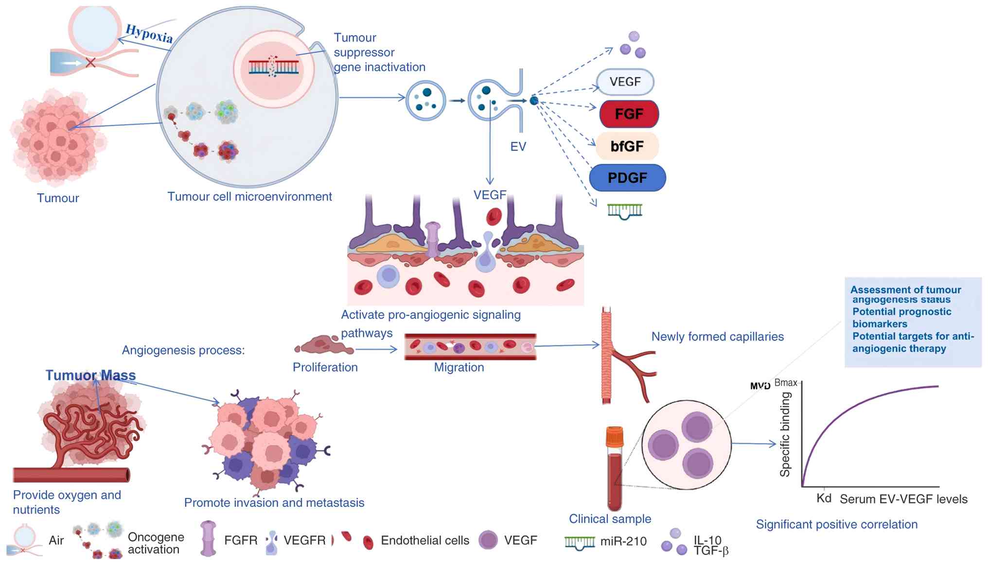 | Figure 4.Schematic illustration of the
mechanism by which EVs regulate tumour angiogenesis. Tumour-derived
EVs are enriched in pro-angiogenic factors, such as VEGF and FGF,
and deliver these factors to vascular endothelial cells. Subsequent
activation of downstream signalling pathways promotes endothelial
cell proliferation, migration and tube formation, providing
material and nutritional support for tumour growth. EV,
extracellular vesicle; VEGF, vascular endothelial growth factor;
VEGFR, VEGF receptor; FGF, fibroblast growth factor; FGFR, FGF
receptor; bFGF, basic fibroblast growth factor; PDGF,
platelet-derived growth factor; TGF-β, transforming growth
factor-β; MVD, microvessel density. Created using BioRender. |
EV-mediated transport of key enzymes
drives metabolic reprogramming and reshapes cellular energy
metabolism
EVs act as central drivers of the horizontal
transmission of metabolic reprogramming phenotypes and systematic
remodelling of the tissue microenvironment. Metabolic reprogramming
is widely recognised as a hallmark of cancer; it not only fuels
malignant tumour cell proliferation through systematic rewiring of
energy and biosynthetic metabolism, but also contributes to the
establishment and maintenance of an immunosuppressive TME. The
primary mechanism underlying tumour immune evasion involves the
direct suppression of tumour-infiltrating immune cell function
through competitive depletion of essential nutrients, including
glucose and glutamine, and accumulation of immunosuppressive
metabolic by-products, notably lactic acid, ultimately facilitating
immune escape and tumour progression (52).
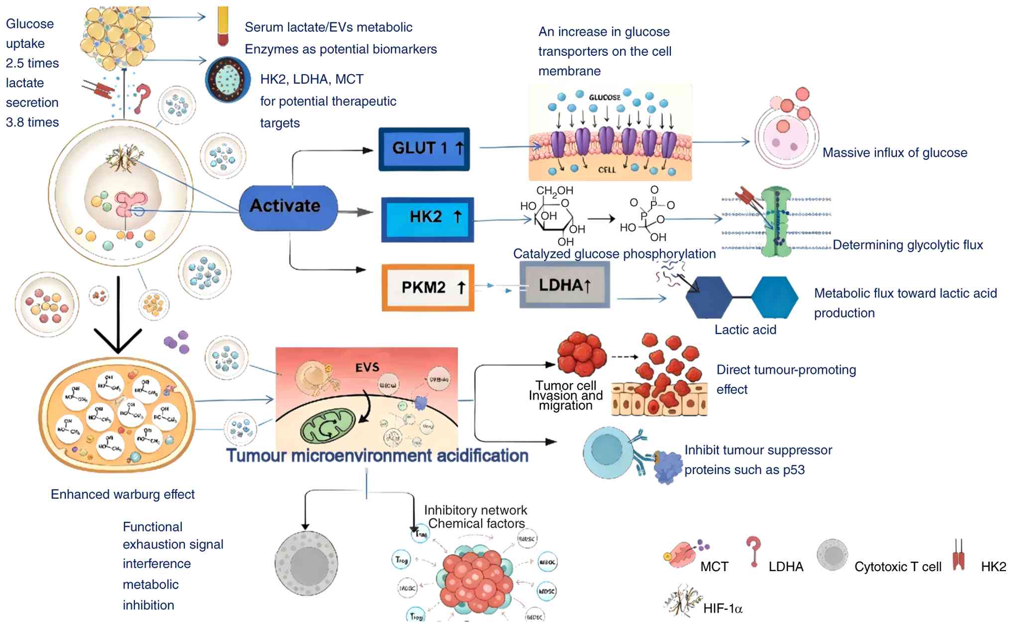
The core mechanism underlying EV-mediated metabolic
reprogramming lies in the selective packaging and delivery of a
broad panel of key metabolic enzymes regulated by transcription
factors. Specifically, activation of key transcriptional
regulators, such as HIF-1α and c-Myc, induces the expression of
glucose transporter 1 and other membrane transport proteins,
thereby markedly increasing cellular glucose uptake and supplying
essential carbon substrates for downstream metabolic pathways
(53). Subsequently, intracellular
glucose is phosphorylated by hexokinase 2 (HK2), an enzyme
consistently overexpressed in malignant tumours. This step commits
glucose to glycolysis and serves as a critical determinant of
glycolytic flux (54). Furthermore,
tumour cells preferentially express the M2 isoform of pyruvate
kinase (PKM2), which, through its distinct kinetic properties and
non-metabolic regulatory functions, synergises with upstream
alterations to establish a metabolic configuration conducive to
rapid proliferation and biomass accumulation (55).
By simultaneously delivering key downstream
metabolic enzymes, EVs function as central executors that drive the
cascade amplification of glycolysis and the systemic acidification
of the tissue microenvironment (56). Downstream of PKM2, lactate
dehydrogenase A (LDHA) is upregulated as a pivotal metabolic enzyme
that amplifies glycolytic flux, sustains high-rate glycolysis and
drives substantial lactic acid accumulation, thereby providing an
essential energetic substrate for rapid tumour cell proliferation
(6). Lactic acid is actively
exported from tumour cells through monocarboxylate transporters,
not only eliminating metabolic waste but also inducing pronounced
acidification of the TME. This acidification is not merely a
passive by-product of heightened glycolytic activity. Rather, it
functions as an active modulator that creates conditions favourable
for tumour invasion, metastasis and immune evasion. Specifically,
TME acidosis promotes tumour cell migration, local invasion and
distant metastasis, while concurrently impairing anti-tumour
immunity, most notably by suppressing cytotoxic T lymphocyte
function through disruption of T-cell receptor signalling and
critical metabolic pathways. Moreover, lactic acid recruits
immunosuppressive cell populations, including Tregs and
myeloid-derived suppressor cells, thereby reinforcing an
immunosuppressive niche that facilitates tumour immune escape
(57). This mechanistic framework
has been experimentally validated in pancreatic ductal
adenocarcinoma, in which pancreatic cancer-derived EVs deliver HK2
to recipient cells, resulting in ~2.5-fold increases in glucose
uptake and lactate secretion (58).
Collectively, this metabolic reprogramming establishes a localised,
persistently acidic TME that critically supports tumour progression
and immune evasion.
Regarding translational clinical applications, from
a liquid biopsy perspective, HK2 and LDHA expression levels in EVs
may serve as biomarkers for tumour metabolic subtyping and guide
individualised therapy (59). For
instance, patients with pancreatic cancer and high HK2 expression
in circulating EVs show a better therapeutic response to glycolysis
inhibitors such as 2-deoxyglucose. Meanwhile, serum EV-associated
lactate levels may be used to assess TME acidification and predict
immunotherapeutic efficacy. In therapeutic target selection, as
described above, inhibition of HK2, delivered via pancreatic cancer
cell-derived EVs, reduces glucose uptake in recipient cells by 62%
and decreases lactate secretion by 71%, while markedly augmenting
the activity of tumour-infiltrating T cells. Accordingly, targeting
EV-mediated metabolic enzymes represents a viable strategy to
abrogate metabolic reprogramming in the TME (60). With respect to engineered EVs,
metabolic enzymes including HK2 and LDHA have become critical
research targets. EVs loaded with glycolysis inhibitors enable
precise targeting of tumour cell metabolic pathways, thereby
avoiding systemic metabolic adverse effects. To date, engineered
EVs encapsulating LDHA inhibitors have completed preclinical
validation in pancreatic cancer models, demonstrating pronounced
suppression of tumour growth and prolonged overall survival.
Collectively, these translational and pre-clinical
observations highlight the central contribution of EV-packaged
metabolic enzymes to tumour progression, prompting further
mechanistic dissection of how tumour-derived EVs propagate
metabolic reprogramming across the tumour microenvironment.
The aforementioned studies demonstrate that key
metabolic enzymes, particularly HK2 and LDHA, serve as core
functional cargos of tumour-derived EVs and play pivotal roles in
EV-mediated intercellular transmission of metabolic reprogramming
by modulating cellular energy metabolism. Mediated by
tumour-derived EVs, this metabolic reprogramming, notably
characterised by enhanced glycolytic flux and increased lactate
secretion, extends from individual tumour cells to the entire
tissue niche, establishing a mechanistically coherent pathogenic
axis linking intracellular metabolic alterations to systemic
remodelling of the TME. This reprogramming exerts dual biological
effects: First, it supplies the bioenergetic and biosynthetic
substrates required for rapid tumour cell proliferation; second, it
fosters an immunosuppressive TME through extracellular
acidification, thereby facilitating immune evasion, a hallmark of
malignant progression. Consequently, blocking the EV-mediated
intercellular transfer of key metabolic enzymes, such as HK2 and
LDHA, has emerged as a promising therapeutic strategy that can
simultaneously suppress tumour metabolic reprogramming and the
formation of an immunosuppressive microenvironment, exert dual
anti-tumour effects, and offer novel mechanistic insights and
potential avenues for clinical intervention in malignant tumours
(Fig. 5).
EVs mediate intercellular transfer of
resistance-associated proteins, thereby fuelling therapeutic
resistance
Multidrug resistance (MDR) in cancer markedly
undermines the efficacy of clinical anticancer therapies (61). Accumulating evidence indicates that
EVs, as critical vehicles for intercellular communication among
tumour cells, contribute to MDR through multiple molecular
mechanisms. Among these, multidrug resistance protein 1
(P-glycoprotein) has a central mechanistic role.
P-gp, a drug-resistance-associated transmembrane
transporter, plays a pivotal role in mediating MDR in malignant
tumours. Overexpression or hyperactivation of P-gp in tumour cells
confers a survival advantage under chemotherapeutic pressure by
facilitating the ATP-dependent efflux of diverse anticancer agents,
thereby reducing intracellular drug accumulation and diminishing
therapeutic efficacy (62).
Furthermore, EVs secreted by drug-resistant tumour cells frequently
exhibit elevated surface expression of clinically relevant
biomarkers, including HER2 and CD20, that mirror those on the
parental tumour cell membrane (63). In the context of targeted therapies
such as trastuzumab (anti-HER2) and rituximab (anti-CD20), these
EVs act as molecular decoys by competitively binding therapeutic
antibodies, sequestering drug molecules and consequently lowering
the bioavailable concentration of active drug reaching tumour
cells, ultimately contributing to therapeutic resistance and
clinical treatment failure (64).
The ABC transporter family represents a
well-established molecular mechanism underlying MDR. Proteomic
profiling has demonstrated notable upregulation of three key ABC
transporters, ABCB1 (P-gp/MDR1), ABCC1 (MRP1) and ABCG2 (BCRP), in
EVs isolated from drug-resistant tumours (65,66).
These membrane-bound efflux pumps use energy derived from ATP
hydrolysis to actively export chemotherapeutic agents from cells,
thereby substantially diminishing intracellular drug accumulation
and compromising therapeutic efficacy. Notably, MDR1 expression in
EVs derived from paclitaxel-resistant ovarian cancer cell lines was
~5.8-fold higher than that observed in their drug-sensitive
counterparts (67).
Moreover, EVs can deliver DNA damage
repair-associated proteins, including topoisomerase IIα (TOP2A), to
recipient cells. TOP2A is a critical regulator of DNA replication
and cell proliferation, with enzymatic activity tightly coordinated
with cell cycle progression. The expression of TOP2A peaks during
the G2/M phase and declines rapidly after mitotic
completion (68,69). Recent evidence has demonstrated that
EV-mediated transfer of TOP2A disrupts canonical DNA damage repair
mechanisms and confers a 3- to 4-fold increase in cisplatin
resistance in HeLa cells (70).
Regarding clinical translational applications, for
liquid biopsy biomarkers, as noted in previous studies, high
preoperative serum EV expression of MDR1 in patients with ovarian
cancer indicates a 5.8-fold elevated risk of paclitaxel resistance
(71). EV-associated TOP2A levels
can be used to predict the therapeutic response to cisplatin and
guide the selection of postoperative adjuvant chemotherapy regimens
(72). Targeting drug
resistance-related proteins carried by EVs can reverse tumour
therapeutic resistance. Combined administration of MDR1 inhibitors
and EV-depletion agents increases intracellular paclitaxel
concentration in drug-resistant ovarian cancer cells by 3.5-fold
and restores chemosensitivity (73). Blocking EV-mediated intercellular
transfer of TOP2A enhances the antitumour activity of cisplatin by
3- to 4-fold. For engineered EV applications, EVs loaded with drug
resistance-reversing small interfering RNAs can specifically
deliver gene-silencing molecules that target drug resistance
(74). In multidrug-resistant
breast cancer models, this strategy restores tumour cell
sensitivity to doxorubicin to the level observed in drug-sensitive
cell lines (75).
In summary, from our perspective, drug
resistance-associated proteins collectively contribute to tumour
treatment resistance through multiple interconnected mechanisms,
including pre-emptive drug binding, active efflux of
chemotherapeutic agents and disruption of DNA repair pathways.
These mechanisms establish a functional link between EV-mediated
drug resistance and clinical treatment outcomes, positioning EVs as
critical biomolecular carriers for investigating tumour drug
resistance. Furthermore, they provide promising avenues for
developing strategies to reverse resistance and optimise
therapeutic regimens. Tumour-derived EVs confer therapeutic
resistance via drug resistance proteins through drug binding,
chemotherapeutic efflux and DNA repair disruption. However, the
actual contribution of EV-mediated transmission of drug resistance
to clinical tumour drug resistance remains unclear. Relevant
studies remain limited, clinical data are insufficient and
differences in transmission efficiency among distinct EV
subpopulations during the propagation of drug resistance have not
been fully elucidated. Detection of drug resistance-related protein
expression levels in circulating EVs enables early prediction of
tumour therapeutic response. The combined application of
EV-depletion agents and conventional targeted agents is expected to
overcome tumour drug resistance and improve therapeutic efficacy
(Fig. 6; Table II).
| Table II.Core mechanisms, key molecules and
clinical translational applications of EVs in malignant
tumours. |
Table II.
Core mechanisms, key molecules and
clinical translational applications of EVs in malignant
tumours.
| Core functional
mechanisms | Key
molecules/genes | Clinical
applications in liquid biopsy | Clinical
applications of engineered therapeutics | Validated
clinical/experimental data |
|---|
| Mediating tumour
invasion and organ-specific metastasis | Integrin αvβ3
(ITGAV/ITGB3), integrin α6β4 (ITGA6/ITGB4), integrin αvβ5
(ITGAV/ITGB5), MMP-14 (MMP14), ADAM10 (ADAM10), β1 integrin
(ITGB1) | Risk stratification
of tumour metastasis; prediction of organotropic metastasis
tendency; dynamic monitoring of postoperative recurrence | Targeted blockade
of integrins on the EV surface to inhibit pre-metastatic niche
formation; engineered EVs loaded with matrix remodelling inhibitors
to block local tumour invasion | Integrin αvβ3
carried by breast cancer-derived EVs is markedly associated with
lung metastasis risk; MMP-14 expression in cervical cancer-derived
EVs is positively associated with tumour stage and depth of
invasion |
| Mediating tumour
immune escape | PD-L1 (CD274) and
TGF-β (TGFB1) | Prediction of
immunotherapeutic efficacy; assessment of immune escape risk;
prognostic stratification of patients | Development of
PD-L1/TGF-β dual-targeted EV inhibitors to block immunosuppressive
signalling; engineered EVs loaded with immune activators to reverse
the local immunosuppressive microenvironment | EV inhibitors
targeting the PD-L1/ TGF-β dual axis have entered phase II clinical
trials, reducing the incidence of immunotherapy resistance by ~38%;
serum EV PD-L1 levels in patients with melanoma are negatively
associated with the response rate to anti-PD-1 therapy |
| Driving tumour
angiogenesis | VEGF (VEGFA), bFGF
(FGF2), PDGF (PDGFA/B), IL-8 (CXCL8), miR-210 (MIR210) | Assessment of
tumour angiogenic activity | Targeted
elimination of pro- angiogenic EVs to disrupt tumour nutrient
supply; engineered EVs loaded with anti-angiogenic drugs to
precisely suppress neovascularization | Serum EV–VEGF
levels in patients with colorectal cancer are markedly positively
associated with tumour tissue MVD scores; EV–VEGF concentration in
cerebrospinal fluid of patients with glioblastoma is markedly
elevated relative to normal levels |
| Remodelling tumour
energy metabolism and microenvironmental acidification | HK2 (HK2), LDHA
(LDHA), GLUT1 (SLC2A1), PKM2 (PKM2), MCT1 (SLC16A1) | Assessment of
tumour metabolic activity; grading of tumour malignancy; prediction
of patient overall survival | Blockade of
EV-mediated metabolic enzyme transfer to suppress tumour glycolytic
flux; engineered EVs loaded with metabolic inhibitors to reverse
the acidic immunosuppressive microenvironment | HK2 delivered by
pancreatic cancer cell-derived EVs increases glucose uptake
capacity of recipient cells by ~2.5-fold and lactate secretion by
~3.8-fold; serum EV-LDHA levels are markedly negatively associated
with overall survival in patients with pancreatic cancer |
| Mediating tumour
therapeutic resistance | MDR1 (ABCB1), ABCC1
(ABCC1), ABCG2 (ABCG2), TOP2A (TOP2A), HER2 (ERBB2) and CD20
(MS4A1) | Early prediction of
chemotherapy/ targeted therapy resistance; basis for dynamic
adjustment of treatment regimens; real-time monitoring of
therapeutic efficacy | Targeted
elimination of drug resistance-associated EVs to reverse tumour
multidrug resistance; engineered EVs loaded with chemotherapeutic/
genetic drugs to overcome tumour drug resistance barriers; blockade
of competitive binding between EVs and targeted agents | MDR1 expression in
EVs from paclitaxel-resistant ovarian cancer cell lines is
~5.8-fold that in drug- sensitive strains; EV-mediated TOP2A
transfer increases cisplatin resistance of tumour cells by 3–4
folds; detection of EV protein biomarkers in prostate cancer helps
avoid ~26% of unnecessary biopsies |
| Mediating tumour
invasion and organ-specific metastasis | Integrin αvβ3
(ITGAV/ITGB3), integrin α6β4 (ITGA6/ITGB4), integrin αvβ5
(ITGAV/ITGB5), MMP-14 (MMP14), ADAM10 (ADAM10) and β1 integrin
(ITGB1) | Risk stratification
of tumour metastasis; prediction of organotropic metastasis
tendency; dynamic monitoring of postoperative recurrence | Targeted blockade
of integrins on the EV surface to inhibit pre-metastatic niche
formation; engineered EVs loaded with matrix remodelling inhibitors
to block local tumour invasion | Integrin αvβ3
carried by breast cancer-derived EVs is markedly associated with
lung metastasis risk MMP-14 expression in cervical cancer-derived
EVs is positively associated with tumour stage and depth of
invasion |
Clinical applications of exosomes in
malignant tumours
Diagnostic and screening
applications
Exosomes serve as promising biological biomarkers
for liquid biopsy, exhibiting well-documented clinical utility in
tumour screening and adjunctive diagnosis, progressing steadily
from foundational research toward integration into clinical
decision-making frameworks.
Highly sensitive detection of protein
biomarkers
Liquid biopsy platforms leveraging
exosome-associated proteins demonstrate superior analytical
sensitivity and specificity. Notably, immune checkpoint molecules,
including PD-L1, which is consistently overexpressed on
tumour-derived exosomes, represent robust and clinically relevant
detection targets (76).
Robust clinical diagnostic
performance
Substantial clinical evidence substantiates the high
diagnostic accuracy of exosome-based assays in prostate cancer
detection. It is estimated that implementation of this technology
could reduce unnecessary prostate biopsies by ~26%, thereby
mitigating patient discomfort and minimizing wastage of healthcare
resources associated with invasive procedures (77). This underscores the transformative
potential of exosome-based liquid biopsy as a dynamic, non-invasive
monitoring modality.
Applications of therapeutic
interventions
Exosomes function not merely as passive biomarkers
but also as active therapeutic delivery vehicles and
mechanistically relevant intervention targets. Their clinical
translation is now advancing into the validation phase,
particularly in addressing tumour drug resistance and improving
therapeutic efficacy.
Engineering targeted delivery
systems
Exosomes engineered via surface conjugation of
targeting ligands, such as EGFR-specific single-chain variable
fragments, demonstrate markedly enhanced accumulation at tumour
sites (78). This strategy offers a
structurally grounded solution to two longstanding challenges in
oncology: Suboptimal target specificity and dose-limiting systemic
toxicity associated with conventional chemotherapeutics.
Reprogramming the tumour immune
microenvironment
Dual-targeting exosome-based inhibitors co-blocking
PD-L1 and TGF-β have progressed to phase II clinical trial stage
(79). Interim data indicate a ~38%
reduction in the incidence of resistance to immune checkpoint
blockade therapy (80). These
findings substantiate that exosomes are not only integral
components of resistance pathways but also promising,
mechanism-informed therapeutic levers for overcoming immune evasion
and reversing treatment resistance.
Challenges in transformative
manufacturing and quality assurance
Although exosomes hold considerable promise for
clinical applications, their translation from laboratory research
to large-scale clinical implementation is hindered by stringent
industrial quality control requirements, constituting a critical
bottleneck in their clinical translation.
Summary and prospects
The present review systematically elaborates on the
core roles of EVs in the occurrence and development of malignant
tumours, their clinical translational value, existing bottlenecks
and future research directions.
EVs play a pivotal role in the core pathobiological
mechanisms underlying malignant tumours. Specifically, EVs
contribute to tumour progression through five well-documented
functional axes: i) Metastasis-associated proteins that drive
tumour cell invasion and dissemination; ii) immunosuppressive
molecules that facilitate immune evasion; iii) pro-angiogenic
factors that remodel the TME to support neovascularisation; iv)
metabolic enzymes involved in cancer-specific metabolic
reprogramming, thereby altering cellular energy metabolism; and v)
drug resistance-associated proteins that confer resistance to
conventional therapeutic agents.
A comprehensive investigation of the core mechanisms
by which EVs influence malignant tumours has markedly advanced the
understanding of tumour pathogenesis and established a robust
theoretical foundation for developing EV-based liquid biopsy
approaches and engineered therapeutic interventions.
Liquid biopsy technologies using EV-associated
proteins have achieved substantial advances, particularly in the
sensitive and specific detection of protein-based biomarkers, and
show considerable clinical promise (64)
However, current research still faces notable
challenges in addressing the functional heterogeneity of
EV-associated proteins. First, EVs exhibit favourable biophysical
and biological properties, including high biocompatibility, low
immunogenicity and intrinsic structural stability, making them
promising candidates for therapeutic applications (81). Second, these attributes confer
distinct advantages over certain cell-based regenerative therapies,
which are often associated with safety concerns, such as
tumourigenicity and immune rejection, as well as practical
limitations in scalability and standardisation. Although several
EV-based therapeutics have advanced to clinical trials for disease
management, translation to large-scale, good manufacturing
practice-compliant production remains hindered by technical
bottlenecks, including mechanical shear-induced damage during
isolation and risks of microbial or co-isolated contaminant
carryover (82). Furthermore, when
EVs are deployed as biopharmaceutical agents or targeted delivery
vehicles, rigorous quality control is indispensable, both to ensure
precise dosing and batch-to-batch consistency, and to preserve EV
integrity, stability and bioactivity after extensive purification.
While engineering approaches, such as surface ligand conjugation
and membrane modification, can enhance targeting specificity,
comprehensive preclinical and clinical safety assessments remain
essential. Critically, establishing standardised, physiologically
relevant storage conditions that maintain both the structural
fidelity and functional competence of EVs is essential to realise
their full therapeutic potential (83).
Despite these challenges, research on the roles of
EVs in malignant tumours remains a dynamic and promising field. We
propose that future in-depth investigations of EVs in malignant
tumours should focus on the following aspects: First, integrating
multi-omics technologies with artificial intelligence approaches to
construct heterogeneity atlases of EVs, thereby enabling precise
identification and sorting of distinct functional subpopulations;
second, establishing standardised systems for EV isolation,
characterisation and quality control to accelerate the translation
of basic research findings into clinical applications; and third,
conducting systematic, well-designed clinical trials to
comprehensively evaluate the clinical value of EVs in tumour
diagnosis, prognostic assessment and therapy.
With continuous technological advances and a
progressively deeper understanding of EV biology, EV research is
expected to bring major breakthroughs in the precision diagnosis
and treatment of malignant tumours, and in the advancement of
liquid biopsy technologies.
Acknowledgements
Not applicable.
Funding
The present study received support from the supplementary
funding for the 76th batch of the China Postdoctoral Science
Foundation (grant no. 12425030), the Science and Technology
Research Project of the Education Department of Jiangxi Province
(grant no. GJJ2500828) and The First Batch of University-Level
Scientific and Technological Innovation Teams of Jiangxi University
of Chinese Medicine, Innovative Research on Integrated Traditional
Chinese and Western Medicine for Prevention and Treatment of
Children's Diseases and Health Protection (grant no.
CXTD22014).
Availability of data and materials
Not applicable.
Authors' contributions
RS, QL, YL, KW, ZW and QL contributed to the present
study conception and design, material preparation, data collection
and analysis, and writing the initial draft of the manuscript. All
authors have read and approved the final manuscript. Data
authentication is not applicable.
Ethics approval and consent to
participate
Not applicable.
Patient consent for publication
Not applicable.
Competing interests
The authors declare that they have no competing
interests.
References
|
1
|
No authors listed. The global challenge of
cancer. Nat Cancer. 1:1–2. 2020.PubMed/NCBI
|
|
2
|
Yildirim DT, Baki YA, Salzet M, Bertelli
M, Beccari T, Prakash S and Luisa P: Messaging malignancy:
Tumour-derived exosomes at the nexus of immune escape, vascular
remodelling and metastatic competence. EuroBiotech J. 9:216–237.
2025. View Article : Google Scholar
|
|
3
|
Shi Y, Yao F, Yin Y, Wu C, Xia D, Zhang K,
Jin Z, Liu X, He J and Zhang Z: Extracellular vesicles derived from
immune cells: Role in tumor therapy. Int Immunopharmacol.
133:1121502024. View Article : Google Scholar : PubMed/NCBI
|
|
4
|
Manno M, Bongiovanni A, Margolis L,
Bergese P and Arosio P: The physico-chemical landscape of
extracellular vesicles. Nat Rev Bioeng. 3:68–82. 2025. View Article : Google Scholar
|
|
5
|
Record M, Silvente-Poirot S, Poirot M and
Wakelam MJO: Extracellular vesicles: Lipids as key components of
their biogenesis and functions. J Lipid Res. 59:1316–1324. 2018.
View Article : Google Scholar : PubMed/NCBI
|
|
6
|
Kumar MA, Baba SK, Sadida HQ, Marzooqi SA,
Jerobin J, Altemani FH, Algehainy N, Alanazi MA, Abou-Samra AB,
Kumar R, et al: Extracellular vesicles as tools and targets in
therapy for diseases. Signal Transduct Target Ther. 9:272024.
View Article : Google Scholar : PubMed/NCBI
|
|
7
|
Li J, Yu S, Rao M and Cheng B:
Tumor-derived extracellular vesicles: Key drivers of
immunomodulation in breast cancer. Front Immunol. 16:15485352025.
View Article : Google Scholar : PubMed/NCBI
|
|
8
|
Chen J, Fei X, Wang J and Cai Z:
Tumor-derived extracellular vesicles: Regulators of tumor
microenvironment and the enlightenment in tumor therapy. Pharmacol
Res. 159:1050412020. View Article : Google Scholar : PubMed/NCBI
|
|
9
|
Liu X, To KKW, Zeng Q and Fu L: Effect of
extracellular vesicles derived from tumor cells on immune evasion.
Adv Sci (Weinh). 12:e24173572025. View Article : Google Scholar : PubMed/NCBI
|
|
10
|
Rabas N, Ferreira RMM, Di Blasio S and
Malanchi I: Cancer-induced systemic pre-conditioning of distant
organs: Building a niche for metastatic cells. Nat Rev Cancer.
24:829–849. 2024. View Article : Google Scholar : PubMed/NCBI
|
|
11
|
Huang D, Shen H, Jin Q, Chen T, Xie Y, Rao
D and Liu M: The role and prospects of extracellular vesicles in
advanced drug and vaccine delivery. Front Immunol 17: 1781365,
2026. Zhao Z, Ukidve A, Kim J and Mitragotri S: Targeting
strategies for tissue-specific drug delivery. Cell. 181:151–167.
2020. View Article : Google Scholar
|
|
12
|
Zhao H, Achreja A, Iessi E, Logozzi M,
Mizzoni D, Di Raimo R, Nagrath D and Fais S: The key role of
extracellular vesicles in the metastatic process. Biochim Biophys
Acta Rev Cancer. 1869:64–77. 2018. View Article : Google Scholar : PubMed/NCBI
|
|
13
|
Asadujjaman M, Nam UR, Lee DE, Choi JU,
Kim SH, Jang DJ and Jee JP: Fusion nanoparticle system of
extracellular vesicles and docetaxel-loaded liposomes: An
innovative therapeutic strategy to enhance anticancer efficacy. J
Pharm Investig. 56:613–632. 2026. View Article : Google Scholar
|
|
14
|
Cooper J and Giancotti FG: Integrin
signaling in cancer: Mechanotransduction, stemness, epithelial
plasticity, and therapeutic resistance. Cancer Cell. 35:347–367.
2019. View Article : Google Scholar : PubMed/NCBI
|
|
15
|
Liu Y, Mao D, Wang H, Che X and Chen Y:
Formation of pre-metastatic niches induced by tumor extracellular
vesicles in lung metastasis. Pharmacol Res. 188:1066692023.
View Article : Google Scholar : PubMed/NCBI
|
|
16
|
Wan J, Zhang G, Li X, Qiu X, Ouyang J, Dai
J and Min S: Matrix metalloproteinase 3: A promoting and
destabilizing factor in the pathogenesis of disease and cell
differentiation. Front Physiol. 12:6639782021. View Article : Google Scholar : PubMed/NCBI
|
|
17
|
Shimoda M and Khokha R: Metalloproteinases
in extracellular vesicles. Biochim Biophys Acta Mol Cell Res.
1864:1989–2000. 2017. View Article : Google Scholar : PubMed/NCBI
|
|
18
|
Guo W, Liu W, Wang J and Fan X:
Extracellular vesicles and macrophages in tumor microenvironment:
Impact on cervical cancer. Heliyon. 10:e350632024. View Article : Google Scholar : PubMed/NCBI
|
|
19
|
Duro MG, Tavares LA, Furtado IP, Saint-Pol
J and D'Angelo G: Protrusion-derived extracellular vesicles
(PD-EVs) and their diverse origins: Key players in cellular
communication, cancer progression, and T cell modulation. Biol
Cell. 117:e700182025. View Article : Google Scholar : PubMed/NCBI
|
|
20
|
Su C, Mo J, Dong S, Liao Z, Zhang B and
Zhu P: Integrinβ-1 in disorders and cancers: Molecular mechanisms
and therapeutic targets. Cell Commun Signal. 22:712024. View Article : Google Scholar : PubMed/NCBI
|
|
21
|
Liu F, Wu Q, Dong Z and Liu K: Integrins
in cancer: Emerging mechanisms and therapeutic opportunities.
Pharmacol Ther. 247:1084582023. View Article : Google Scholar : PubMed/NCBI
|
|
22
|
Sun L, Guo S, Xie Y and Yao Y: The
characteristics and the multiple functions of integrin β1 in human
cancers. J Transl Med. 21:7872023. View Article : Google Scholar : PubMed/NCBI
|
|
23
|
Steiner D, Sultan L, Sullivan T, Liu H,
Xiao X, LeClerc A, Melvin S, Alekseyev YO, Liu G, Mazzilli SA, et
al: Vascular invasion-associated gene expression is detectable in
pre-surgical biopsies of stage I lung adenocarcinoma. Nat Commun.
17:25812026. View Article : Google Scholar : PubMed/NCBI
|
|
24
|
Urabe F, Patil K, Ramm GA, Ochiya T and
Soekmadji C: Extracellular vesicles in the development of
organ-specific metastasis. J Extracell Vesicles. 10:e121252021.
View Article : Google Scholar : PubMed/NCBI
|
|
25
|
Hao Z, Zhang M, Du Y, Liu J, Zeng G, Li H
and Peng X: Invadopodia in cancer metastasis: Dynamics, regulation,
and targeted therapies. J Transl Med. 23:5482025. View Article : Google Scholar : PubMed/NCBI
|
|
26
|
Kong X, Zhang J, Chen S, Wang X, Xi Q,
Shen H and Zhang R: Immune checkpoint inhibitors: Breakthroughs in
cancer treatment. Cancer Biol Med. 21:451–472. 2024.PubMed/NCBI
|
|
27
|
Coleborn E, Ghebosu R, Wolfram J and
Souza-Fonseca-Guimaraes F: Cancer-derived extracellular vesicles in
natural killer cell immune evasion: Molecular mechanisms and
therapeutic insights. Mol Ther. 33:4113–4129. 2025. View Article : Google Scholar : PubMed/NCBI
|
|
28
|
Wang YA, Li XL, Mo YZ, Fan CM, Tang L,
Xiong F, Guo C, Xiang B, Zhou M, Ma J, et al: Effects of tumor
metabolic microenvironment on regulatory T cells. Mol Cancer.
17:1682018. View Article : Google Scholar : PubMed/NCBI
|
|
29
|
Chen G, Huang AC, Zhang W, Zhang G, Wu M,
Xu W, Yu Z, Yang J, Wang B, Sun H, et al: Exosomal PD-L1
contributes to immunosuppression and is associated with anti-PD-1
response. Nature. 560:382–386. 2018. View Article : Google Scholar : PubMed/NCBI
|
|
30
|
Huai G, Markmann JF, Deng S and Rickert
CG: TGF-β-secreting regulatory B cells: Unsung players in immune
regulation. Clin Transl Immunology. 10:e12702021. View Article : Google Scholar : PubMed/NCBI
|
|
31
|
Wang Q, Xiong F, Wu G, Wang D, Liu W, Chen
J, Qi Y, Wang B and Chen Y: SMAD proteins in TGF-β signalling
pathway in cancer: Regulatory mechanisms and clinical applications.
Diagnostics (Basel). 13:27692023. View Article : Google Scholar : PubMed/NCBI
|
|
32
|
Kusterer LJ, Reeh M, Kumar Y, Clauditz T,
Woestemeier A, Pantel K, Melling N, Perez D, Izbicki J, Riethdorf S
and Konczalla L: PD-L1-positive circulating tumor cells associate
with tumor malignancy and impaired circulating immunity in patients
with gastrointestinal tumors. Sci Rep. 16:158702026. View Article : Google Scholar : PubMed/NCBI
|
|
33
|
Tufail M, Jiang CH and Li N: Immune
evasion in cancer: Mechanisms and cutting-edge therapeutic
approaches. Signal Transduct Target Ther. 10:2272025. View Article : Google Scholar : PubMed/NCBI
|
|
34
|
Sobczyńska-Rak A, Żylińska B, Nowicka B,
Rak E and Rzepka T: Role and mechanisms of angiogenesis in tumours.
Biology (Basel). 14:7562025.PubMed/NCBI
|
|
35
|
Zhang S, Yang J and Shen L: Extracellular
vesicle-mediated regulation of tumor angiogenesis-implications for
anti-angiogenesis therapy. J Cell Mol Med. 25:2776–2785. 2021.
View Article : Google Scholar : PubMed/NCBI
|
|
36
|
Richmond G, Nguyen R, Sedgwick A, Schorey
JS and D'Souza-Schorey C: Large extracellular vesicles regulate
endothelial angiogenic potential via paracrine and autocrine
signaling. bioRxiv [Preprint]. 12:6949052025.
|
|
37
|
Hosseini R, Hosseinzadeh N, Asef-Kabiri L,
Akbari A, Ghezelbash B, Sarvnaz H and Akbari ME: Small
extracellular vesicle TGF-β in cancer progression and immune
evasion. Cancer Gene Ther. 30:1309–1322. 2023. View Article : Google Scholar : PubMed/NCBI
|
|
38
|
Chavda VP, Sugandhi VV, Pardeshi CV, Patil
RJ, Joshi M, Patel B, Khadela A, Bezbaruah R, Bhattacharjee B,
Balar PC and Vora LK: Engineered exosomes for cancer theranostics:
Next-generation tumor targeting. J Drug Deliv Sci Technol.
85:1045792023. View Article : Google Scholar
|
|
39
|
Yang C, Xue Y, Duan Y, Mao C and Wan M:
Extracellular vesicles and their engineering strategies, delivery
systems, and biomedical applications. J Control Release.
365:1089–1123. 2024. View Article : Google Scholar : PubMed/NCBI
|
|
40
|
Sun XT, Ding YT, Yan XG, Wu LY, Li Q,
Cheng N, Qiu YD and Zhang MY: Angiogenic synergistic effect of
basic fibroblast growth factor and vascular endothelial growth
factor in an in vitro quantitative microcarrier-based
three-dimensional fibrin angiogenesis system. World J
Gastroenterol. 10:2524–2528. 2004. View Article : Google Scholar : PubMed/NCBI
|
|
41
|
Shibuya M: Vascular endothelial growth
factor (VEGF) and its receptor (VEGFR) signaling in angiogenesis: A
crucial target for anti- and pro-angiogenic therapies. Genes
Cancer. 2:1097–1105. 2011. View Article : Google Scholar : PubMed/NCBI
|
|
42
|
Ahir BK, Engelhard HH and Lakka SS: Tumor
development and angiogenesis in adult brain tumor: Glioblastoma.
Mol Neurobiol. 57:2461–2478. 2020. View Article : Google Scholar : PubMed/NCBI
|
|
43
|
McKiernan J, Donovan MJ, O'Neill V,
Bentink S, Noerholm M, Belzer S, Skog J, Kattan MW, Partin A,
Andriole G, et al: A novel urine exosome gene expression assay to
predict high-grade prostate cancer at initial biopsy. JAMA Oncol.
2:882–889. 2016. View Article : Google Scholar : PubMed/NCBI
|
|
44
|
Xie J, Xing S, Jiang H, Zhang J, Li D, Niu
S, Huang Z and Yin H: Extracellular vesicles-derived CXCL4 is a
candidate serum tumor biomarker for colorectal cancer. iScience.
27:1096122024. View Article : Google Scholar : PubMed/NCBI
|
|
45
|
Kusakabe T, Wada Y, Umezu T, Kuroda M,
Okochi H, Nishibe T, Inoue A, Ochiya T and Fukuda S: MicroRNAs in
BM-MSC-derived extracellular vesicles promote angiogenesis: An in
vitro model study. Biomedicines. 13:23532025. View Article : Google Scholar : PubMed/NCBI
|
|
46
|
Li G, Wang B, Ding X, Zhang X, Tang J and
Lin H: Plasma extracellular vesicle delivery of miR-210-3p by
targeting ATG7 to promote sepsis-induced acute lung injury by
regulating autophagy and activating inflammation. Exp Mol Med.
53:1180–1191. 2021. View Article : Google Scholar : PubMed/NCBI
|
|
47
|
Ma L, Guo H, Zhao Y, Liu Z, Wang C, Bu J,
Sun T and Wei J: Liquid biopsy in cancer: Current status,
challenges and future prospects. Signal Transduct Target Ther.
9:3362024. View Article : Google Scholar : PubMed/NCBI
|
|
48
|
Lee C, Kim MJ, Kumar A, Lee HW, Yang Y and
Kim Y: Vascular endothelial growth factor signaling in health and
disease: From molecular mechanisms to therapeutic perspectives.
Signal Transduct Target Ther. 10:1702025. View Article : Google Scholar : PubMed/NCBI
|
|
49
|
Liu ZL, Chen HH, Zheng LL, Sun LP and Shi
L: Angiogenic signaling pathways and anti-angiogenic therapy for
cancer. Signal Transduct Target Ther. 8:1982023. View Article : Google Scholar : PubMed/NCBI
|
|
50
|
Yang M, Mu Y, Yu X, Gao D, Zhang W, Li Y,
Liu J, Sun C and Zhuang J: Survival strategies: How tumor hypoxia
microenvironment orchestrates angiogenesis. Biomed Pharmacother.
176:1167832024. View Article : Google Scholar : PubMed/NCBI
|
|
51
|
Akagi K, Ikeda Y, Miyazaki M, Abe T,
Kinoshita J, Maehara Y and Sugimachi K: Vascular endothelial growth
factor-C (VEGF-C) expression in human colorectal cancer tissues. Br
J Cancer. 83:887–891. 2000. View Article : Google Scholar : PubMed/NCBI
|
|
52
|
Gangadaran P, Rajendran RL, Oh JM, Hong
CM, Jeong SY, Lee SW, Lee J and Ahn BC: Extracellular vesicles
derived from macrophage promote angiogenesis in vitro and
accelerate new vasculature formation in vivo. Exp Cell Res.
394:1121462020. View Article : Google Scholar : PubMed/NCBI
|
|
53
|
Pickart L and Margolina A: Regenerative
and protective actions of the GHK-Cu peptide in the light of the
new gene data. Int J Mol Sci. 19:19872018. View Article : Google Scholar : PubMed/NCBI
|
|
54
|
Al-Ostoot FH, Salah S, Khamees HA and
Khanum SA: Tumor angiogenesis: Current challenges and therapeutic
opportunities. Cancer Treat Res Commun. 28:1004222021.PubMed/NCBI
|
|
55
|
Zhang H, Li S, Wang D, Liu S, Xiao T, Gu
W, Yang H, Wang H, Yang M and Chen P: Metabolic reprogramming and
immune evasion: the interplay in the tumor microenvironment.
Biomark Res. 12:962024. View Article : Google Scholar : PubMed/NCBI
|
|
56
|
Yang Y, Wu Y, Chen H, Xu Z, Lu R, Zhang S,
Zhan R, Xi Q and Jin Y: Research progress on the interaction
between glucose metabolic reprogramming and lactylation in tumors.
Front Immunol. 16:15951622025. View Article : Google Scholar : PubMed/NCBI
|
|
57
|
He K, Tao F, Lu Y, Fang M, Huang H and
Zhou Y: The role of HK2 in tumorigenesis and development: Potential
for targeted therapy with natural products. Int J Med Sci.
22:790–805. 2025. View Article : Google Scholar : PubMed/NCBI
|
|
58
|
Mazurek S: Pyruvate kinase type M2: A key
regulator within the tumour metabolome and a tool for metabolic
profiling of tumours. Ernst Schering Found Symp Proc. 99–124.
2007.PubMed/NCBI
|
|
59
|
Sharma D, Singh M and Rani R: Role of LDH
in tumor glycolysis: Regulation of LDHA by small molecules for
cancer therapeutics. Semin Cancer Biol. 87:184–195. 2022.
View Article : Google Scholar : PubMed/NCBI
|
|
60
|
Fernández-Lázaro D, Hernández JLG, García
AC, Castillo ACD, Hueso MV and Cruz-Hernández JJ: Clinical
perspective and translational oncology of liquid biopsy.
Diagnostics (Basel). 10:4432020. View Article : Google Scholar : PubMed/NCBI
|
|
61
|
Jackson Cullison SR, Flemming JP, Karagoz
K, Wermuth PJ and Mahoney MG: Mechanisms of extracellular vesicle
uptake and implications for the design of cancer therapeutics. J
Extracell Biol. 3:e700172024. View Article : Google Scholar : PubMed/NCBI
|
|
62
|
Zhou B, Xu K, Zheng X, Chen T, Wang J,
Song Y, Shao Y and Zheng S: Application of exosomes as liquid
biopsy in clinical diagnosis. Signal Transduct Target Ther.
5:1442020. View Article : Google Scholar : PubMed/NCBI
|
|
63
|
Murphy DE, de Jong OG, Brouwer M, Wood MJ,
Lavieu G, Schiffelers RM and Vader P: Extracellular vesicle-based
therapeutics: Natural versus engineered targeting and trafficking.
Exp Mol Med. 51:1–12. 2019. View Article : Google Scholar : PubMed/NCBI
|
|
64
|
Shan Z, Su X, Liu L and Duan S: Liquid
biopsy based on EV biomarkers: A new frontier for early diagnosis
and prognosis assessment of cancer at ESMO 2024. Nano TransMed.
4:1000842025. View Article : Google Scholar : PubMed/NCBI
|
|
65
|
Liu YY, Chen Y, Wang QQ, Zhang X, Ma XX,
Wu XD and Su J: Yatein-induced DNA damage of breast cancer cells by
interfering with DNA topoisomerase IIα. J Asian Nat Prod Res.
28:889–900. 2026. View Article : Google Scholar : PubMed/NCBI
|
|
66
|
Li J, Xia Y, Peng J and Chen Z:
Exosome-based liquid biopsy: A new frontier in early cancer
diagnosis. Biosci Trends. 19:589–593. 2025. View Article : Google Scholar : PubMed/NCBI
|
|
67
|
Dogan AB, Marsh SR, Tschetter RJ, E Beard
C, Amin MR, Jane Jourdan L and Gourdie RG: Stabilizing milk-derived
extracellular vesicles (mEVs) through lyophilization: A novel
trehalose and tryptophan formulation for maintaining structure and
bioactivity during long-term storage. J Biol Eng. 19:42025.
View Article : Google Scholar : PubMed/NCBI
|
|
68
|
Gomerdinger VF, Nabar N and Hammond PT:
Advancing engineering design strategies for targeted cancer
nanomedicine. Nat Rev Cancer. 25:657–683. 2025. View Article : Google Scholar : PubMed/NCBI
|
|
69
|
Arroyo M, Fernández-Mimbrera MA, Gollini
E, Esteve-Codina A, Sánchez A and Marchal JA: TOP2A inhibition and
its cellular effects related to cell cycle checkpoint adaptation
pathway. Sci Rep. 15:38312025. View Article : Google Scholar : PubMed/NCBI
|
|
70
|
Morotomi-Yano K and Yano KI: Nucleolar
translocation of human DNA topoisomerase II by ATP depletion and
its disruption by the RNA polymerase I inhibitor BMH-21. Sci Rep.
11:215332021. View Article : Google Scholar : PubMed/NCBI
|
|
71
|
Lokman NA, Macpherson AM, Thompson AR,
Price ZK, Goonetilleke L, Condina MR, Young C, Hoffmann P, Oehler
MK and Ricciardelli C: Proteomics analysis of serum extracellular
vesicle identifies UCHL1 as a potential therapeutic target for high
grade serous ovarian cancer. Br J Cancer. 134:195–207. 2026.
View Article : Google Scholar : PubMed/NCBI
|
|
72
|
Shen T, Sheng L, Zhan H and Chen S: The
analysis of TOP2A as a potential target for predicting the
prognosis of brain glioma in the TCGA database and in single-center
study. Discov Oncol. 16:22912025. View Article : Google Scholar : PubMed/NCBI
|
|
73
|
Zheng X, Andruska N, Lambrecht MJ, He S,
Parissenti A, Hergenrother PJ, Nelson ER and Shapiro DJ: Targeting
multidrug-resistant ovarian cancer through estrogen receptor α
dependent ATP depletion caused by hyperactivation of the unfolded
protein response. Oncotarget. 9:14741–14753. 2016. View Article : Google Scholar : PubMed/NCBI
|
|
74
|
Mimi MA and Hasan MM: Extracellular
vesicles as mediators of intercellular communication: Implications
for drug discovery and targeted therapies. Future Pharmacol.
5:482025. View Article : Google Scholar
|
|
75
|
Yu S, Zheng J, Zhang Y, Meng D, Wang Y, Xu
X, Liang N, Shabiti S, Zhang X, Wang Z, et al: The mechanisms of
multidrug resistance of breast cancer and research progress on
related reversal agents. Bioorg Med Chem. 95:1174862023. View Article : Google Scholar : PubMed/NCBI
|
|
76
|
López Mujica MEJ and Ferapontova EE:
Electrochemical biosensors for cancer diagnosis and prognosis using
protein biomarkers: Current trends, advances, and clinical
translation potential. Sensors (Basel). 26:11392026. View Article : Google Scholar : PubMed/NCBI
|
|
77
|
Liu Z, Wu J, Lu Q, Li Y and Yang Y:
Evolution of the prostate cancer diagnostic paradigm: integrated
evidence and clinical pathways from targeted biopsy to biopsy-free
strategy. Front Oncol. 16:18355802026. View Article : Google Scholar : PubMed/NCBI
|
|
78
|
Liang Y, Duan L, Lu J and Xia J:
Engineering exosomes for targeted drug delivery. Theranostics.
11:3183–3195. 2021. View Article : Google Scholar : PubMed/NCBI
|
|
79
|
Nguyen DH, You SH, Ngo HT, Van Nguyen K,
Tran KV, Chu TH, Kim SY, Ha SJ, Hong Y and Min JJ: Reprogramming
the tumor immune microenvironment using engineered dual-drug loaded
Salmonella. Nat Commun. 15:66802024. View Article : Google Scholar : PubMed/NCBI
|
|
80
|
Kalbasi A and Ribas A: Tumour-intrinsic
resistance to immune checkpoint blockade. Nat Rev Immunol.
20:25–39. 2020. View Article : Google Scholar : PubMed/NCBI
|
|
81
|
Carney RP, Mizenko RR, Bozkurt BT, Lowe N,
Henson T, Arizzi A, Wang A, Tan C and George SC: Harnessing
extracellular vesicle heterogeneity for diagnostic and therapeutic
applications. Nat Nanotechnol. 20:14–25. 2025. View Article : Google Scholar : PubMed/NCBI
|
|
82
|
Zhen X, Chen W and Tao W: mRNA-based
technology for engineered regenerative medicine. Cell Biomaterials.
2:1001852026. View Article : Google Scholar
|
|
83
|
Chaudhari AP, Budayr OM, Bonacquisti EE,
Kussatz CC, Bannon MS, Law KJ, Liu Y, Bolton ML, Glassman PM, Huang
L and Nguyen J: The status of extracellular vesicles as drug
carriers and therapeutics. Nat Rev Bioeng. 4:301–318. 2026.
View Article : Google Scholar : PubMed/NCBI
|