Introduction
Pancreatic ductal adenocarcinoma (PDAC) is one of
the most aggressive malignancies of the digestive system,
characterized by an insidious clinical onset, rapid disease
progression and limited therapeutic options. Epidemiological data
indicate that in Western countries, the incidence and mortality
rates of PDAC are nearly equivalent, with a 5-year overall survival
rate of only 5–10%, reflecting its dismal prognosis (1). At the molecular level, KRAS represents
the most prevalent and pivotal driver gene in PDAC, with activating
mutations detected in ~90% patients (2). Constitutive activation of KRAS leads
to the persistent activation of downstream signaling through
pathways, such as MAPK and PI3K/AKT, thereby promoting tumor cell
proliferation, metabolic reprogramming, invasion and metastasis
(3,4). Previous studies have further
demonstrated that the mutational burden of KRAS is closely
associated with clinical outcomes in PDAC, with higher variant
allele frequencies associating with worse survival (5). Consequently, KRAS mutations not only
serve a central role in PDAC tumorigenesis but also constitute a
major barrier to the development of effective targeted
therapies.
As well as KRAS, TP53 is another frequently mutated
gene in PDAC, with an estimated mutation frequency of ~70%
(6). As a key tumor suppressor
gene, TP53 serves a central role in maintaining genomic stability,
regulating cell cycle progression and coordinating cellular
responses to DNA damage. The clinical and prognostic implications
of TP53 alterations may vary according to the specific variant
type. A genomic study identified diverse TP53 alteration types in
PDAC, including missense, truncating, frameshift and splice-site
variants (7). In a clinical cohort
of patients with PDAC receiving FOLFIRINOX, TP53 mutation status
was associated with adverse survival outcomes (8). However, synonymous variants should be
interpreted cautiously and should not be assumed to have the same
biological consequences as pathogenic missense, truncating,
frameshift or splice-site variants.
Increasing attention has been directed toward the
role of BRCA gene mutations in the precision treatment of PDAC over
the past decade. A previous review estimated that pathogenic
germline mutations in BRCA1 or BRCA2 are present in 5–9% patients
with PDAC (9). Tumors harboring
BRCA mutations are generally characterized by pronounced genomic
instability and are associated with unfavorable clinical outcomes
(10). Importantly, loss of BRCA
function results in defective homologous recombination repair
(HRR), rendering tumor cells highly dependent on alternative,
error-prone DNA repair mechanisms. This intrinsic vulnerability
confers marked sensitivity to DNA damaging therapeutic strategies,
particularly platinum-based chemotherapy and poly(ADP-ribose)
polymerase (PARP) inhibitors (11).
Consequently, BRCA mutated PDAC represents a biologically and
clinically distinct molecular subgroup with well-defined
therapeutic relevance.
By contrast, the mutation rate of EGFR in PDAC
remains relatively low compared with the high-frequency genomic
alterations aforementioned. Based on circulating tumor DNA
analyses, the reported incidence of EGFR mutations is ~13.3%.
However, EGFR protein overexpression is considerably more
prevalent, occurring in 32–68% PDAC cases (12). Unlike non-small cell lung cancer, in
which activating kinase domain mutations represent the dominant
oncogenic mechanism, EGFR dysregulation in pancreatic cancer
predominantly manifests as protein overexpression and/or gene
amplification, whereas classic driver mutations within the tyrosine
kinase domain are exceedingly rare (13). These fundamental differences suggest
that the oncogenic role of EGFR and its associated therapeutic
response patterns in PDAC may be distinct from those observed in
lung cancer. Notably, to the best of the authors' knowledge, among
patients with KRAS wild-type PDAC, clinical reports with tumors
harboring potentially actionable EGFR alterations, together with
germline BRCA1 mutations, remains limited, particularly when
accompanied by additional variants of uncertain or unclear
functional significance.
The present report aimed to describe the therapeutic
implications of a somatic EGFR exon 19 deletion-insertion
alteration and a heterozygous germline BRCA1 splice-site alteration
in KRAS wild-type PDAC. By integrating a clinically documented case
with the relevant literature, the report discusses the rationale,
limitations and unresolved questions associated with individualized
treatment using an EGFR tyrosine kinase inhibitor in combination
with a PARP inhibitor in this rare molecular context.
Case report
A 60-year-old female patient presented to Yichang
Central Hospital (Yichang, China) in January 2020 with a 2-month
history of persistent epigastric pain that had progressively
worsened over the preceding 2 weeks. The pain was described as a
dull, non-radiating discomfort in the upper abdomen and was
occasionally accompanied by dizziness. The patient denied fever,
nausea, vomiting or jaundice. On physical examination, the patient
was in stable condition. Mild tenderness was noted in the
epigastrium without rebound tenderness and no palpable abdominal
mass was detected.
Laboratory investigations revealed an elevated
carbohydrate antigen 19-9 (CA19-9) level of 95.2 U/ml (reference
range, <39 U/ml), whilst carcinoembryonic antigen (CEA) remained
within normal limits at 2.52 ng/ml (reference range, <13.6
ng/ml). Routine tests, including complete blood count, liver
function tests, serum biochemistry and urinalysis, were
unremarkable. These findings raised the suspicion of a pancreatic
malignancy in the absence of evident systemic dysfunction.
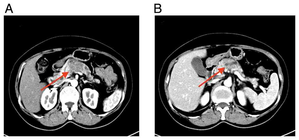
To further investigate the cause of the symptoms,
contrast-enhanced computed tomography (CT) angiography of the
abdomen was performed. Imaging revealed a mass-like hypodense
lesion located in the body of the pancreas, which was highly
suggestive of pancreatic malignancy. The lesion was associated with
involvement of the proximal splenic vein and significant luminal
narrowing, suggesting potential local vascular compression or
invasion. Representative axial CT images are shown in Fig. 1A and B.
Following comprehensive evaluation, which revealed
no major organ dysfunction and no radiologically evident
abnormalities of the hepatic artery or portal vein that would
preclude surgery, the patient underwent laparoscopic distal
pancreatectomy combined with splenectomy under general anesthesia
on January 8, 2020, within the same month as her initial
presentation. During intraoperative exploration, a firm mass
located in the pancreatic body was identified. Several enlarged
lymph nodes along the superior border of the pancreas were also
observed, raising suspicion for regional lymph node involvement.
The surgical procedure was successfully completed without major
intraoperative complications.
Postoperative histopathological examination
confirmed moderately differentiated PDAC involving the pancreatic
body and tail. H&E staining revealed irregular glandular
structures consisting of atypical tumor cells with enlarged
hyperchromatic nuclei and visible mitotic figures within a
fibrotic/desmoplastic stroma (Fig.
2A-C). Immunohistochemical staining showed positivity for
cytokeratin (CK) 7 and CK19, negativity for CK20 and caudal type
homeobox 2 and loss/reduced expression of SMAD4 in tumor cells
(Fig. 2D-R). Examination of the
surgical specimen found that the pancreatic resection margin,
vascular margins, omental tissue and spleen were free of tumor
invasion, indicating that complete macroscopic tumor resection had
been achieved. However, regional lymph node metastasis was
detected, with tumor involvement found in 1/8 peripancreatic lymph
nodes and in all 3 examined regional lymph nodes. According to the
American Joint Committee on Cancer 8th edition TNM staging system,
the postoperative pathological stage was determined to be pT2N2M0
(stage III). The presence of infiltrative glandular structures
within the desmoplastic stroma, together with regional lymph node
metastases, supported a diagnosis of invasive PDAC rather than a
high-grade in situ lesion.
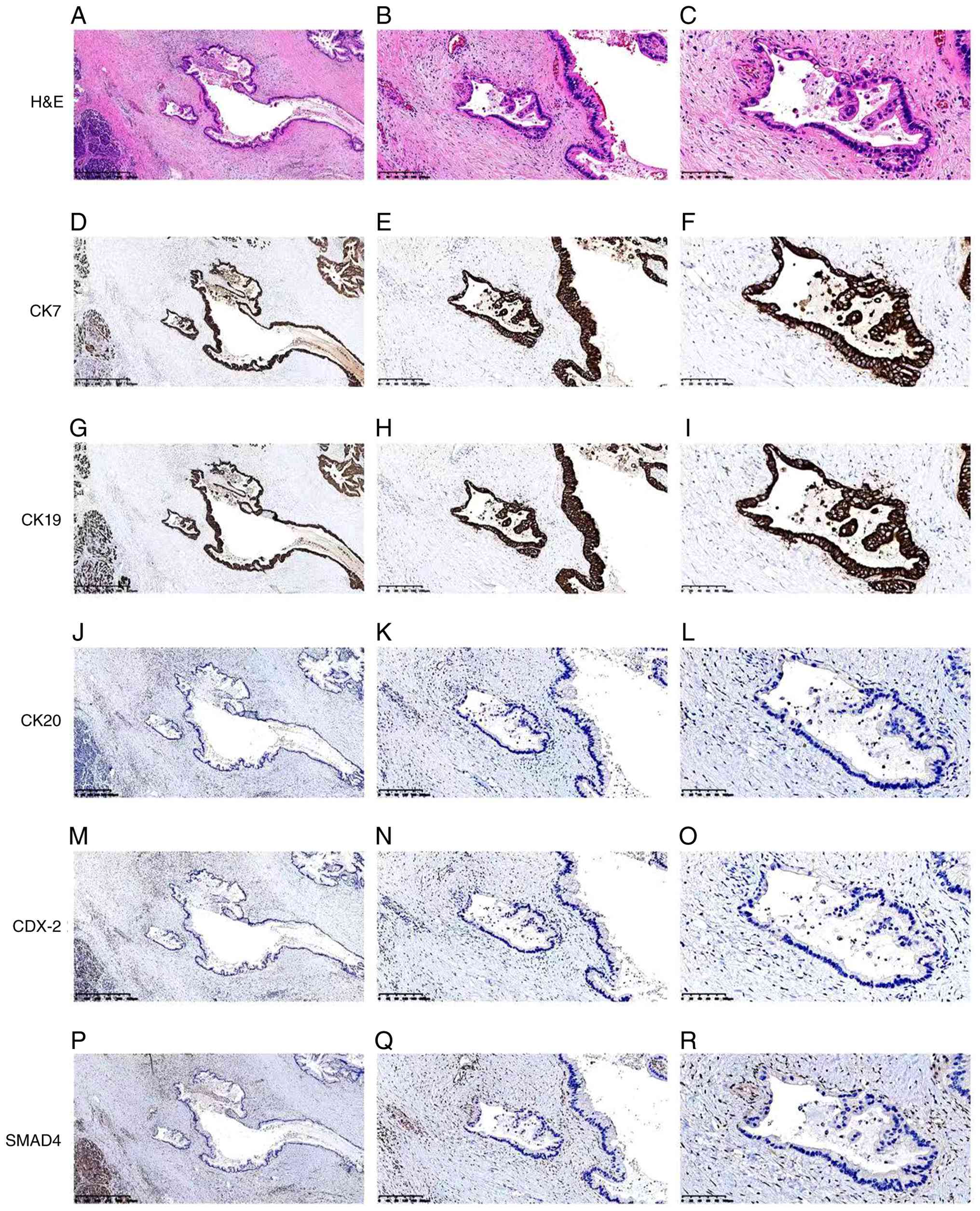 | Figure 2.Low- and high-power histopathological
and immunohistochemical findings of the resected pancreatic tumor.
H&E staining shows irregular glandular structures consisting of
atypical tumor cells with enlarged hyperchromatic nuclei and
visible mitotic figures within a fibrotic/desmoplastic stroma. (A)
Magnification, ×40, (B) magnification, ×100 and (C) magnification,
×200. Immunohistochemical staining shows CK7 positivity (D)
magnification, ×40, (E) magnification, ×100 and (F) magnification,
×200. CK19 positivity (G) magnification, ×40, (H) magnification,
×100 and (I) magnification, ×200, CK20 negativity (J)
magnification, ×40, (K) magnification, ×100 and (L) magnification,
×200. CDX2 negativity (M) magnification, ×40, (N) magnification,
×100 and (O) magnification, ×200. Loss or markedly reduced SMAD4
expression in tumor cells (P) magnification, ×40, (Q)
magnification, ×100 and (R) magnification, ×200. H&E,
hematoxylin and eosin; CK, cytokeratin; CDX2, caudal type homeobox
2; SMAD4, SMAD family member 4. |
Following postoperative recovery, the patient
received adjuvant chemotherapy. Specifically, between February and
June 2020, the patient underwent five cycles of gemcitabine
combined with S-1 (GS regimen), with gemcitabine administered
intravenously at 1.6 g on day 1 and 1.4 g on day 8, and S-1
administered orally at a total daily dose of 125 mg in two divided
doses on days 1–14. This was followed by two additional cycles of
S-1 monotherapy administered between July and August 2020 as
maintenance therapy. The patient tolerated the chemotherapy regimen
relatively well and completed the planned treatment without severe
toxicities. After the completion of adjuvant therapy, she entered a
routine surveillance program with periodic imaging examinations and
tumor marker monitoring.
The patient remained clinically stable for 4 years
and 9 months following surgery. However, during routine follow-up
in September 2024, contrast-enhanced abdominal CT revealed a newly
developed oval soft-tissue lesion in the portal vein-superior
mesenteric artery space, measuring ~1.9 cm in maximal diameter and
showing prominent contrast enhancement. Given the prior history of
resected PDAC of this patient, the typical locoregional nodal
location of the lesion and the concomitant increase in CA19-9 to
216 U/ml, these findings were highly suggestive of clinically
suspected locoregional recurrence. Representative CT images of the
suspected recurrence are shown in Fig.
3A.
To identify potential therapeutic targets after
postoperative recurrence, molecular profiling was performed using
archived formalin-fixed paraffin-embedded surgical tumor tissue
obtained from the primary pancreatic lesion, with matched
peripheral blood used as the germline control. The tumor specimen
was initially collected in January 2020 and submitted to 3D
Medicines Inc. for molecular testing in September 2024. The
estimated tumor cell content was 30%. A probe-based hybrid capture
next-generation sequencing assay was used to detect
tumor-associated genomic alterations, including clinically relevant
somatic variants, germline variants, microsatellite status and DNA
damage repair pathway-related alterations. The sequencing quality
met the quality control criteria, with an average sequencing depth
of 1,989.30× and a Q30 base percentage of 96.04%.
Molecular profiling identified a somatic EGFR exon
19 deletion-insertion mutation (NM_005228.5: c.2237_2256delinsTG;
p.E746_S752delinsV; variant allele frequency, 16.43%), a somatic
synonymous TP53 variant (NM_000546.6: c.375G>A; p.T125=; variant
allele frequency, 10.51%) and a heterozygous germline BRCA1
splice-site mutation (NM_007294.4: c.4485–2A>C). No KRAS
mutation was detected by NGS, indicating a KRAS wild-type molecular
profile. KRAS status was therefore defined based on molecular
testing rather than immunohistochemical staining. The tumor was
microsatellite stable. The complete NGS findings, including
clinically relevant variants and variants of uncertain
significance, are summarized in Table
I.
 | Table I.Summary of next-generation sequencing
findings. |
Table I.
Summary of next-generation sequencing
findings.
| Category | Gene | Variant/result | Status | VAF/result | Interpretation |
|---|
| Clinically relevant
somatic variant | EGFR | NM_005228.5; exon 19;
c.2237_2256delinsTG; p.E746_S752delinsV | Somatic | VAF, 16.43% | EGFR exon 19
deletion-insertion alteration; potentially actionable, although
functional relevance in PDAC remains insufficiently
characterized. |
| Clinically relevant
somatic variant | TP53 | NM_000546.6; exon 4;
c.375G>A; p.T125= | Somatic | VAF, 10.51% | Synonymous variant;
functional significance uncertain. |
| Clinically relevant
germline variant | BRCA1 | NM_007294.4;
splice-site region; c.4485–2A>C | Germline | Heterozygous | Germline BRCA1
splice-site mutation; DNA damage repair pathway-related
alteration. |
| Driver gene
status | KRAS | No mutation
detected | - | No mutation
detected | KRAS mutation was not
detected by NGS. |
| Microsatellite
status | MSI | Microsatellite
status | - | MSS | Microsatellite
stable. |
| Variant of uncertain
significance | RHBDF2 | NM_001005498.4; exon
18; c.1988G>A; p.R663H | Not classified in the
report | VAF, 8.36% | Variant of uncertain
significance. |
| Variant of uncertain
significance | SMAD4 | NM_005359.6; exon 3;
c.297G>T; p.W99C | Not classified in the
report | VAF, 12.35% | Variant of uncertain
significance. |
| Variant of uncertain
significance | SMARCA4 | NM_003072.5; exon 6;
c.1111G>A; p.E371K | Not classified in the
report | VAF, 7.99% | Variant of uncertain
significance. |
Given the rarity of this molecular profile and the
lack of standard targeted treatment options for this clinical
scenario, this finding was discussed at a multidisciplinary team
(MDT) meeting. Biopsy of the suspected recurrent lesion was
considered to obtain histological confirmation and updated
molecular profiling. Several potential approaches, including
endoscopic ultrasound-guided fine-needle aspiration or biopsy,
CT-guided percutaneous biopsy and diagnostic laparoscopy with
biopsy, were discussed by the MDT. However, the lesion was located
in the portal vein-superior mesenteric artery space and was
adjacent to major vascular structures, making tissue acquisition
technically challenging and potentially associated with a risk of
bleeding or vascular injury depending on the access route. After
detailed discussion of the potential benefits and risks of these
invasive diagnostic procedures, the patient explicitly declined
further biopsy or other invasive tissue sampling. Liquid biopsy was
also discussed as a less invasive option for additional molecular
reassessment; however, the patient declined further molecular
testing after discussion, and the MDT therefore proceeded with
non-invasive imaging and tumor marker follow-up. She also refused
further cytotoxic chemotherapy and radiotherapy due to concerns
regarding treatment-related toxicity. After comprehensive
discussion, the MDT recommended an individualized exploratory
targeted treatment strategy based on the clinical course of the
patient, radiological findings, CA19-9 elevation and the molecular
profile of the archived primary tumor tissue.
In October 2024, the patient initiated an
individualized exploratory combination regimen consisting of the
third-generation EGFR tyrosine kinase inhibitor aumolertinib (110
mg once daily) and the PARP inhibitor olaparib at an initial dose
of 300 mg twice daily. After 4 weeks of full-dose olaparib, the
patient developed grade 2 nausea/vomiting, grade 3 neutropenia and
grade 2 thrombocytopenia according to the Common Terminology
Criteria for Adverse Events version 5.0 (CTCAE v5.0; http://ctep.cancer.gov/protocolDevelopment/electronic_applications/ctc.htm#ctc_50).
After supportive care, the olaparib dose was reduced to 150 mg
twice daily, whilst aumolertinib was maintained at 110 mg once
daily. Following dose adjustment, these toxicities were manageable
and did not require permanent treatment discontinuation.
After ~3 months of therapy, follow-up imaging
demonstrated a reduction in the size of the suspected recurrent
lesion, accompanied by a marked decrease in CA19-9 from the
pretreatment level of 216 U/ml. With continued therapy, the lesion
regressed. By the most recent follow-up in December 2025, after
approximately 14 months of treatment, the CA19-9 level gradually
returned to the normal range and decreased to 20 U/ml. Serial
imaging demonstrated further radiological regression of the
suspected lesion, accompanied by durable disease control.
Representative follow-up CT images after targeted therapy are shown
in Fig. 3B.
At the most recent follow-up in December 2025, the
patient remained on aumolertinib at 110 mg once daily combined with
olaparib at the reduced dose of 150 mg twice daily and continued to
derive sustained clinical benefit. No evidence of disease
progression or distant metastasis had been observed. The patient
has achieved a progression-free survival >12 months, with
acceptable tolerability following dose modification.
The present case documented a rare KRAS wild-type
PDAC molecular context involving a somatic EGFR exon 19
deletion-insertion mutation and a heterozygous germline BRCA1
splice-site mutation, which informed an individualized exploratory
treatment strategy with aumolertinib and olaparib. The additional
synonymous TP53 variant was reported as part of the molecular
profile but was not regarded as a confirmed functional driver or
therapeutic determinant.
Discussion
PDAC is one of the most aggressive malignancies of
the digestive system, characterized by profound molecular
heterogeneity and an exceptionally poor prognosis. Tumor initiation
and progression are typically driven by the stepwise accumulation
of multiple genetic alterations involving both oncogenes and tumor
suppressor genes. Amongst these, KRAS mutations represent the
dominant molecular event in PDAC, occurring in ~90% cases and
serving as a central driver of tumorigenesis and disease
progression (7,14). These mutations are frequently
accompanied by alterations in key tumor suppressor genes, including
TP53, CDKN2A and SMAD4 (13),
resulting in a highly unstable genomic background. This complex
molecular landscape has substantially limited the clinical success
of targeted therapies in PDAC.
Importantly, KRAS mutations are not only strongly
associated with unfavorable prognosis (15,16),
but their long-standing classification as ‘undruggable’ has
represented a major therapeutic bottleneck in PDAC (17). However, 5–10% patients with PDAC
harbor KRAS wild-type tumors whose molecular pathogenesis differs
fundamentally from that of the classical KRAS-mutant subtype. In a
recent large-scale clinicogenomic analysis, Varghese et al
(5) demonstrated that KRAS
wild-type PDAC represents a distinct genomic subgroup associated
with specific somatic and germline features, with improved overall
survival. Accumulating molecular pathology data suggest that this
subgroup is more likely to exhibit defects in DNA damage repair
pathways, dysregulation of receptor tyrosine kinase signaling or
other potentially actionable driver events, such as BRAF
alterations and NRG1 fusions (18).
With the increasing clinical adoption of NGS, this distinct
molecular subset defined by KRAS wild-type status and the presence
of targetable genomic alterations is gaining recognition, opening
novel opportunities for individualized treatment in patients with
recurrent or treatment-refractory disease.
Among the currently known actionable molecular
alterations, mutations involving BRCA and other HRR genes hold
particular clinical relevance. BRCA1 and BRCA2 are essential
components of the HRR pathway, where their inactivation leads to
impaired repair of DNA double-strand breaks, increased genomic
instability and tumorigenesis. Germline and somatic BRCA1/2
mutations have both been implicated in PDAC development, with
germline carriers exhibiting a markedly elevated lifetime risk of
pancreatic cancer (19). In a
cohort of 854 patients with apparently sporadic PDAC, deleterious
germline BRCA1/2 mutations were identified in 15 patients (1.8%)
(19). Although the prevalence of
BRCA mutations in PDAC is relatively low, these alterations have
established therapeutic implications. Defective HRR constitutes the
molecular basis for sensitivity to DNA-damaging therapies. Wang
et al (20) evaluated
preclinical models and a molecularly annotated patient cohort of
HRR-deficient PDAC and identified genomic features associated with
sensitivity to platinum-based therapy, while also demonstrating
heterogeneity in treatment response. Furthermore, the POLO trial
confirmed that maintenance therapy with the PARP inhibitor olaparib
significantly prolonged progression-free survival in patients with
metastatic BRCA-mutant PDAC (median progression-free survival, 7.4
vs. 3.8 months) (21), firmly
establishing PARP inhibition as a standard therapeutic strategy now
incorporated into CSCO and other international guidelines.
Nevertheless, genomic instability driven by BRCA
mutations rarely occurs in isolation and frequently coexists with
aberrations in other oncogenic signaling pathways. In KRAS
wild-type PDAC, increasing attention has been directed toward
dysregulation of receptor tyrosine kinase signaling, particularly
the EGFR pathway. Unlike non-small cell lung cancer, activating
EGFR kinase-domain mutations are exceedingly rare in PDAC (22). Instead, EGFR abnormalities
predominantly manifest as protein overexpression or gene
amplification (13). Biologically,
EGFR signaling promotes tumor cell proliferation, invasion and
survival through the activation of various downstream pathways,
such as RAS/RAF/MAPK and PI3K/AKT (13). However, clinical translation of
EGFR-targeted therapy in PDAC has been disappointing. Although the
addition of erlotinib to gemcitabine demonstrated a statistically
significant survival benefit in a previous phase III trial, the
magnitude of benefit was modest and clinically limited (23,24).
In addition, individual reports have suggested that selected
patients with EGFR-mutated pancreatic adenocarcinoma may derive
benefit from third-generation EGFR-TKIs (25), although acquired resistance,
including EGFR C797S, may subsequently emerge (26).
The limited efficacy of EGFR-targeted therapy in
PDAC is widely attributed to the high prevalence of downstream KRAS
mutations, which enable persistent pathway activation independent
of EGFR inhibition. In addition, compensatory activation of
paralogous alternative signaling pathways, such as insulin-like
growth factor-1 receptors, further contributes to therapeutic
resistance (27). In this context,
combination treatment strategies have emerged as a rational
approach. Notably, EGFR inhibition may be more biologically
plausible in selected KRAS wild-type PDAC cases, where downstream
signaling may remain at least partially dependent on upstream
receptor tyrosine kinase activity. Furthermore, emerging evidence
implicating EGFR signaling in early pancreatic carcinogenesis,
particularly during acinar-to-ductal metaplasia, provides
additional biological rationale for its role as a therapeutic
target in selected molecular contexts (13,17).
In the present case, the EGFR alteration was
identified as the uncommon exon 19 deletion-insertion variant
p.E746_S752delinsV. EGFR exon 19 deletion and deletion-insertion
alterations are established sensitizing alterations in non-small
cell lung cancer. However, uncommon exon 19 deletion-insertion
subtypes may show heterogeneous sensitivity to different EGFR-TKIs
(28). The p.E746_S752delinsV
variant has been described among the uncommon EGFR exon 19
deletion-insertion alterations in lung cancer, where an isolated
clinical report suggested that a tumor harboring this variant may
respond to EGFR-TKI treatment (28,29).
Nevertheless, these data are largely derived from lung cancer,
where the functional relevance and predictive value of this
specific EGFR alteration in PDAC remain insufficiently
characterized (26,29). No in vitro or in
silico functional validation was performed in the present case.
Therefore, aumolertinib was selected as an empirical molecularly
guided treatment option in the context of KRAS wild-type PDAC with
a potentially actionable EGFR alteration, rather than on the basis
of confirmed functional sensitivity of this variant in PDAC.
TP53 alterations are frequent molecular events in
PDAC and have been associated with tumor biology and prognosis when
pathogenic variants are present (27,30).
However, the TP53 alteration identified in the present case was a
somatic synonymous variant, rather than a deleterious missense,
truncating, frameshift or splice-site mutation. Therefore, this
variant was included only as part of the molecular profile and was
not used to support the therapeutic rationale for aumolertinib plus
olaparib.
To date, to the best of our knowledge therapeutic
experience in KRAS wild-type PDAC harboring potentially actionable
EGFR alterations together with germline BRCA1 mutations remains
limited. The present case was therefore notable because it combines
a KRAS wild-type background, a somatic EGFR exon 19
deletion-insertion mutation and a heterozygous germline BRCA1
splice-site mutation. The additional TP53 synonymous variant was
included in the molecular profile but was not used as a primary
rationale for treatment selection. This molecular context provided
a clinical rationale for considering an individualized exploratory
targeted approach with aumolertinib and olaparib after
multidisciplinary discussion. However, the biological interaction
between EGFR inhibition and PARP inhibition in PDAC remains
insufficiently defined, where the present case cannot establish a
mechanistic synergistic effect between these two agents.
A noteworthy aspect of the present case was the dose
adjustment of olaparib during combination targeted therapy.
Olaparib was initially administered at 300 mg twice daily. However,
the patient developed intolerance during treatment, including grade
2 nausea/vomiting, grade 3 neutropenia and grade 2 thrombocytopenia
according to CTCAE v5.0. Therefore, the dose of olaparib was
reduced to 150 mg twice daily as an individualized
toxicity-management strategy, whilst aumolertinib was continued.
This dose reduction reflected real-world tolerability
considerations during off-label combination therapy rather than a
preplanned low-dose strategy. The observed clinical course cannot
determine the independent contribution of aumolertinib, olaparib or
their combination. Nevertheless, the durable disease control
observed in the present patient, together with the uncommon
convergence of KRAS wild-type status, a potentially actionable EGFR
alteration and germline BRCA1 mutation, may help define a
clinically testable question for future studies evaluating EGFR-TKI
and PARP inhibitor-based strategies in selected molecularly defined
patients with PDAC.
Several limitations of the present case should be
acknowledged. NGS was performed on archived formalin-fixed
paraffin-embedded tumor tissue obtained during the initial surgery
in 2020, rather than at the time of postoperative recurrence.
Although such specimens can generally preserve DNA integrity for
extended periods and are widely used in molecular analyses, the use
of archival tissue may not fully reflect potential genomic
evolution during disease progression. In addition, histological or
cytological confirmation of the suspected recurrent lesion was not
obtained. The lesion was located in the portal vein-superior
mesenteric artery space and was adjacent to major vascular
structures. Although several tissue-acquisition approaches,
including EUS-guided fine-needle (FN) aspiration/FN biopsy,
CT-guided percutaneous biopsy and diagnostic laparoscopy with
biopsy, were considered, each would require an invasive procedure
and careful assessment of bleeding or vascular injury risk. The
patient explicitly declined further invasive diagnostic procedures.
Consequently, recurrence was diagnosed clinically based on the
prior history of resected PDAC, the appearance of a newly developed
enhancing lesion in a typical locoregional nodal region,
concomitant CA19-9 elevation and serial radiological follow-up.
Although the imaging appearance, typical nodal location, rising
CA19-9 level and subsequent radiological regression during targeted
therapy supported the clinical diagnosis of recurrence, alternative
benign or inflammatory etiologies could not be completely excluded
in the absence of histological confirmation. The functional
relevance of the EGFR p.E746_S752delinsV alteration and the
biological significance of the TP53 synonymous variant in PDAC also
remain uncertain. The subsequent dose reduction of olaparib was
performed because of CTCAE v5.0-defined treatment-related
intolerance during off-label combination therapy, reflecting
real-world toxicity management rather than a predefined dosing
strategy. The independent contribution of aumolertinib, olaparib or
their combination cannot be determined from a single case.
Acknowledgements
Not applicable.
Funding
Funding: No funding was received.
Availability of data and materials
The next-generation sequencing data generated in the
present study have been deposited in the Genome Sequence Archive
for Human (GSA-Human) under accession no. HRA020193 (BioProject
accession no. PRJCA071184) and are publicly available at https://ngdc.cncb.ac.cn/gsa-human/browse/HRA020193.
Additional data generated in the present study may be requested
from the corresponding author.
Authors' contributions
SYW and WY analyzed the data, and wrote and revised
the manuscript. SYW and QH designed the study protocol. QH was the
primary care physician of the patient and developed and implemented
the treatment plan. SYW, WY and QH performed the literature review
and analyzed and interpreted the data in the paper. WY and QH
obtained the patient data and performed the histological
examination of the tumor. WY and QH reviewed the manuscript. QH was
responsible for the analysis and interpretation of the imaging
data. SYW, WY and QH confirm the authenticity of all the raw data.
All authors have read and approved the final version of the
manuscript.
Ethics approval and consent to
participate
Written informed consent for the publication of
clinical details and images was obtained from the patient.
Patient consent for publication
Not applicable.
Competing interests
The authors declare that they have no competing
interests.
References
|
1
|
Siegel RL, Miller KD, Wagle NS and Jemal
A: Cancer statistics, 2023. CA Cancer J Clin. 73:17–48.
2023.PubMed/NCBI
|
|
2
|
Luo J: KRAS mutation in pancreatic cancer.
Semin Oncol. 48:10–18. 2021. View Article : Google Scholar : PubMed/NCBI
|
|
3
|
Stickler S, Rath B and Hamilton G:
Targeting KRAS in pancreatic cancer. Oncol Res. 32:799–805. 2024.
View Article : Google Scholar : PubMed/NCBI
|
|
4
|
Dilly J, Hoffman MT, Abbassi L, Li Z,
Paradiso F, Parent BD, Hennessey CJ, Jordan AC, Morgado M, Dasgupta
S, et al: Mechanisms of resistance to oncogenic KRAS inhibition in
pancreatic cancer. Cancer Discov. 14:2135–2161. 2024. View Article : Google Scholar : PubMed/NCBI
|
|
5
|
Varghese AM, Perry MA, Chou JF, Nandakumar
S, Muldoon D, Erakky A, Zucker A, Fong C, Mehine M, Nguyen B, et
al: Clinicogenomic landscape of pancreatic adenocarcinoma
identifies KRAS mutant dosage as prognostic of overall survival.
Nat Med. 31:466–477. 2025. View Article : Google Scholar : PubMed/NCBI
|
|
6
|
Cicenas J, Kvederaviciute K, Meskinyte I,
Meskinyte-Kausiliene E, Skeberdyte A and Cicenas J: KRAS, TP53,
CDKN2A, SMAD4, BRCA1, and BRCA2 mutations in pancreatic cancer.
Cancers (Basel). 9:422017. View Article : Google Scholar : PubMed/NCBI
|
|
7
|
Waddell N, Pajic M, Patch AM, Chang DK,
Kassahn KS, Bailey P, Johns AL, Miller D, Nones K, Quek K, et al:
Whole genomes redefine the mutational landscape of pancreatic
cancer. Nature. 518:495–501. 2015. View Article : Google Scholar : PubMed/NCBI
|
|
8
|
Kim MK, Cho IR, Kim Y, Choi JH, Jung K,
Kim J, Kim S, Yun H, Yoon J, Oh DY, et al: Prognostic value of the
TP53 mutation in patients with pancreatic ductal adenocarcinoma
receiving FOLFIRINOX. Ther Adv Med Oncol. 16:175883592412904822024.
View Article : Google Scholar : PubMed/NCBI
|
|
9
|
Wong W, Raufi AG, Safyan RA, Bates SE and
Manji GA: BRCA mutations in pancreas cancer: Spectrum, current
management, challenges and future prospects. Cancer Manag Res.
12:2731–2742. 2020. View Article : Google Scholar : PubMed/NCBI
|
|
10
|
Katona BW, Lubinski J, Pal T, Huzarski T,
Foulkes WD, Moller P, Eisen A, Randall Armel S, Neuhausen SL, Raj
R, et al: The incidence of pancreatic cancer in women with a BRCA1
or BRCA2 mutation. Cancer. 131:e356662025. View Article : Google Scholar : PubMed/NCBI
|
|
11
|
Kowalewski A, Szylberg Ł, Saganek M,
Napiontek W, Antosik P and Grzanka D: Emerging strategies in
BRCA-positive pancreatic cancer. J Cancer Res Clin Oncol.
144:1503–1507. 2018. View Article : Google Scholar : PubMed/NCBI
|
|
12
|
Cheng H, Liu C, Jiang J, Luo G, Lu Y, Jin
K, Guo M, Zhang Z, Xu J, Liu L, et al: Analysis of ctDNA to predict
prognosis and monitor treatment responses in metastatic pancreatic
cancer patients: Clinical value of ctDNA in metastatic pancreatic
cancer. Int J Cancer. 140:2344–2350. 2017. View Article : Google Scholar : PubMed/NCBI
|
|
13
|
Mustafa M, Abbas K, Alam M, Habib S,
Zulfareen Hasan GM, Islam S, Shamsi A and Hassan I: Investigating
underlying molecular mechanisms, signaling pathways, emerging
therapeutic approaches in pancreatic cancer. Front Oncol.
14:14278022024. View Article : Google Scholar : PubMed/NCBI
|
|
14
|
Waters AM and Der CJ: KRAS: The critical
driver and therapeutic target for pancreatic cancer. Cold Spring
Harb Perspect Med. 8:a0314352018. View Article : Google Scholar : PubMed/NCBI
|
|
15
|
Ye L, Fei Q, Li T, Lin C, Ou H, Zhang Y,
Zhou Y, Zhang J, Jin K, Yu X and Wu W: Genomic mutation and
clinicopathological features of KRAS wild-type tumors in Chinese
cohort with pancreatic adenocarcinoma. Genomics. 117:1110482025.
View Article : Google Scholar : PubMed/NCBI
|
|
16
|
Kent OA: Increased mutant KRAS gene dosage
drives pancreatic cancer progression: Evidence for wild-type KRAS
as a tumor suppressor? Hepatobiliary Surg Nutr. 7:403–405. 2018.
View Article : Google Scholar : PubMed/NCBI
|
|
17
|
Cheng DK, Oni TE, Thalappillil JS, Park Y,
Ting HC, Alagesan B, Prasad NV, Addison K, Rivera KD, Pappin DJ, et
al: Oncogenic KRAS engages an RSK1/NF1 pathway to inhibit wild-type
RAS signaling in pancreatic cancer. Proc Natl Acad Sci USA.
118:e20169041182021. View Article : Google Scholar : PubMed/NCBI
|
|
18
|
Mouawad A, Habib S, Boutros M, Attieh F
and Kourie HR: How to find a needle in a haystack: A systematic
review on targeting KRAS wild-type pancreatic cancer. Future Oncol.
20:3539–3547. 2024. View Article : Google Scholar : PubMed/NCBI
|
|
19
|
Shindo K, Yu J, Suenaga M, Fesharakizadeh
S, Cho C, Macgregor-Das A, Siddiqui A, Witmer PD, Tamura K, Song
TJ, et al: Deleterious germline mutations in patients with
apparently sporadic pancreatic adenocarcinoma. J Clin Oncol.
35:3382–3390. 2017. View Article : Google Scholar : PubMed/NCBI
|
|
20
|
Wang Y, Park JYP, Pacis A, Denroche RE,
Jang GH, Zhang A, Cuggia A, Domecq C, Monlong J, Raitses-Gurevich
M, et al: A preclinical trial and molecularly annotated patient
cohort identify predictive biomarkers in homologous
recombination-deficient pancreatic cancer. Clin Cancer Res.
26:5462–5476. 2020. View Article : Google Scholar : PubMed/NCBI
|
|
21
|
Kindler HL, Hammel P, Reni M, Van Cutsem
E, Macarulla T, Hall MJ, Park JO, Hochhauser D, Arnold D, Oh DY, et
al: Overall survival results from the POLO trial: A phase III study
of active maintenance olaparib versus placebo for germline
BRCA-mutated metastatic pancreatic cancer. J Clin Oncol.
40:3929–3939. 2022. View Article : Google Scholar : PubMed/NCBI
|
|
22
|
Witkiewicz AK, McMillan EA, Balaji U, Baek
G, Lin WC, Mansour J, Mollaee M, Wagner KU, Koduru P, Yopp A, et
al: Whole-exome sequencing of pancreatic cancer defines genetic
diversity and therapeutic targets. Nature Commun. 6:67442015.
View Article : Google Scholar : PubMed/NCBI
|
|
23
|
Lee HS and Park SW: Systemic chemotherapy
in advanced pancreatic cancer. Gut Liver. 10:340–347. 2016.
View Article : Google Scholar : PubMed/NCBI
|
|
24
|
Schlick K, Kiem D and Greil R: Recent
advances in pancreatic cancer: Novel prognostic biomarkers and
targeted therapy-a review of the literature. Biomolecules.
11:14692021. View Article : Google Scholar : PubMed/NCBI
|
|
25
|
Ma X, Liu X, Ou K, Zhang M, Gao L and Yang
L: Advanced pancreatic cancer with KRAS wild-type and
EGFR-sensitive mutation respond favorably to furmonertinib: A case
report. Front Oncol. 13:11511782023. View Article : Google Scholar : PubMed/NCBI
|
|
26
|
Papa B, Dorwal P, Htain P, Robin J, Tan CP
and Singhal N: Acquired EGFR resistance mutation C797S in
pancreatic adenocarcinoma following partial response to
Third-Generation EGFR inhibitor therapy. JCO Precis Oncol.
8:e24001322024. View Article : Google Scholar : PubMed/NCBI
|
|
27
|
Qian Y, Gong Y, Fan Z, Luo G, Huang Q,
Deng S, Cheng H, Jin K, Ni Q, Yu X and Liu C: Molecular alterations
and targeted therapy in pancreatic ductal adenocarcinoma. J Hematol
Oncol. 13:1302020. View Article : Google Scholar : PubMed/NCBI
|
|
28
|
Chen Y, Xu J, Zhang L, Song Y, Wen W, Lu
J, Zhao Z, Kong W, Liu W, Guo A, et al: A multicenter-retrospective
study of non-small-cell lung carcinoma harboring uncommon epidermal
growth factor receptor (EGFR) mutations: Different subtypes of EGFR
exon 19 deletion-insertions exhibit the clinical characteristics
and prognosis of non-small cell lung carcinoma. Transl Lung Cancer
Res. 11:238–249. 2022. View Article : Google Scholar : PubMed/NCBI
|
|
29
|
Zhuang W, Zhang C, Tang Y, Tian D, Lan Z,
Zeng C and Qiao G: Clinical and pathologic complete response to
gefitinib in a patient with SqCLC harboring EGFR p.E746_S752delinsV
mutation. Onco Targets Ther. 14:4805–4808. 2021. View Article : Google Scholar : PubMed/NCBI
|
|
30
|
Pan M, Jiang C, Zhang Z, Achacoso N,
Alexeeff S, Solorzano AV, Tse P, Chung E, Sundaresan T, Suga JM, et
al: TP53 gain-of-function and non-gain-of-function mutations are
associated with differential prognosis in advanced pancreatic
ductal adenocarcinoma. JCO Precis Oncol. 7:e22005702023. View Article : Google Scholar : PubMed/NCBI
|