Introduction
Gliomas represent the most prevalent and aggressive
primary intracranial tumors, accounting for >40% of all primary
brain tumors (1). Among these,
glioblastoma (GBM), classified as grade IV glioma in the World
Health Organization classification, represents the highest level of
malignancy, characterized by highly invasive growth and worse
prognosis (2). Despite the
incorporation of multimodal treatment strategies, which include
maximal safe resection supplemented by temozolomide chemotherapy
and radiotherapy, the clinical prognosis for patients with GBM
remains dismal. The median survival period is typically <15
months, with the majority of patients experiencing tumor recurrence
within 1 year following surgery (3,4).
Consequently, the exploration of innovative therapeutic molecules
and strategies with the potential to effectively impede GBM
progression has emerged as a pivotal scientific imperative in this
domain, necessitating prompt and comprehensive resolution.
Melatonin (MT; N-acetyl-5-methoxytryptamine), a
naturally occurring indoleamine hormone, is primarily secreted by
the pineal gland and serves a key role in regulating circadian
rhythms, sleep-wake cycles and various metabolic processes
(5–7). The amphiphilic properties of MT, which
are both lipophilic and hydrophilic, enable its diffusion through
biological membranes, including the blood-brain barrier, resulting
in its distribution throughout tissues and body fluids (8,9).
Beyond classical receptor-mediated signaling, MT has been revealed
to exert its broad physiological and pharmacological activities
through direct, receptor-independent interactions with
intracellular targets (10,11). MT has been demonstrated to possess
particularly potent neuroprotective effects in central nervous
system disorders, including Parkinson's disease, Alzheimer's
disease and ischemic brain injury models (12,13).
MT has been revealed to possess dual antioxidant properties. In
normal cells, such as neurons, MT acts as an effective free radical
scavenger and antioxidant enzyme inducer (14,15).
However, in several tumor models, MT paradoxically exerts
pro-oxidant effects, thereby inhibiting cancer cell proliferation
and promoting apoptotic cell death (16–18).
The antitumor effect of MT has been suggested in a variety of
malignancies, including prostate cancer, colorectal cancer,
hepatocellular carcinoma and breast cancer (19). However, the molecular mechanisms
underlying the action of MT in GBM, particularly its regulation of
key signaling pathways, remain to be elucidated.
High mobility group box 1 (HMGB1) is a nuclear
non-histone protein that is widely expressed in eukaryotic cells
(20,21). It serves a key role in maintaining
nuclear structural homeostasis and regulating gene transcription by
modulating chromosome structure and function, as well as sustaining
cellular autophagy (22). In
conditions of cellular stress, injury or necrosis, HMGB1 can be
actively secreted or passively released into the extracellular
space (23,24), serving as a key damage-associated
molecular pattern molecule. By binding to its receptors [such as
Toll-like receptor 4 (TLR4)], it activates downstream pro-survival
and pro-inflammatory signaling pathways, such as NF-κB, which
ultimately drive tumor cell proliferation, migration, invasion and
treatment resistance (25). TLR4 is
expressed at high levels in glioma cells and tumor-associated glial
cells within the brain. The activation of the NF-κB pathway,
triggered by its binding to HMGB1 (26), is considered a pivotal step in
promoting the malignant progression of gliomas. However, the
potential of MT to inhibit GBM by regulating the HMGB1/TLR4/NF-κB
signaling axis remains to be elucidated. Therefore, the present
study aimed to investigate the anti-GBM effects of MT and to
explore whether the SIRT1/HMGB1/TLR4/NF-kB axis is involved in this
process. To address this aim, in vitro functional assays
were performed to evaluate the effects of MT on GBM cell viability,
apoptosis, migration and invasion. In addition, network
pharmacology, protein-protein interaction (PPI) analysis and
molecular docking were used to identify potential molecular targets
and pathways, and western blotting together with EX527-mediated
SIRT1 inhibition was performed to assess the involvement of
SIRT1/HMGB1/TLR4/NF-κB-related signaling.
Materials and methods
Cell culture
U87MG cells, a commonly used GBM cell line of
unknown origin (cat. no. CL-0238), and LN229 human GBM cells (cat.
no. CL-0578) were purchased from Procell Life Science &
Technology Co., Ltd. The U87MG cell line used in the present study
was the American Type Culture Collection version, which is most
probably a GBM cell line of unknown origin. The authenticity of the
U87MG cell line was verified by short tandem repeat profiling. Both
cell types were grown in Dulbecco's Modified Eagle Medium (DMEM;
Gibco; Thermo Fisher Scientific, Inc.) supplemented with 10% fetal
bovine serum (FBS; Gibco; Thermo Fisher Scientific, Inc.) and 1%
penicillin/streptomycin. Cell cultures were kept in a humidified
incubator at 37°C under a 5% CO2 atmosphere. For
subculturing, cells were detached using 0.25% trypsin and passaged
following standard protocols.
Reagents and antibodies
MT and the sirtuin 1 (SIRT1) inhibitor EX527 were
purchased from Beyotime Biotechnology and were dissolved in
dimethyl sulfoxide (DMSO) to prepare storage solutions for use. The
primary antibodies used in the present study and their dilution
ratios are as follows: Anti-Bax (1:2,000; cat. no. 50599-2-Ig),
anti-Bcl-2 (1:2,000; 68103-1-Ig), anti-cadherin CDH1 (1:2,000;
20874-1-AP), anti-CDH2 (1:2,000; 22018-1-AP), anti-MMP3 (1:2,000;
17873-1-AP), anti-Vimentin (1:2,000; 10366-1-AP), anti-SIRT1
(1:2,000; 13161-1-AP), anti-TLR4 (1:2,000; 66350-1-Ig), anti-p65
(1:2,000; 66535-1-Ig) and anti-phosphorylated-p65 (p-p65; 1:2,000;
82335-1-RR) were all purchased from Proteintech Group, Inc.
Anti-HMGB1 (1:1,000; YM4697) and anti-acetylated-HMGB1 (acHMGB1;
1:1,000; YK0060) were purchased from Immunoway Biotechnology
Company and anti-β-actin (1:10,000; Proteintech Group, Inc.;
81115-1-RR) was used as an internal control. All antibodies were
used according to the manufacturer's recommended conditions.
Cell viability assay
The assessment of cell viability was conducted by
employing the Cell Counting Kit-8 (CCK-8; Biosharp Life Sciences).
In summary, cells were seeded into 96-well plates. Following cell
attachment, fresh DMEM containing 10% FBS and MT at various
concentrations (0, 1, 3 and 5 mM) was added, and cells were
cultured for 24 and 48 h. At each designated time point, the CCK-8
reagent was added to each well (representing 10% of the medium
volume). The plates were then incubated at 37°C in the dark for a
period of 2 h. Subsequently, the absorbance was measured at 450 nm
by means of a microplate reader.
Calcein AM/propidium iodide (PI) assay
for cell viability and cytotoxicity
The viability and cytotoxicity of cultured cells
were determined by Calcein-AM/PI double-staining (Beyotime
Biotechnology). U87MG and LN229 cells were seeded in 6-well plates
at a density of 2×10⁵ cells/well. Following the
completion of treatments, the medium was discarded, and the cell
monolayer was washed twice with pre-cooled PBS. Subsequent to the
addition of the Calcein-AM/PI working solution, incubation
proceeded at 37°C for 30 min, protected from light. Observation
under a fluorescence microscope distinguished viable cells,
characterized by green fluorescence, from non-viable cells, which
were labeled with red fluorescence.
Western blot analysis
After treatment with 1 mM MT or 5 µM EX527 for 48 h,
cells were washed twice with pre-chilled PBS and lysed for 30 min
on ice using RIPA lysis buffer (containing 1% protease inhibitor
and 1% phosphatase inhibitor). Cell lysates were collected,
centrifuged at 4°C and 12,000 g for 20 min, and the supernatant was
collected. Following protein quantification with a BCA kit, 25 µg
of total protein was loaded per lane. samples were diluted in 5X
SDS loading buffer and heat-denatured at 100°C for 5–10 min. The
proteins were separated via SDS-PAGE on a 10% gel and then
transferred to a PVDF membrane. After blocking with 5% non-fat milk
at room temperature for 2 h, the membrane was incubated with
respective primary antibodies overnight at 4°C. Subsequent to
washing with TBST containing 0.1% Tween-20, the membrane was
exposed to an HRP-conjugated secondary antibody, including
HRP-conjugated goat anti-rabbit IgG (cat. no. SA00001-2;
Proteintech Group, Inc; 1:10,000) and HRP-conjugated goat
anti-mouse IgG (cat. no. SA00001-1; Proteintech Group, Inc;
1:10,000), for 2 h at room temperature. Final detection was
achieved using a chemiluminescence substrate (BeyoECL Plus; cat.
no. P0018S; Beyotime Biotechnology), and band intensities were
quantified based on grayscale values analyzed with ImageJ (v1.53;
National Institutes of Health).
Wound healing assay
To assess cell migration capacity, U87MG and LN229
cells were seeded at high density in 6-well plates. Once the
monolayer exhibited >90% confluence, a linear incision was made
across the monolayer using a 200 µl sterile pipette tip. The cell
debris was then washed away twice with PBS, and the cells were then
cultured in serum-free DMEM containing the indicated treatments.
The images were obtained from the same location with an inverted
microscope at two distinct time points: 0 and 24 h
post-scratch.
Transwell invasion assay
Cell invasion assays were performed using
Matrigel-coated Transwell inserts with an 8.0 µm pore size.
Briefly, Matrigel was added to the upper surface of the Transwell
membrane and allowed to solidify at 37°C for 1 h before cell
seeding. U87MG and LN229 cells were serum-starved overnight in
serum-free DMEM, harvested and seeded into the upper chamber at a
density of 1×10⁵ cells/well in serum-free DMEM
containing 1 mM MT. The lower chamber contained DMEM supplemented
with 10% FBS as a chemoattractant. After incubation at 37°C in a
humidified atmosphere with 5% CO2 for 48 h, non-invading
cells on the upper surface of the membrane were removed. The
invaded cells on the lower surface were fixed with 4%
paraformaldehyde at room temperature for 20 min and stained with
0.1% crystal violet at room temperature for 20 min. The number of
invaded cells was counted in three randomly selected fields under
an inverted light microscope.
Network pharmacology, PPI network and
molecular docking analysis
To predict potential targets and mechanisms of MT
against GBM, potential targets of MT were obtained from the
Comparative Toxicogenomics Database (CTD; http://ctdbase.org/) (27) and disease-related targets were
retrieved from the GeneCards database (https://www.genecards.org/) (28) using ‘Glioblastoma’ as the keyword.
The intersection of these two target sets yielded common
interaction targets. These shared targets underwent Gene Ontology
(GO) functional annotation and Kyoto Encyclopedia of Genes and
Genomes (KEGG) pathway enrichment analysis using the DAVID online
database (version 2021 update; http://david.ncifcrf.gov/). The GO and KEGG databases
were accessed through DAVID for functional annotation and pathway
enrichment analysis (http://geneontology.org/; http://www.kegg.jp/). Enrichment results were
visualized using the ‘ggplot2’ package (version 4.0.3; http://CRAN.R-project.org/package=ggplot2) in RStudio
software (2025.09.2+418; Posit Software, PBC).
To further explore the relationships among the
overlapping targets, a PPI network was constructed using the STRING
database (https://string-db.org/) with Homo
sapiens as the species. The PPI data were imported into
Cytoscape software (version 3.10.0; Cytoscape Consortium;
http://cytoscape.org/) for network visualization
and topological analysis. Core targets were ranked according to
degree centrality, and the top 35 nodes were selected for further
visualization.
Molecular docking was performed to evaluate the
potential interaction between MT and SIRT1. The three-dimensional
structure of SIRT1 was obtained from the Protein Data Bank (PDB;
http://www.rcsb.org/), and the chemical structure
of MT was obtained from PubChem. The docking process was performed
using AutoDock Vina (version 1.2.7; Center for Computational
Structural Biology, Scripps Research), while Discovery Studio
(version 2019; BIOVIA, Dassault Systèmes S.E.) was employed to
generate two-dimensional interaction maps illustrating hydrogen
bonds, hydrophobic contacts and π-π overlap interactions.
Statistical analysis
Data analysis and visualization were performed using
GraphPad Prism 8.0 (Dotmatics). Data are presented as mean ± SD
from at least three independent biological replicates. Comparisons
between two groups were performed using an unpaired Student's
t-test. Comparisons among multiple groups were performed using
one-way ANOVA followed by Tukey's post hoc test. P<0.05 was
considered to indicate a statistically significant difference.
Results
MT inhibits the viability of GBM cells
and promotes their apoptosis
To elucidate the direct effect of MT on GBM cell
viability, cells were first treated with varying concentrations of
MT. CCK-8 assay results indicated that GBM cell viability decreased
in a concentration-dependent manner after treatment with 1, 3 and 5
mM MT for 24 and 48 h (Fig. 1A and
B). To further validate the effect of MT on cell survival,
Calcein-AM/PI dual staining was performed. Fluorescence microscopy
revealed a notable reduction in green-fluorescent viable cells and
a corresponding increase in red-fluorescent dead cells in
MT-treated groups, indicating MT markedly decreased the relative
survival rate of GBM cells (Fig. 1C and
D). Subsequently, it was investigated whether MT modulates cell
survival by regulating apoptosis. Western blot analysis suggested
that MT treatment significantly upregulated the expression of the
pro-apoptotic protein Bax while markedly suppressing the expression
of the anti-apoptotic protein Bcl-2 (Fig. 1E and F).
MT inhibits the migratory and invasive
capabilities of GBM cells
The effects of MT on GBM cell migration and invasion
were evaluated using a series of functional assays. Transwell
assays demonstrated that MT treatment reduced the number of invaded
U87MG and LN229 cells after 48 h (Figs.
2A and S1A). Wound healing
assays further indicated that treatment with MT for 24 h
significantly reduced the relative migration of both GBM cell lines
(Figs. 2B and S1B). To investigate the molecular changes
associated with the effects of MT on migration and invasion, the
expression levels of epithelial-mesenchymal transition and
associated invasive proteins were examined. Western blot analysis
revealed that MT treatment significantly upregulated the expression
of the epithelial marker CDH1. Conversely, the expression of the
mesenchymal markers CDH2 and vimentin, as well as the matrix
metalloproteinase MMP3, were significantly downregulated (Fig. 2C and D). These molecular changes
collectively suggest the functional role of MT in inhibiting GBM
cell migration and invasion.
 | Figure 2.MT inhibits the migration and
invasion capabilities of GBM cells. (A) Representative images of
the Transwell invasion assay showing the invasive capacity of GBM
cells after treatment with 1 mM MT for 48 h. Cells were stained
with crystal violet. Original magnification, ×50. (B)
Representative images of the wound healing assay showing the
migratory capacity of GBM cells after treatment with 1 mM MT for 24
h. Scale bars, 200 µm. Representative western blot bands and
quantitative analysis of migration- and invasion-related proteins,
including CDH1/E-cadherin, CDH2/N-cadherin, vimentin and MMP3, in
(C) U87MG and (D) LN229 cells after treatment with 1 mM MT for 48
h. Data are presented as mean ± SD from three independent
biological replicates (n=3). Statistical significance was
determined using an unpaired Student's t-test. **P<0.01 and
***P<0.001. GBM, glioblastoma; MT, melatonin; VIM, vimentin;
CDH, cadherin. |
Network pharmacology predicts
potential targets and pathways for MT in GBM
To explore the potential mechanisms underlying the
anti-GBM effects of MT, network pharmacology, PPI network analysis
and molecular docking were performed. MT-related targets were
obtained from the CTD, whereas GBM-related targets were retrieved
from GeneCards. Intersection of the two datasets identified 388
overlapping targets as potential targets of MT against GBM
(Fig. 3A).
GO enrichment analysis indicated that these targets
were mainly associated with transcriptional and gene expression
regulation, apoptosis, signal transduction and inflammatory
responses. The targets were predominantly localized in the
cytoplasm, nucleus, plasma membrane, extracellular region and
mitochondria and were enriched in molecular functions such as
‘protein binding’, ‘DNA binding’, ‘ATP binding’ and ‘DNA-binding
transcription factor activity’ (Fig.
3B-D). KEGG analysis further revealed enrichment in cancer and
inflammation-related pathways, including TLR signaling, NF-κB
signaling, glioma-related pathways and apoptosis (Fig. 3E).
A PPI network was subsequently constructed, and
degree centrality analysis was performed using Cytoscape. The top
35 hub genes are presented in Fig.
3F. Notably, NF-κB1, SIRT1 and TLR4 were included among these
hub genes, providing a network-level basis for further
investigation of SIRT1- and TLR4/NF-κB-related signaling.
Molecular docking suggested that MT could be
accommodated within the binding pocket of SIRT1 and interact with
surrounding amino acid residues (Fig.
3G and H), supporting SIRT1 as a plausible upstream candidate
target of MT. Collectively, the PPI topology, pathway enrichment
and docking results suggest that MT may regulate TLR4/NF-κB-related
signaling through SIRT1. Due to the deacetylase activity of SIRT1
and the mechanistic relevance of HMGB1 acetylation to TLR4/NF-κB
signaling (29,30), the SIRT1/HMGB1/TLR4/NF-κB axis was
selected for subsequent experimental validation.
MT increases SIRT1 expression and
attenuates HMGB1/TLR4/NF-κB-related signaling in GBM cells
To elucidate the antitumor regulatory mechanism of
MT, western blotting was performed to analyze the effects of MT
treatment on the expression of proteins involved in relevant
signaling pathways. Previous studies have suggested that MT can
upregulated the expression of SIRT1 (31–33).
The present experimental results demonstrated that MT treatment
significantly upregulated the protein expression level of SIRT1 in
U87MG cells (Fig. 4A and B) and
concurrently markedly reduced the levels of acHMGB1, its key
receptor TLR4 and p-p65 (Fig. 4A and
B). To validate the universality of this regulatory
relationship, the experiment was repeated using another cell line,
LN229, with results consistent with those observed in U87MG cells:
MT similarly significantly increased SIRT1 expression in LN229
cells (Fig. 4C and D) while
suppressing the protein levels of acHMGB1, TLR4 and p-p65 (Fig. 4C and D). Collectively, the data
suggests that MT may inhibit the activity of the HMGB1/TLR4/NF-κB
signaling pathway by activating SIRT1 expression. This synergistic
effect suggests that MT attenuates HMGB1/TLR4/NF-κB-dependent
signaling by enhancing SIRT1 expression.
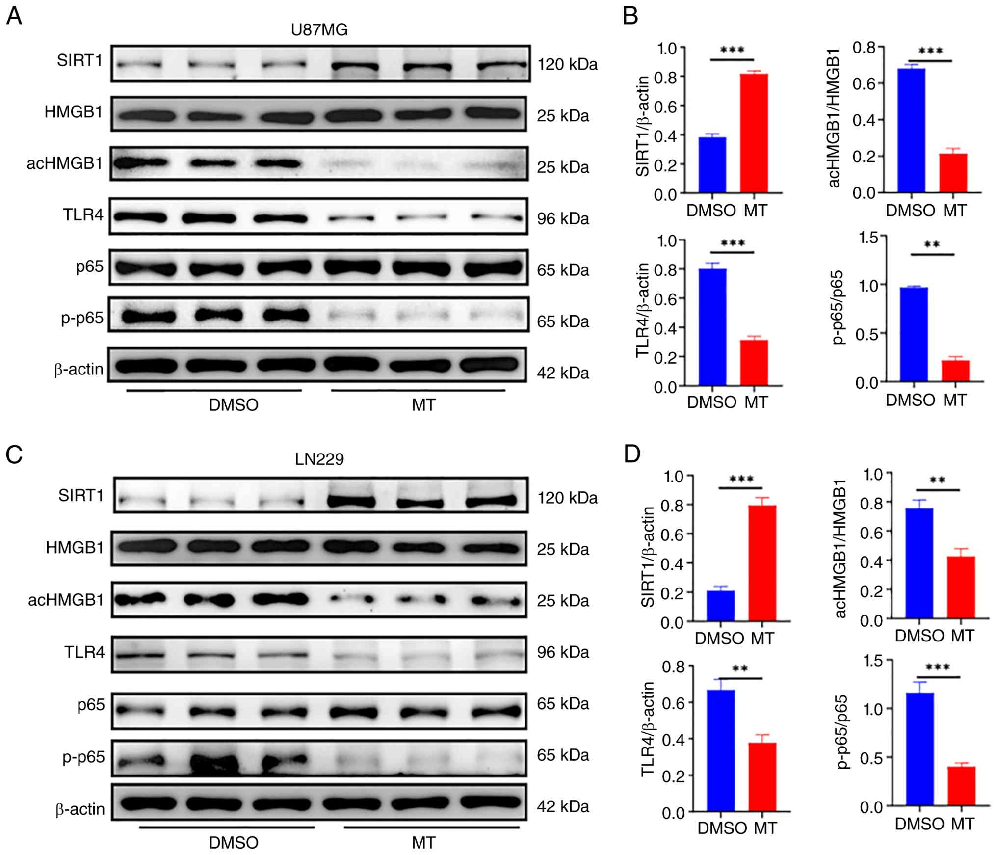 | Figure 4.MT increases SIRT1 expression and
attenuates HMGB1/TLR4/NF-κB-related signaling in glioblastoma
cells. (A) Western blot bands showing the expression of SIRT1,
HMGB1, acHMGB1, TLR4, p65 and p-p65 in U87MG cells after treatment
with 1 mM MT for 48 h. (B) Semi-quantitative analysis of SIRT1,
acHMGB1/HMGB1, TLR4 and p-p65/p65 protein levels in U87MG cells.
(C) Western blot bands showing the expression of SIRT1, HMGB1,
acHMGB1, TLR4, p65 and p-p65 in LN229 cells after treatment with 1
mM MT for 48 h. (D) Semi-quantitative analysis of SIRT1,
acHMGB1/HMGB1, TLR4 and p-p65/p65 protein levels in LN229 cells.
Data are presented as mean ± SD from three independent biological
replicates (n=3). Statistical significance was determined using an
unpaired Student's t-test. **P<0.01 and ***P<0.001. MT,
melatonin; SIRT1, sirtuin-1; HMGB1, high mobility group box 1;
TLR4, Toll-like receptor 4; acHMGB1; acetylated HMGB1; p-p65,
phosphorylated p65. |
Pharmacological inhibition of SIRT1
enhances GBM cell migration and HMGB1/TLR4/NF-κB-related
signaling
The aforementioned results demonstrated that MT
upregulated SIRT1 expression and suppressed the HMGB1/TLR4/NF-κB
signaling pathway. To further determine whether SIRT1 contributed
to the anti-migratory effect of MT, U87MG and LN229 cells were
treated with DMSO, MT, EX527 or MT combined with EX527. Wound
healing assays indicated that MT markedly reduced the relative
migration of both GBM cell lines, whereas EX527 markedly enhanced
cell migration. Importantly, the relative migration rate in the
MT+EX527 group was notably higher than that in the MT-alone group,
indicating that pharmacological inhibition of SIRT1 partially
reversed the inhibitory effect of MT on GBM cell migration
(Figs. 5A and B and S1C). Western blot analysis further
suggested that EX527 treatment decreased SIRT1 expression in both
U87MG and LN229 cells. This reduction was accompanied by increased
levels of acHMGB1, TLR4 and p-p65 (Fig.
5C-F). These findings indicate that pharmacological inhibition
of SIRT1 induces molecular changes opposite to those observed after
MT treatment and suggest that SIRT1 may negatively regulate HMGB1
acetylation and downstream TLR4/NF-κB pathway activity in GBM
cells. Together, these results support the involvement of SIRT1 in
the MT-associated regulation of the HMGB1/TLR4/NF-κB signaling
axis.
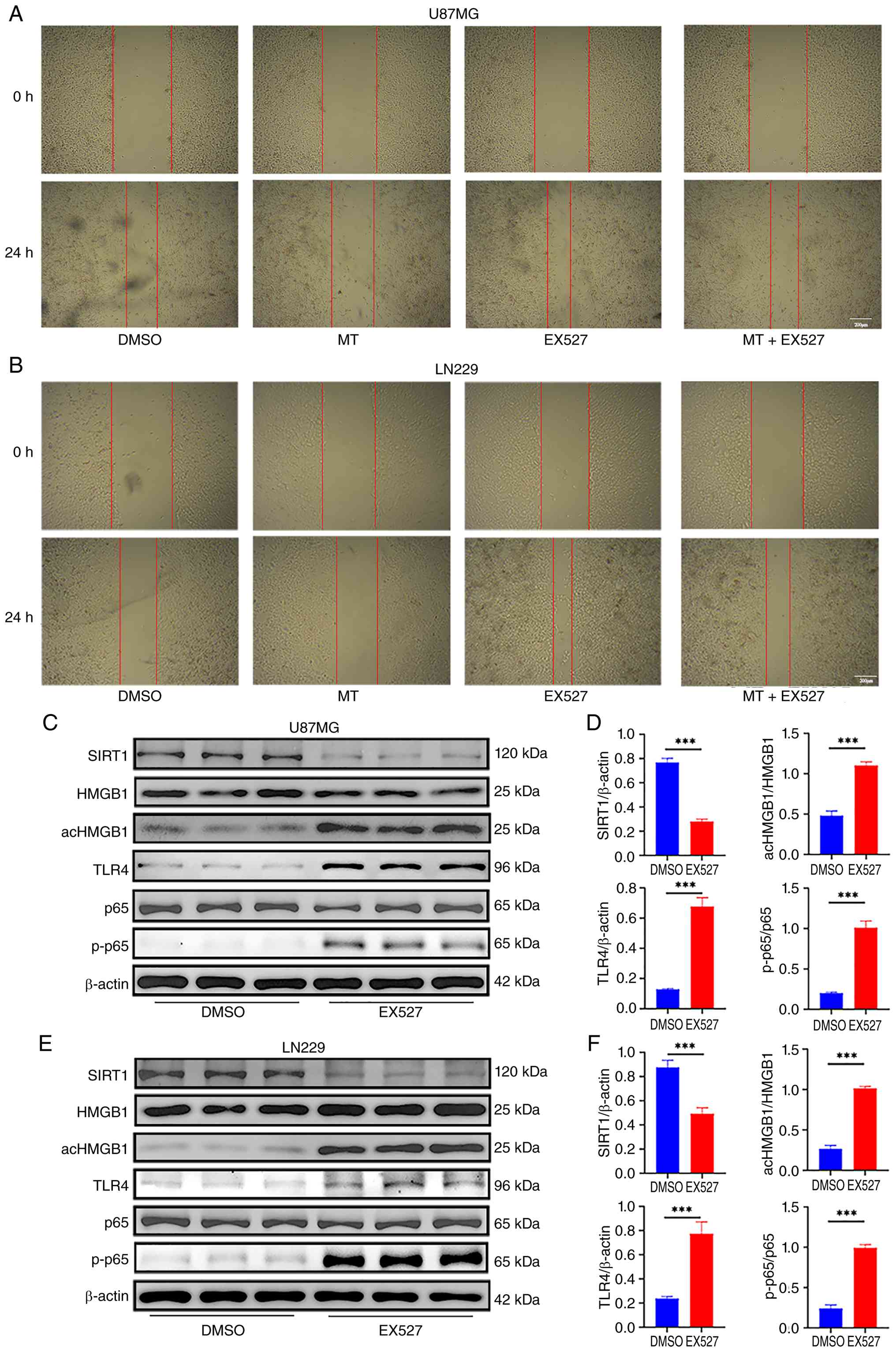 | Figure 5.Inhibition of SIRT1 enhances the
HMGB1/TLR4/NF-κB signaling pathway in glioblastoma cells.
Representative wound healing images of (A) U87MG and (B) LN229
cells treated with DMSO, 1 mM MT, 5 µM EX527 or the combination of
MT and EX527 for 24 h. Images were captured at 0 and 24 h after
scratching. Scale bars, 200 µm. (C) Western blot bands showing the
expression of SIRT1, HMGB1, acHMGB1, TLR4, p65 and p-p65 in U87MG
cells after treatment with EX527 for 48 h. (D) Semi-quantitative
analysis of SIRT1, ac-HMGB1/HMGB1, TLR4 and p-p65/p65 protein
levels in U87MG cells. (E) Western blot bands showing the
expression of SIRT1, HMGB1, acHMGB1, TLR4, p65 and p-p65 in LN229
cells after treatment with EX527 for 48 h. (F) Semi-quantitative
analysis of SIRT1, acHMGB1/HMGB1, TLR4 and p-p65/p65 protein levels
in LN229 cells. Data are presented as mean ± standard deviation
from three independent biological replicates (n=3). Statistical
significance was determined using an unpaired Student's t-test.
***P<0.001. MT, melatonin; SIRT1, sirtuin-1; HMGB1, high
mobility group box 1; TLR4, Toll-like receptor 4; acHMGB1;
acetylated HMGB1; p-p65, phosphorylated p65. |
Discussion
The present study provides evidence that MT may
suppress the malignant phenotype of GBM cells and suggests the
involvement of the SIRT1/HMGB1/TLR4/NF-κB signaling axis. MT
inhibited GBM cell viability, migration and invasion and promoted
apoptosis. These effects were accompanied by increased SIRT1
expression, reduced HMGB1 acetylation, decreased TLR4 expression
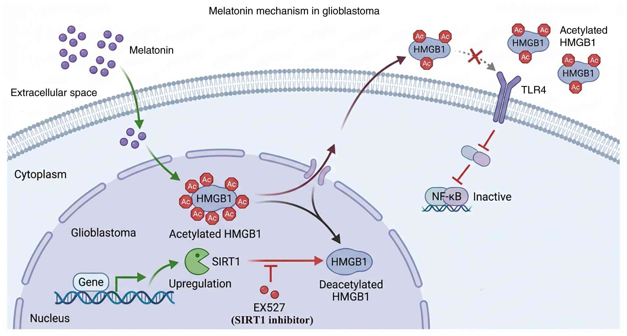
and suppressed p65 phosphorylation (Fig. 6).
GBM is the most aggressive malignant tumor in the
central nervous system, accounting for ~49.1% of all primary brain
tumors (34). The incidence rate is
~3.2 cases per 100,000 people annually, and it is projected to
continue rising over the coming decades, posing a notable public
health challenge (35,36). Despite comprehensive treatment
approaches including surgery, radiotherapy and chemotherapy,
patients with GBM continue to face an unfavorable prognosis, with a
median survival of only ~12 months and an extremely low 5-year
survival rate (37). Furthermore,
the high recurrence and mortality rates of GBM, coupled with its
resistance to the first-line chemotherapeutic agent temozolomide
and high recurrence rates, further underscore the urgent need to
develop novel therapeutic strategies.
MT is an endogenous indoleamine primarily
synthesized and secreted by the pineal gland under circadian
regulation. It has attracted considerable attention because of its
antioxidant, neuroprotective and broad-spectrum anticancer
properties (38,39). The anticancer mechanisms of MT
involve multiple levels, including inducing autophagy, modulating
mitochondrial function, triggering endoplasmic reticulum stress and
regulating various forms of cell death (40). The molecular effects of MT vary
across types of tumors. For example, MT induces tumor wasting
through the peroxisome proliferator-activated receptor γ
coactivator 1-α/uncoupling protein 1 pathway in renal cell
carcinoma (41) and disrupts folate
metabolism in head and neck squamous cell carcinoma (42). SIRT1 has emerged as an important
mediator of several biological effects of MT. Previous studies have
demonstrated that MT can alleviate endoplasmic reticulum stress and
apoptosis by increasing SIRT1 expression (31) and modulate inflammatory or
neuroprotective signaling through SIRT1-related pathways (32). These findings suggest that SIRT1 may
serve as a key hub for the biological functions of MT.
HMGB1 is a highly conserved nucleoprotein that
serves a key role in maintaining genomic stability (43). During cellular stress, injury or
death, HMGB1 can be actively secreted or passively released from
the nucleus into the extracellular space, where it functions as a
damage-associated molecular pattern molecule (44). Extracellular HMGB1 activates
downstream signaling cascades, including NF-κB, by binding to
receptors such as the advanced glycation end-product receptor or
TLRs, thereby promoting tumor growth, metastasis and the formation
of an immune-inflammatory microenvironment (45,46).
MT also affected EMT-like and invasion-related proteins in GBM
cells. In the present study, MT increased CDH1/E-cadherin
expression while decreasing CDH2/N-cadherin, vimentin and MMP3
expression, which was consistent with the observed reduction in
migration and invasion. These findings suggest that MT may suppress
the invasive phenotype of GBM cells by modulating EMT-like and
matrix degradation-related proteins.
The biological effects of MT are highly dependent on
cellular context. In normal or non-transformed cells, MT frequently
exerts antioxidant, mitochondrial-protective and cytoprotective
effects (14,15), whereas in tumor cells it may inhibit
proliferation, disturb tumor metabolism and promote apoptosis
(16). The effect of MT on immune
cells may also differ from those observed in tumor cells and may
involve the regulation of cytokine production, inflammatory
signaling, immune-cell recruitment and cell-cell interactions
(47). Of particular relevance, Lai
et al (33) reported that MT
increased SIRT1 expression in GBM cells and reduced C-C motif
chemokine ligand 2-mediated monocyte adhesion by downregulating the
adhesion molecules vascular cell adhesion molecule-1 and
intercellular adhesion molecule 1, indicating that MT may modulate
interactions between GBM cells and infiltrating monocytes within
the tumor microenvironment. However, as the present study did not
include immune-cell co-culture or direct analysis of
tumor-associated microglia/macrophages, these potential
microenvironmental effects remain to be validated. Previous
mechanistic studies have established the biological link between
TLR4 signaling and p65 nuclear translocation (48,49).
In particular, the cytokine-active disulfide form of HMGB1 has been
revealed to bind the myeloid differentiation factor 2 (MD-2)/TLR4
complex and induce NF-κB activation, including increased nuclear
accumulation of the p50 and p65 subunits (48). Genetic deletion or silencing of MD-2
markedly attenuated HMGB1-dependent TLR4 signaling. In addition,
studies have demonstrated that TLR4 activation induces NF-κB
nuclear translocation, with MyD88 serving as a major upstream
mediator of this process (48,49).
These findings provide a mechanistic basis for the proposed
connection between HMGB1/TLR4 signaling and downstream p65
activation. In the present study, MT reduced HMGB1 acetylation and
attenuated TLR4/NF-κB-related signaling raises the possibility that
this pathway may also influence damage-associated inflammatory
signaling and myeloid-cell responses in the GBM
microenvironment.
The present findings support the involvement of
SIRT1 in the regulation of the HMGB1/TLR4/NF-κB-related signaling
axis. MT increased SIRT1 expression while reducing HMGB1
acetylation, TLR4 expression and p65 phosphorylation. Importantly,
EX527 partially restored the migratory capacity of MT-treated GBM
cells, providing functional evidence that SIRT1 contributes to the
anti-migratory effect of MT. EX527 treatment alone also decreased
SIRT1 expression and increased acHMGB1, TLR4 and p-p65 levels,
further supporting a negative regulatory role of SIRT1 in this
pathway. Nevertheless, several limitations should be acknowledged.
First, the present study was limited to two established GBM cell
lines, and validation in patient-derived GBM cells and in
vivo models is warranted. Second, NF-κB-related signaling was
evaluated mainly through p65 phosphorylation without direct
assessment of p65 nuclear translocation. In addition, molecular
docking only predicted a potential interaction between MT and SIRT1
and did not directly demonstrate physical binding. Future studies
using patient-derived models, orthotopic GBM models, p65
immunofluorescence or nuclear/cytoplasmic fractionation, and more
complete molecular rescue experiments will be required to further
validate the proposed mechanism and its therapeutic relevance.
In conclusion, the present study suggests that MT
inhibits GBM cell viability, migration and invasion and promotes
apoptosis, at least partly, through upregulation of SIRT1,
reduction of HMGB1 acetylation and attenuation of
TLR4/NF-κB-related signaling. PPI analysis and molecular docking
provided a computational basis for selecting this candidate
pathway, whereas the EX527 experiments provided additional evidence
supporting the regulatory involvement of SIRT1. These findings
expand the current understanding of the anti-GBM mechanisms of MT
and support further investigation of the SIRT1/HMGB1/TLR4/NF-κB
axis as a potential therapeutic target. However, in vivo
validation and more complete functional rescue experiments are
required before MT can be considered a clinically applicable
adjunctive treatment for GBM.
Supplementary Material
Supporting Data
Acknowledgements
Not applicable.
Funding
The present work was supported by the Scientific Research
Foundation of Education Department of Anhui Province (grant nos.
2024AH040093 and 2025AHGXZK50002), Health Research Program of
Chuzhou (grant no. CZWJ2024A001) and Chuzhou Science and Technology
Program (grant no. 2024YF007).
Availability of data and materials
The data generated in the present study may be
requested from the corresponding author.
Authors' contributions
BS, CZ, YW and SH contributed to the conception and
design of the present study. BS, CZ, YW, SH and NL analyzed the
data. CZ, YW, SH, NL and LZ drafted the manuscript and performed
the experimental operations. BS, YW, CZ and LZ contributed to the
critical revision of the manuscript. BS and LZ confirm the
authenticity of all the raw data. All authors read and approved the
final version of the manuscript.
Ethics approval and consent to
participate
Not applicable.
Patient consent for publication
Not applicable.
Competing interests
The authors declare that they have no competing
interests.
Glossary
Abbreviations
Abbreviations:
|
GBM
|
glioblastoma
|
|
MT
|
melatonin
|
|
HMGB1
|
high mobility group box 1
|
|
TLR4
|
Toll-like receptor 4
|
|
CTD
|
Comparative Toxicogenomics
Database
|
|
GO
|
Gene Ontology
|
|
KEGG
|
Kyoto Encyclopedia of Genes and
Genomes
|
References
|
1
|
Ostrom QT, Gittleman H, Liao P,
Vecchione-Koval T, Wolinsky Y, Kruchko C and Barnholtz-Sloan JS:
CBTRUS statistical report: Primary brain and other central nervous
system tumors diagnosed in the United States in 2010–2014. Neuro
Oncol. 19 (Suppl_5):v1–v88. 2017. View Article : Google Scholar : PubMed/NCBI
|
|
2
|
Yu D, Wang S, Wang J, Zhang K, Niu Z and
Lin N: EZH2-STAT3 signaling pathway regulates GSDMD-mediated
pyroptosis in glioblastoma. Cell Death Discov. 10:3412024.
View Article : Google Scholar : PubMed/NCBI
|
|
3
|
Stupp R, Mason WP, van den Bent MJ, Weller
M, Fisher B, Taphoorn MJ, Belanger K, Brandes AA, Marosi C, Bogdahn
U, et al: Radiotherapy plus concomitant and adjuvant temozolomide
for glioblastoma. N Engl J Med. 352:987–996. 2005. View Article : Google Scholar : PubMed/NCBI
|
|
4
|
Yabo YA, Niclou SP and Golebiewska A:
Cancer cell heterogeneity and plasticity: A paradigm shift in
glioblastoma. Neuro Oncol. 24:669–682. 2022. View Article : Google Scholar : PubMed/NCBI
|
|
5
|
Reiter RJ: Pineal melatonin: Cell biology
of its synthesis and of its physiological interactions. Endocr Rev.
12:151–180. 1991. View Article : Google Scholar : PubMed/NCBI
|
|
6
|
Foulkes NS, Borjigin J, Snyder SH and
Sassone-Corsi P: Rhythmic transcription: The molecular basis of
circadian melatonin synthesis. Trends Neurosci. 20:487–492. 1997.
View Article : Google Scholar : PubMed/NCBI
|
|
7
|
Lanoix D, Beghdadi H, Lafond J and
Vaillancourt C: Human placental trophoblasts synthesize melatonin
and express its receptors. J Pineal Res. 45:50–60. 2008. View Article : Google Scholar : PubMed/NCBI
|
|
8
|
Acuna-Castroviejo D, Escames G, Venegas C,
Díaz-Casado ME, Lima-Cabello E, López LC, Rosales-Corral S, Tan DX
and Reiter RJ: Extrapineal melatonin: Sources, regulation, and
potential functions. Cell Mol Life Sci. 71:2997–3025. 2014.
View Article : Google Scholar : PubMed/NCBI
|
|
9
|
Moretti E, Favero G, Rodella LF and
Rezzani R: Melatonin's antineoplastic potential against
glioblastoma. Cells. 9:5992020. View Article : Google Scholar : PubMed/NCBI
|
|
10
|
Brzezinski A: Melatonin in humans. N Engl
J Med. 336:186–195. 1997. View Article : Google Scholar : PubMed/NCBI
|
|
11
|
Hardeland R: Melatonin, hormone of
darkness and more: Occurrence, control mechanisms, actions and
bioactive metabolites. Cell Mol Life Sci. 65:2001–2018. 2008.
View Article : Google Scholar : PubMed/NCBI
|
|
12
|
Rosales-Corral SA, Acuna-Castroviejo D,
Coto-Montes A, Boga JA, Manchester LC, Fuentes-Broto L, Korkmaz A,
Ma S, Tan DX and Reiter RJ: Alzheimer's disease: Pathological
mechanisms and the beneficial role of melatonin. J Pineal Res.
52:167–202. 2012. View Article : Google Scholar : PubMed/NCBI
|
|
13
|
Cervantes M, Morali G and
Letechipia-Vallejo G: Melatonin and ischemia-reperfusion injury of
the brain. J Pineal Res. 45:1–7. 2008. View Article : Google Scholar : PubMed/NCBI
|
|
14
|
Tan DX, Manchester LC, Sainz RM, Mayo JC,
Leon J, Hardeland R, Poeggeler B and Reiter RJ: Interactions
between melatonin and nicotinamide nucleotide: NADH preservation in
cells and in cell-free systems by melatonin. J Pineal Res.
39:185–194. 2005. View Article : Google Scholar : PubMed/NCBI
|
|
15
|
Rodriguez C, Mayo JC, Sainz RM, Antolín I,
Herrera F, Martín V and Reiter RJ: Regulation of antioxidant
enzymes: A significant role for melatonin. J Pineal Res. 36:1–9.
2004. View Article : Google Scholar : PubMed/NCBI
|
|
16
|
Bizzarri M, Proietti S, Cucina A and
Reiter RJ: Molecular mechanisms of the pro-apoptotic actions of
melatonin in cancer: A review. Expert Opin Ther Targets.
17:1483–1496. 2013. View Article : Google Scholar : PubMed/NCBI
|
|
17
|
Franco DG, Moretti IF and Marie SKN:
Mitochondria transcription factor A: A putative target for the
effect of melatonin on U87MG malignant glioma cell line. Molecules.
23:11292018. View Article : Google Scholar : PubMed/NCBI
|
|
18
|
Florido J, Martinez-Ruiz L,
Rodriguez-Santana C, López-Rodríguez A, Hidalgo-Gutiérrez A,
Cottet-Rousselle C, Lamarche F, Schlattner U, Guerra-Librero A,
Aranda-Martínez P, et al: Melatonin drives apoptosis in head and
neck cancer by increasing mitochondrial ROS generated via reverse
electron transport. J Pineal Res. 73:e128242022. View Article : Google Scholar : PubMed/NCBI
|
|
19
|
Mediavilla MD, Sanchez-Barcelo EJ, Tan DX,
Manchester L and Reiter RJ: Basic mechanisms involved in the
anti-cancer effects of melatonin. Curr Med Chem. 17:4462–4481.
2010. View Article : Google Scholar : PubMed/NCBI
|
|
20
|
Klune JR, Dhupar R, Cardinal J, Billiar TR
and Tsung A: HMGB1: Endogenous danger signaling. Mol Med.
14:476–484. 2008. View Article : Google Scholar : PubMed/NCBI
|
|
21
|
Thomas JO: HMG1 and 2: Architectural
DNA-binding proteins. Biochem Soc Trans. 29:395–401. 2001.
View Article : Google Scholar : PubMed/NCBI
|
|
22
|
Chen R, Kang R and Tang D: The mechanism
of HMGB1 secretion and release. Exp Mol Med. 54:91–102. 2022.
View Article : Google Scholar : PubMed/NCBI
|
|
23
|
Andersson U, Yang H and Harris H:
Extracellular HMGB1 as a therapeutic target in inflammatory
diseases. Expert Opin Ther Targets. 22:263–277. 2018. View Article : Google Scholar : PubMed/NCBI
|
|
24
|
Rapoport BL, Steel HC, Theron AJ, Heyman
L, Smit T, Ramdas Y and Anderson R: High mobility group box 1 in
human cancer. Cells. 9:16642020. View Article : Google Scholar : PubMed/NCBI
|
|
25
|
Jin X, Liu MY, Zhang DF, Zhong X, Du K,
Qian P, Gao H and Wei MJ: Natural products as a potential modulator
of microglial polarization in neurodegenerative diseases. Pharmacol
Res. 145:1042532019. View Article : Google Scholar : PubMed/NCBI
|
|
26
|
Tewari R, Choudhury SR, Ghosh S, Mehta VS
and Sen E: Involvement of TNFα-induced TLR4-NF-κB and TLR4-HIF-1α
feed-forward loops in the regulation of inflammatory responses in
glioma. J Mol Med (Berl). 90:67–80. 2012. View Article : Google Scholar : PubMed/NCBI
|
|
27
|
Davis AP, Wiegers TC, Sciaky D, Barkalow
F, Strong M, Wyatt B, Wiegers J, McMorran R, Abrar S and Mattingly
CJ: Comparative Toxicogenomics Database's 20th anniversary: Update
2025. Nucleic Acids Res. 53:D1328–D1334. 2025. View Article : Google Scholar : PubMed/NCBI
|
|
28
|
Stelzer G, Rosen N, Plaschkes I, Zimmerman
S, Twik M, Fishilevich S, Stein TI, Nudel R, Lieder I, Mazor Y, et
al: The GeneCards Suite: From gene data mining to disease genome
sequence analyses. Curr Protoc Bioinformatics. 54:1.30.1–1.30.33.
2016. View
Article : Google Scholar : PubMed/NCBI
|
|
29
|
Rabadi MM, Xavier S, Vasko R, Kaur K,
Goligorsky MS and Ratliff BB: High-mobility group box 1 is a novel
deacetylation target of Sirtuin1. Kidney Int. 87:95–108. 2015.
View Article : Google Scholar : PubMed/NCBI
|
|
30
|
Wei L, Zhang W, Li Y and Zhai J: The
SIRT1-HMGB1 axis: Therapeutic potential to ameliorate inflammatory
responses and tumor occurrence. Front Cell Dev Biol. 10:9865112022.
View Article : Google Scholar : PubMed/NCBI
|
|
31
|
He B, Zhang W, Qiao J, Peng Z and Chai X:
Melatonin protects against COPD by attenuating apoptosis and
endoplasmic reticulum stress via upregulating SIRT1 expression in
rats. Can J Physiol Pharmacol. 97:386–391. 2019. View Article : Google Scholar : PubMed/NCBI
|
|
32
|
Yang C, Kang F, Huang X, Zhang W, Wang S,
Han M, Zhang Z and Li J: Melatonin attenuates bone cancer pain via
the SIRT1/HMGB1 pathway. Neuropharmacology. 220:1092542022.
View Article : Google Scholar : PubMed/NCBI
|
|
33
|
Lai SW, Liu YS, Lu DY and Tsai CF:
Melatonin modulates the microenvironment of glioblastoma multiforme
by targeting Sirtuin 1. Nutrients. 11:13432019. View Article : Google Scholar : PubMed/NCBI
|
|
34
|
Zhang C, Hou S, Hu B, Wu Z, Li X, Yu D,
Shi X, Wang S, Wang Y, Sun Z, et al: Self-assembling nanoparticles
orchestrate cuproptosis-immunotherapy synergy to suppress
postoperative glioma recurrence. ACS Appl Mater Interfaces.
17:64322–64339. 2025. View Article : Google Scholar : PubMed/NCBI
|
|
35
|
Alexopoulos G, Zhang J, Karampelas I,
Patel M, Kemp J, Coppens J, Mattei TA and Mercier P: Long-term time
series forecasting and updates on survival analysis of Glioblastoma
multiforme: A 1975–2018 population-based study. Neuroepidemiology.
56:75–89. 2022. View Article : Google Scholar : PubMed/NCBI
|
|
36
|
Babaei Rikan S, Sorayaie Azar A, Naemi A,
Bagherzadeh Mohasefi J, Pirnejad H and Wiil UK: Survival prediction
of glioblastoma patients using modern deep learning and machine
learning techniques. Sci Rep. 14:23712024. View Article : Google Scholar : PubMed/NCBI
|
|
37
|
Hertler C, Felsberg J, Gramatzki D, Le
Rhun E, Clarke J, Soffietti R, Wick W, Chinot O, Ducray F, Roth P,
et al: Long-term survival with IDH wildtype glioblastoma: First
results from the ETERNITY brain tumor Funders' collaborative
consortium (EORTC 1419). Eur J Cancer. 189:1129132023. View Article : Google Scholar : PubMed/NCBI
|
|
38
|
Wichert K, Hoppe R, Ickstadt K, Behrens T,
Winter S, Herold R, Terschüren C, Lo WY, Guénel P, Truong T, et al:
Polymorphisms in genes of melatonin biosynthesis and signaling
support the light-at-night hypothesis for breast cancer. Eur J
Epidemiol. 38:1053–1068. 2023. View Article : Google Scholar : PubMed/NCBI
|
|
39
|
Socaciu AI, Ionut R, Socaciu MA, Ungur AP,
Bârsan M, Chiorean A, Socaciu C and Râjnoveanu AG: Melatonin, an
ubiquitous metabolic regulator: Functions, mechanisms and effects
on circadian disruption and degenerative diseases. Rev Endocr Metab
Disord. 21:465–478. 2020. View Article : Google Scholar : PubMed/NCBI
|
|
40
|
Tarocco A, Caroccia N, Morciano G,
Wieckowski MR, Ancora G, Garani G and Pinton P: Melatonin as a
master regulator of cell death and inflammation: Molecular
mechanisms and clinical implications for newborn care. Cell Death
Dis. 10:3172019. View Article : Google Scholar : PubMed/NCBI
|
|
41
|
Xiao W, Xiong Z, Xiong W, Yuan C, Xiao H,
Ruan H, Song Z, Wang C, Bao L, Cao Q, et al: Melatonin/PGC1A/UCP1
promotes tumor slimming and represses tumor progression by
initiating autophagy and lipid browning. J Pineal Res.
67:e126072019. View Article : Google Scholar : PubMed/NCBI
|
|
42
|
Cui L, Zhao X, Jin Z, Wang H, Yang SF and
Hu S: Melatonin modulates metabolic remodeling in HNSCC by
suppressing MTHFD1L-formate axis. J Pineal Res. 71:e127672021.
View Article : Google Scholar : PubMed/NCBI
|
|
43
|
Tang D, Kang R, Livesey KM, Kroemer G,
Billiar TR, Van Houten B, Zeh HJ III and Lotze MT: High-mobility
group box 1 is essential for mitochondrial quality control. Cell
Metab. 13:701–711. 2011. View Article : Google Scholar : PubMed/NCBI
|
|
44
|
Yanai H, Matsuda A, An J, Koshiba R,
Nishio J, Negishi H, Ikushima H, Onoe T, Ohdan H, Yoshida N and
Taniguchi T: Conditional ablation of HMGB1 in mice reveals its
protective function against endotoxemia and bacterial infection.
Proc Natl Acad Sci USA. 110:20699–20704. 2013. View Article : Google Scholar : PubMed/NCBI
|
|
45
|
van Beijnum JR, Nowak-Sliwinska P, van den
Boezem E, Hautvast P, Buurman WA and Griffioen AW: Tumor
angiogenesis is enforced by autocrine regulation of high-mobility
group box 1. Oncogene. 32:363–374. 2013. View Article : Google Scholar : PubMed/NCBI
|
|
46
|
He S, Cheng J, Sun L, Wang Y, Wang C, Liu
X, Zhang Z, Zhao M, Luo Y, Tian L, et al: HMGB1 released by
irradiated tumor cells promotes living tumor cell proliferation via
paracrine effect. Cell Death Dis. 9:6482018. View Article : Google Scholar : PubMed/NCBI
|
|
47
|
Hekmatirad S, Moloudizargari M, Fallah M,
Rahimi A, Poortahmasebi V and Asghari MH: Cancer-associated immune
cells and their modulation by melatonin. Immunopharmacol
Immunotoxicol. 45:788–801. 2023. View Article : Google Scholar : PubMed/NCBI
|
|
48
|
Yang H, Wang H, Ju Z, Ragab AA, Lundbäck
P, Long W, Valdes-Ferrer SI, He M, Pribis JP, Li J, et al: MD-2 is
required for disulfide HMGB1-dependent TLR4 signaling. J Exp Med.
212:5–14. 2015. View Article : Google Scholar : PubMed/NCBI
|
|
49
|
Sakai J, Cammarota E, Wright JA, Cicuta P,
Gottschalk RA, Li N, Fraser IDC and Bryant CE:
Lipopolysaccharide-induced NF-κB nuclear translocation is primarily
dependent on MyD88, but TNFα expression requires TRIF and MyD88.
Sci Rep. 7:14282017. View Article : Google Scholar : PubMed/NCBI
|