Introduction
Tumor angiogenesis is a hallmark of cancer and a key
determinant of tumor growth, metastatic dissemination and
therapeutic response. Vascular endothelial growth factor
(VEGF)-targeted agents have become integral components of systemic
therapy in several solid malignancies, and the concept of vascular
normalization has provided an important framework for understanding
how anti-angiogenic treatment can transiently improve perfusion and
drug delivery (1,2). However, the clinical benefit of
VEGF-directed therapy is frequently limited by adaptive resistance,
recurrence of vascular abnormalities and the emergence of an
increasingly hypoxic and immunosuppressive tumor microenvironment
(1,2). These limitations have stimulated
interest in additional pro-angiogenic pathways and in the
contribution of neural and immune cues to the organization of the
tumor vascular bed. Recent cancer-neuroscience studies further
indicate that nerves, vascular cells and immune populations form
reciprocal networks that influence tumor growth, perfusion, stromal
organization and treatment response (3–5).
Brain-derived neurotrophic factor (BDNF) is a
prototypical member of the neurotrophin family and a key regulator
of neuronal development, synaptic plasticity and neurovascular unit
homeostasis (6–8). In physiological settings, BDNF and its
high-affinity receptor, tropomyosin receptor kinase B (TrkB), are
expressed in the central and peripheral nervous systems as well as
in vascular endothelial cells and hematopoietic progenitors, where
they promote cell survival, migration and tissue revascularization
after injury (9–14). Accumulating evidence indicates that
the BDNF/TrkB axis is aberrantly activated in multiple solid tumors
and is associated with aggressive biological behaviors, including
enhanced proliferation, invasion, epithelial-mesenchymal transition
(EMT), perineural invasion and treatment resistance (3,15). A
2025 study independently demonstrated that endothelial TrkB
signaling promotes capillary regeneration and BDNF-induced tube
formation, strengthening the biological basis for direct vascular
effects (16). Nevertheless,
whether BDNF/TrkB is rate-limiting for angiogenesis remains tumor-
and model-dependent.
The present review summarizes current knowledge on
BDNF/TrkB signaling in solid malignancies, with a specific focus on
angiogenesis and neurovascular-immune interactions. The molecular
structure and physiological functions of BDNF/TrkB are first
outlined, with emphasis on its roles in the neurovascular unit and
tissue revascularization. The major downstream signaling cascades
through which BDNF/TrkB interfaces with classical pro-angiogenic
networks are then described, and tumor-specific mechanisms are
highlighted in neuroblastoma, gliomas, breast cancer,
hepatocellular carcinoma, colorectal cancer, pancreatic ductal
adenocarcinoma and gynecologic and urogenital tumors. These data
are subsequently integrated within a three-level framework
encompassing endothelial and hematopoietic progenitor compartments,
tumor-intrinsic BDNF/TrkB signaling involving VEGF and
hypoxia-inducible factor-1α (HIF-1α), and the neurovascular-immune
interface. Finally, the potential intersection of BDNF/TrkB with
VEGF-targeted therapy and vascular normalization strategies is
discussed, and the evidence required for therapeutic translation is
outlined (Fig. 1). To improve
transparency, PubMed/MEDLINE and Web of Science were searched from
database inception to July 15, 2026, using combinations of
BDNF/TrkB, angiogenesis, neurovascular, biomarker and
tumor-specific terms. Primary studies were prioritized for
mechanistic and clinical claims, and recent reviews were used to
identify additional relevant literature.
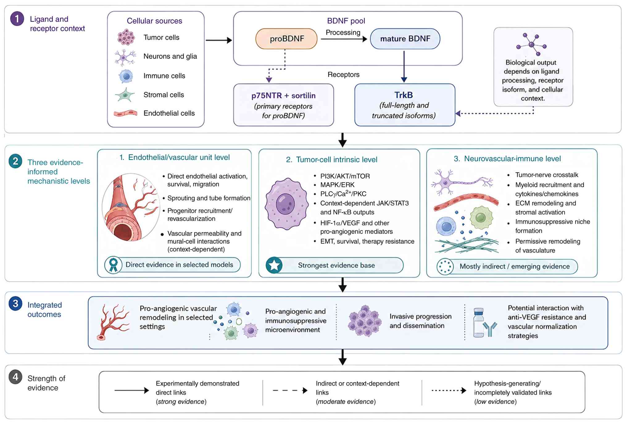 | Figure 1.Evidence-layered framework of
BDNF/TrkB signaling in tumor angiogenesis and neurovascular-immune
interactions. The framework comprises four sections: (1) Ligand and receptor context, showing the
cellular sources of BDNF, processing of proBDNF to mature BDNF and
receptor context; (2) three
evidence-informed mechanistic levels, comprising
endothelial/vascular-unit actions, tumor-cell-intrinsic signaling
and neurovascular-immune interactions; (3) integrated outcomes, including
pro-angiogenic vascular remodeling, a pro-angiogenic and
immunosuppressive microenvironment, invasive progression and
potential interactions with anti-VEGF resistance and vascular
normalization strategies; and (4)
strength of evidence. Solid arrows indicate experimentally
demonstrated direct links, dashed arrows indicate indirect or
context-dependent links and dotted arrows indicate
hypothesis-generating or incompletely validated links. AKT, AKT
serine/threonine kinase; BDNF, brain-derived neurotrophic factor;
Ca2+, calcium ion; ECM, extracellular matrix; EMT,
epithelial-mesenchymal transition; ERK, extracellular
signal-regulated kinase; HIF-1α, hypoxia-inducible factor-1α; JAK,
Janus kinase; MAPK, mitogen-activated protein kinase; mTOR,
mechanistic target of rapamycin; NF-κB, nuclear factor-κB; p75NTR,
p75 neurotrophin receptor; PI3K, phosphoinositide 3-kinase; PKC,
protein kinase C; PLC-γ, phospholipase C-γ; proBDNF, precursor
BDNF; STAT3, signal transducer and activator of transcription 3;
TrkB, tropomyosin receptor kinase B; VEGF, vascular endothelial
growth factor. |
Molecular structure and physiological
functions of BDNF/TrkB
BDNF is a secreted polypeptide that is widely
expressed in both the central and peripheral nervous systems.
Precursor BDNF (proBDNF) is proteolytically processed to the mature
form, and these two species differ markedly in their biological
functions, proBDNF preferentially forms complexes with the p75
neurotrophin receptor (p75NTR) and sortilin, whereas mature BDNF
selectively activates the tyrosine kinase receptor TrkB to support
cell survival and growth (6,8).
Together, they constitute a bidirectional signaling system that is
tightly regulated in space and time. The frequently used
distinction between pro-apoptotic proBDNF and pro-survival mature
BDNF is useful but not absolute. In clear-cell renal cell
carcinoma, proBDNF/p75NTR signaling promoted survival and
migration, illustrating that ligand processing, receptor abundance
and cellular context can reverse the expected response (17). Evidence that proBDNF acts as an
endogenous anti-angiogenic factor in human solid tumors remains
limited; therefore, total BDNF measurements should not be
interpreted as a direct indication of either pro- or
anti-angiogenic activity.
TrkB belongs to the receptor tyrosine kinase family
and exists as full-length and truncated isoforms. Full-length TrkB
contains an intact intracellular kinase domain; upon BDNF binding,
it undergoes autophosphorylation and activates several downstream
pathways, including phosphoinositide 3-kinase (PI3K)/AKT,
mitogen-activated protein kinase (MAPK)/extracellular
signal-regulated kinase (ERK) and PLC-γ, which collectively
regulate cell survival, differentiation and metabolic state.
Although lacking the canonical kinase domain, truncated TrkB
isoforms alter ligand availability by binding neurotrophins and may
also mediate kinase-independent intracellular signaling (8). Consequently, total neurotrophic
receptor tyrosine kinase 2 (NTRK2) mRNA or pan-TrkB
immunohistochemistry may reflect biologically distinct receptor
isoforms and may not necessarily indicate kinase-active TrkB
signaling.
Under physiological conditions, the BDNF/TrkB axis
is essential for neural development and functional maintenance. By
regulating neural stem/progenitor proliferation and
differentiation, axonal guidance, dendritic spine formation and
long-term potentiation, BDNF participates in learning, memory and
stress adaptation. BDNF is also a regulator of neurovascular unit
homeostasis, endothelial cells synthesize BDNF, and TrkB signaling
contributes to endothelial survival, vascular stabilization,
blood-brain barrier function and tissue revascularization (9–14,16,18).
In pathological settings such as ischemia, trauma, chronic
inflammation and persistent pain, BDNF expression and
receptor-mediated signaling are frequently dysregulated. These
physiological actions provide a mechanistic basis for studying BDNF
in cancer, while also indicating potential neurological and
cardiovascular on-target effects if TrkB is inhibited
systemically.
BDNF downstream signaling pathways in
cancer
Upon binding to full-length TrkB, BDNF can activate
multiple canonical and context-dependent signaling pathways that
regulate tumor cell proliferation, invasion, resistance to
apoptosis and immune escape. These pathways intersect with
classical pro-angiogenic signaling through HIF-1α, VEGF and
inflammatory mediators and can thereby affect endothelial and
stromal cells. Because PI3K, ERK, PLC-γ and signal transducer and
activator of transcription 3 (STAT3) are shared by numerous
receptors, pathway activation alone does not prove BDNF/TrkB
dependence; the strongest studies combine ligand or receptor
manipulation with a downstream functional endpoint. Key pathways
and evidence-linked outputs are summarized in Fig. 2.
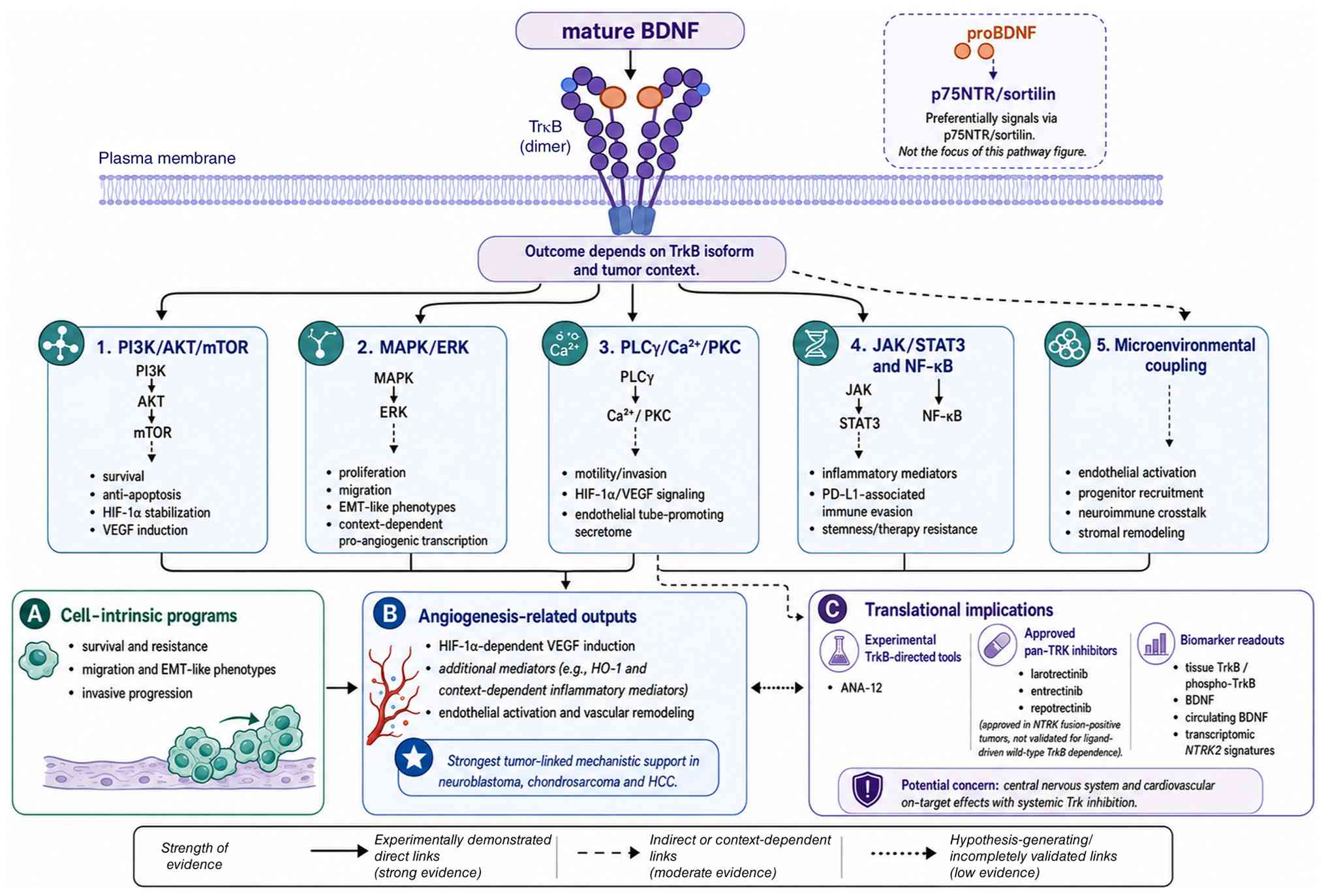 | Figure 2.BDNF/TrkB signaling pathways,
evidence-linked outputs and translational implications. Mature BDNF
binding to full-length TrkB activates PI3K/AKT/mTOR, MAPK/ERK and
PLCγ/Ca2+/PKC pathways, whereas links to JAK/STAT3,
NF-κB and microenvironmental coupling are more context-dependent.
(A) Cell-intrinsic programs include survival and resistance to
apoptosis, migration and EMT-like phenotypes and invasive
progression. (B) Angiogenesis-related outputs include
HIF-1α-dependent VEGF induction, additional mediators, endothelial
activation and vascular remodeling. The strongest tumor-linked
mechanistic evidence is reported in neuroblastoma, chondrosarcoma
and hepatocellular carcinoma. (C) Translational implications
include experimental TrkB-directed tools and approved pan-TRK
inhibitors for NTRK fusion-positive tumors, candidate biomarker
readouts and potential on-target toxicity. Solid, dashed and dotted
connectors denote direct, indirect/context-dependent and
hypothesis-generating relationships, respectively. AKT, AKT
serine/threonine kinase; ANA-12, a small-molecule TrkB antagonist;
BDNF, brain-derived neurotrophic factor; Ca2+, calcium
ion; EMT, epithelial-mesenchymal transition; ERK, extracellular
signal-regulated kinase; HCC, hepatocellular carcinoma; HIF-1α,
hypoxia-inducible factor-1α; HO-1, heme oxygenase-1; JAK, Janus
kinase; MAPK, mitogen-activated protein kinase; mTOR, mechanistic
target of rapamycin; NF-κB, nuclear factor-κB; NTRK, neurotrophic
receptor tyrosine kinase; NTRK2, neurotrophic receptor tyrosine
kinase 2; p75NTR, p75 neurotrophin receptor; PD-L1, programmed
death-ligand 1; PI3K, phosphoinositide 3-kinase; PKC, protein
kinase C; PLC-γ, phospholipase C-γ; proBDNF, precursor BDNF; STAT3,
signal transducer and activator of transcription 3; TrkB,
tropomyosin receptor kinase B; TRK, tropomyosin receptor kinase;
VEGF, vascular endothelial growth factor. |
PI3K/AKT/mechanistic target of
rapamycin (mTOR) pathway
Following BDNF stimulation, TrkB activates the
PI3K/AKT/mTOR pathway to upregulate anti-apoptotic proteins such as
Bcl-2 and inhibit caspase-9, thereby enhancing tumor cell survival,
proliferation and resistance to chemotherapy. In neuroblastoma,
BDNF/TrkB-PI3K signaling protects cells from chemotherapy-induced
apoptosis and increases HIF-1α-dependent VEGF expression (19,20).
This constitutes a defined molecular link between BDNF stimulation
and a pro-angiogenic secretory program, although direct reduction
of the intratumoral vasculature after selective TrkB inhibition has
not been consistently evaluated.
MAPK/ERK pathway
TrkB can engage the RAS-RAF-MEK-ERK cascade to
promote cell-cycle progression, migration and EMT-like phenotypes.
ERK signaling may also cooperate with hypoxia-responsive
transcription and thereby influence VEGF production in a
context-dependent manner. In endothelial cells, the recent
alveolar-capillary study identified MAPK/ERK as a downstream
mediator of TrkB-dependent tube formation and regenerative
angiogenesis (16). In cancer
models, however, ERK activation should be interpreted as supportive
rather than sufficient evidence for a vascular mechanism unless
endothelial or in vivo vessel endpoints are included.
PLC-γ/Ca2+/PKC pathway
BDNF/TrkB can activate PLC-γ, leading to hydrolysis
of phosphatidylinositol 4,5-bisphosphate into diacylglycerol and
inositol trisphosphate, elevation of intracellular Ca2+
and subsequent activation of protein kinase C (PKC). This branch
regulates cytoskeletal reorganization, motility and invasion. In
human chondrosarcoma, a TrkB-PLC-γ-PKCα-HIF-1α cascade increased
VEGF-A expression, endothelial tube formation and in vivo
angiogenic activity (21). BDNF
signaling via the MEK/ERK/mTOR pathway increased VEGF-C-dependent
lymphangiogenesis by suppressing miR-624-3p (22). Together, these studies establish
chondrosarcoma as one of the clearest models linking BDNF/TrkB to
angiogenesis and lymphangiogenesis, while providing a framework for
testing the same pathway in additional tumor types.
Janus kinase (JAK)/STAT3 and other
context-dependent pathways
BDNF/TrkB can interact with cytokine receptor
signaling and stress-responsive transcriptional programs, including
JAK/STAT3 and NF-κB, thereby affecting inflammatory mediator
production, stemness and treatment resistance. In therapy-induced
neuroendocrine prostate cancer, stromal-tumor crosstalk generated a
BDNF-associated immunosuppressive program linked to STAT3
activation and programmed death-ligand 1 (PD-L1) expression
(23). These findings position BDNF
within a neuroimmune program that may influence the vascular
microenvironment through inflammatory and immunosuppressive
outputs. Current evidence more strongly supports STAT3 and NF-κB
than β-catenin as context-dependent downstream programs relevant to
this interface.
Overall, BDNF/TrkB signaling connects malignant-cell
survival, EMT, perineural invasion and immune escape with HIF-1α,
VEGF and inflammatory mediators. Across this pathway spectrum, the
vascular evidence forms a continuum, direct causality is strongest
when pathway perturbation changes endothelial or vessel endpoints,
whereas VEGF or HIF-1α changes identify candidate pro-angiogenic
programs that can be evaluated functionally.
Tumor-specific roles of BDNF/TrkB in solid
malignancy
Across numerous solid tumors, the BDNF/TrkB axis is
upregulated and promotes proliferation, invasion, EMT, resistance
to apoptosis and perineural invasion. Clinically, high BDNF/TrkB
expression is frequently associated with aggressive disease and
unfavorable outcome. In this section, representative tumor types
are considered with emphasis on their tumor-specific phenotypes and
on whether angiogenesis has been evaluated directly or inferred.
Table I summarizes the experimental
models, principal tumor phenotypes, angiogenic endpoints, biomarker
findings, therapeutic relevance and major limitations of the
available evidence.
 | Table I.Tumor-specific evidence matrix for
BDNF/TrkB-related phenotypes, angiogenesis, biomarkers and
therapeutic relevance. |
Table I.
Tumor-specific evidence matrix for
BDNF/TrkB-related phenotypes, angiogenesis, biomarkers and
therapeutic relevance.
| Tumor type | Principal
models/evidence | Tumor
phenotype | Angiogenic
evidence | Biomarker
evidence | Therapeutic
relevance | Main
limitations | Overall evidence
tier |
|---|
| Neuroblastoma | Cell lines;
xenografts; clinical serum study (19,20,24–27) | Survival,
chemoresistance, metastatic traits | HIF-1α-dependent
VEGF induction; limited direct endothelial or in-tumor vessel
endpoints | Tissue TrkB and
serum BDNF associated with aggressive disease/relapse | Experimental TRK
inhibitors enhance therapy in xenografts | Few functional
vascular endpoints; limited prospective biomarker validation | Strong for
progression; moderate for angiogenesis |
| Glioma | Human tissues;
xenografts; neuronal activity/synapse models (28–31) | Neural plasticity
and tumor progression | Direct
TrkB-specific vascular evidence remains limited | Tissue BDNF/TrkB;
heterogeneous meta-analytic evidence | Biological
rationale for TrkB perturbation; no validated TrkB-directed
strategy and VEGF-associated pathways | Highly vascular
disease, but TrkB-specific vascular effects have not been isolated
from neuronal angiogenesis | Strong for neural
progression; preliminary-to-moderate for |
| Breast cancer | Retrospective
tissues; TNBC/HER2 models; brain metastasis; syngeneic TNBC;
perioperative cohort (32–42) | Anoikis resistance,
EMT-like plasticity, stemness/recurrence, endothelial interaction,
brain metastasis, axonogenesis | Cancer-endothelial
effects in vitro; direct primary-tumor vascular evidence
limited | Tissue BDNF/NTRK2,
transcriptomic signatures and exploratory perioperative circulating
BDNF measurements in a small cohort; no validated clinical
assay | TrkB perturbation
remains preclinical in ligand-driven disease; approved pan-TRK
inhibitors are validated for NTRK fusion-positive tumors, not
ligand-driven wild-type TrkB | Subtype
heterogeneity; isoform and antibody issues; small circulating
cohort; recent axonogenesis study did not assess vessels | Strong for
progression and neural-nichebiology; preliminary-to-moderate for
angiogenesis |
| Hepatocellular
carcinoma | Clinical tissues;
cell lines; endothelial co-implantation xenografts; phase Ib
combination study with real-world validation (43–47) | Proliferation,
invasion, stemness | Increased
microvessel density and tumor growth; reduction after TrkB
inhibition in a co-implantation model | Tissue and serum
associations; exploratory serum BDNF association with benefit from
PD-L1 inhibition plus an anti-angiogenic TKI (45–47) | TrkB inhibition
remains preclinical; serum BDNF showed an exploratory
treatment-associated prognostic signal in advanced HCC (47) | Limited independent
mechanistic replication; non-physiological co-implantation design;
small, regimen-specific clinical cohorts and data-derived serum
cut-off | Moderate-to-strong
within limited literature; clinical biomarker evidence
preliminary |
| Chondrosarcoma | Cell lines;
endothelial assays; in vivo angiogenic/lymphangiogenic
models (21,22) | Migration and
invasion | VEGF-A-dependent
angiogenesis and VEGF-C-dependent lymphangiogenesis | Limited clinical
validation | Pathway inhibition
in preclinical studies | Evidence
concentrated in one tumor type and research program | Moderate-to-strong
mechanistic evidence |
| Colorectal
cancer | Clinical tissues;
cell lines; peritoneal dissemination models (48–53) | Migration, anoikis
resistance, EGFR-inhibitor resistance | VEGF/HO-1 changes
reported; direct vascular endpoints sparse | Tissue
associations; no validated circulating assay | Combined
TrkB/autophagy or EGFR strategies are preclinical | Angiogenesis often
inferred; heterogeneous models | Moderate for
progression; preliminary for angiogenesis |
| PDAC/PNI-associated
tumors | Clinical tissues;
neural co-culture; perineural invasion models (54–59) | Perineural
migration, survival, pain and neural remodeling | Direct
microvascular endpoints rarely assessed | Exploratory
tissue/pathway associations with PNI and BDNF-linked pain; no
validated assay | Experimental
neural/TrkB targeting | Desmoplastic
stroma; vascular inference exceeds measured evidence | Moderate for neural
progression; preliminary for angiogenesis |
|
Gynecologic/urogenital tumors | Fallopian tube
precursor models; ovarian/bladder/cervical cell systems; small
clinical cohorts (60–66) | Survival,
attachment, invasion and EMT | Direct angiogenic
evidence limited | Serum multi-protein
association and tissue expression; no validated assay | Preclinical pathway
inhibition | Small cohorts; lack
of vessel endpoints and prospective validation | Moderate for tumor
phenotype; preliminary for angiogenesis |
Neural-origin and central nervous
system tumors
Neuroblastoma
In neuroblastoma, BDNF/TrkB signaling has been
associated with aggressive disease and relapse (19,20,24–27).
Mechanistic studies show that it enhances survival, chemoresistance
and metastatic traits through PI3K/AKT/mTOR and induces
HIF-1α-dependent VEGF expression (19,20).
Pharmacological inhibition of tropomyosin receptor kinase (TRK)
signaling suppresses the growth of TrkB-expressing neuroblastoma
xenografts and can enhance chemotherapy or radiation responses
(24–26). A clinical study also associated
serum BDNF with childhood neuroblastoma relapse (27) but circulating BDNF has not been
prospectively validated as a decision-making biomarker. Together,
these findings establish a functional BDNF/TrkB-HIF-1α-VEGF axis in
neuroblastoma. Future studies incorporating perfusion and
vessel-density endpoints should define the extent to which this
axis controls the tumor vasculature in vivo.
Gliomas
High-grade gliomas, particularly glioblastoma, show
upregulation of mature BDNF and TrkB in tumor tissues, and pooled
clinical analyses support potential biomarker relevance while
emphasizing substantial heterogeneity (28,29).
Neuronal activity and glioma synaptic adaptation promote tumor
growth, and TrkB participates in plasticity-associated signaling
(30,31). In this markedly vascular tumor, TrkB
is positioned to connect neuronal plasticity with VEGF-associated
and microenvironmental signaling. Selective TrkB perturbation
combined with perfusion, vessel-maturity and vascular-normalization
endpoints will be important for defining the magnitude of this
contribution.
Breast cancer
In primary breast cancer, tissue BDNF expression has
been associated with adverse pathological features and clinical
outcome, while a 2024 analysis reported that high NTRK2 protein
expression may be associated with poorer prognosis (32,33).
These studies are mainly retrospective and use different antibodies
and scoring systems; they therefore support prognostic association
rather than a validated clinical assay. Mechanistically, loss of
miR-200c can release a TrkB/neurotrophin 3 autocrine loop that
promotes anoikis resistance and motility in triple-negative breast
cancer (34,35). In recurrent triple-negative breast
cancer, BDNF promoted Krüppel-like factor 4-dependent self-renewal
of TrkB-positive cancer stem cells associated with
post-chemotherapy recurrence (36).
BDNF acts in autocrine and paracrine manners to increase the
migration of triple-negative breast cancer and endothelial cells,
while higher tumor TrkB expression is associated with poorer
disease-free and overall survival (37). In human epidermal growth factor
receptor 2 (HER2)-positive models, TrkB cooperates with HER2 to
support brain-tropic behavior (38). Direct evidence that ligand-driven
BDNF/TrkB signaling mediates endocrine-therapy resistance in
primary estrogen receptor-positive breast cancer remains
insufficient. Together, the available findings support subtype- and
stage-dependent roles in epithelial plasticity, survival and
metastatic competence. Cancer-endothelial co-culture data provide a
vascular link, and in vivo studies of primary tumors should
clarify the magnitude and context of this effect.
In brain-tropic breast cancer models,
estradiol-responsive astrocytes increase BDNF secretion, which
activates TrkB on triple-negative breast cancer cells and promotes
metastatic outgrowth; TrkB blockade reduces the number and size of
brain lesions (39). Transcriptomic
studies have also identified NTRK2-centered prognostic and
immune-associated networks in breast cancer, although bulk RNA data
do not define the relevant TrkB isoform or cellular source
(40). Most recently,
macrophage-derived BDNF was shown to be necessary and sufficient
for tumor axonogenesis and to support tumor growth in syngeneic
triple-negative breast cancer models (41). This study provides strong causal
evidence for a BDNF-dependent tumor-immune-nerve circuit, but its
consequences for tumor angiogenesis have not yet been directly
measured. A small perioperative study of 18 patients found that
serum BDNF decreased 24 h after breast cancer surgery and was
associated with platelet count and C-reactive protein, whereas
plasma BDNF did not change markedly (42).
Highly vascularized abdominal
tumors
Hepatocellular carcinoma
In hepatocellular carcinoma, BDNF/TrkB activation
promotes proliferation, migration, resistance to apoptosis and
cancer stem cell maintenance (43).
Co-implantation of hepatocellular carcinoma cells with
BDNF-overexpressing endothelial cells increased intratumoral
microvessel density and tumor burden, whereas TrkB inhibition
reduced neovascularization and tumor growth (44). Serum BDNF is notably influenced by
platelet count and has not been established as a stand-alone
hepatocellular carcinoma biomarker (45). Increased BDNF and TrkB expression
has also been associated with multifocal disease, poor
differentiation and metastasis (46). More recently, a phase Ib study of
anlotinib plus the PD-L1 inhibitor TQB2450, supported by a separate
real-world validation cohort, associated higher baseline serum BDNF
with clinical benefit and longer progression-free and overall
survival (47). This
treatment-associated biomarker signal remains exploratory because
it was derived from small cohorts, a specific combination regimen
and a data-derived cut-off. These findings provide direct in
vivo evidence for BDNF-supported tumor vascularization in the
co-implantation model and identify a clinically relevant, but
unvalidated, circulating biomarker signal in advanced disease.
Validation in orthotopic and immunocompetent models and prospective
treatment cohorts would further define the biological and
translational generalizability of these observations.
Colorectal cancer
In colorectal cancer, elevated BDNF/TrkB expression
is associated with local invasion, liver metastasis, peritoneal
dissemination and unfavorable prognosis (48). BDNF/TrkB signaling contributes to
anoikis resistance, protection from epidermal growth factor
receptor inhibition and autophagy-associated treatment resistance
(48–50). BDNF also enhances the migration of
HCT116 and SW480 cells through heme oxygenase-1 (HO-1), and pathway
inhibition partially reverses this effect (51). The reported HO-1, VEGF and matrix
metalloproteinase responses suggest a pro-angiogenic secretory
component within the broader BDNF/TrkB program. Endothelial,
perfusion and in vivo vascular endpoints are now needed to
define its functional contribution. Recent reviews of colorectal
cancer neuroscience further support interactions among nerves,
immune cells and vascular remodeling, placing BDNF within a wider
neurovascular network rather than as an isolated mediator (52,53).
Other gastrointestinal tumors
In other digestive malignancies, current data place
BDNF/TrkB primarily within tumor-survival and tumor-nerve-immune
programs. Reviews of digestive system cancer neuroscience situate
neurotrophin signaling within broader neural and immune networks
(53), while an experimental
pancreatic cancer study supports tumor-promoting BDNF/TrkB activity
(54). These observations provide a
basis for testing whether such programs converge on endothelial
remodeling in tumor-specific models, rather than implying a uniform
angiogenic mechanism across gastrointestinal cancers.
Perineural-invasive and stroma-rich
tumors
Pancreatic ductal adenocarcinoma
Pancreatic ductal adenocarcinoma is characterized by
pronounced perineural invasion and a desmoplastic stroma (55). BDNF and TrkB are upregulated in
pancreatic cancer cells and peripancreatic nerves, and experimental
studies indicate that the pathway enhances survival, migration and
neural remodeling (54,55). A β2-adrenergic-neurotrophin
feed-forward loop also promotes pancreatic cancer growth and nerve
density (56). In this stroma-rich
tumor, the vascular relevance of BDNF/TrkB is most plausibly linked
to its effects on perineural invasion, stromal remodeling and
neurogenic signaling. Orthotopic studies that integrate these
processes with perfusion and vessel-function measurements should
define how the pathway shapes the pancreatic tumor vasculature.
Other perineural invasion-associated
tumors
In oral squamous cell carcinoma and prostate cancer,
BDNF/TrkB promotes tumor-cell migration, anoikis resistance or
neural interactions. In an in vitro model, TrkB inhibition
reduces reciprocal Schwann cell-cancer cell migration but increases
Schwann cell-associated cancer cell dispersion, and tumor-derived
BDNF contributes to oral cancer pain through peripheral TrkB
activation (57,58). In prostate cancer, BDNF/TrkB
promotes EMT and anoikis resistance (59). These findings support perineural
invasion and neural niche adaptation and provide a rationale for
examining how neurogenic inflammation and stromal remodeling
influence local vascular behavior.
Gynecologic and urogenital tumors
Ovarian cancer
Studies on the origin of high-grade serous ovarian
carcinoma highlight the fallopian tube fimbrial epithelium as a key
site of initiation. Follicular fluid-derived BDNF activates TrkB on
fallopian tube epithelial cells, promoting survival, migration and
attachment of p53-mutant precursors (60,61).
In established ovarian cancer cells, BDNF signals through
TrkB-PLCγ1 to promote proliferation, migration and resistance to
apoptosis (62). Elevated serum
BDNF has also been included in a multi-protein model associated
with recurrence (63). These data
support dissemination and early colonization, processes that
subsequently require vascular support. Direct assessment of
BDNF-dependent vascularization in ovarian implants should clarify
whether the pathway also contributes to this later phase.
Other genitourinary tumors
In bladder transitional cell carcinoma, BDNF/TrkB
expression is higher in tumor tissue than in non-malignant
urothelium; exogenous BDNF promotes proliferation and migration,
whereas TrkB-Fc neutralization increases apoptosis (64,65).
In cervical cancer, BDNF/TrkB signaling promotes EMT, migration and
invasion, and TrkB knockdown reverses invasive phenotypes (66). These tumor-promoting effects may
create a permissive context for vascular remodeling, and dedicated
microvascular studies should define the extent of that
contribution.
Summary and link to angiogenesis-focused
mechanisms
The tumor entities outlined above can be grouped
according to the nature of their evidence. Neuroblastoma,
chondrosarcoma and hepatocellular carcinoma provide the clearest
mechanistic or in vivo links between BDNF/TrkB and
VEGF-family signaling, endothelial responses or microvessel
formation. In pancreatic, perineural-invasive, ovarian and other
genitourinary tumors, BDNF/TrkB is more strongly linked to neural
invasion, stromal remodeling, dissemination or survival of
metastatic cells. These distinctions are summarized in Table I and guide the mechanistic synthesis
below.
BDNF in tumor angiogenesis and
neurovascular-immune interactions
Accumulating evidence indicates that BDNF/TrkB
signaling can contribute to tumor angiogenesis through multiple,
partially overlapping levels. At the endothelial and hematopoietic
progenitor level, BDNF has direct vascular activity. At the
tumor-cell level, BDNF/TrkB can amplify VEGF and other
pro-angiogenic mediators. Finally, in selected neurotropic or
stroma-rich tumors, BDNF shapes neurovascular and immune
interactions that may influence vascular remodeling. These
complementary mechanisms extend the angiogenic framework from
direct endothelial effects to tumor-, neural- and immune-mediated
vascular remodeling.
Endothelial and hematopoietic
progenitor level
Multiple developmental, ischemia and
endothelial-cell models have shown that BDNF, acting through TrkB,
promotes endothelial survival, migration and tube formation
(11–14,16).
In murine ischemia, BDNF recruited TrkB-positive endothelial cells
and mobilized hematopoietic progenitors, increasing capillary
formation and reperfusion (12).
BDNF also induced endothelial proliferation and migration and
increased angiogenin secretion in human endothelial cells (13,14).
The 2025 alveolar-capillary study independently demonstrated that
TrkB activation enhanced endothelial tube formation and capillary
recovery, whereas TrkB inhibition impaired these endpoints
(16). Together, these studies
establish BDNF/TrkB as a functional endothelial and reparative
vascular pathway that may be co-opted in selected tumors. The
relative contribution of BDNF/TrkB alongside VEGF is expected to
depend on the oncogenic, matrix, pericyte and immune context of the
tumor vasculature.
Tumor-cell level: HIF-1α/VEGF and
related mediators
Beyond acting on endothelial and progenitor cells,
BDNF can increase pro-angiogenic mediators within tumor cells. In
neuroblastoma, BDNF-induced TrkB activation upregulates HIF-1α and
VEGF (19). In chondrosarcoma,
BDNF-TrkB signaling activates PLC-γ-PKCα-HIF-1α, increases VEGF-A,
stimulates endothelial tube formation and promotes angiogenic
activity in vivo (21). BDNF
signaling via the MEK/ERK/mTOR pathway increases VEGF-C-dependent
lymphangiogenesis (22). In
hepatocellular carcinoma, endothelial BDNF increases intratumoral
microvessel density and tumor growth (44). These tumor systems provide some of
the strongest mechanistic evidence for BDNF/TrkB-dependent vascular
effects. In colorectal and other cancers, HO-1, VEGF and
inflammatory changes identify candidate angiogenic programs that
can now be tested using functional endothelial and in vivo
vascular endpoints.
Neurovascular-immune level
Tumor-associated nerve fibers and their
neurotransmitters, neuropeptides and neurotrophic factors influence
endothelial permeability, pericyte coverage, cytokine production
and immune-cell positioning (3–5). In
pancreatic and oral cancers, BDNF/TrkB contributes to perineural
migration, neural remodeling and pain (54–58).
In glioma, neural activity and tumor synapses provide
growth-promoting signals (30,31).
The 2026 triple-negative breast cancer study demonstrated that
macrophage-derived BDNF promotes tumor axonogenesis and growth
(41), adding a BDNF-specific
immune-to-nerve mechanism to the broader neurovascular-immune
framework. Whether this macrophage-BDNF axis also alters vascular
density, perfusion or immune trafficking remains to be
determined.
Relationship to other neuroangiogenic
mediators
BDNF operates within a larger neuroangiogenic
network that includes VEGF-A/VEGFR2, angiopoietin/TIE2, fibroblast
growth factors, CXCL12/CXCR4, nerve growth factor, adrenergic and
cholinergic signals, neuropeptides and inflammatory cytokines
(1,3–5). VEGF
has substantially stronger clinical and mechanistic validation as a
driver of tumor angiogenesis, whereas BDNF is best supported as a
context-dependent modifier that can act directly on endothelial
cells or indirectly through tumor, neural and immune compartments.
Comparative experiments using matched perturbations and vascular
endpoints are required to determine whether BDNF is redundant with,
upstream of or independent of these mediators in a given tumor.
Potential relationship with vascular
normalization and anti-VEGF therapy
Anti-VEGF therapies can transiently suppress
angiogenesis and induce vascular normalization, thereby improving
tumor perfusion and the delivery of drugs and immune effector
cells. With prolonged treatment, adaptive resistance, renewed
vascular abnormalities and increased invasiveness may develop
(2). BDNF/TrkB directly supports
endothelial survival and can increase VEGF-family signaling in
selected tumors (11,16,19,21,22,44).
These findings provide a rationale for testing BDNF/TrkB as a
context-dependent contributor to adaptive vascular resistance. The
importance of BDNF/TrkB is likely to vary by tumor type and should
be evaluated through perfusion, permeability, pericyte coverage,
hypoxia and immune-cell trafficking rather than tumor volume
alone.
TRK-directed drugs have entered oncology practice,
but their validated indication is molecularly distinct from most
BDNF/TrkB studies. Larotrectinib, entrectinib and repotrectinib
inhibit TRK family kinases and are used in NTRK fusion-positive
cancers; NTRK fusions produce constitutive signaling and should not
be equated with ligand-driven wild-type TrkB expression. At the
time of the literature search, no BDNF/TrkB-selected clinical trial
combining a TRK inhibitor with an anti-VEGF agent was identified.
No validated BDNF or TrkB assay currently selects patients for such
combination therapy. Systemic TRK inhibition may cause central
nervous system effects, including dizziness, cognitive or mood
changes, and may affect cardiovascular homeostasis; these risks
support tumor-selective, transient or source-specific strategies
(67–72).
BDNF/TrkB as a biomarker in human
cancer
BDNF/TrkB has been investigated as a diagnostic,
prognostic and predictive biomarker in human cancer, but validation
varies across analytes and tumor types. Tissue expression,
circulating BDNF and transcriptomic NTRK2 signatures are not
interchangeable because they reflect different cellular sources,
receptor isoforms and pre-analytical variables. Table II summarizes current biomarker
approaches and limitations.
| Table II.Biomarker-development framework for
BDNF/TrkB in human cancer. |
Table II.
Biomarker-development framework for
BDNF/TrkB in human cancer.
| Biomarker
class | Potential
application | Current
evidence | Key analytical
issues | Required
validation | Current status |
|---|
| Tissue BDNF | Prognostic
association; spatial source assessment | Associations
reported in several tumors (28,29,32,33,46,48,64) | Antibody
specificity, cellular source, scoring thresholds | Large multi-center
cohorts; standardized IHC |
Investigational |
| Total TrkB IHC | Prognostic
association | Associations with
grade, invasion or survival (28,29,32,33,46,48,64) | Full-length and
truncated isoforms may be combined | Isoform-specific
antibodies and reporting |
Investigational |
| Phospho-TrkB | Functional pathway
activation | Limited
tumor-cohort data; functional rationale from receptor biology
(7,8) | Pre-analytical
phosphoprotein loss; antibody validation | Controlled fixation
and analytical validation | Experimental |
| Serum/plasma
BDNF | Non-invasive
monitoring, prognosis or exploratory treatment-associated
stratification | Small studies in
neuroblastoma, HCC, ovarian and breast cancer, including an
exploratory HCC immunotherapy/anti-angiogenic treatment cohort and
a small perioperative breast cancer cohort (27,42,45,47,63,73,74) | Platelet release;
serum/plasma non-equivalence; processing and clinical
confounders | Standardized
collection, platelet adjustment, prospective longitudinal and
treatment-interaction cohorts | Not clinically
validated; treatment-associated HCC signal is exploratory |
| Bulk NTRK2
transcript |
Prognostic/immune-associated
signatures | TCGA-derived and
other transcriptomic analyses, including breast cancer (40) | Isoforms, protein
activation and cell of origin unresolved | Protein/isoform
confirmation and external validation |
Hypothesis-generating |
| Single-cell/spatial
BDNF-NTRK2 | Cell-source and
niche mapping | Emerging
opportunity; limited direct validation (4,5,41) | RNA dropout;
spatial resolution; protein confirmation | Integrated spatial
transcriptomic/proteomic studies | Research
priority |
| Composite
neurovascular-immune panel | Prognosis or
treatment selection | Conceptual
rationale from neurovascular-immune frameworks (3–5,40) | Assay complexity
and model overfitting | Predefined panel,
prospective treatment interaction | Future
direction |
| NTRK fusion
assay | Selection for
pan-TRK inhibitors | Clinically
validated across fusion-positive solid tumors (68–72) | Distinct from
ligand-driven wild-type TrkB | Approved molecular
testing workflows | Validated
predictive biomarker for fusion-positive disease |
Tissue BDNF and TrkB
Tissue immunohistochemistry provides spatial
information, and BDNF/TrkB expression has been associated with
tumor grade, invasion, metastasis or survival in glioma, breast,
hepatocellular, colorectal and bladder cancers (28,29,32,33,46,48,64).
Most studies, however, are retrospective, use small or
single-center cohorts and apply non-standardized scoring
thresholds. Pan-TrkB antibodies may not distinguish full-length
from truncated TrkB isoforms. Phospho-TrkB may better reflect
pathway activation but is sensitive to tissue handling and requires
validated controls. Future studies should report antibody clone,
epitope, isoform coverage, scoring method and interobserver
reproducibility.
Circulating BDNF
Serum and plasma BDNF are not interchangeable.
Platelets store and release substantial BDNF, and concentrations
vary with clotting time, platelet count, centrifugation, storage,
exercise, age, inflammation, neuropsychiatric status and anticancer
treatment (45,73,74).
In a small perioperative breast cancer cohort of 18 patients, serum
BDNF decreased 24 h after surgery and associated with platelet
count and C-reactive protein, whereas plasma BDNF did not change
markedly (42). Small studies have
associated circulating BDNF with relapse or clinicopathological
features in neuroblastoma, hepatocellular and ovarian cancer
(27,45,63). A
2025 phase Ib hepatocellular carcinoma study, supported by a
separate real-world cohort, associated higher baseline serum BDNF
with benefit from PD-L1 blockade plus an anti-angiogenic tyrosine
kinase inhibitor (47). This
treatment-associated signal remains exploratory and is not a
validated patient-selection assay. Accordingly, studies should
standardize pre-analytical procedures, measure platelet counts
concurrently and include longitudinal sampling.
Transcriptomic, single-cell and
spatial biomarkers
Bulk NTRK2 analyses can identify prognostic or
immune-associated patterns in large datasets (40), but bulk RNA does not resolve
receptor isoforms, pathway activation or cellular source.
Single-cell RNA sequencing and spatial transcriptomics could
determine whether BDNF and NTRK2 are expressed by malignant cells,
endothelial cells, neurons, Schwann cells, macrophages or
fibroblasts and whether these populations occupy vascular or
perineural niches. Multi-marker panels combining ligand and
isoform-resolved receptor measurements with VEGF-A/HIF-1α,
endothelial and pericyte markers, neural density, macrophage states
and clinical subtype may be more informative than BDNF alone.
Prognostic and predictive
applications
Prognostic association is distinct from predictive
utility. No validated assay currently selects patients for TrkB
inhibition, anti-VEGF plus TrkB inhibition or immune-checkpoint
blockade based on ligand-driven BDNF/TrkB activity. By contrast,
NTRK fusion testing is a validated predictive biomarker for pan-TRK
inhibitors because it identifies a constitutively active oncogenic
fusion. A predictive biomarker for ligand-driven wild-type
BDNF/TrkB will require evidence of pathway activation, analytical
validation, a prespecified treatment interaction and prospective
confirmation.
Discussion and perspectives
Across preclinical and clinical studies, BDNF/TrkB
signaling emerges as a pleiotropic regulator that links tumor-cell
behavior to angiogenesis and microenvironmental remodeling. The
evidence is strongest in a limited set of models, such as
neuroblastoma, hepatocellular carcinoma and chondrosarcoma, where
BDNF/TrkB activation has been mechanistically coupled to
HIF-1α/VEGF upregulation, endothelial responses, microvessel
formation or lymphangiogenesis. Work on neurotropic tumors and
perineural invasion also provides converging support for BDNF as a
mediator of tumor-nerve interactions: Recent studies have provided
independent causal evidence for TrkB-dependent regenerative
angiogenesis and macrophage-derived BDNF-driven tumor axonogenesis,
respectively (16,41).
The evidence base remains heterogeneous. Direct
endothelial and tumor-vascular effects are established in selected
systems, while numerous additional studies rely on in vitro
models or subcutaneous xenografts without longitudinal assessment
of vascular function. In several solid tumors, BDNF/TrkB is most
strongly linked to proliferation, EMT, anoikis resistance and
treatment resistance, providing plausible routes through which the
pathway may subsequently influence vascular remodeling. Clinical
data are largely correlative, and high BDNF/TrkB expression is
rarely separated from confounding oncogenic pathways. Key
conclusions regarding angiogenesis still rely on a relatively small
number of foundational studies, with limited independent
replication, variable assessment of TrkB isoforms, heterogeneous
antibodies and potential publication bias. These considerations
define the settings in which the strongest vascular findings should
now be validated and extended.
The conceptual framework in the present review
complements, rather than replaces, the classical VEGF-centered view
of tumor angiogenesis. At the endothelial and hematopoietic
progenitor level, BDNF/TrkB can support neovessel formation and
vascular repair. Within tumor cells, it interfaces with PI3K/AKT,
ERK, PLCγ and context-dependent JAK/STAT3 pathways, with effects
including increased HIF-1α/VEGF signaling and immunosuppressive
phenotypes in selected models (19,21,23).
In neurotropic and stroma-rich tumors, BDNF contributes to
tumor-nerve-immune circuits. The three-level organization is
consistent with recent neurovascular-immune and peripheral
nerve-cancer frameworks (4,5), while specifically distinguishing
BDNF/TrkB evidence according to the cellular compartment and the
directness of the vascular endpoint.
These features position BDNF/TrkB at the
intersection of angiogenesis, neurogenesis and immune escape.
Attenuating BDNF/TrkB signaling could, in principle, reduce
selected pro-angiogenic stimuli and weaken neural support for tumor
progression. Conversely, BDNF has important physiological roles in
the nervous and cardiovascular systems, including vascular repair
and neuroprotection. Approved pan-TRK inhibitors demonstrate that
TRK kinases are clinically druggable, but their success in NTRK
fusion-positive cancers does not validate BDNF concentration or
TrkB immunohistochemistry as patient-selection biomarkers in
fusion-negative tumors. Clinical translation will require tumor-
and source-specific pathway dependence, a validated biomarker and a
therapeutic window that avoids unacceptable neurological and
cardiovascular toxicity.
Several research priorities emerge from these
limitations. Future preclinical studies should incorporate
quantitative and functional microvascular readouts, such as vessel
density and maturity, perfusion, permeability, hypoxia and
immune-cell trafficking, when evaluating BDNF/TrkB in tumor models.
Integrative models of the tumor neurovascular-immune unit are
needed to dissect how nerves, endothelial cells, myeloid
populations and stromal fibroblasts interact during dissemination,
early colonization and relapse. Cell-type-specific and inducible
approaches should distinguish BDNF derived from tumor cells,
endothelial cells, macrophages, neurons and Schwann cells, while
full-length TrkB and truncated isoforms should be measured
separately. From a translational perspective, it will be important
to clarify whether circulating or tissue BDNF/TrkB signatures can
serve as prognostic or predictive biomarkers in patients receiving
anti-angiogenic or immunomodulatory therapy. Early-generation TrkB
antagonists, such as ANA-12, together with ligand traps, genetic
silencing and pan-TRK inhibitors, provide tools for well-designed
preclinical studies. Future tumor-specific investigations should
account for molecular subtype, disease stage, metastatic site and
treatment context. Biomarker studies should standardize serum or
plasma processing, control for platelet count and use single-cell
or spatial approaches to define the cellular source of BDNF/TrkB
activity. Combination studies with anti-VEGF or immune-checkpoint
therapy should proceed only in models demonstrating a measurable
BDNF-dependent vascular or immune phenotype.
Overall, BDNF/TrkB is best regarded as a
context-dependent regulator of malignant-cell survival, neural and
immune interactions and, in selected models, angiogenesis or
lymphangiogenesis. The available evidence supports continued
investigation but not classification as a universal VEGF-parallel
angiogenic pathway or immediate use as a stand-alone clinical
biomarker. The translational value of BDNF/TrkB will depend on
identifying the relevant ligand source, receptor isoform, tumor
subtype and disease stage and on demonstrating that pathway
inhibition changes a functional vascular or therapeutic
endpoint.
Acknowledgements
Not applicable.
Funding
The present review was funded by the Zhongguancun Precision
Medicine Foundation (grant no. 2024HZ29).
Availability of data and materials
Not applicable.
Authors' contributions
GZ and DZ conceived the review topic and drafted the
manuscript. GZ, DZ, HW, SY, MN, SN and CW contributed to the
literature search, literature screening, critical appraisal and
evidence synthesis. HW, SY, MN, SN and CW also participated in
secondary language review, formatting verification and manuscript
revision. JL and ML critically revised the manuscript for important
intellectual content and supervised the project. All authors read
and approved the final manuscript. Data authentication is not
applicable.
Ethics approval and consent to
participate
Not applicable.
Patient consent for publication
Not applicable.
Competing interests
The authors declare that they have no competing
interests.
Glossary
Abbreviations
Abbreviations:
|
AKT
|
AKT serine/threonine kinase
|
|
BDNF
|
brain-derived neurotrophic factor
|
|
Ca2+
|
calcium ion
|
|
EMT
|
epithelial-mesenchymal transition
|
|
ERK
|
extracellular signal-regulated
kinase
|
|
HER2
|
human epidermal growth factor receptor
2
|
|
HIF-1α
|
hypoxia-inducible factor-1α
|
|
HO-1
|
heme oxygenase-1
|
|
JAK
|
Janus kinase
|
|
MAPK
|
mitogen-activated protein kinase
|
|
mTOR
|
mechanistic target of rapamycin
|
|
NF-κB
|
nuclear factor-κB
|
|
NTRK
|
neurotrophic receptor tyrosine
kinase
|
|
NTRK2
|
neurotrophic receptor tyrosine kinase
2
|
|
p75NTR
|
p75 neurotrophin receptor
|
|
PD-L1
|
programmed death-ligand 1
|
|
PI3K
|
phosphoinositide 3-kinase
|
|
PKC
|
protein kinase C
|
|
PLC-γ
|
phospholipase C-γ
|
|
proBDNF
|
precursor BDNF
|
|
STAT3
|
signal transducer and activator of
transcription 3
|
|
TrkB
|
tropomyosin receptor kinase B
|
|
TRK
|
tropomyosin receptor kinase
|
|
VEGF
|
vascular endothelial growth
factor
|
|
VEGFR2
|
VEGF receptor 2
|
References
|
1
|
Ghalehbandi S, Yuzugulen J, Pranjol MZI
and Pourgholami MH: The role of VEGF in cancer-induced angiogenesis
and research progress of drugs targeting VEGF. Eur J Pharmacol.
949:1755862023. View Article : Google Scholar : PubMed/NCBI
|
|
2
|
Liu Z, Zhao Q, Zheng Z, Liu S, Meng L,
Dong L and Jiang X: Vascular normalization in immunotherapy: A
promising mechanism combined with radiotherapy. Biomed
Pharmacother. 139:1116072021. View Article : Google Scholar : PubMed/NCBI
|
|
3
|
Shalabi S, Belayachi A and Larrivée B:
Involvement of neuronal factors in tumor angiogenesis and the
shaping of the cancer microenvironment. Front Immunol.
15:12846292024. View Article : Google Scholar : PubMed/NCBI
|
|
4
|
Li D, Zhao Q, Liu L and Zeng F: The
neuro-vascular-immune triad: The interactive network in the tumor
microenvironment. Cell Commun Signal. 24:2852026. View Article : Google Scholar : PubMed/NCBI
|
|
5
|
Shao F, Wang J, Li A, Wu R, Chen J, Tuo Z,
Wusiman D, Yoo KH, Wei W, Wang Z, et al: Peripheral nerve-cancer
interactions in the tumor microenvironment: A three-dimensional
framework integrating mechanisms, modulators, and therapeutic
strategies. Research (Wash D C). 9:12212026.PubMed/NCBI
|
|
6
|
Balaratnasingam S and Janca A:
Brain-derived neurotrophic factor: A novel neurotrophin involved in
psychiatric and neurological disorders. Pharmacol Ther.
134:116–124. 2012. View Article : Google Scholar : PubMed/NCBI
|
|
7
|
Enkavi G, Girych M, Moliner R, Vattulainen
I and Castrén E: TrkB transmembrane domain: Bridging structural
understanding with therapeutic strategy. Trends Biochem Sci.
49:445–456. 2024. View Article : Google Scholar : PubMed/NCBI
|
|
8
|
Huang EJ and Reichardt LF: Trk receptors:
Roles in neuronal signal transduction. Annu Rev Biochem.
72:609–642. 2003. View Article : Google Scholar : PubMed/NCBI
|
|
9
|
Kermani P and Hempstead B: BDNF actions in
the cardiovascular system: Roles in development, adulthood and
response to injury. Front Physiol. 10:4552019. View Article : Google Scholar : PubMed/NCBI
|
|
10
|
Nakahashi T, Fujimura H, Altar CA, Li J,
Kambayashi J, Tandon NN and Sun B: Vascular endothelial cells
synthesize and secrete brain-derived neurotrophic factor. FEBS
Lett. 470:113–117. 2000. View Article : Google Scholar : PubMed/NCBI
|
|
11
|
Donovan MJ, Lin MI, Wiegn P, Ringstedt T,
Kraemer R, Hahn R, Wang S, Ibañez CF, Rafii S and Hempstead BL:
Brain derived neurotrophic factor is an endothelial cell survival
factor required for intramyocardial vessel stabilization.
Development. 127:4531–4540. 2000. View Article : Google Scholar : PubMed/NCBI
|
|
12
|
Kermani P, Rafii D, Jin DK, Whitlock P,
Schaffer W, Chiang A, Vincent L, Friedrich M, Shido K, Hackett NR,
et al: Neurotrophins promote revascularization by local recruitment
of TrkB+ endothelial cells and systemic mobilization of
hematopoietic progenitors. J Clin Invest. 115:653–663. 2005.
View Article : Google Scholar : PubMed/NCBI
|
|
13
|
Sun C, Hu Y, Chu Z, Huang J and Zhang L:
The effect of brain-derived neurotrophic factor on angiogenesis. J
Huazhong Univ Sci Technolog Med Sci. 29:139–143. 2009. View Article : Google Scholar : PubMed/NCBI
|
|
14
|
Mori A, Nishioka Y, Yamada M, Nishibata Y,
Masuda S, Tomaru U, Honma N, Moriyama T and Ishizu A: Brain-derived
neurotrophic factor induces angiogenin secretion and nuclear
translocation in human umbilical vein endothelial cells. Pathol Res
Pract. 214:521–526. 2018. View Article : Google Scholar : PubMed/NCBI
|
|
15
|
Meng L, Liu B, Ji R, Jiang X, Yan X and
Xin Y: Targeting the BDNF/TrkB pathway for the treatment of tumors
(Review). Oncol Lett. 17:2031–2039. 2019.PubMed/NCBI
|
|
16
|
Ma Q, Liu H, Liu M, Wang Y, Lai Y, Zhao Y,
Li M, Liu Y, Ma C, Lai J, et al: TrkB signaling promotes alveolar
capillary angiogenesis following perinatal hyperoxic damage. Am J
Physiol Lung Cell Mol Physiol. 328:L617–L630. 2025. View Article : Google Scholar : PubMed/NCBI
|
|
17
|
De la Cruz-Morcillo MA, Berger J,
Sánchez-Prieto R, Saada S, Naves T, Guillaudeau A, Perraud A,
Sindou P, Lacroix A, Descazeaud A, et al: p75 neurotrophin receptor
and pro-BDNF promote cell survival and migration in clear cell
renal cell carcinoma. Oncotarget. 7:34480–34497. 2016. View Article : Google Scholar : PubMed/NCBI
|
|
18
|
Kaplan L, Chow BW and Gu C: Neuronal
regulation of the blood-brain barrier and neurovascular coupling.
Nat Rev Neurosci. 21:416–432. 2020. View Article : Google Scholar : PubMed/NCBI
|
|
19
|
Nakamura K, Martin KC, Jackson JK, Beppu
K, Woo CW and Thiele CJ: Brain-derived neurotrophic factor
activation of TrkB induces vascular endothelial growth factor
expression via hypoxia-inducible factor-1alpha in neuroblastoma
cells. Cancer Res. 66:4249–4255. 2006. View Article : Google Scholar : PubMed/NCBI
|
|
20
|
Jaboin J, Kim CJ, Kaplan DR and Thiele CJ:
Brain-derived neurotrophic factor activation of TrkB protects
neuroblastoma cells from chemotherapy-induced apoptosis via the
phosphatidylinositol 3-kinase pathway. Cancer Res. 62:6756–6763.
2002.PubMed/NCBI
|
|
21
|
Lin CY, Hung SY, Chen HT, Tsou HK, Fong
YC, Wang SW and Tang CH: Brain-derived neurotrophic factor
increases vascular endothelial growth factor expression and
enhances angiogenesis in human chondrosarcoma cells. Biochem
Pharmacol. 91:522–533. 2014. View Article : Google Scholar : PubMed/NCBI
|
|
22
|
Lin CY, Wang SW, Chen YL, Chou WY, Lin TY,
Chen WC, Yang CY, Liu SC, Hsieh CC, Fong YC, et al: Brain-derived
neurotrophic factor promotes VEGF-C-dependent lymphangiogenesis by
suppressing miR-624-3p in human chondrosarcoma cells. Cell Death
Dis. 8:e29642017. View Article : Google Scholar : PubMed/NCBI
|
|
23
|
Liu YN, Chen WY, Liu MK, Yeh HL, Chen WH,
Jiang KC, Li HR, Chen ZQ, Wang WH, Abou-Kheir W and Wen YC:
Immunosuppressive role of BDNF in therapy-induced neuroendocrine
prostate cancer. Mol Oncol. 18:1665–1686. 2024. View Article : Google Scholar : PubMed/NCBI
|
|
24
|
Iyer R, Varela CR, Minturn JE, Ho R,
Simpson AM, Light JE, Evans AE, Zhao H, Thress K, Brown JL and
Brodeur GM: AZ64 inhibits TrkB and enhances the efficacy of
chemotherapy and local radiation in neuroblastoma xenografts.
Cancer Chemother Pharmacol. 70:477–486. 2012. View Article : Google Scholar : PubMed/NCBI
|
|
25
|
Iyer R, Wehrmann L, Golden RL, Naraparaju
K, Croucher JL, MacFarland SP, Guan P, Kolla V, Wei G, Cam N, et
al: Entrectinib is a potent inhibitor of Trk-driven neuroblastomas
in a xenograft mouse model. Cancer Lett. 372:179–186. 2016.
View Article : Google Scholar : PubMed/NCBI
|
|
26
|
Croucher JL, Iyer R, Li N, Molteni V,
Loren J, Gordon WP, Tuntland T, Liu B and Brodeur GM: TrkB
inhibition by GNF-4256 slows growth and enhances chemotherapeutic
efficacy in neuroblastoma xenografts. Cancer Chemother Pharmacol.
75:131–141. 2015. View Article : Google Scholar : PubMed/NCBI
|
|
27
|
Xiong X, Zeng M, Peng X, Feng C, Li C,
Weng W and Li Y: Serum brain-derived neurotrophic factor (BDNF) as
predictors of childhood neuroblastoma relapse. BMC Cancer.
23:6702023. View Article : Google Scholar : PubMed/NCBI
|
|
28
|
Xiong J, Zhou L, Lim Y, Yang M, Zhu YH, Li
ZW, Fu DL and Zhou XF: Mature brain-derived neurotrophic factor and
its receptor TrkB are upregulated in human glioma tissues. Oncol
Lett. 10:223–227. 2015. View Article : Google Scholar : PubMed/NCBI
|
|
29
|
Hasani F, Masrour M, Khamaki S, Jazi K,
Ghoodjani E and Teixeira AL: Brain-derived neurotrophic factor
(BDNF) as a potential biomarker in brain glioma: A systematic
review and meta-analysis. Brain Behav. 15:e702662025. View Article : Google Scholar : PubMed/NCBI
|
|
30
|
Venkatesh HS, Johung TB, Caretti V, Noll
A, Tang Y, Nagaraja S, Gibson EM, Mount CW, Polepalli J, Mitra SS,
et al: Neuronal activity promotes glioma growth through
neuroligin-3 secretion. Cell. 161:803–816. 2015. View Article : Google Scholar : PubMed/NCBI
|
|
31
|
Taylor KR, Barron T, Hui A, Spitzer A,
Yalçin B, Ivec AE, Geraghty AC, Hartmann GG, Arzt M, Gillespie SM,
et al: Glioma synapses recruit mechanisms of adaptive plasticity.
Nature. 623:366–374. 2023. View Article : Google Scholar : PubMed/NCBI
|
|
32
|
Patani N, Jiang WG and Mokbel K:
Brain-derived neurotrophic factor expression predicts adverse
pathological & clinical outcomes in human breast cancer. Cancer
Cell Int. 11:232011. View Article : Google Scholar : PubMed/NCBI
|
|
33
|
Zhang R, Zhao J and Zhao L: High NTRK2
protein expression levels may be associated with poorer prognosis
of breast cancer patients. J Int Med Res. 52:30006052412813222024.
View Article : Google Scholar : PubMed/NCBI
|
|
34
|
Howe EN, Cochrane DR, Cittelly DM and
Richer JK: Targets of miR-200c mediate suppression of cell motility
and anoikis resistance. Breast Cancer Res. 13:R452011. View Article : Google Scholar : PubMed/NCBI
|
|
35
|
Howe EN, Cochrane DR, Cittelly DM and
Richer JK: miR-200c targets a NF-κB up-regulated TrkB/NTF3
autocrine signaling loop to enhance anoikis sensitivity in triple
negative breast cancer. PLoS One. 7:e499872012. View Article : Google Scholar : PubMed/NCBI
|
|
36
|
Yin B, Ma ZY, Zhou ZW, Gao WC, Du ZG, Zhao
ZH and Li QQ: The TrkB+ cancer stem cells contribute to
post-chemotherapy recurrence of triple-negative breast cancers in
an orthotopic mouse model. Oncogene. 34:761–770. 2015. View Article : Google Scholar : PubMed/NCBI
|
|
37
|
Tsai YF, Tseng LM, Hsu CY, Yang MH, Chiu
JH and Shyr YM: Brain-derived neurotrophic factor (BDNF)-TrKB
signaling modulates cancer-endothelial cells interaction and
affects the outcomes of triple negative breast cancer. PLoS One.
12:e01781732017. View Article : Google Scholar : PubMed/NCBI
|
|
38
|
Choy C, Ansari KI, Neman J, Hsu S, Duenas
MJ, Li H, Vaidehi N and Jandial R: Cooperation of neurotrophin
receptor TrkB and Her2 in breast cancer cells facilitates brain
metastases. Breast Cancer Res. 19:512017. View Article : Google Scholar : PubMed/NCBI
|
|
39
|
Contreras-Zárate MJ, Day NL, Ormond DR,
Borges VF, Tobet S, Gril B, Steeg PS and Cittelly DM: Estradiol
induces BDNF/TrkB signaling in triple-negative breast cancer to
promote brain metastases. Oncogene. 38:4685–4699. 2019. View Article : Google Scholar : PubMed/NCBI
|
|
40
|
Lan M, Qin S, Wei J, Wu L, Lu Z and Huang
W: The SLC26A4-AS1/NTRK2 axis in breast cancer: Insights into the
ceRNA network and implications for prognosis and immune
microenvironment. Discov Oncol. 16:3292025. View Article : Google Scholar : PubMed/NCBI
|
|
41
|
Abbadi J, Velayutham R, Annan AC, Nikpoor
AR, Ahmadi M, Rocha BGS, Farriester JW, Reel JM, Holland EC,
Szulzewsky F, et al: Macrophage-secreted brain-derived neurotrophic
factor promotes tumor growth in triple-negative breast cancer by
inducing axonogenesis. Cell Death Differ. Jul 2–2026.(Epub ahead of
print). doi: 10.1038/s41418-026-01796-5, 2026. View Article : Google Scholar : PubMed/NCBI
|
|
42
|
Zoladz JA, Nowak LR, Majerczak J, Kulpa J,
Pilc A and Duda K: Breast cancer surgery decreases serum
brain-derived neurotrophic factor concentrations in middle aged
women: Relationship to the serum C-reactive protein concentration.
J Physiol Pharmacol. 70:495–502. 2019.PubMed/NCBI
|
|
43
|
Kim MS, Lee WS and Jin W: TrkB inhibition
of DJ-1 degradation promotes the growth and maintenance of cancer
stem cell characteristics in hepatocellular carcinoma. Cell Mol
Life Sci. 80:3032023. View Article : Google Scholar : PubMed/NCBI
|
|
44
|
Lam CT, Yang ZF, Lau CK, Tam KH, Fan ST
and Poon RTP: Brain-derived neurotrophic factor promotes
tumorigenesis via induction of neovascularization: Implication in
hepatocellular carcinoma. Clin Cancer Res. 17:3123–3133. 2011.
View Article : Google Scholar : PubMed/NCBI
|
|
45
|
Yang ZF, Ho DW, Lau CK, Tam KH, Lam CT, Yu
WC, Poon RTP and Fan ST: Significance of the serum brain-derived
neurotrophic factor and platelets in hepatocellular carcinoma.
Oncol Rep. 16:1237–1243. 2006.PubMed/NCBI
|
|
46
|
Guo D, Hou X, Zhang H, Sun W, Zhu L, Liang
J and Jiang X: More expressions of BDNF and TrkB in multiple
hepatocellular carcinoma and anti-BDNF or K252a induced apoptosis,
supressed invasion of HepG2 and HCCLM3 cells. J Exp Clin Cancer
Res. 30:972011. View Article : Google Scholar : PubMed/NCBI
|
|
47
|
Gong C, Huang J, Cao D, Liu T, Cai Q, You
S, Sun Y, Jiang Z, Wang W, Yang L, et al: Serum BDNF helps identify
favorable subgroups in HCC patients treated with PD-L1 inhibitors
and anti-angiogenic TKIs. Ther Adv Med Oncol.
17:175883592514070522025. View Article : Google Scholar : PubMed/NCBI
|
|
48
|
Tanaka K, Okugawa Y, Toiyama Y, Inoue Y,
Saigusa S, Kawamura M, Araki T, Uchida K, Mohri Y and Kusunoki M:
Brain-derived neurotrophic factor (BDNF)-induced
tropomyosin-related kinase B (Trk B) signaling is a potential
therapeutic target for peritoneal carcinomatosis arising from
colorectal cancer. PLoS One. 9:e964102014. View Article : Google Scholar : PubMed/NCBI
|
|
49
|
de Farias CB, Heinen TE, dos Santos RP,
Abujamra AL, Schwartsmann G and Roesler R: BDNF/TrkB signaling
protects HT-29 human colon cancer cells from EGFR inhibition.
Biochem Biophys Res Commun. 425:328–332. 2012. View Article : Google Scholar : PubMed/NCBI
|
|
50
|
Mazouffre C, Geyl S, Perraud A, Blondy S,
Jauberteau MO, Mathonnet M and Verdier M: Dual inhibition of
BDNF/TrkB and autophagy: A promising therapeutic approach for
colorectal cancer. J Cell Mol Med. 21:2610–2622. 2017. View Article : Google Scholar : PubMed/NCBI
|
|
51
|
Huang SM, Lin C, Lin HY, Chiu CM, Fang CW,
Liao KF, Chen DR and Yeh WL: Brain-derived neurotrophic factor
regulates cell motility in human colon cancer. Endocr Relat Cancer.
22:455–464. 2015. View Article : Google Scholar : PubMed/NCBI
|
|
52
|
Zeng Z, Cai S, Ye C, Li T, Tian Y, Liu E,
Cai J, Yuan X, Yang H, Liang Q, et al: Neural influences in
colorectal cancer progression and therapeutic strategies. Int J
Colorectal Dis. 40:1202025. View Article : Google Scholar : PubMed/NCBI
|
|
53
|
Sun H, Wang T, Jiang X, Li M, He X, Ma Y,
Li X, Jin W and Jiao Z: Integrating neuroscience and oncology:
Neuroimmune crosstalk in the initiation and progression of
digestive system tumors. Mol Cancer. 24:2152025. View Article : Google Scholar : PubMed/NCBI
|
|
54
|
Oyama Y, Nagao S, Na L, Yanai K,
Umebayashi M, Nakamura K, Nagai S, Fujimura A, Yamasaki A, Nakayama
K, et al: TrkB/BDNF signaling could be a new therapeutic target for
pancreatic cancer. Anticancer Res. 41:4047–4052. 2021. View Article : Google Scholar : PubMed/NCBI
|
|
55
|
Selvaggi F, Melchiorre E, Casari I,
Cinalli S, Cinalli M, Aceto GM, Cotellese R, Garajova I and Falasca
M: Perineural invasion in pancreatic ductal adenocarcinoma: From
molecules toward drugs of clinical relevance. Cancers (Basel).
14:57932022. View Article : Google Scholar : PubMed/NCBI
|
|
56
|
Renz BW, Takahashi R, Tanaka T, Macchini
M, Hayakawa Y, Dantes Z, Maurer HC, Chen X, Jiang Z, Westphalen CB,
et al: β2 Adrenergic-neurotrophin feedforward loop promotes
pancreatic cancer. Cancer Cell. 33:75–90.e7. 2018. View Article : Google Scholar : PubMed/NCBI
|
|
57
|
Ein L, Mei C, Bracho O, Bas E, Monje P,
Weed D, Sargi Z, Thomas G and Dinh C: Modulation of BDNF-TRKB
interactions on schwann cell-induced oral squamous cell carcinoma
dispersion in vitro. Anticancer Res. 39:5933–5942. 2019. View Article : Google Scholar : PubMed/NCBI
|
|
58
|
Grayson M, Arris D, Wu P, Merlo J, Ibrahim
T, Fang-Mei C, Valenzuela V, Ganatra S and Ruparel S: Oral squamous
cell carcinoma-released brain-derived neurotrophic factor
contributes to oral cancer pain by peripheral tropomyosin receptor
kinase B activation. Pain. 163:496–507. 2022. View Article : Google Scholar : PubMed/NCBI
|
|
59
|
Li T, Yu Y, Song Y, Li X, Lan D, Zhang P,
Xiao Y and Xing Y: Activation of BDNF/TrkB pathway promotes
prostate cancer progression via induction of epithelial-mesenchymal
transition and anoikis resistance. FASEB J. 34:9087–9101. 2020.
View Article : Google Scholar : PubMed/NCBI
|
|
60
|
Kang M, Chong KY, Hartwich TMP, Bi F,
Witham AK, Patrick D, Morrisson MJ, Cady SL, Cerchia AP, Kelk D, et
al: Ovarian BDNF promotes survival, migration, and attachment of
tumor precursors originated from p53 mutant fallopian tube
epithelial cells. Oncogenesis. 9:552020. View Article : Google Scholar : PubMed/NCBI
|
|
61
|
Zhu M, Wang N, Wang S, Wang Y, Yang X, Fan
J and Chen Y: Effects of follicular fluid on physiological
characteristics and differentiation of fallopian tube epithelial
cells implicating for ovarian cancer pathogenesis. Int J Mol Sci.
24:101542023. View Article : Google Scholar : PubMed/NCBI
|
|
62
|
Xu Y, Jiang WG, Wang HC, Martin T, Zeng
YX, Zhang J and Qi YS: BDNF activates TrkB/PLCγ1 signaling pathway
to promote proliferation and invasion of ovarian cancer cells
through inhibition of apoptosis. Eur Rev Med Pharmacol Sci.
23:5093–5100. 2019.PubMed/NCBI
|
|
63
|
Mysona D, Pyrzak A, Purohit S, Zhi W,
Sharma A, Tran L, Tran P, Bai S, Rungruang B, Ghamande S and She
JX: A combined score of clinical factors and serum proteins can
predict time to recurrence in high grade serous ovarian cancer.
Gynecol Oncol. 152:574–580. 2019. View Article : Google Scholar : PubMed/NCBI
|
|
64
|
Lai PC, Chiu TH and Huang YT:
Overexpression of BDNF and TrkB in human bladder cancer specimens.
Oncol Rep. 24:1265–1270. 2010.PubMed/NCBI
|
|
65
|
Huang YT, Lai PC, Wu CC, Hsu SH, Cheng CC,
Lan YF and Chiu TH: BDNF mediated TrkB activation is a survival
signal for transitional cell carcinoma cells. Int J Oncol.
36:1469–1476. 2010.PubMed/NCBI
|
|
66
|
Yuan Y, Ye HQ and Ren QC: Upregulation of
the BDNF/TrKB pathway promotes epithelial-mesenchymal transition,
as well as the migration and invasion of cervical cancer. Int J
Oncol. 52:461–472. 2018.PubMed/NCBI
|
|
67
|
Cazorla M, Prémont J, Mann A, Girard N,
Kellendonk C and Rognan D: Identification of a low-molecular weight
TrkB antagonist with anxiolytic and antidepressant activity in
mice. J Clin Invest. 121:1846–1857. 2011. View Article : Google Scholar : PubMed/NCBI
|
|
68
|
Drilon A, Laetsch TW, Kummar S, DuBois SG,
Lassen UN, Demetri GD, Nathenson M, Doebele RC, Farago AF, Pappo
AS, et al: Efficacy of larotrectinib in TRK fusion-positive cancers
in adults and children. N Engl J Med. 378:731–739. 2018. View Article : Google Scholar : PubMed/NCBI
|
|
69
|
Doebele RC, Drilon A, Paz-Ares L, Siena S,
Shaw AT, Farago AF, Blakely CM, Seto T, Cho BC, Tosi D, et al:
Entrectinib in patients with advanced or metastatic NTRK
fusion-positive solid tumours: Integrated analysis of three phase
1–2 trials. Lancet Oncol. 21:271–282. 2020. View Article : Google Scholar : PubMed/NCBI
|
|
70
|
U.S. Food and Drug Administration, .
Highlights of Prescribing Information:. VITRAKVI®
(larotrectinib) capsules, for oral use; VITRAKVI®
(larotrectinib) oral solution. https://www.accessdata.fda.gov/drugsatfda_docs/label/2025/210861s012lbl.pdfSeptember
15–2026
|
|
71
|
U.S. Food and Drug Administration, .
Highlights of Prescribing Information:. ROZLYTREK®
(entrectinib) capsules, for oral use; ROZLYTREK®
(entrectinib) oral pellets. https://www.accessdata.fda.gov/drugsatfda_docs/label/2023/212725Orig1s009Correctedlbl.pdfSeptember
15–2026
|
|
72
|
U.S. Food & Drug Administration, . FDA
grants accelerated approval to repotrectinib for adult and
pediatric patients with NTRK gene fusion-positive solid tumors.
https://www.fda.gov/drugs/resources-information-approved-drugs/fda-grants-accelerated-approval-repotrectinib-adult-and-pediatric-patients-ntrk-gene-fusion-positiveSeptember
15–2026
|
|
73
|
Gejl AK, Enevold C, Bugge A, Andersen MS,
Nielsen CH and Andersen LB: Associations between serum and plasma
brain-derived neurotrophic factor and influence of storage time and
centrifugation strategy. Sci Rep. 9:96552019. View Article : Google Scholar : PubMed/NCBI
|
|
74
|
Polacchini A, Metelli G, Francavilla R,
Baj G, Florean M, Mascaretti LG and Tongiorgi E: A method for
reproducible measurements of serum BDNF: Comparison of the
performance of six commercial assays. Sci Rep. 5:179892015.
View Article : Google Scholar : PubMed/NCBI
|

















