Introduction
Lung cancer is the malignancy with the highest
incidence and mortality rates globally (1). For patients with advanced non-small
cell lung cancer (NSCLC) harboring driver gene-negative mutations,
traditional platinum-based doublet chemotherapy demonstrates
limited efficacy, with median survival time rarely exceeding 12
months (2). The advent of immune
checkpoint inhibitors has transformed the therapeutic landscape.
Ivonescimab (AK112), a novel programmed cell death protein 1
(PD-1)/vascular endothelial growth factor (VEGF) bispecific
antibody, simultaneously blocks the PD-1/programmed death-ligand 1
(PD-L1) immune checkpoint pathway and inhibits VEGF-mediated tumor
angiogenesis, exhibiting synergistic antitumor potential (3). Phase III clinical trials of
ivonescimab (for example, the AK112-301 and HARMONi-A trials)
demonstrated superior survival benefits over conventional
chemotherapy in advanced NSCLC, particularly among populations with
high PD-L1 expression (4–6). Radiotherapy not only directly kills
tumor cells but also alters the tumor microenvironment, creating an
‘abscopal effect’ with immunotherapy. However, two key issues
remain unresolved: i) The real-world efficacy dynamics of
ivonescimab in the ‘super-dominant population’ with a PD-L1 tumor
proportion score (TPS) ≥90%; and ii) the optimal timing and
synergistic mechanisms for sequential radiotherapy after
immunotherapy. The present study reports a case of advanced lung
adenocarcinoma with a PD-L1 TPS of 90% and concurrent TP53
mutation, in which the patient achieved a sustained response after
sequential radiotherapy following ivonescimab-induced monotherapy,
providing preliminary clinical evidence for these issues.
Case report
Patient basic information and chief
complaint
A 54-year-old female farmer presented to the
Affiliated Hospital of Qinghai University (Xining, China) in
January 2025 with the symptoms of chest tightness, dyspnea and
chest pain that had persisted for 1 month, with exacerbation over
the past 5 days.
Presenting history
The patient developed a persistent stabbing pain in
the right thoracic and back region without apparent cause at the
end of January 2025, with a numerical pain rating scale (NRS) score
of 6 (7). Symptoms markedly
worsened after physical exertion, accompanied by progressively
worsening dyspnea and nocturnal paroxysmal dyspnea requiring
orthopnea, as well as a dry cough, palpitations and hyperhidrosis.
In mid-January 2025, a chest computed tomography (CT) scan
performed at an external hospital revealed a mediastinal mass with
multiple right lung metastases, massive right pleural effusion,
interstitial lung disease with inflammation in both lungs and
localized bilateral pleural thickening.
Medical history and personal
history
The patient had no smoking history or drug allergy
history, with a declared allergy to seafood.
Physical examination
The patient's body temperature was 36.1°C, pulse
rate was 78 beats/min, respiration rate was 16 breaths/min, blood
pressure was 124/82 mmHg and body surface area was 1.5
m2. The Eastern Cooperative Oncology Group performance
status (ECOG PS) score was 2 (8). A
physical examination of the chest revealed reduced right thoracic
expansion compared with that on the contralateral side, with absent
breath sounds in the right lung.
Diagnostic evaluation
To establish a definitive diagnosis, the patient
underwent a series of examinations.
Imaging findings
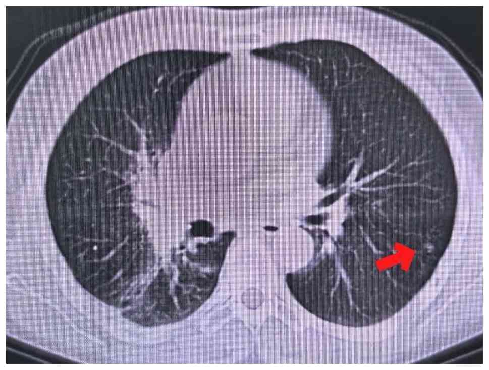
Chest CT performed on day 22 after the initial
presentation (Fig. 1) revealed the
following: i) An irregular soft-tissue mass was observed in the
dorsal segment of the right lower lobe, suggestive of central lung
cancer (cT4 stage), accompanied by adjacent obstructive pneumonia;
ii) multiple nodules of varying sizes were present in both lungs,
indicating intrapulmonary metastasis (M1a) according to the eighth
edition of the TNM staging system (9); and iii) multiple enlarged lymph nodes
were detected in the mediastinum and right hilar region, suggesting
lymph node metastasis (N2) (9).
Systemic examination
No definitive metastatic signs were observed on
cranial magnetic resonance imaging with contrast enhancement,
pelvic CT, or whole-body bone scintigraphy. Abdominal CT revealed a
small cyst in the S4 segment of the liver and bilateral renal
cysts.
Pathological diagnosis
Percutaneous lung biopsy guided by CT revealed a
poorly differentiated non-small cell carcinoma.
The biopsy specimen was fixed in 10%
neutral-buffered formalin at room temperature (~25°C) for 12–24 h,
processed through graded alcohols, cleared in xylene and embedded
in paraffin wax. Serial sections were cut at a thickness of 4 µm
and mounted on poly-L-lysine-coated glass slides. For
immunohistochemical staining, sections were deparaffinized in
xylene and rehydrated through graded ethanol. Antigen retrieval was
performed using citrate buffer (pH 6.0) at 95°C for 20 min in a
water bath. Endogenous peroxidase activity was blocked by
incubation with 3% hydrogen peroxide in methanol at room
temperature for 10 min. Non-specific binding was blocked by
incubation with 5% normal goat serum (cat. no. ab7481; Abcam) at
room temperature for 30 min. The sections were then incubated with
primary antibodies at 4°C overnight in a humidified chamber. The
primary antibodies used were as follows: Pan-CK (clone AE1/AE3;
cat. no. ab27976; 1:200; Abcam), CK7 (clone OV-TL 12/30; cat. no.
ab9021; 1:100; Abcam), thyroid transcription factor-1 (clone
8G7G3/1; cat. no. M3575; 1:100; Dako; Agilent Technologies, Inc.),
napsin A (clone IP64; cat. no. MCA2119; 1:200; Bio-Rad
Laboratories, Inc.), p40 (clone BC28; cat. no. ab202493; 1:200;
Abcam), p63 (clone D2K8X; cat. no. 13540; 1:200; Cell Signaling
Technology, Inc.), SMARCA4/BRG1 (clone EPNCIR111A; cat. no.
ab110641; 1:100; Abcam), synaptophysin (clone DAK-SYNAP; cat. no.
M0776; 1:100; Dako; Agilent Technologies, Inc.) and Ki-67 (clone
MIB-1; cat. no. M7240; 1:100; Dako; Agilent Technologies, Inc.).
After washing with phosphate-buffered saline, the sections were
incubated with horseradish peroxidase-conjugated goat anti-rabbit
or anti-mouse secondary antibody (cat. nos. ab205718 and ab205719;
1:200; Abcam) at room temperature for 30 min. Immunoreactivity was
visualized using 3,3′-diaminobenzidine chromogen (cat. no. ab64238;
Abcam) for 2–5 min at room temperature, followed by counterstaining
with Mayer's hematoxylin for 1 min. The sections were then
dehydrated, cleared and coverslipped with mounting medium. All
stained sections were examined under a light microscope, and images
were captured at ×400 magnification.
Molecular pathology testing
Next-generation sequencing (NGS) was conducted on
formalin-fixed paraffin-embedded (FFPE) tumor tissue obtained from
the CT-guided percutaneous lung biopsy. DNA was extracted using a
laboratory-developed solid tumor gene panel FFPE extraction kit,
and libraries were prepared using a laboratory-developed test (LDT)
solid tumor gene panel library preparation kit. Sequencing was
performed on an Illumina NovaSeq 6000 platform with 150-bp
paired-end reads.
Raw reads were preprocessed using Fastp (version
0.21.0; OpenGene; URL: http://github.com/OpenGene/fastp) to remove adapter
sequences and low-quality bases, with quality control assessed
using FastQC (version 0.11.9; Babraham Bioinformatics; http://www.bioinformatics.babraham.ac.uk/projects/fastqc/).
Clean reads were aligned to the GRCh37/hg19 reference genome using
BWA-MEM (version 0.7.17; Li H; URL: http://bio-bwa.sourceforge.net). PCR duplicates were
removed using Samblaster (version 0.1.24; Gregory Faust; http://github.com/GregoryFaust/samblaster). Variant
calling was performed using a custom in-house algorithm integrating
Pisces (version 5.3.0.0; Illumina; http://github.com/Illumina/Pisces) and GATK (version
4.0.4.0; Broad Institute; http://github.com/broadinstitute/gatk/releases).
Variant filtering criteria included a variant allele frequency
(VAF) >1.3%, a mean sequencing depth >500×, and specific
SNP/InDel depth >50×. Functional annotation was performed using
a custom pipeline integrating Annovar (version 2022-01-13; Wang K;
http://annovar.openbioinformatics.org), VEP (version
111.0; Ensembl; http://useast.ensembl.org/info/docs/tools/vep/index.html)
and SnpEff (version 5.3; Cingolani P; http://github.com/snpEff/snpEff).
PD-L1 expression was evaluated as the TPS, defined
as the percentage of tumor cells showing partial or complete
membranous staining at any intensity. A TP53 missense mutation was
identified, and no driver gene mutations were detected for EGFR,
ALK or ROS1.
Clinical diagnosis
The final diagnosis was adenocarcinoma of the middle
and lower lobes of the right lung (cT4N2bM1a, stage IVa), according
to the eighth edition of the TNM staging system (9). with pulmonary metastasis, multiple
lymph node metastases in the mediastinum and hilar region, and
malignant pleural effusion.
Treatment process and efficacy
evaluation
The patient's treatment plan was developed through
multidisciplinary team discussions. The timeline of diagnosis and
treatment is summarized in Table
I.
 | Table I.Timeline of diagnosis and
treatment. |
Table I.
Timeline of diagnosis and
treatment.
| Date | Action |
|---|
| January 2025 | Diagnosis (cT4N2bM1a,
stage IVa) |
| February 2025 | Intrapleural
perfusion (60 mg cisplatin + 200 mg bevacizumab) on days 1, 6 and
9; each instilled via chest tube, clamped for 2 h with
positioning |
| February-June
2025 | Ivonescimab
monotherapy (900 mg every 3 weeks for four cycles) |
| April
2025 | PR after two
cycles |
| June
2025 | June 2025: PR after
four cycles (47% reduction) |
| June-July 2025 | Sequential thoracic
radiotherapy (5,000 cGy/25 fractions) |
| Late July 2025 | Grade I radiation
pneumonitis, resolved with steroids |
| August 2025 | Last follow-up: PFS,
8 months; ECOG PS, 0 |
Initial supportive therapy and local
treatment (February 2025)
The patient was admitted to the Affiliated Hospital
of Qinghai University with concurrent pulmonary infection and
massive right-sided pleural effusion. Empirical anti-infective
therapy with levofloxacin (500 mg once daily for 7 days) was
administered, followed by closed thoracic drainage, resulting in
symptom relief. To control the pleural effusion, three intrapleural
perfusion treatments of 60 mg cisplatin + 200 mg bevacizumab were
performed on days 1, 6 and 9 of the intrapleural perfusion regimen.
Post-treatment, the pleural effusion was well controlled.
Systemic immunotherapy (February to
June 2025)
Based on the patient's high PD-L1 expression status,
ivonescimab (AK112) was initiated as monotherapy on day 6 of the
intrapleural perfusion regimen (concurrent with the second
intrapleural perfusion), at a dose of 900 mg administered
intravenously every 3 weeks. The treatment course proceeded
smoothly.
Efficacy evaluation
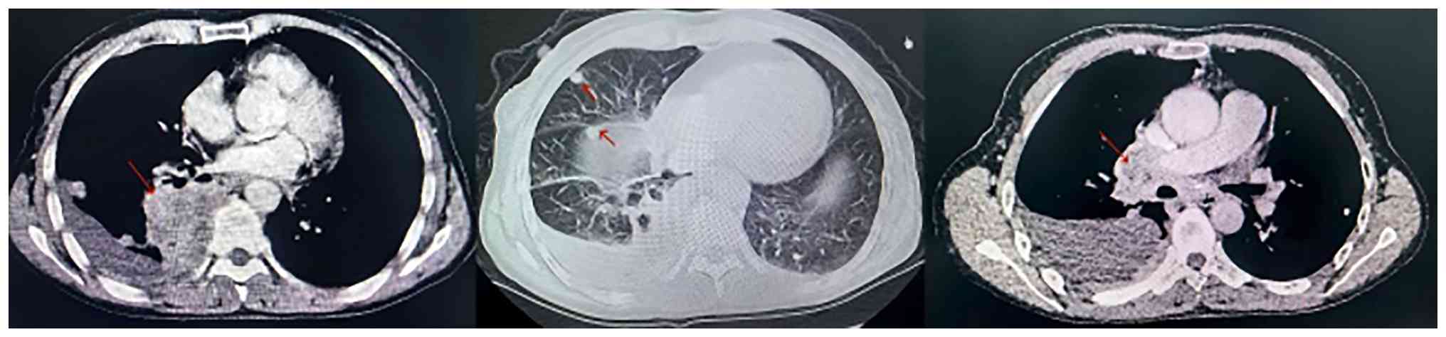
Baseline imaging performed on day 22 after initial
presentation is shown in Fig. 1.
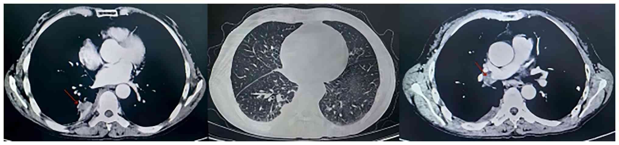
Chest enhanced CT re-examination was performed after two cycles of
treatment (April 2025) (Fig. 2) and
four cycles of treatment (June 2025) (Fig. 3). Percutaneous lung biopsy guided by
CT revealed a poorly differentiated non-small cell carcinoma
(Fig. 4A and B).
Immunohistochemical staining results were as follows: Thyroid
transcription factor-1 (+) (Fig.
4C), napsin A (+) (Fig. 4D),
tumor protein p40 (−) (Fig. 4E),
tumor protein p63 (−) (Fig. 4F),
pan-cytokeratin (pan-CK) (+) (Fig.
4G), CK7 (+) (Fig. 4H),
SMARCA4/BRG1 (−) (Fig. 4I),
synaptophysin (−) (Fig. 4J) and a
Ki-67 proliferation index ~70% (Fig.
4K). These findings supported a diagnosis of pulmonary
adenocarcinoma. According to the Response Evaluation Criteria in
Solid Tumors 1.1, both assessments demonstrated a partial response
(PR). Specifically, the sum of the longest diameters of the target
lesions decreased by 33 and 47%, respectively, compared with
baseline. The dynamic changes in serum tumor markers are shown in
Table II, with marked reductions
in carcinoembryonic antigen (CEA), carbohydrate antigen 125
(CA125), cytokeratin 19 fragment (CYFRA21-1) and neuron-specific
enolase (NSE) levels compared with pre-treatment levels. Notably,
SCC remained within the normal range throughout the treatment
course.
| Table II.Dynamic changes in serum tumor markers
with normal reference ranges. |
Table II.
Dynamic changes in serum tumor markers
with normal reference ranges.
| Tumor marker | Normal value | Baseline (day
22) | After 2 cycles (~day
90) | After 4 cycles (~day
150) |
|---|
| CEA, ng/ml | <5.0 | 46.76 | 7.18 | 3.83 |
| CA125, U/ml | <35.0 | 108.10 | 11.20 | 9.30 |
| CYFRA21-1, ng/ml | <3.3 | 27.86 | 2.14 | 2.71 |
| SCC, ng/ml | <1.5 | 0.80 | 1.10 | 1.10 |
| NSE, ng/ml | <16.3 | 29.99 | 12.47 | 10.11 |
Sequential radiotherapy (June to July
2025)
Based on achieving a good response to systemic
therapy, sequential chest radiotherapy was initiated in June 2025
to consolidate local lesion control. Intensity-modulated
radiotherapy was employed, with the following target volumes
defined: Gross tumor volume 1 (GTV1) was the imaging-confirmed
primary lesion in the right lung, with a prescription dose of 1,000
cGy/5 fractions; and GTV2 included the right lung metastatic lymph
nodes and high-risk lymphatic drainage prophylaxis areas, with a
prescription dose of 5,000 cGy/25 fractions. The radiotherapy plan
was successfully completed.
Management and follow-up of adverse
events
Approximately 20 days after radiotherapy (end of
July 2025), a follow-up CT scan revealed stable primary lesions but
new ground-glass opacities and linear opacities in the lungs,
suggestive of radiation pneumonitis. Pulmonary function tests
revealed mild reductions in small airway function, with maximal
mid-expiratory flow at 62% of predicted and maximal expiratory flow
at 25% recorded at 65% of predicted, while forced expiratory volume
in 1 sec (FEV1) and forced vital capacity (FVC) remained within
normal limits (FEV1 89% of predicted, FVC 92% of predicted). After
comprehensive evaluation, the diagnosis was Grade I (10) radiation pneumonitis (Fig. 5). Treatment included intravenous
dexamethasone (5 mg on days 1–3), followed by sequential oral
methylprednisolone starting at 20 mg/day, tapered by 4 mg every 3
days to a maintenance dose of 4 mg/day, then reduced by 2 mg every
3 days to discontinuation over a total of 4 weeks. Both imaging
abnormalities and symptoms resolved within a short period. As of
the last follow-up in August 2025, the patient had achieved 8
months of progression-free survival, with the ECOG PS score
improving from a baseline of 2 points to 0 points, and a notable
improvement in the quality of life.
Discussion
The present case provides real-world evidence for
the efficacy and safety of ivonescimab followed by consolidative
radiotherapy in a patient with advanced lung adenocarcinoma
exhibiting a PD-L1 TPS of 90% and a concurrent TP53 mutation.
Several clinically relevant observations merit further
discussion.
First, the depth of response observed with
ivonescimab monotherapy in this ‘super-responder’ population is
noteworthy. Following two cycles of treatment, the patient achieved
a PR, with target lesion reduction reaching 47% after four cycles.
This rapid and sustained tumor shrinkage aligns with findings from
the phase III HARMONi-2 trial, which demonstrated superior
progression-free survival with ivonescimab vs. pembrolizumab in
patients with PD-L1 TPS ≥50% (4).
In addition to imaging responses, serial monitoring of serum tumor
markers, including CEA, CA125, CYFRA21-1 and NSE, revealed
consistent decreases, further supporting the systemic antitumor
activity of ivonescimab. These findings reinforce the potential of
ivonescimab as a potent first-line option in advanced NSCLC with
high PD-L1 expression.
Second, the sequential strategy of ‘immunotherapy
induction followed by radiotherapy consolidation’ proved feasible
and safe in the present case. After confirming a PR to four cycles
of ivonescimab, the patient received thoracic intensity-modulated
radiotherapy (50 Gy/25 fractions) for local consolidation. This
approach was designed to leverage the synergistic potential of
radiotherapy, including direct cytotoxicity, antigen release and
enhancement of antitumor immunity, while minimizing the risk of
overlapping toxicity. The patient developed only grade 1 radiation
pneumonitis, which resolved rapidly with low-dose glucocorticoids.
This favorable safety profile is consistent with the 2025 Chinese
expert consensus, which supports the use of consolidative
radiotherapy after effective systemic immunotherapy in selected
patients with oligoresidual disease (10). Notably, this case illustrates that
delaying radiotherapy until after an immunotherapy-induced response
may optimize the therapeutic window and reduce the likelihood of
severe immune-related adverse events. A recent comprehensive review
by Burr et al (11)
summarized the preclinical and clinical evidence for combining
radiotherapy with immunotherapy in NSCLC, concluding that the
timing of these modalities is crucial to optimizing outcomes.
To contextualize the present findings, other case
reports have described similar responses in NSCLC patients with
high PD-L1 expression treated with sequential immunotherapy and
radiotherapy. Xia et al (12) reported a case of lung adenocarcinoma
with PD-L1 TPS ≥50% in which an extracranial abscopal effect was
observed following brain radiotherapy alone, accompanied by a
durable partial response to subsequent pembrolizumab therapy. This
observation further supports the concept that radiotherapy can
serve as an immunomodulator, even in the setting of high PD-L1
expression, whereby the abscopal effect may contribute to the
efficacy of sequential strategies.
Several clinical factors may have influenced the
treatment outcomes for the present patient. The patient had a
baseline pulmonary infection that was treated with levofloxacin.
This resolved before immunotherapy initiation and did not appear to
affect the final response. The malignant pleural effusion was
managed with intrapleural cisplatin plus bevacizumab, which likely
contributed to local symptom control but is not known to exert
systemic antitumor effects. The patient had no other comorbidities
(diabetes, hypertension or autoimmune diseases), which could have
confounded the safety profile.
The presence of a TP53 missense mutation in the
present patient raises the possibility of a predictive biomarker
for combined immunotherapy and radiotherapy. TP53 is the most
frequently mutated gene in lung cancer and has been associated with
increased tumor mutational burden, genomic instability and enhanced
immunogenicity (13). Preclinical
evidence suggests that p53 dysfunction may impair DNA damage
repair, thereby increasing radiosensitivity, while also promoting
immunogenic cell death via activation of the cyclic GMP-AMP
synthase/stimulator of IFN genes pathway (14). In a cohort of 220 patients with
metastatic non-squamous NSCLC, Mathiot et al (15) demonstrated that TP53 mutations were
associated with significantly improved overall survival (24.7 vs.
12.0 months; P=0.017) and progression-free survival (9.6 vs. 3.2
months, P=0.048) times in patients receiving immune checkpoint
inhibitor monotherapy. This finding is further corroborated by
another large German cohort study of 696 patients with non-squamous
NSCLC with high PD-L1 expression (≥50%) receiving first-line
pembrolizumab monotherapy. Bischoff et al (16) demonstrated that patients with TP53
mutations, particularly when co-occurring with KRAS G12C mutations,
experienced markedly longer progression-free survival (33.7 months)
and overall survival (65.3 months) compared with those with
wild-type TP53 (16). Notably, the
improved outcomes were associated with a proinflammatory tumor
microenvironment, characterized by high tumor-infiltrating
lymphocyte densities and upregulation of IFNγ target genes
(16). This large-scale, real-world
evidence strongly supports the notion that TP53 mutations can be
used to identify a subset of NSCLC patients with heightened
sensitivity to immune checkpoint inhibitor monotherapy. In this
context, the coexistence of high PD-L1 expression and TP53 mutation
may have contributed to the deep and durable response observed in
the present patient. Although hypothesis-generating, this case
supports further investigation into TP53 mutation status as a
potential biomarker for selecting candidates for combined modality
approaches.
In summary, the present case highlights the clinical
potential of a sequential regimen involving ivonescimab induction
followed by consolidative radiotherapy in patients with advanced
lung adenocarcinoma and high PD-L1 expression. The strategy
achieved meaningful tumor regression, maintained quality of life
(ECOG PS improved from 2 to 0) and demonstrated a manageable safety
profile. Future prospective studies are warranted to validate
optimal sequencing, fractionation schedules and biomarker-driven
patient selection for this promising therapeutic approach.
In conclusion, for patients with advanced lung
adenocarcinoma exhibiting high PD-L1 expression, a promising
therapeutic strategy involves systemic treatment based on
ivonescimab followed by sequential radical local radiotherapy after
achieving marked efficacy. The combined data from this case and the
HARMONi-2 study demonstrate that this strategy can induce a deep
and durable tumor response in the favorable population, improve
quality of life and maintain controllable safety, providing
valuable reference for clinical practice.
Acknowledgements
Not applicable.
Funding
Funding: No funding was received.
Availability of data and materials
The data generated in the present study may be found
in the NCBI Sequence Read Archive under accession number
PRJNA1498320 or at the following URL: https://www.ncbi.nlm.nih.gov/bioproject/PRJNA1498320.
The remaining data generated in the present study may be requested
from the corresponding author.
Authors' contributions
QZ was responsible for the conceptualization and
design of the study, the analysis and interpretation of clinical
patient data, drafting the original manuscript, and reviewing and
editing the final version. BY was responsible for the acquisition,
review and interpretation of the clinical imaging data (CT scans
and MRI) and pathological data to confirm the diagnosis
(cT4N2bM1a), and for providing advice on patient treatment. HC was
responsible for the acquisition and analysis of laboratory data
(serum tumor markers), and the visualization of clinical figures
and tables. JJ was responsible for designing the treatment
protocol, providing clinical advice on systemic immunotherapy and
radiotherapy, and supervising the treatment course. RC was
responsible for conceptualization, critical revision of the
manuscript for important intellectual content, and final approval
of the version to be published. QZ and RC confirm the authenticity
of all the raw data. All authors have read and approved the final
manuscript.
Ethics approval and consent to
participate
Not applicable.
Patient consent for publication
Written informed consent was obtained from the
patient for publication of this case report and any accompanying
images.
Competing interests
The authors declare that they have no competing
interests.
References
|
1
|
Zhou J, Xu Y, Liu J, Feng L, Yu J and Chen
D: Global burden of lung cancer in 2022 and projections to 2050:
Incidence and mortality estimates from GLOBOCAN. Cancer Epidemiol.
93:1026932024. View Article : Google Scholar : PubMed/NCBI
|
|
2
|
Hayashi H, Ogura M, Niwa T, Yokoyama T,
Tanizaki J, Ozaki T, Yoshioka H, Kurata T, Tamura Y, Fujisaka Y, et
al: Phase I/II study of cisplatin plus Nab-paclitaxel with
concurrent thoracic radiotherapy for patients with locally advanced
non-small cell lung cancer. Oncologist. 26:19–e52. 2021. View Article : Google Scholar : PubMed/NCBI
|
|
3
|
Dhillon S: Ivonescimab: First approval.
Drugs. 84:1135–1142. 2024. View Article : Google Scholar : PubMed/NCBI
|
|
4
|
Xiong A, Wang L, Chen J, Wu L, Liu B, Yao
J, Zhong H, Li J, Cheng Y, Sun Y, et al: Ivonescimab versus
pembrolizumab for PD-L1-positive non-small cell lung cancer
(HARMONi-2): A randomised, double-blind, phase 3 study in China.
Lancet. 405:839–849. 2025. View Article : Google Scholar : PubMed/NCBI
|
|
5
|
Long R, Kuang W and Zhou Q: Ivonescimab
plus chemotherapy in advanced or metastatic non-squamous non-small
cell lung cancer with EGFR variant in China: A cost-effectiveness
analysis. Transl Lung Cancer Res. 14:1622–1634. 2025. View Article : Google Scholar : PubMed/NCBI
|
|
6
|
Al Matairi A, Hammadeh BM, Aldalati AY,
Qtaishat FA, Nashwan AJ and Alzibdeh A: Efficacy and safety of
ivonescimab in the treatment of advanced non-small cell lung cancer
(NSCLC): A systematic review. Cureus. 17:e773812025.PubMed/NCBI
|
|
7
|
Karcioglu O, Topacoglu H and Dikme O and
Dikme O: A systematic review of the pain scales in adults: Which to
use? Am J Emerg Med. 36:707–714. 2018. View Article : Google Scholar : PubMed/NCBI
|
|
8
|
Oken MM, Creech RH, Tormey DC, Horton J,
Davis TE, McFadden ET and Carbone PP: Toxicity and response
criteria of the eastern cooperative oncology group. Am J Clin
Oncol. 5:649–655. 1982. View Article : Google Scholar : PubMed/NCBI
|
|
9
|
Detterbeck FC, Boffa DJ, Kim AW and Tanoue
LT: The eighth edition lung cancer stage classification. Chest.
151:193–203. 2017. View Article : Google Scholar : PubMed/NCBI
|
|
10
|
Multidisciplinary Committee on Oncology,
Chinese Medical Doctor Association; Expert Committee on Quality
Control of Lung Cancer, . National Quality Control Center for
Cancer: Chinese expert consensus on the combination of immune
checkpoint inhibitors and radiotherapy for lung cancer (2025
edition). Zhonghua Zhong Liu Za Zhi. 47:39–64. 2025.(In Chinese).
PubMed/NCBI
|
|
11
|
Burr JL, Johnson KC, Carmicheal JJ, Lin C
and Ganti AK: Combination Immunotherapy with radiotherapy in
non-small cell lung cancer: A review of evidence. Cancer Med.
13:e704022024. View Article : Google Scholar : PubMed/NCBI
|
|
12
|
Xia Y, Zhu H, Huang S, Guan X, Chen X,
Zhang Q, Meng L, Xue H, Xiang H, Lai S and Lou J: Remarkable
response to pembrolizumab in PD-L1 overexpressing (≥ 50%) NSCLC and
extracranial abscopal effect induced by brain radiotherapy: A case
report. Front Oncol. 15:16525152025. View Article : Google Scholar : PubMed/NCBI
|
|
13
|
Li L, Li M and Wang X: Cancer
type-dependent correlations between TP53 mutations and antitumor
immunity. DNA Repair (Amst). 88:1027852020. View Article : Google Scholar : PubMed/NCBI
|
|
14
|
Ghosh M, Saha S, Li J, Montrose DC and
Martinez LA: p53 engages the cGAS/STING cytosolic DNA sensing
pathway for tumor suppression. Mol Cell. 83:266–280.e6. 2023.
View Article : Google Scholar : PubMed/NCBI
|
|
15
|
Mathiot L, Nigen B, Goronflot T, Hiret S,
Doucet L, Pons-Tostivint E, Bennouna J, Denis MG, Herbreteau G and
Raimbourg J: Prognostic impact of TP53 mutations in metastatic
nonsquamous non-small-cell lung cancer. Clin Lung Cancer.
25:244–253.e2. 2024. View Article : Google Scholar : PubMed/NCBI
|
|
16
|
Bischoff P, Reck M, Overbeck T,
Christopoulos P, Rittmeyer A, Lüders H, Kollmeier J, Kulhavy J,
Kemper M, Reinmuth N, et al: Outcome of first-line treatment with
pembrolizumab according to KRAS/TP53 mutational status for
nonsquamous programmed death-ligand 1-high (≥50%) NSCLC in the
German national network genomic medicine lung cancer. J Thorac
Oncol. 19:803–817. 2024. View Article : Google Scholar : PubMed/NCBI
|

















![Histopathological and
immunohistochemical findings of the lung biopsy specimen. (A and B)
Hematoxylin and eosin-stained sections showing clusters of poorly
differentiated adenocarcinoma cells (original magnification, ×400;
scale bar, 100 µm). (C) Thyroid transcription factor-1 positivity.
(D) Napsin A positivity. (E) Negative expression of p40. (F)
Negative expression of p63. (G) Pan-cytokeratin positivity. (H) CK7
positivity. (I) Loss of SMARCA4/BRG1 expression. (J) Negative
expression of synaptophysin. (K) Ki-67 proliferation index of ~70%.
[(C-K) Scale bar, 100 µm; original magnification, ×400].](/article_images/ol/32/5/ol-32-05-15894-g03.jpg)