Introduction
Hepatocellular carcinoma (HCC), as a highly
prevalent primary malignant liver tumor worldwide, has long ranked
among the leading causes of cancer incidence and mortality, posing
a significant public health challenge (1,2).
Chronic hepatitis C virus infection represents a major etiological
factor for HCC development, with genetic polymorphisms affecting
both treatment outcomes and disease progression toward cirrhosis
and hepatocarcinogenesis (3–5).
Despite advances in surgical techniques, local regional therapies
and systemic treatments, the long-term prognosis for patients with
HCC remains poor due to frequent tumor recurrence, early metastasis
and significant molecular heterogeneity (6). Furthermore, the lack of reliable
biomarkers for early diagnosis and precise prognosis assessment
continues to hinder the effective management of HCC (7,8).
Therefore, identifying novel molecular biomarkers closely
associated with tumor progression, immune microenvironment
remodeling and patient prognosis is crucial for improving HCC risk
stratification and developing personalized treatment
strategies.
Nucleolar proteins play a central role in ribosomal
biosynthesis and regulate key processes such as cell cycle
progression, stress responses and genomic stability (9). The function of nuclear proteins has
been increasingly recognized as a hallmark feature of tumorigenesis
and malignant transformation (10,11).
Nucleolar protein 11 (NOL11), as a conserved nucleolar protein,
participates in ribosomal RNA transcription and processing, playing
a crucial role in maintaining nucleolar integrity and cellular
homeostasis (12). Research
indicates that abnormal expression of NOL11 can lead to the
development of various diseases, including liver cirrhosis
(13–15). However, its expression pattern,
biological function and clinical significance in HCC remain poorly
understood.
The present study aimed to systematically
investigate the expression pattern, prognostic significance, immune
relevance and biological function of NOL11 in HCC using
transcriptomic and clinical data from The Cancer Genome Atlas
(TCGA). Specifically, the study sought to analyze the association
between NOL11 expression and clinicopathological characteristics,
patient survival, immune cell infiltration and immune-related
signaling pathways. The present study further aimed to construct
and validate a NOL11-related prognostic risk model and to evaluate
its potential clinical value in survival prediction. In addition,
the associations of the risk model with tumor mutational burden
(TMB) and immune checkpoint molecule expression were explored to
assess its possible relevance to immunotherapy responsiveness.
Finally, in vitro experiments were performed to verify the
effects of NOL11 on HCC cell proliferation, migration and invasion.
These analyses were expected to provide new insight into the role
of NOL11 in HCC progression and to support its potential utility as
a prognostic biomarker and therapeutic target.
Materials and methods
Data acquisition and processing
The data retrieval and bioinformatics analyses were
conducted between December 2024 and April 2025. RNA transcriptome
data, clinical information and somatic mutation data were obtained
from the TCGA HCC project (https://portal.gdc.cancer.gov/) (16). Transcriptome data were uniformly
converted to fragments per kilobase million format and standardized
to ensure comparability across samples (17). During clinical information
integration, samples with missing or incomplete key survival time
were excluded to guarantee the reliability of subsequent analyses.
The final research cohort comprised 370 HCC tissue samples and 50
adjacent normal tissue samples. In addition, the infiltration
levels of 22 immune cell types in the HCC samples were estimated
using the CIBERSORT algorithm (https://cibersort.stanford.edu/), and NOL11 expression
across clinicopathological subgroups was analyzed using the UALCAN
database (https://ualcan.path.uab.edu/). The protein expression
of NOL11 in normal liver and HCC tissues was examined using the
Human Protein Atlas (HPA) database (https://www.proteinatlas.org/).
Functional enrichment and protein
interaction network analysis
To elucidate the potential biological functions of
NOL11 in HCC, this study comprehensively employed Gene Ontology
(GO) and Kyoto Encyclopedia of Genes and Genomes (KEGG) for
functional annotation and pathway enrichment analysis using the
clusterProfiler package in R (https://biocon-ductor.org/packages/release/bioc/html/clusterProfiler.html).
All analyses set a P-value <0.05 as the threshold for
statistically significant enrichment, with results visualized using
the ggplot2 package. Additionally, the protein interaction network
for NOL11 was constructed via the search tool for the retrieval of
interacting genes (STRING) database (http://string-db.org), setting a minimum interaction
confidence of 0.6 to identify highly associated protein
interactions (18).
Construction and validation of risk
model
This analysis first identified NOL11 co-expressed
genes significantly associated with HCC prognosis through
univariate Cox regression analysis. To optimize model stability and
prevent overfitting, these genes were further incorporated into a
least absolute shrinkage and selection operator (LASSO) Cox
regression analysis performed using the glmnet package in R
(https://cran.r-project.org/web/packages/glmnet/index.html),
to construct a risk model. Based on the expression levels of the
selected genes and their corresponding coefficients in the LASSO
regression, a risk score was calculated for each patient using the
following formula: Risk score=∑(Gene expression level ×
Coefficient). Patients were then stratified into high-risk or
low-risk groups using the median risk score across the entire
cohort as the cutoff point. To further evaluate the prognostic
discriminatory power of this risk model, survival analysis was
performed on the TCGA-HCC cohort to compare overall survival (OS)
differences between the two groups. Additionally, receiver
operating characteristic (ROC) curves and calibration curves were
used to assess the model's accuracy and consistency in predicting
1-, 3- and 5-year survival rates. Principal component analysis
(PCA) and t-distributed stochastic neighborhood embedding (t-SNE)
were employed to visualize distribution differences between the
high- and low-risk groups. Finally, univariate and multivariate Cox
regression analyses were performed to validate whether the risk
score could serve as an independent prognostic factor for HCC, with
P<0.05 set as the threshold for statistical significance.
Construction and prediction of
nomograms
Based on risk scores and other independent clinical
prognostic factors, this study utilized the regplot package in R to
construct a clinical prognostic nomogram for HCC patients. This
nomogram quantifies individual patient risk and predicts 1-, 3- and
5-year OS rates. The accuracy and consistency of the nomogram in
predicting survival probability were evaluated by plotting ROC
curves and calibration curves.
TMB and immune checkpoint
analysis
This study first employed the maftools software
package (19) to conduct systematic
analysis and visualization of somatic mutation data from patients
with HCC, revealing their genetic mutation spectrum
characteristics. Building upon this foundation, differential
analysis, correlation analysis and survival analysis methods were
further integrated to systematically evaluate the association
between risk scores and TMB, and explore their synergistic role in
HCC prognosis. Additionally, by comparing expression differences of
key immune checkpoint molecules [such as programmed cell death 1
(PD-1), programmed cell death ligand 1 (PD-L1), cytotoxic
T-lymphocyte antigen (CTLA)-4, lymphocyte activating (LAG)-3,
indoleamine 2,3-dioxygenase 1 (IDO1) and kinase insert domain
receptor (KDR)] between high- and low-risk groups, the correlation
between risk scores and immune checkpoint expression levels was
analyzed to explore the potential value of the risk model in
predicting responses to immunotherapy.
Short hairpin RNA (shRNA) plasmid
transfection and establishment of NOL11-knockdown cell lines
shRNA expression plasmids targeting human NOL11
(shNOL11-1 and shNOL11-2), together with a non-targeting
negative-control plasmid (shCtrl), were designed and synthesized by
GenePharma. The target sequences of the shRNAs, excluding the
plasmid vector backbone region, were as follows: shNOL11-1,
5′-CCAAACGCATGTGCTTTCTTACAGT-3′; shNOL11-2,
5′-GTCTACTTCTGGATGCGAATT-3′; and shCtrl, 5′-TTCTCCGAACGTGTCACGT-3′.
The shCtrl sequence was a scrambled non-targeting sequence with no
significant homology to known human genes. The shRNA constructs
carried a puromycin-resistance cassette to allow the selection of
stably transfected cells.
The human HCC cell lines Huh-7 and Hep3B were
obtained from Procell Life Science & Technology Co., Ltd. Both
cell lines were cultured in Dulbecco's modified Eagle's medium
(DMEM; Gibco; Thermo Fisher Scientific, Inc.) supplemented with 10%
fetal bovine serum (FBS; Zhejiang Tianhang Biotechnology Co., Ltd.)
and 1% penicillin-streptomycin (Gibco; Thermo Fisher Scientific,
Inc.) at 37°C in a humidified atmosphere containing 5%
CO2. Huh-7 and Hep3B cells were seeded in six-well
plates and cultured in antibiotic-free complete medium until they
reached ~70–80% confluence. Cells were then transfected with
shNOL11-1, shNOL11-2 or shCtrl plasmids using Lipofectamine 2000
(Invitrogen; Thermo Fisher Scientific, Inc.) according to the
manufacturer's instructions. Briefly, 2 µg of plasmid DNA and 4 µl
of Lipofectamine 2000 were separately diluted in Opti-MEM
reduced-serum medium (Gibco; Thermo Fisher Scientific, Inc.). After
incubation for 5 min at room temperature, the diluted DNA and
Lipofectamine 2000 solutions were combined and incubated for an
additional 20 min to allow complex formation. The DNA-lipid
complexes were then added dropwise to the cells. After 4–6 h of
incubation at 37°C in a humidified atmosphere containing 5%
CO2, the transfection medium was replaced with fresh
complete culture medium. At 48 h after transfection, cells were
cultured in complete medium supplemented with puromycin for the
selection of stably transfected cells. The puromycin-containing
medium was replaced every 2–3 days until non-transfected control
cells were completely eliminated. The resulting puromycin-resistant
cell populations were expanded and used for subsequent experiments.
The knockdown efficiency of NOL11 was confirmed by western
blotting.
Western blot analysis
Western blotting was performed to determine NOL11
protein expression in Huh-7 and Hep3B cells. Total cellular
proteins were extracted using RIPA lysis buffer (cat. no. P0013B;
Beyotime Biotechnology) supplemented with protease inhibitors
(phenylmethylsulfonyl fluoride; cat. no. ST506; Beyotime
Biotechnology). Protein concentrations were quantified using a
bicinchoninic acid protein assay kit (cat. no. P0010; Beyotime
Biotechnology). Equal amounts of protein (35 µg per lane) were
resolved by 10% sodium dodecyl sulfate-polyacrylamide gel
electrophoresis and subsequently transferred onto polyvinylidene
difluoride membranes (MilliporeSigma). After transfer, the
membranes were blocked with 5% bovine serum albumin (cat. no.
A8020; Beijing Solarbio Science & Technology Co., Ltd.) at room
temperature for 1 h and incubated overnight at 4°C with the
following primary antibodies: Anti-NOL11 antibody (1:2,000
dilution; cat. no. ab180132; Abcam) and anti-β-actin antibody
(1:25,000 dilution; cat. no. ab49900; Abcam). After washing with
Tris-buffered saline containing 0.1% Tween-20, the membranes were
incubated with the appropriate horseradish peroxidase-conjugated
secondary antibodies (goat anti-rabbit IgG, 1:3,000 dilution; cat.
no. A0208; and goat anti-mouse IgG, 1:3,000 dilution; cat. no.
A0216; both from Beyotime Institute of Biotechnology) for 1 h at
room temperature. Immunoreactive bands were visualized using a
chemiluminescence detection system (MilliporeSigma). β-actin was
used as the internal loading control.
Cell functional assays
To evaluate the function of NOL11 in HCC cells, a
series of experiments including cell proliferation, colony
formation, migration and invasion assays were performed. Cell
viability was assessed using the MTT assay (cat. no. M8180; Beijing
Solarbio Science & Technology Co., Ltd.): Huh-7 and Hep3B cells
were seeded at 1×10³ cells per well in a 96-well plate.
MTT solution (20 µl; 5 mg/ml) was added to designated wells on each
of 5 consecutive days, with a separate set of wells assayed at each
time-point, and absorbance was measured at 570 nm following
dissolution of the formazan crystals in dimethyl sulfoxide. For the
colony-formation assay, equal numbers of cells (1,000 cells per
well) were seeded into 6-well plates. After 10 days of culture,
cells were stained with 0.1% crystal violet (cat. no. C8470;
Beijing Solarbio Science & Technology Co., Ltd.) at room
temperature for 20 min and colonies (≥50 cells) were counted. Cell
migration and invasion capabilities were evaluated using Transwell
chambers (8-µm pore size; cat. no. 3422; Corning, Inc.). The
migration assay: cells (1×10⁵ cells per well) were resuspended in
200 µl serum-free medium and seeded into the upper chamber with
uncoated 8-µm pores, while 600 µl complete medium containing 10%
FBS was added to the lower chamber; the invasion assay utilized a
pre-coated Matrigel matrix (cat. no. M8370; Beijing Solarbio
Science & Technology Co., Ltd.). After incubation for 24 and 48
h, respectively, cells were fixed with 4% paraformaldehyde at room
temperature for 15 min and stained with 0.1% crystal violet at room
temperature for 20 min and the number of cells that had
transgressed through the membrane was counted under a light
microscope (×200 magnification) using ImageJ software (version
1.54; National Institutes of Health; http://imagej.net/ij/) to assess metastatic
potential.
Statistical analysis
Bioinformatics analyses were performed using R
software (version 4.2.3). Differential gene expression analysis was
conducted using the limma package. Survival curves were generated
using the Kaplan-Meier method and compared using the log-rank test.
Univariate and multivariate Cox proportional hazards regression
analyses were performed to evaluate the independent prognostic
value of clinicopathological variables and the risk score.
Comparisons of immune cell infiltration levels among the four NOL11
copy number states were performed using the Kruskal-Wallis test.
All statistical tests were two-sided and P<0.05 was considered
statistically significant. In vitro experiments were
performed as three independent replicates. Quantitative data are
presented as the mean ± standard deviation. Statistical analyses
for in vitro experiments were performed using GraphPad Prism
version 10.4 (Dotmatics). Comparisons among shCtrl, shNOL11-1 and
shNOL11-2 groups were performed using one-way analysis of variance
(ANOVA) followed by Dunnett's multiple-comparisons test, with the
shCtrl group designated as the reference group. For MTT cell-growth
curves involving multiple groups measured over multiple
time-points, two-way ANOVA followed by Dunnett's
multiple-comparisons test was used. A two-sided P<0.05 was
considered to indicate statistical significance.
Results
High expression of NOL11 is associated
with poor prognosis in HCC
Based on HCC transcriptome and clinical data from
the TCGA database (including 50 normal tissues and 370 tumor
tissues), NOL11 expression and its clinical significance in HCC
were assessed. Results revealed significantly higher mRNA
expression levels of NOL11 in HCC tissues compared to normal
tissues (Fig. 1A). This trend was
further validated in paired homologous patient samples, where NOL11
expression was markedly elevated in tumor tissues relative to
adjacent normal tissues (Fig. 1B).
Survival analysis demonstrated that patients with high NOL11
expression exhibited shorter OS compared to those with low
expression, with statistically significant differences (Fig. 1C). Subgroup analysis using the
UALCAN database revealed that NOL11 transcription levels in HCC
were significantly higher than those in normal controls across
different ethnic groups, genders, ages, grades, nodal metastasis
statuses, TP53 mutation statuses, histological subtypes and stages
(Fig. 1D-K). Additionally, NOL11
expression increased gradually with higher tumor grade, lymph node
metastasis and TP53-mutant status. With respect to clinical stage,
NOL11 expression increased progressively from stage 1 to stage 3
but was lower at stage 4 than at stage 3.
Correlation between NOL11 and
tumor-infiltrating immune cells in HCC
To elucidate the relationship between NOL11 and the
immune microenvironment, a systematic analysis of the composition
of tumor-infiltrating immune cells in HCC samples was conducted.
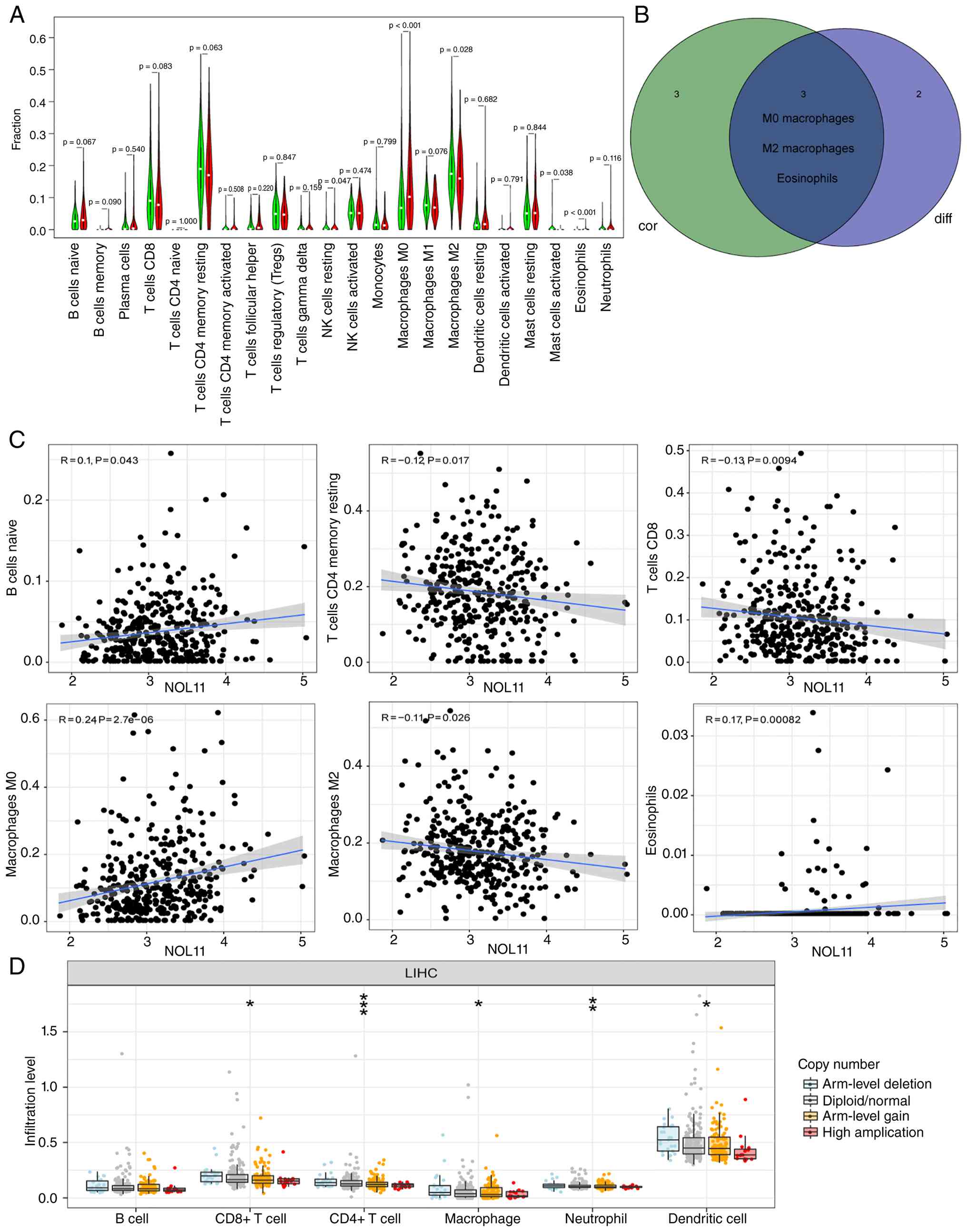
The results revealed that five immune cell types exhibited
significant differences between the NOL11 low-expression and
high-expression groups (Fig. 2A).
Additionally, NOL11 expression was positively correlated with the
infiltration of B cells naive, Macrophages M0 and Eosinophils,
while it was negatively correlated with T cells CD4 memory resting,
T cells CD8 and Macrophages M2 (Fig.
2C). Combining the differential and correlation analyses, the
immune cell types identified as differentially infiltrated and
those significantly correlated with NOL11 expression were
intersected, and three cell types, namely M0 macrophages, M2
macrophages and eosinophils, were found to overlap between the two
analyses, suggesting that NOL11 may primarily regulate immune
activity in the tumor microenvironment by modulating the
infiltration levels of these three cell types (Fig. 2B). Further analysis revealed that
the CNV of NOL11 was also closely associated with the infiltration
levels of multiple immune cell types. In general, the infiltration
levels of CD8+ T cells, CD4+ T cells, macrophages, neutrophils and
dendritic cells were lower in tumors with arm-level gain or
high-level amplification of NOL11 than in those with arm-level
deletion, with stepwise decreases being most evident for dendritic
cells, macrophages, CD8+ T cells and CD4+ T cells (P<0.05 for
CD8+ T cells, macrophages and dendritic cells; P<0.01 for
neutrophils; P<0.001 for CD4+ T cells), whereas B-cell
infiltration did not differ evidently among the NOL11 copy number
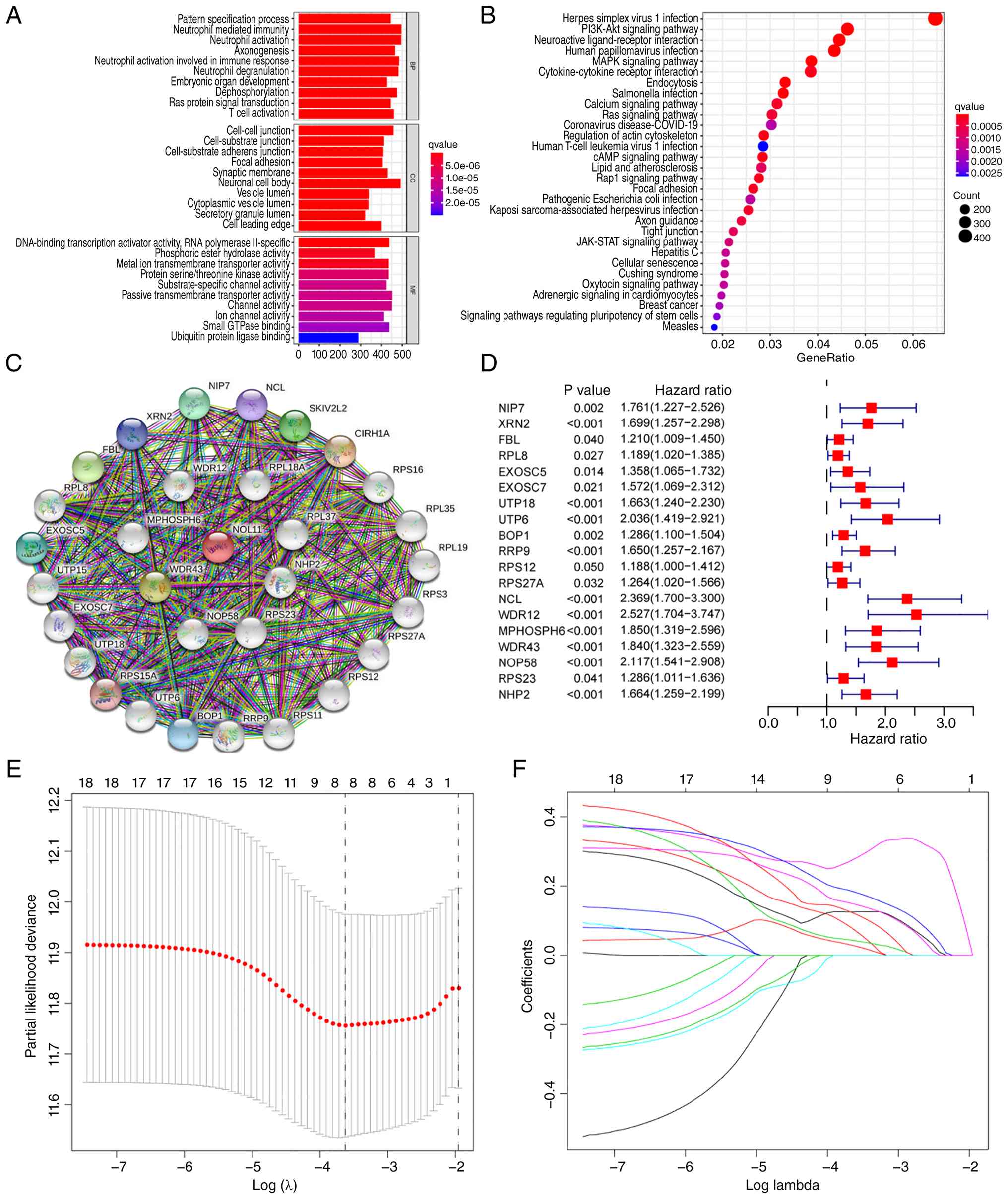
states (Fig. 2D). GO enrichment
analysis indicated that NOL11 participates in multiple
immune-related biological processes (Fig. 3A). KEGG pathway enrichment analysis
further suggested that NOL11 may be associated with tumor-related
pathways such as the PI3K-Akt signaling pathway and the MAPK
pathway (Fig. 3B). Collectively,
these results indicate that NOL11 expression levels may serve as a
potential indicator reflecting the immune status of the tumor
microenvironment.
Construction and validation of risk
model
A protein interaction network for NOL11 was
constructed based on the STRING database, from which the 30 genes
most closely interacting with NOL11 were identified (Fig. 3C). To further evaluate the potential
prognostic value of these co-expressed genes in HCC, univariate Cox
regression analysis was performed, revealing that 19 of these genes
were significantly associated with HCC patient prognosis (Fig. 3D). Subsequently, a risk prediction
model comprising eight genes was constructed using LASSO-Cox
regression analysis (Fig. 3E and
F). The risk score calculation formula is as follows: Risk
score= (XRN2 expression × 0.0748) + (BOP1 expression × 0.0277) +
(RRP9 expression × 0.0426) + (NCL expression × 0.2772) + (WDR12
expression × 0.1253) + (MPHOSPH6 expression × 0.1213) + (NOP58
expression × 0.1833) + (NHP2 expression × 0.1482). Patients were
stratified into high-risk and low-risk groups based on median risk
scores. The results demonstrated that high-risk patients exhibited
higher mortality rates and significantly shorter OS compared to
low-risk patients (Fig. 4A and B).
ROC curve and calibration curve analyses further confirmed that
this NOL11-related gene-based risk model possesses high predictive
accuracy for patient prognosis (Fig. 4C
and D). PCA and t-SNE dimensionality reduction revealed that
the risk score effectively distinguished patients with HCC across
subgroups, with significant differences in clinicopathological
characteristics and prognosis between groups (Fig. 4E and F). Finally, both univariate
and multivariate Cox regression analyses confirmed that the risk
score represents an independent prognostic factor for patients with
HCC (Fig. 4G and H).
Construction and prediction of
nomograms
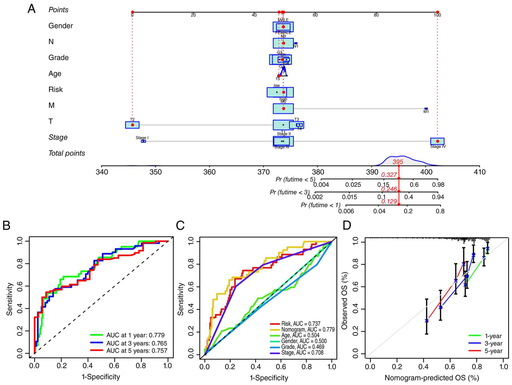
To develop a clinical tool capable of quantitatively
predicting 1-, 3- and 5-year OS in patients with HCC, a predictive
nomogram was constructed based on independent prognostic factors
(Fig. 5A). By accumulating the
scores corresponding to each variable in the nomogram, a total
score for each patient is obtained. This total score is then
projected onto the survival probability axis to derive the
patient's expected survival rates at 1, 3 and 5 years. ROC analysis
revealed area under curve values of 0.779, 0.765 and 0.757 for
predicting 1-, 3- and 5-year survival rates, respectively (Fig. 5B). Compared with other clinical
prognostic factors, this nomogram demonstrated higher accuracy in
predicting HCC patient survival rates (Fig. 5C). Calibration curves further
demonstrated that the predicted survival rates at 1-, 3- and 5-year
time-points closely aligned with the ideal reference line,
indicating excellent predictive consistency and reliability
(Fig. 5D). In summary, this
nomogram serves as an intuitive and accurate prognostic assessment
tool with potential clinical utility, facilitating personalized
survival prediction for patients with HCC.
Estimation of TMB and immune
checkpoint expression levels by risk model
Fig. 6A and B
display significantly enriched signaling pathways in the high-risk
and low-risk groups. Analysis of the HCC mutation spectrum based on
the TCGA cohort revealed that although mutation frequencies varied
across genes between the high-risk and low-risk groups, TP53,
CTNNB1 and TTN were consistently the three most frequently mutated
genes in both groups (Fig. 6C and
D). Further calculation of TMB scores and comparison between
groups revealed significantly higher TMB in the high-risk group
compared to the low-risk group, with risk scores positively
correlated with TMB (Fig. 6E and
F). Survival analysis revealed significantly shorter OS in
patients with high TMB compared to those with low TMB (Fig. 6G). Subgroup analysis further
demonstrated the poorest prognosis in patients with high TMB
combined with the high-risk subgroup, while the best prognosis was
observed in those with low TMB combined with the low-risk subgroup
(Fig. 6H). Considering that
patients with elevated TMB may exhibit greater sensitivity to
immune checkpoint inhibitor therapy, the relationship between risk
scores and immune checkpoint gene expression was further analyzed.
The results demonstrated that PD-1, CTLA-4 and IDO1 were
significantly upregulated in the high-risk group, with their
expression levels positively correlated with risk scores. The
expression levels of PD-L1, LAG3 and KDR did not differ
significantly between the two groups, and the correlation for LAG3
was not significant. KDR expression was numerically higher in the
low-risk group (Fig. 7).
 | Figure 7.Estimation of immune checkpoint
expression levels by risk model. High risk scores are correlated
with upregulated (A) PD-1, (B) PD-L1, (C) CTLA4, (D) LAG3 and (E)
IDO1 in patients with HCC. Low risk scores correlate with
upregulated (F) KDR in patients with HCC. HCC, hepatocellular
carcinoma; PD-1, programmed cell death 1; PD-L1, programmed cell
death ligand 1; CTLA, cytotoxic T-lymphocyte antigen; LAG3,
lymphocyte activating 3; IDO1, indoleamine 2,3-dioxygenase 1; KDR,
kinase insert domain receptor. |
Knockdown of NOL11 inhibits the
proliferation and migration of HCC cells in vitro
To further validate the expression level of NOL11 in
HCC, immunohistochemical images were viewed in the HPA database,
revealing obviously elevated NOL11 protein expression in HCC
tissues compared with normal liver tissues (Fig. 8). To investigate the biological role
of NOL11 in HCC cells, NOL11 expression was silenced in Huh-7 and
Hep3B cells using two independent shRNA plasmids, shNOL11-1 and
shNOL11-2. Western blotting confirmed that both shNOL11 constructs
substantially reduced NOL11 protein expression compared with the
non-targeting shCtrl (Fig. 9A). In
functional assays, NOL11 knockdown markedly impaired cell
proliferation (Fig. 9B). MTT
assays, which reflect the number of viable cells and thus cell
proliferation rather than cell death, further confirmed that NOL11
knockdown significantly reduced the number of viable HCC cells
(Fig. 9C). Colony-formation assays
revealed markedly reduced colony formation in both Huh-7 and Hep3B
cells following NOL11 knockdown (Fig.
9D and E). Given the clinical association between NOL11
overexpression and HCC metastasis (Fig.
1), cell migration and invasion assays were subsequently
performed. The results indicated that NOL11 knockdown significantly
inhibited the migration capacity in both Huh-7 and Hep3B cells
(Fig. 9F and G). Similarly, the
cell invasion assays demonstrated markedly reduced invasive ability
of tumor cells following NOL11 interference (Fig. 9H and I). These findings collectively
demonstrated that NOL11 plays a crucial regulatory role in the
proliferation, migration, and invasion processes of HCC cells.
 | Figure 9.Knockdown of NOL11 inhibits the
proliferation and migration of HCC cells in vitro. (A)
Western blot analysis of NOL11 expression in Huh-7 and Hep3B cells
transfected with shNOL11-1, shNOL11-2, or the non-targeting control
plasmid shCtrl. β-actin was used as the loading control. (B) Cell
proliferation (cell-growth curves) of HCC cells in the control and
experimental groups. (C) Cell viability of HCC cells in the control
and experimental groups, as determined using the MTT assay. Cell
colony formation assay was performed in control and experimental
(D) Huh-7 and (E) Hep3B HCC cells. Transwell migration assays in
control and experimental (F) Huh-7 and (G) Hep3B HCC cells.
Transwell invasion assays were performed in control and
experimental (H) Huh-7 and (I) Hep3B HCC cells. Representative
Transwell images were captured at ×200 magnification (scale bars,
100 µm). Data are presented as the mean ± SD from three independent
experiments. One-way ANOVA followed by Dunnett's
multiple-comparisons test was used for comparisons among three
groups in panels C-I, with shCtrl as the reference group. Two-way
ANOVA followed by Dunnett's multiple-comparisons test was used for
the cell-growth curves in panel B. *P<0.05, **P<0.01 and
***P<0.001 vs. shCtrl. NOL11, nucleolar protein 11; HCC,
hepatocellular carcinoma; shNOL11, short hairpin RNA targeting
shNOL11; Ctrl, control; OD, optical density. |
Discussion
In the present study, the expression pattern,
prognostic significance, immune relevance and biological function
of NOL11 in HCC were comprehensively characterized. Through
integrative analyses of transcriptomic and clinical data from the
TCGA cohort, it was demonstrated that NOL11 is significantly
upregulated in HCC tissues and closely associated with aggressive
clinicopathological characteristics and unfavorable OS.
Furthermore, a robust NOL11-related risk model was constructed and
validated, which effectively stratifies patient prognosis and
reflects distinct immune and mutational landscapes. Functional
experiments further confirmed that NOL11 promotes HCC cell
proliferation, migration and invasion, supporting its oncogenic
role in HCC progression.
Nucleolar proteins, as core participants in
ribosomal biosynthesis, cell cycle regulation and stress responses,
are increasingly recognized for their regulatory roles in
tumorigenesis (20). NOL11, an
evolutionarily conserved nucleolar protein, is known to play a
critical role in the transcription and processing of precursor rRNA
and is associated with hepatic pathological processes such as
cirrhosis. In recent years, multiple studies have progressively
revealed the potential role of NOL11 in the development and
progression of various malignant tumors (21,22).
However, its role in HCC has not been fully elucidated. The present
study confirms that NOL11 is abnormally overexpressed in HCC and is
closely associated with tumor grade, lymph node metastasis and
clinical staging, suggesting that this protein may be involved in
the development and progression of HCC. Mechanistically, functional
enrichment analyses revealed that NOL11-associated genes are
involved in multiple tumor-related signaling pathways, including
the PI3K-Akt and MAPK pathways (23), which are well-established drivers of
HCC proliferation, survival and metastasis. Consistent with these
bioinformatics findings, in vitro functional assays
demonstrated that silencing NOL11 significantly suppressed HCC cell
proliferation, colony formation, migration and invasion. These
results provide direct experimental evidence that NOL11 exerts
pro-tumorigenic effects and contributes to the malignant phenotype
of HCC cells.
The tumor immune microenvironment plays a pivotal
role in HCC progression, therapeutic response and patient prognosis
(24,25). HCC typically develops in a
background of chronic inflammation and immune dysregulation,
resulting in a complex and often immunosuppressive microenvironment
characterized by dysfunctional T cells, tumor-associated
macrophages and impaired antitumor immunity (26). Increasing evidence indicates that
immune cell composition and functional status within the tumor
microenvironment critically influence tumor growth, metastatic
potential and resistance to therapy (27–29).
In recent years, immune checkpoint inhibitors targeting PD-1/PD-L1
and CTLA-4 pathways have emerged as promising treatment options for
advanced HCC (24,30). However, only a subset of patients
achieves durable clinical benefit, highlighting the urgent need for
reliable biomarkers to predict immunotherapy responsiveness. TMB,
immune checkpoint expression and immune cell infiltration patterns
have been proposed as predictive indicators, yet their clinical
utility remains limited when considered individually (30,31).
In this context, the present study revealed a close
association between the NOL11-related risk model and the immune
landscape of HCC. High-risk patients exhibited significantly higher
TMB and poorer survival outcomes, and those with concomitant high
TMB and high-risk scores showed the worst prognosis. Furthermore,
key immune checkpoint molecules, including PD-1, CTLA-4 and IDO1,
were markedly upregulated in the high-risk group, whereas PD-L1,
LAG3 and KDR did not differ significantly between the two groups,
with KDR expression being numerically higher in the low-risk group.
These findings suggest that the NOL11-related risk model may
reflect an immunologically active but functionally suppressed tumor
microenvironment and may have potential value in identifying
patients who could benefit from immune checkpoint blockade therapy.
To facilitate clinical translation, the risk score was further
integrated with independent prognostic factors to construct a
nomogram for individualized survival prediction. This nomogram
demonstrated superior predictive accuracy and excellent calibration
for 1-, 3- and 5-year OS compared with traditional clinical
parameters alone (32),
underscoring its potential utility as a practical prognostic
assessment tool in clinical settings. Future research should focus
on validating the prognostic and immunological relevance of NOL11
in larger independent and multicenter HCC cohorts, as well as
clarifying the molecular mechanisms underlying its role in tumor
progression and immune regulation. From a translational
perspective, exploration of NOL11-associated molecular networks may
help identify potential therapeutic targets. In this context, drug
repurposing may represent a practical, cost-effective and
potentially sustainable strategy for accelerating the discovery of
clinically relevant compounds and facilitating the translation of
molecular findings into therapeutic applications (33–35).
The present study has several notable strengths.
First, it integrated large-scale transcriptomic and clinical data
with multiple bioinformatics approaches to systematically evaluate
the expression pattern and prognostic significance of NOL11 in HCC.
Second, beyond conventional survival analyses, this study further
explored the associations of NOL11 with immune cell infiltration,
immune-related pathways, tumor mutational burden and immune
checkpoint molecules, thereby providing a broader view of its
potential biological and clinical relevance. Third, a NOL11-related
prognostic model was constructed to assess its potential value in
individualized risk stratification. Finally, the bioinformatics
findings were supported by in vitro experiments, which
strengthened the biological relevance of the study. Despite these
strengths, several limitations should be acknowledged. First, all
transcriptomic and clinical analyses were based on retrospective
public datasets, primarily from TCGA, and lacked independent
multicenter external validation (36). Second, although the present in
vitro experiments confirmed the role of NOL11 in HCC cell
proliferation, migration and invasion, the underlying molecular
mechanisms were not fully elucidated. Therefore, future studies
incorporating large prospective cohorts, external validation
datasets and in vivo functional experiments are warranted to
further confirm the prognostic significance and therapeutic
potential of NOL11 in HCC.
In conclusion, the present study demonstrates that
NOL11 plays a critical role in HCC progression by promoting tumor
aggressiveness and shaping the immune microenvironment. The
NOL11-related risk model provides a powerful tool for prognostic
stratification and may aid in predicting immunotherapy
responsiveness. These findings expand the current understanding of
nucleolar protein dysregulation in HCC and support NOL11 as a
promising prognostic biomarker and potential therapeutic
target.
Acknowledgements
Not applicable.
Funding
Funding: No funding was received.
Availability of data and materials
The data generated in the present study may be
requested from the corresponding author.
Authors' contributions
HX designed the study. SW and XS performed the
bioinformatics analysis and data visualization. LY conducted the
in vitro experiments. HX and SW drafted the manuscript. HX
and SW checked and confirmed the authenticity of all the raw data.
All authors read and approved the final manuscript.
Ethics approval and consent to
participate
Not applicable.
Patient consent for publication
Not applicable.
Competing interests
The authors declare that they have no competing
interests.
Glossary
Abbreviations
Abbreviations:
|
AUC
|
area under the curve
|
|
CIBERSORT
|
Cell-type Identification By Estimating
Relative Subsets Of RNA Transcripts
|
|
CNV
|
copy number variation
|
|
CTLA-4
|
cytotoxic t-lymphocyte associated
protein 4
|
|
GO
|
gene ontology
|
|
HCC
|
hepatocellular carcinoma
|
|
HPA
|
Human Protein Atlas
|
|
IDO1
|
indoleamine 2,3-dioxygenase 1
|
|
KDR
|
kinase insert domain receptor
|
|
KEGG
|
Kyoto Encyclopedia of Genes and
Genomes
|
|
LAG-3
|
lymphocyte activation gene 3
|
|
LASSO
|
Least Absolute Shrinkage and Selection
Operator
|
|
NOL11
|
nucleolar protein 11
|
|
OS
|
overall survival
|
|
PCA
|
principal component analysis
|
|
PD-1
|
programmed cell death protein 1
|
|
PD-L1
|
programmed death-ligand 1
|
|
ROC
|
receiver operating characteristic
|
|
shRNA
|
short hairpin RNA
|
|
STRING
|
Search Tool for the Retrieval of
Interacting Genes
|
|
TCGA
|
The Cancer Genome Atlas
|
|
TMB
|
tumor mutational burden
|
|
t-SNE
|
t-distributed stochastic neighborhood
embedding
|
References
|
1
|
Sung H, Ferlay J, Siegel RL, Laversanne M,
Soerjomataram I, Jemal A and Bray F: Global cancer statistics 2020:
GLOBOCAN estimates of incidence and mortality worldwide for 36
cancers in 185 countries. CA Cancer J Clin. 71:209–249.
2021.PubMed/NCBI
|
|
2
|
Tan EY, Danpanichkul P, Yong JN, Yu Z, Tan
DJH, Lim WH, Koh B, Lim RYZ, Tham EKJ, Mitra K, et al: Liver cancer
in 2021: Global burden of disease study. J Hepatol. 82:851–860.
2025. View Article : Google Scholar : PubMed/NCBI
|
|
3
|
El-Derany MO, Hamdy NM, Al-Ansari NL and
El-Mesallamy HO: Integrative role of vitamin D related and
interleukin-28B genes polymorphism in predicting treatment outcomes
of chronic hepatitis C. BMC Gastroenterol. 16:192016. View Article : Google Scholar : PubMed/NCBI
|
|
4
|
El-Mesallamy HO, Hamdy NM, Rizk HH and
El-Zayadi AR: Apelin serum level in Egyptian patients with chronic
hepatitis C. Mediators Inflamm. 2011:7030312011. View Article : Google Scholar : PubMed/NCBI
|
|
5
|
Youssef SS and Hamdy NM: SOCS1 and pattern
recognition receptors: TLR9 and RIG-I; novel haplotype associations
in Egyptian fibrotic/cirrhotic patients with HCV genotype 4. Arch
Virol. 162:3347–3354. 2017. View Article : Google Scholar : PubMed/NCBI
|
|
6
|
Ganesan P and Kulik LM: Hepatocellular
carcinoma: New developments. Clin Liver Dis. 27:85–102. 2023.
View Article : Google Scholar : PubMed/NCBI
|
|
7
|
European Association for the Study of the
Liver, . EASL clinical practice guidelines on the management of
hepatocellular carcinoma. J Hepatol. 82:315–374. 2025. View Article : Google Scholar : PubMed/NCBI
|
|
8
|
Park JW, Chen M, Colombo M, Roberts LR,
Schwartz M, Chen PJ, Kudo M, Johnson P, Wagner S, Orsini LS and
Sherman M: Global patterns of hepatocellular carcinoma management
from diagnosis to death: The BRIDGE study. Liver Int. 35:2155–2166.
2015. View Article : Google Scholar : PubMed/NCBI
|
|
9
|
Andersen JS, Lam YW, Leung AKL, Ong SE,
Lyon CE, Lamond AI and Mann M: Nucleolar proteome dynamics. Nature.
433:77–83. 2005. View Article : Google Scholar : PubMed/NCBI
|
|
10
|
Takada H and Kurisaki A: Emerging roles of
nucleolar and ribosomal proteins in cancer, development, and aging.
Cell Mol Life Sci. 72:4015–4025. 2015. View Article : Google Scholar : PubMed/NCBI
|
|
11
|
Popławski P, Bogusławska J, Hanusek K and
Piekiełko-Witkowska A: Nucleolar proteins and non-coding RNAs:
Roles in renal cancer. Int J Mol Sci. 22:131262021. View Article : Google Scholar : PubMed/NCBI
|
|
12
|
Hayashi Y, Fujimura A, Kato K, Udagawa R,
Hirota T and Kimura K: Nucleolar integrity during interphase
supports faithful Cdk1 activation and mitotic entry. Sci Adv.
4:eaap77772018. View Article : Google Scholar : PubMed/NCBI
|
|
13
|
Kang MG, Lee HS, Tantisira KG and Park HW:
Genetic signatures of acute asthma exacerbation related with
ineffective response to corticosteroid. Allergy Asthma Immunol Res.
12:626–640. 2020. View Article : Google Scholar : PubMed/NCBI
|
|
14
|
Zhou X, Wang Y, Chen J and Pan J:
Constructing a 10-core genes panel for diagnosis of pediatric
sepsis. J Clin Lab Anal. 35:e236802021. View Article : Google Scholar : PubMed/NCBI
|
|
15
|
Freed EF, Prieto JL, McCann KL, McStay B
and Baserga SJ: NOL11, implicated in the pathogenesis of North
American Indian childhood cirrhosis, is required for pre-rRNA
transcription and processing. PLoS Genet. 8:e10028922012.
View Article : Google Scholar : PubMed/NCBI
|
|
16
|
Blum A, Wang P and Zenklusen JC: SnapShot:
TCGA-analyzed tumors. Cell. 173:5302018. View Article : Google Scholar : PubMed/NCBI
|
|
17
|
Zhao Y, Li MC, Konaté MM, Chen L, Das B,
Karlovich C, Williams PM, Evrard YA, Doroshow JH and McShane LM:
TPM, FPKM, or normalized counts? A comparative study of
quantification measures for the analysis of RNA-seq data from the
NCI patient-derived models repository. J Transl Med. 19:2692021.
View Article : Google Scholar : PubMed/NCBI
|
|
18
|
Szklarczyk D, Kirsch R, Koutrouli M,
Nastou K, Mehryary F, Hachilif R, Gable AL, Fang T, Doncheva NT,
Pyysalo S, et al: The STRING database in 2023: Protein-protein
association networks and functional enrichment analyses for any
sequenced genome of interest. Nucleic Acids Res. 51(D1): D638–D646.
2023. View Article : Google Scholar : PubMed/NCBI
|
|
19
|
Mayakonda A, Lin DC, Assenov Y, Plass C
and Koeffler HP: Maftools: Efficient and comprehensive analysis of
somatic variants in cancer. Genome Res. 28:1747–1756. 2018.
View Article : Google Scholar : PubMed/NCBI
|
|
20
|
Takafuji T, Kayama K, Sugimoto N and
Fujita M: GRWD1, a new player among oncogenesis-related
ribosomal/nucleolar proteins. Cell Cycle. 16:1397–1403. 2017.
View Article : Google Scholar : PubMed/NCBI
|
|
21
|
Zang W, Fan D, Lu Z, Gao X, Xing D, Zhang
G, Liu L, Yi J, Chen J, Hu Y and Xue W: The cancer-testis lncRNA
LINC01940 promotes gastric cancer malignant progression and
chemoresistance by enhancing ribosome biogenesis via TAF15-mediated
NOL11 SUMOylation. Cell Mol Biol Lett. 31:142025. View Article : Google Scholar : PubMed/NCBI
|
|
22
|
Dong Y, Zhang T, Li X, Yu F and Guo Y:
Comprehensive analysis of coexpressed long noncoding RNAs and genes
in breast cancer. J Obstet Gynaecol Res. 45:428–437. 2019.
View Article : Google Scholar : PubMed/NCBI
|
|
23
|
Dimri M and Satyanarayana A: Molecular
signaling pathways and therapeutic targets in hepatocellular
carcinoma. Cancers (Basel). 12:4912020. View Article : Google Scholar : PubMed/NCBI
|
|
24
|
Llovet JM, Castet F, Heikenwalder M, Maini
MK, Mazzaferro V, Pinato DJ, Pikarsky E, Zhu AX and Finn RS:
Immunotherapies for hepatocellular carcinoma. Nat Rev Clin Oncol.
19:151–172. 2022. View Article : Google Scholar : PubMed/NCBI
|
|
25
|
Huang J, Kang W, Pan S, Yu C, Jie Z and
Chen C: NOL12 as an oncogenic biomarker promotes hepatocellular
carcinoma growth and metastasis. Oxid Med Cell Longev.
2022:68911552022. View Article : Google Scholar : PubMed/NCBI
|
|
26
|
Liu Y, Xun Z, Ma K, Liang S, Li X, Zhou S,
Sun L, Liu Y, Du Y, Guo X, et al: Identification of a tumour immune
barrier in the HCC microenvironment that determines the efficacy of
immunotherapy. J Hepatol. 78:770–782. 2023. View Article : Google Scholar : PubMed/NCBI
|
|
27
|
Huang J, Chen W, Jie Z and Jiang M:
Comprehensive analysis of immune implications and prognostic value
of SPI1 in gastric cancer. Front Oncol. 12:8205682022. View Article : Google Scholar : PubMed/NCBI
|
|
28
|
Huang J, Chen W, Chen C, Jie Z and Xiao T:
Comprehensive analysis and prognosis prediction of
N6-methyladenosine-related lncRNAs in immune microenvironment
infiltration of gastric cancer. Int J Gen Med. 15:2629–2643. 2022.
View Article : Google Scholar : PubMed/NCBI
|
|
29
|
Huang J, Chen M, Pei W, Xu Z, Ning J and
Chen C: Distinct tumor microenvironment landscapes in gastric
cancer classified by cuproptosis-related lncRNAs. J Cancer.
13:3687–3700. 2022. View Article : Google Scholar : PubMed/NCBI
|
|
30
|
Ji JH, Ha SY, Lee D, Sankar K, Koltsova
EK, Abou-Alfa GK and Yang JD: Predictive biomarkers for
immune-checkpoint inhibitor treatment response in patients with
hepatocellular carcinoma. Int J Mol Sci. 24:76402023. View Article : Google Scholar : PubMed/NCBI
|
|
31
|
Pavelescu LA, Enache RM, Roşu OA, Profir
M, Creţoiu SM and Gaspar BS: Predictive biomarkers and resistance
mechanisms of checkpoint inhibitors in malignant solid tumors. Int
J Mol Sci. 25:96592024. View Article : Google Scholar : PubMed/NCBI
|
|
32
|
Shahini E, Pasculli G, Solimando AG,
Tiribelli C, Cozzolongo R and Giannelli G: Updating the clinical
application of blood biomarkers and their algorithms in the
diagnosis and surveillance of hepatocellular carcinoma: A critical
review. Int J Mol Sci. 24:42862023. View Article : Google Scholar : PubMed/NCBI
|
|
33
|
Mostafa AM, Hamdy NM, Abdel-Rahman SZ and
El-Mesallamy HO: Effect of vildagliptin and pravastatin combination
on cholesterol efflux in adipocytes. IUBMB Life. 68:535–543. 2016.
View Article : Google Scholar : PubMed/NCBI
|
|
34
|
Khodair AI, El-Hallouty SM, Cagle-White B,
Abdel Aziz MH, Hanafy MK, Mowafy S, Hamdy NM and Kassab SE:
Camptothecin structure simplification elaborated new
imidazo[2,1-b]quinazoline derivative as a human topoisomerase I
inhibitor with efficacy against bone cancer cells and colon
adenocarcinoma. Eur J Med Chem. 265:1160492024. View Article : Google Scholar : PubMed/NCBI
|
|
35
|
El-Mesallamy HO, El Magdoub HM, Chapman
JM, Hamdy NM, Schaalan MF, Hammad LN and Berger SH: Biomolecular
study of human thymidylate synthase conformer-selective inhibitors:
New chemotherapeutic approach. PLoS One. 13:e01938102018.
View Article : Google Scholar : PubMed/NCBI
|
|
36
|
Zhang W, Xie X, Huang Z, Zhong X, Liu Y,
Cheong KL, Zhou J and Tang S: The integration of single-cell
sequencing, TCGA, and GEO data analysis revealed that PRRT3-AS1 is
a biomarker and therapeutic target of SKCM. Front Immunol.
13:9191452022. View Article : Google Scholar : PubMed/NCBI
|