Introduction
Clear cell renal cell carcinoma (ccRCC) accounts for
75–80% of all renal cancer cases and is characterized by aggressive
biological features and high metastatic potential (1). Abdominal wall metastasis is a rare
metastatic pattern of ccRCC and is typically associated with
advanced disease and poor prognosis (2). Unlike more common metastatic sites
such as the lungs, liver and bones, abdominal wall metastases occur
in anatomically complex regions adjacent to key neurovascular
structures, posing unique therapeutic challenges (3,4).
From a vascular anatomical perspective, tumors
arising in the posterolateral abdominal wall may recruit blood
supply from multiple arterial territories. The posterior abdominal
wall is supplied in part by the lumbar arteries arising from the
abdominal aorta (5), while the
iliolumbar artery, a branch of the internal iliac system, and the
deep circumflex iliac artery, typically arising from the external
iliac artery, also contribute to the perfusion of the lower lateral
abdominal wall and iliac region (6). Furthermore, collateral communication
may exist among the aortic, internal iliac and external iliac
arterial systems (7). Therefore,
abdominal wall metastases in this region may demonstrate a complex
and variable multi-source arterial supply rather than a dependence
on a single dominant feeder, which provides an anatomical basis for
angiography-guided, superselective drug-eluting bead transarterial
chemoembolization (DEB-TACE) (8).
Conventional systemic therapy often provides
suboptimal control of abdominal wall metastases. Although
combinations of immune checkpoint inhibitors and multi-target
tyrosine kinase inhibitors (TKIs) have become the standard of care
for advanced ccRCC; however, their efficacy remains limited in
patients with a high tumor burden or refractory lesions (9–11).
Surgical resection of deep-seated lesions adjacent to vital
structures can be challenging because these tumors may be closely
associated with surrounding muscles, neurovascular structures and
major vessels. Furthermore, abdominal wall metastases may exhibit
complex anatomical relationships and variable collateral vascular
networks, increasing the risk of bleeding, incomplete resection and
postoperative functional impairment (12–14).
DEB-TACE has been increasingly investigated in solid
tumors, including emerging combinations with programmed cell death
protein-1 (PD-1) blockade (15,16).
This technique involves superselective arterial catheterization to
deliver DEBs directly to tumor-feeding arteries, achieving
sustained intratumoral drug concentrations with sustained release
while simultaneously occluding the arterial blood supply, thereby
producing a dual antitumor effect (16). The most common adverse events
include post-embolization syndrome (namely, pain, fever and
nausea/vomiting), transient liver dysfunction, and less commonly,
non-target embolization, liver abscess, bile duct injury, pulmonary
embolism or renal dysfunction (17).
Selective internal radiation therapy (SIRT), also
referred to as transarterial radioembolization, represents another
catheter-based locoregional option for liver-dominant malignancies;
in contrast to DEB-TACE, it delivers cytotoxic radiation using
intra-arterially administered radioactive microspheres and has been
used in both primary and metastatic hepatic tumors, including
hepatocellular carcinoma, intrahepatic cholangiocarcinoma, and
liver metastases from colorectal cancer, neuroendocrine tumors and
other solid malignancies (18–20).
The integration of locoregional intervention
therapies with systemic treatment has exhibited notable synergistic
potential. Immune checkpoint inhibitors restore antitumor immune
activity, whereas multi-target TKIs inhibit tumor angiogenesis and
proliferation-associated signaling pathways, including the vascular
endothelial growth factor (VEGF)/VEGF receptor (VEGFR),
platelet-derived growth factor (PDGF)/PDGF receptor and fibroblast
growth factor (FGF)/FGF receptor signaling axes, as well as
downstream pathways involved in tumor cell proliferation and
survival, such as the RAS/RAF/MEK/ERK and PI3K/AKT/mTOR pathways
(21,22). Local therapy-induced tumor necrosis
may promote the release of tumor antigens, potentially enhancing
the efficacy of immune checkpoint inhibitors. The anti-angiogenic
effects of TKIs can suppress hypoxia-induced neovascularization
following TACE, further reinforcing the therapeutic effects of
embolization (23,24). The present study reports a detailed
case of a patient with advanced ccRCC and abdominal wall and liver
metastases, who was successfully treated with DEB-TACE in
combination with axitinib and pucotenlimab, highlighting the
potential effectiveness of this multimodal therapeutic strategy in
the future.
Case report
A 65-year-old man was admitted to the Department of
Oncology, The Affiliated Hospital of Yanbian University (Yanbian
Hospital) (Yanji, China) in September 2024 with a painless,
progressively enlarging mass in the left abdominal wall that had
been present for ~2 years. At presentation, the Eastern Cooperative
Oncology Group (ECOG) performance status of the patient was 1,
indicating that the patient was ambulatory and capable of light
work (25). The patient underwent
radical left nephrectomy at The First Hospital of Jilin University
(Changchun, China) in June 2017 because of a renal mass detected
during routine medical examination. Postoperative histopathological
examination confirmed ccRCC, classified as pT3aN0M0, grade 1
according to the World Health Organization/International Society of
Urological Pathology nuclear grading system (26). No lymphovascular invasion was
identified and the surgical margins were negative. In August 2022,
the patient incidentally noticed a painless mass on the left flank
of the body and subsequently commenced oral therapy with pazopanib
(0.8 g/day). Pazopanib treatment was administered for ~2 years.
During outpatient evaluation at The Affiliated Hospital of Yanbian
University (Yanbian Hospital) in September 2024, disease
progression was confirmed based on enlargement of the abdominal
wall lesion and the development of multiple liver metastases.
At initial evaluation in September 2024, physical
examination revealed a hard, poorly defined, immobile mass
measuring ~8×7 cm in the left abdominal wall. Laboratory testing
demonstrated elevated tumor marker levels, including
carcinoembryonic antigen (CEA; 38.5 ng/ml; reference range, 0–5
ng/ml) and carbohydrate antigen 19-9 (CA19-9; 52.1 U/ml; reference
range, 0–37 U/ml).
Baseline contrast-enhanced computed tomography (CT)
imaging performed in September 2024 revealed multiple hypervascular
hepatic nodules and a large, irregularly enhancing mass in the left
abdominal wall (Fig. 1A and B). To
establish a definitive diagnosis, a CT-guided core needle biopsy of
the left abdominal wall mass was performed. Histopathological
examination was performed on formalin-fixed, paraffin-embedded
tissue sections. For hematoxylin and eosin staining, the sections
were deparaffinized, rehydrated through graded ethanol solutions,
stained with hematoxylin and eosin, dehydrated, cleared and mounted
according to standard procedures. For immunohistochemistry, the
sections were deparaffinized and rehydrated, followed by antigen
retrieval, blocking of endogenous peroxidase activity and
nonspecific binding, incubation with primary antibodies, incubation
with the corresponding secondary antibody/detection reagent,
chromogenic visualization, hematoxylin counterstaining,
dehydration, clearing and mounting, as described previously
(27–31). Histopathological examination of the
biopsy specimen (Fig. 1C) revealed
nests and alveolar structures of clear cells with abundant pale to
clear cytoplasm and delicate fibrovascular stroma, consistent with
metastatic clear cell carcinoma. Immunohistochemical staining
revealed positive expression levels of the CD10 (Fig. 1D) and RCC(glycoprotein 200) markers
(Fig. 1E), supporting a renal
origin. Ki-67 exhibited scattered nuclear positivity (Fig. 1F), indicating a relatively low
proliferative activity in the sampled tissue. Combined with the
oncological history of the patient, imaging findings and
pathological evidence, these findings supported the diagnosis of
abdominal wall metastasis from ccRCC. The final diagnosis confirmed
advanced ccRCC with metastasis to abdominal wall and liver.
Before initiating second-line therapy in September
2024, International Metastatic Renal Cell Carcinoma Database
Consortium (IMDC) risk stratification was performed at The
Affiliated Hospital of Yanbian University (Yanbian Hospital)
according to the established IMDC prognostic model (32). The interval from the initial
diagnosis of ccRCC in 2017 to initiation of second-line systemic
therapy in September 2024 was >1 year. The patient had a
Karnofsky performance status of 90%, corresponding to an ECOG
performance status grade of 1 (33), corrected serum calcium of 2.40
mmol/l (reference range, 2.25–2.75 mmol/l), neutrophil count of
4.5×10⁹/l (reference range, 2.0–7.7×10⁹/l),
platelet count of 210×109/l (reference range,
100–300×109/l) and hemoglobin of 112 g/l (reference
range, 110–160 g/l). None of the six IMDC adverse prognostic
factors were present. According to the IMDC risk model, the patient
had an IMDC score of 0 and was classified as having favorable-risk
disease.
The multidisciplinary team considered surgical
resection and stereotactic body radiotherapy; however, these
options were not selected because of the anatomical complexity of
the lesion, surgical risk and patient preference. Consensus was
subsequently reached to proceed with DEB-TACE combined with
systemic therapy. Written informed consent was obtained from the
patient after providing a detailed explanation of the treatment
plan. In September 2024, the patient underwent the first DEB-TACE
procedure under fluoroscopic guidance and local infiltration
anesthesia using 5 ml lidocaine. The right femoral artery was
accessed using the Seldinger technique. A 5F pigtail catheter
(Terumo Corporation) was used for angiography of the celiac trunk
and its branches. Intraprocedural digital subtraction angiography
revealed a complex tumor vascular supply. Liver metastases were
supplied by branches of the left and right hepatic arteries
(Fig. 2A, D and E), whereas the
abdominal wall metastasis received blood supply from two left
lumbar arteries arising from the abdominal aorta (Fig. 2B), the iliolumbar artery originating
from the left internal iliac artery (Fig. 2C and F) and the deep circumflex
iliac artery branching from the left external iliac artery
(Fig. 2F). A microcatheter
(2.0F-130 cm; APT Medical) was used to superselectively catheterize
each tumor-feeding artery. Embolization was performed using
CalliSphere® microspheres (300–500 µm; Suzhou Hengrui
Medical Devices Co., Ltd.) loaded with 50 mg epirubicin. The beads
were agitated every 5 min during loading, mixed with iodixanol at a
ratio of 1:3, and slowly injected into the tumor-feeding arteries
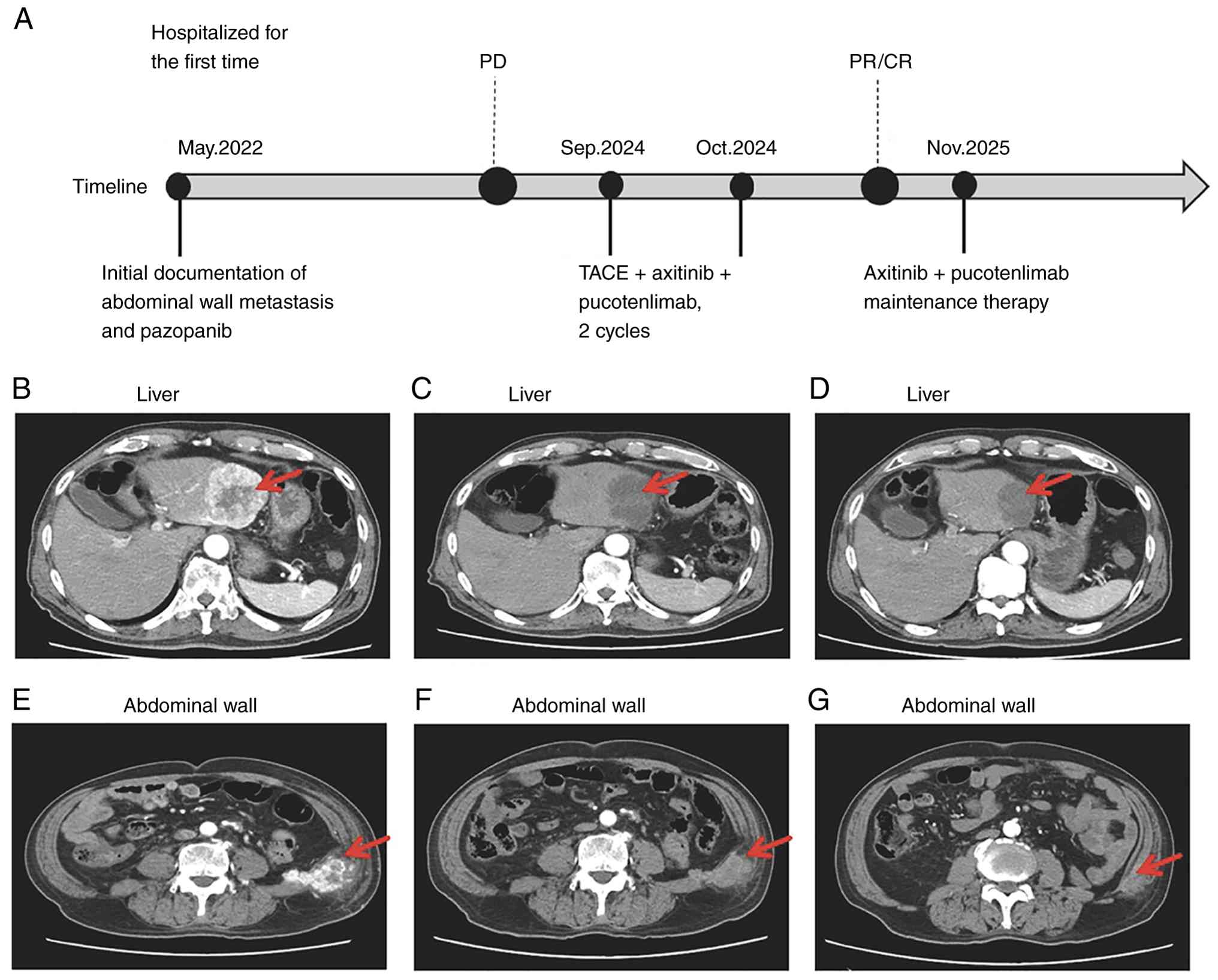
under fluoroscopic monitoring. The detailed treatment timeline is
presented in Fig. 3A.
A total of 2 weeks after the first DEB-TACE session
in October 2024, second-line systemic therapy with axitinib (3 mg
orally, twice daily) and pucotenlimab (200 mg intravenously, every
3 weeks) was initiated after disease progression on pazopanib. The
patient received 17 cycles of pucotenlimab by the last follow-up in
November 2025, with axitinib maintenance therapy continued during
follow-up. No treatment-related adverse events were observed during
follow-up through to November 2025, based on regular outpatient
assessments, including clinical evaluation, patient-reported
symptoms, physical examination and laboratory tests. Therefore, no
adverse-event grade was assigned according to Common Terminology
Criteria for Adverse Events version 5.0 (34).
The first treatment response assessment was
conducted in late October 2024. Comparison of contrast-enhanced CT
scans (Fig. 3C and F) with baseline
images (Fig. 3B and E) revealed a
notable reduction in both the liver and abdominal wall metastases,
consistent with a partial response (PR) according to the Response
Evaluation Criteria in Solid Tumors guideline (RECIST; version 1.1)
(35). The modified RECIST
(mRECIST) guideline (36), which is
not validated as the standard response criterion for metastatic
RCC, was retained only as an exploratory complementary assessment
of viable enhancing tissue after embolization and was not used as
the primary efficacy criterion. By this exploratory assessment, the
lesions revealed notable internal necrosis and were classed as PR,
with one intrahepatic lesion achieving a complete response (CR).
All serial CT scans, target-lesion measurements and RECIST version
1.1 classifications were independently evaluated and validated by a
board-certified radiologist with 20 years of experience in
oncological imaging. Clinically, the patient remained asymptomatic
during follow-up and no new tumor-related symptoms were reported.
CEA and CA19-9 were measured as part of routine clinical laboratory
assessment. However, neither CEA nor CA19-9 has been validated as a
biomarker for treatment-response assessment in ccRCC; therefore,
treatment response in this patient was evaluated primarily
according to radiological criteria (RECIST version 1.1) rather than
changes in serum tumor marker level (37,38).
Accordingly, treatment response was determined radiologically
according to the RECIST version 1.1 guideline and was not inferred
from changes in serum CEA or CA19-9 levels. Baseline CEA and CA19-9
levels were 38.5 ng/ml and 52.1 U/ml, respectively. The CEA and
CA19-9 levels subsequently decreased to 2.53 ng/ml and <2.0 U/ml
in September 2024, to 1.95 ng/ml and <2.0 U/ml in October 2024,
and to 1.85 ng/ml and <2.0 U/ml in November 2024, respectively.
To provide a quantitative assessment of treatment response, target
lesion measurements were recorded according to RECIST version 1.1
at baseline before treatment (September 2024), first radiological
assessment after treatment (October 2024) and the last follow-up
assessment (November 2025). Hepatic segment (S) 6 metastasis
decreased from 65 mm at baseline to 32 mm at the first follow-up
and 18 mm at the last follow-up, corresponding to a 72.3%
reduction. The hepatic S4 metastasis decreased from 38 to 19 mm and
subsequently disappeared completely (0 mm), corresponding to a CR
of the lesion. Abdominal wall metastasis decreased from 73 to 35 mm
and then to 20 mm, corresponding to a 72.6% reduction. Overall, the
sum of the target lesion diameters decreased from 176 mm at
baseline to 86 mm at the first follow-up and 38 mm at the last
follow-up, representing a 78.4% reduction from baseline and meeting
the RECIST guideline for PR (Table
I).
 | Table I.Changes in target lesion size and
RECIST response assessment. |
Table I.
Changes in target lesion size and
RECIST response assessment.
| Lesion site | Baseline (September
2024)a | First follow-up
(October 2024)a | Last follow-up
(November 2025)a | Change, % | RECIST version
1.1 | mRECIST |
|---|
| Hepatic S6
metastasis | 65 | 32 | 18 | −72.3 | PR | PR |
| Hepatic S4
metastasis | 38 | 19 | 0 | −100.0 | CR | CR |
| Abdominal wall
metastasis | 73 | 35 | 20 | −72.6 | PR | NA |
| Sum of
diameters | 176 | 86 | 38 | −78.4 | PR | - |
Maintenance immunotherapy and targeted therapy were
continued from October 2024 to November 2025 (~13 months), with
sustained disease control observed during follow-up. After >1
year of follow-up, the quality of life of the patient had improved
markedly, with the ECOG performance status improving from 1 at
presentation to 0 at the most recent follow-up (November 2025).
Follow-up evaluations were performed through regular outpatient
assessments, including clinical examination, ECOG performance
status evaluation, laboratory testing and contrast-enhanced CT
imaging. In November 2025, a repeat contrast-enhanced abdominal CT
(Fig. 3D and G) demonstrated
sustained disease control.
Discussion
Therapeutic options for ccRCC with abdominal wall
metastasis are limited due to its rarity. Stereotactic body
radiotherapy (SBRT) is an effective and well-established local
option for metastatic RCC (39,40),
and SIRT represents another catheter-based locoregional approach
that has been investigated mainly for hepatic malignancies,
including hepatocellular carcinoma, intrahepatic cholangiocarcinoma
and selected liver metastases (18–20).
However, the relatively high cost and limited financial
accessibility of SIRT markedly restrict its routine clinical use.
Therefore, it was not a feasible option for the patient in the
present case. The multidisciplinary team discussed surgery and
SBRT; however, further surgery was not recommended due to the prior
radical nephrectomy performed in the patient, the intricate
anatomical relationship of the abdominal wall lesion and to avoid
another procedure as per the request of the patient. Similarly,
SBRT was not selected after considering the anatomy of the lesion
and patient preference. DEB-TACE may be particularly useful in
selected patients with bulky, hypervascular and anatomically
complex lesions with multiple tumor-feeding arteries (41–43).
In the present case, angiography demonstrated a multi-source
arterial supply to the abdominal wall metastasis from two left
lumbar arteries, the iliolumbar artery and the deep circumflex
iliac artery, permitting superselective treatment of the identified
feeding vessels. Therefore, rather than implying an advantage over
SBRT, the present case emphasizes DEB-TACE as a technically
appropriate local treatment option for this specific lesion pattern
and clinical context. In contrast to systemic therapy alone, the
rapid and effective local tumor debulking achieved by DEB-TACE in
the aforementioned protocol may have created a more favorable tumor
microenvironment providing an opportunity for subsequent
immunotherapy and targeted therapy to exert their effects. The
precision and high efficacy of DEB-TACE are key to its successful
implementation. The complex vascular network of the abdominal wall
metastases, spanning the abdominal aorta and both the internal and
external iliac arteries, is a major contributor to the failure of
conventional treatments (12–14).
Using angiographic analysis and superselective catheterization,
DEB-TACE successfully addressed all tumor-feeding arteries. The
strategy of superselective embolization targeting multiple
tumor-feeding arteries enabled comprehensive devascularization of
the metastatic lesions.
To further contextualize the magnitude of response
observed, the present case was compared with previously published
outcomes of axitinib-based immune checkpoint inhibitor regimens in
advanced RCC. As current research on axitinib plus pucotenlimab in
RCC is limited, the most relevant benchmarks are derived from other
axitinib-based immune checkpoint inhibitor combinations. This
regimen was selected because axitinib is an established second-line
treatment after prior anti-angiogenic therapy, VEGF/PD-1
co-inhibition is biologically rational in RCC, and pucotenlimab is
locally available (44–47). By contrast, standard
VEGF-immuno-oncology combinations, such as pembrolizumab plus
axitinib were developed mainly for previously untreated advanced
RCC. In the phase III KEYNOTE-426 trial, pembrolizumab plus
axitinib achieved an objective response rate (ORR) of 59.3% and a
median progression-free survival (PFS) time of 15.1 months in
previously untreated advanced ccRCC; in the 5-year update, the ORR
remained 60.6% (48). Similarly, in
the phase III JAVELIN Renal 101 trial, avelumab plus axitinib
achieved an ORR of 55.2% and a median PFS time of 13.8 months
(49). In patients with previously
treated disease, a retrospective multicenter study of second-line
axitinib plus toripalimab after prior VEGFR-TKI failure reported an
ORR of 31.6% and median PFS time of 11.7 months (50). Compared with this background, the
patient in the present case, who had already progressed on
pazopanib, experienced a rapid radiographic PR, marked intratumoral
necrosis by mRECIST, complete radiological response of one hepatic
lesion and durable disease control for >1 year after DEB-TACE
followed by axitinib plus pucotenlimab. Although causality cannot
be established from a single case report, the depth and speed of
the local response suggested that DEB-TACE may have provided
additional benefits beyond systemic therapy alone, particularly by
rapidly reducing tumor vascularity and burden.
No tumor microenvironment or immune biomarker
assessment was performed in the present case report; therefore, it
remains to be elucidated if local tumor devascularization or
debulking affected the tumor microenvironment or systemic therapy.
The abnormal rapid and notable response observed in the present
case may reflect a biologically plausible interaction between
locoregional and systemic therapies rather than the isolated effect
of any single treatment component. DEB-TACE likely contributed not
only through immediate devascularization and localized cytotoxic
drug delivery, but also by inducing extensive tumor necrosis, which
may promote the release of tumor antigens (for example,
tumor-associated antigens and neoantigens) and damage-associated
molecular patterns, such as high mobility group box 1, heat shock
proteins and extracellular ATP, together with proinflammatory
cytokines including interleukin (IL)-1β, IL-6, IL-8 and tumor
necrosis factor-α, thereby enhancing antitumor immune priming
(51–53). This finding provides a potential
mechanistic rationale in combining locoregional therapy with PD-1
blockade in the future. In parallel, anti-angiogenic treatment with
axitinib may help suppress hypoxia-driven neovascularization after
embolization, thereby reinforcing the local effects of TACE and
contributing to systemic antitumor activity (54). Therefore, in the present case, the
observed benefit may be further understood as the result of
complementary local tumor control and systemic immune-targeted
therapy acting in combination, rather than as separate and
disconnected therapeutic effects. Furthermore, locoregional
therapies can induce antigen and proinflammatory cytokine release.
Anti-VEGF/TKI strategies have been proposed to counter
post-embolization angiogenic rebound (55), providing a biologically coherent
framework for the multimodal treatment strategy used in the present
case.
In the comprehensive management of ccRCC,
particularly in cases refractory to systemic therapy, DEB-TACE may
represent a notable adjunctive treatment with rapid tumor-debulking
capability. The principal advantage of DEB-TACE lies in the precise
targeting of lesions. By embolizing tumor-feeding arteries and
locally delivering a sustained local concentration of
chemotherapeutic drugs, DEB-TACE can efficiently induce tumor
ischemia and necrosis, resulting in rapid tumor reduction. In the
present case, multimodality treatment was associated with rapid
tumor shrinkage and complete radiological CR of the treated
abdominal wall lesion, resulting in a marked reduction of the local
tumor burden. This observation should be interpreted cautiously;
however, it is broadly consistent with emerging evidence suggesting
that DEB-TACE may provide meaningful local control in selected RCC
cases, including unresectable disease and rare reports of complete
response (8,15). This rapid local control creates a
key therapeutic window, enabling improved control of
oligometastatic activity and enhancing the potential benefit of
subsequent systemic therapies. Therefore, as a notable adjunctive
modality in the comprehensive treatment of advanced ccRCC, DEB-TACE
offers marked clinical value in potentially overcoming treatment
resistance and improving patient outcomes in the future (56,57).
Previously published case reports relevant to the
present case remains limited and heterogeneous (Table II). A 2015 case report described an
isolated abdominal wall metastasis from ccRCC occurring 2 years
after nephrectomy treated with wide excision (58), whereas a 2025 case report described
an abdominal wall metastasis as the initial presentation of an
otherwise occult ccRCC, followed by excision of the abdominal wall
mass and radical nephrectomy (59).
These case reports emphasized the rarity and variable presentation
of abdominal wall involvement; however, neither study evaluated
transarterial treatment. Regarding DEB-TACE in RCC, Bi et al
(8) reported a 35-patient series of
unresectable RCC in which the 1-month ORR was 47.1% and the disease
control rate was 94.1%, with no treatment-related grade ≥3 adverse
events. Qiu et al (15)
reported a CR after DEB-TACE in a patient with hepatic and
retroperitoneal lymph node metastases from RCC after systemic
treatment failure. Compared with these case reports, the present
case is distinguished by the abnormal complex multi-source arterial
supply of the abdominal wall metastases and the use of DEB-TACE
within a multimodality regimen after pazopanib progression. As
patient populations, disease sites, systemic treatments and
response methods differ markedly among case reports, direct
comparisons of treatment efficacy should be interpreted with
caution.
| Table II.Selected published reports relevant
to abdominal wall metastasis and DEB-TACE in renal cell
carcinoma. |
Table II.
Selected published reports relevant
to abdominal wall metastasis and DEB-TACE in renal cell
carcinoma.
| First author,
yeara | Clinical
setting/disease site | Treatment
strategy | Key reported
findings |
Relevance/limitation | (Refs.) |
|---|
| Kumar et al,
2015 | Isolated abdominal
wall metastasis from ccRCC 2 years after nephrectomy | Wide excision | Surgical management
of a rare delayed isolated abdominal wall metastasis | Illustrates rarity
and presentation; no transarterial treatment | (58) |
| Yu et al,
2025 | Abdominal wall
metastasis as the initial presentation of otherwise occult
ccRCC | Excision of
abdominal wall mass followed by radical nephrectomy | Case report of
abdominal wall involvement at initial presentation | Illustrates
variable presentation; no transarterial treatment | (59) |
| Bi et al,
2022 | 35 patients with
unresectable RCC | Doxorubicin-loaded
CalliSphere® DEB-TACE | 1-month ORR, 47.1%;
disease control rate, 94.1%; no treatment-related grade ≥3 adverse
events | Series supports
feasibility of DEB- TACE in selected RCC; population and disease
setting differ from the present case | (8) |
| Qiu et al,
2024 | RCC with liver and
retroperitoneal lymph node metastases after systemic treatment
failure | DEB-TACE | Complete response
reported in a single case | Supports potential
local activity after systemic treatment failure; single-case
evidence | (15) |
| Present case | ccRCC with
abdominal wall and liver metastases; complex multi-source arterial
supply; progression on pazopanib | SuperselectiveDEB-
TACE followed by axitinib plus pucotenlimab | RECIST version 1.1,
PR; sum of target diameters, 176 to 38 mm; sustained disease
control observed during >1 year of available follow-up | Multimodal
treatment contributions cannot be separated; no mechanistic synergy
can be inferred | - |
In conclusion, the present case highlighted the
potential role of DEB-TACE as part of a multimodal treatment
strategy for patients with advanced ccRCC. However, further
validation in larger prospective clinical studies is warranted.
Future research should focus on optimizing the timing of
combination therapy (for example, concurrent vs. sequential
administration), comparing the efficacy of different drug
combinations, and identifying predictive biomarkers to guide
individualized treatment strategies and potentially improve patient
outcomes.
Acknowledgements
Not applicable.
Funding
Funding: No funding was received.
Availability of data and materials
The data generated in the present study may be
requested from the corresponding author.
Authors' contributions
YG and QX contributed to the acquisition and
analysis of clinical data, interpreted the clinical findings and
drafted the manuscript. DK contributed to the interpretation of
imaging and clinical data and critically revised the manuscript. LP
conceptualized and designed the study, supervised the treatment
strategy, interpreted the data and critically revised the
manuscript. YG and LP confirm the authenticity of all the raw data.
All authors read and approved the final manuscript.
Ethics approval and consent to
participate
Not applicable.
Patient consent for publication
Written informed consent was obtained from the
patient for the publication of their clinical data and any
associated medical images.
Competing interests
The authors declare that they have no competing
interests.
References
|
1
|
Barragan-Carrillo R, Saad E, Saliby RM,
Sun M, Albiges L, Bex A, Heng D, Mejean A, Motzer RJ, Plimack ER,
et al: First and Second-line treatments in metastatic renal cell
carcinoma. Eur Urol. 87:143–154. 2025. View Article : Google Scholar : PubMed/NCBI
|
|
2
|
Singer EA, Rumble RB and Van Veldhuizen
PJ: Management of metastatic clear cell renal cell carcinoma: ASCO
Guideline Q&A. JCO Oncol Pract. 19:127–131. 2023. View Article : Google Scholar : PubMed/NCBI
|
|
3
|
Sepe P, Mennitto A, Corti F and Procopio
G: Immunotherapeutic targets and therapy for renal cell carcinoma.
Immunotargets Ther. 9:273–288. 2020. View Article : Google Scholar : PubMed/NCBI
|
|
4
|
Boyle JJ, Pfail JL, Lichtbroun BJ and
Singer EA: Adjuvant therapy for renal cell carcinoma: End points,
outcomes, and risk assessments. JCO Precis Oncol. 7:e22004072023.
View Article : Google Scholar : PubMed/NCBI
|
|
5
|
Zhang L, Zhang Y, Wen D, Guo S, Qi X, Wang
H, Sun Y, Yang G, Wang Y and Xue S: NFS1 Regulates IDH2 to
attenuate abdominal aortic aneurysms via interacting with SP2. Adv
Sci (Weinh). 13:e52402026.
|
|
6
|
Singh T and Andi K: Iliolumbar artery: A
useful pedicle for the iliac crest free flap in maxillofacial
reconstruction. Br J Oral Maxillofac Surg. 55:270–273. 2017.
View Article : Google Scholar : PubMed/NCBI
|
|
7
|
Hardman RL, Lopera JE, Cardan RA, Trimmer
CK and Josephs SC: Common and rare collateral pathways in
aortoiliac occlusive disease: A pictorial essay. AJR Am J
Roentgenol. 197:W519–W524. 2011. View Article : Google Scholar : PubMed/NCBI
|
|
8
|
Bi Y, Shi X, Ren J, Yi M and Han X:
Transarterial chemoembolization of unresectable renal cell
carcinoma with doxorubicin-loaded CalliSpheres drug-eluting beads.
Sci Rep. 12:81362022. View Article : Google Scholar : PubMed/NCBI
|
|
9
|
Comandone A, Vana F, Comandone T and Tucci
M: Antiangiogenic therapy in clear cell renal carcinoma (CCRC):
Pharmacological basis and clinical results. Cancers (Basel).
13:58962021. View Article : Google Scholar : PubMed/NCBI
|
|
10
|
Rassy E, Flippot R and Albiges L: Tyrosine
kinase inhibitors and immunotherapy combinations in renal cell
carcinoma. Ther Adv Med Oncol. 12:17588359209075042020. View Article : Google Scholar : PubMed/NCBI
|
|
11
|
Climent C, Soriano S, Bonfill T, Lopez N,
Rodriguez M, Sierra M, Andreu P, Fragio M, Busquets M, Carrasco A,
et al: The role of immunotherapy in non-clear cell renal cell
carcinoma. Front Oncol. 13:9418352023. View Article : Google Scholar : PubMed/NCBI
|
|
12
|
Loukas M, Hullett J, Louis RG Jr, Kapos T,
Knight J, Klueber K and Jordan R: The clinical anatomy of the
abdominal wall. Clin Anat. 20:702–709. 2007.
|
|
13
|
Wright N and Burns B: Anatomy, Abdomen and
Pelvis: Posterior Abdominal Wall Arteries. StatPearls Publishing.
updated. 2022
|
|
14
|
Loukas M, Myers C, Shah R, Tubbs RS,
Wartmann C, Apaydin N, Betancor J and Jordan R: Arcuate line of the
rectus sheath: Clinical approach. Anat Sci Int. 83:140–144. 2008.
View Article : Google Scholar : PubMed/NCBI
|
|
15
|
Qiu Z, Ji J, Zhang S and Wang S: Case
report: Drug-eluting bead transcatheter arterial chemoembolization
in liver metastasis and retroperitoneal lymph node metastases of
renal cell carcinoma: Effective local therapy with the first
report. Front Oncol. 14:13714142024. View Article : Google Scholar : PubMed/NCBI
|
|
16
|
Melchiorre F, Patella F, Pescatori L,
Pesapane F, Fumarola E, Biondetti P, Brambillasca P, Monaco C,
Ierardi AM, Franceschelli G and Carrafiello G: DEB-TACE: A standard
review. Future Oncol. 14:2969–2984. 2018. View Article : Google Scholar : PubMed/NCBI
|
|
17
|
Marcacuzco Quinto A, Nutu O, San Román
Manso R, Justo Alonso I, Calvo Pulido J, Manrique Municio A,
García-Sesma Á, Loinaz Segurola C, Martínez Caballero J and Jiménez
Romero LC: Complications of transarterial chemoembolization (TACE)
in the treatment of liver tumors. Cir Esp (Engl Ed). 96:560–567.
2018. View Article : Google Scholar : PubMed/NCBI
|
|
18
|
Faggian G, Imbriaco M, Guarino M, Bagni O,
Filippi L, Quartuccio N and DE Luca S: Role of SIRT in patients
with hepatocellular carcinoma: Current clinical indications and
future perspectives. In Vivo. 40:2139–2149. 2026. View Article : Google Scholar : PubMed/NCBI
|
|
19
|
Gosztonyi B, Pestalozzi B, Kenkel D,
Engel-Bicik I, Kaufmann PA, Treyer V and Siebenhüner AR: A
descriptive analysis of the characteristics, treatment response and
prognosis of hepatic dominant solid tumors undergoing selective
internal radiation therapy (SIRT). J Gastrointest Oncol.
13:3240–3253. 2022. View Article : Google Scholar : PubMed/NCBI
|
|
20
|
Hirsch RD, Mills C, Sawhney R, Sood S,
Bird V, Mishra G, Dev A, Kemp W, Lubel J, Roberts SK, et al: SIRT
compared with DEB-TACE for hepatocellular carcinoma: A Real-world
study (the SITAR Study). J Gastrointest Cancer. 52:907–914. 2021.
View Article : Google Scholar : PubMed/NCBI
|
|
21
|
Larroquette M, Peyraud F, Domblides C,
Lefort F, Bernhard JC, Ravaud A and Gross-Goupil M: Adjuvant
therapy in renal cell carcinoma: Current knowledges and future
perspectives. Cancer Treat Rev. 97:1022072021. View Article : Google Scholar : PubMed/NCBI
|
|
22
|
Ballesteros PÁ, Chamorro J, Román-Gil MS,
Pozas J, Gómez Dos Santos V, Granados ÁR, Grande E, Alonso-Gordoa T
and Molina-Cerrillo J: Molecular mechanisms of resistance to
immunotherapy and antiangiogenic treatments in clear cell renal
cell carcinoma. Cancers (Basel). 13:59812021. View Article : Google Scholar : PubMed/NCBI
|
|
23
|
Özalp FR, Yörükoğlu K, Yıldırım EÇ, Uzun M
and Semiz HS: Prognostic value of B7-H3 expression in metastatic
renal cell carcinoma and its impact on immunotherapy response. BMC
Cancer. 24:14712024. View Article : Google Scholar : PubMed/NCBI
|
|
24
|
Quhal F, Mori K, Bruchbacher A, Resch I,
Mostafaei H, Pradere B, Schuettfort VM, Laukhtina E, Egawa S,
Fajkovic H, et al: First-line immunotherapy-based combinations for
metastatic renal cell carcinoma: A systematic review and network
Meta-analysis. Eur Urol Oncol. 4:755–765. 2021. View Article : Google Scholar : PubMed/NCBI
|
|
25
|
Oken MM, Creech RH, Tormey DC, Horton J,
Davis TE, McFadden ET and Carbone PP: Toxicity and response
criteria of the Eastern Cooperative Oncology Group. Am J Clin
Oncol. 5:649–655. 1982. View Article : Google Scholar : PubMed/NCBI
|
|
26
|
Delahunt B, Cheville JC, Martignoni G,
Humphrey PA, Magi-Galluzzi C, McKenney J, Egevad L, Algaba F, Moch
H, Grignon DJ, et al: The international society of urological
pathology (ISUP) grading system for renal cell carcinoma and other
prognostic parameters. Am J Surg Pathol. 37:1490–1504. 2013.
View Article : Google Scholar : PubMed/NCBI
|
|
27
|
Truong LD and Shen SS: Immunohistochemical
diagnosis of renal neoplasms. Arch Pathol Lab Med. 135:92–109.
2011. View Article : Google Scholar : PubMed/NCBI
|
|
28
|
Sangoi AR, Karamchandani J, Kim J, Pai RK
and McKenney JK: The use of immunohistochemistry in the diagnosis
of metastatic clear cell renal cell carcinoma: A review of PAX-8,
PAX-2, hKIM-1, RCCma, and CD10. Adv Anat Pathol. 17:377–383. 2010.
View Article : Google Scholar : PubMed/NCBI
|
|
29
|
Shi SR, Liu C and Taylor CR:
Standardization of immunohistochemistry for formalin-fixed,
paraffin-embedded tissue sections based on the antigen-retrieval
technique: From experiments to hypothesis. J Histochem Cytochem.
55:105–109. 2007. View Article : Google Scholar : PubMed/NCBI
|
|
30
|
Shi SR, Shi Y and Taylor CR: Antigen
retrieval immunohistochemistry: Review and future prospects in
research and diagnosis over two decades. J Histochem Cytochem.
59:13–32. 2011. View Article : Google Scholar : PubMed/NCBI
|
|
31
|
Fitzgibbons PL, Bradley LA, Fatheree LA,
Alsabeh R, Fulton RS, Goldsmith JD, Haas TS, Karabakhtsian RG,
Loykasek PA, Marolt MJ, et al: Principles of analytic validation of
immunohistochemical assays: Guideline from the College of American
Pathologists Pathology and Laboratory Quality Center. Arch Pathol
Lab Med. 138:1432–1443. 2014. View Article : Google Scholar : PubMed/NCBI
|
|
32
|
Heng DYC, Xie W, Regan MM, Warren MA,
Golshayan AR, Sahi C, Eigl BJ, Ruether JD, Cheng T, North S, et al:
Prognostic factors for overall survival in patients with metastatic
renal cell carcinoma treated with vascular endothelial growth
factor-targeted agents: Results from a large, multicenter study. J
Clin Oncol. 27:5794–5799. 2009. View Article : Google Scholar : PubMed/NCBI
|
|
33
|
Karnofsky DA and Burchenal JH: The
clinical evaluation of chemotherapeutic agents in cancer. MacLeod
CM: Evaluation of Chemotherapeutic Agents New York, Columbia:
University Press; pp. 191–205. 1949
|
|
34
|
National Cancer Institute, . Common
Terminology Criteria for Adverse Events (CTCAE) Version 5.0.
Bethesda (MD): U.S Department of Health and Human Services,
National Institutes of Health; 2017
|
|
35
|
Eisenhauer EA, Therasse P, Bogaerts J,
Schwartz LH, Sargent D, Ford R, Dancey J, Arbuck S, Gwyther S,
Mooney M, et al: New response evaluation criteria in solid tumours:
Revised RECIST guideline (version 1.1). Eur J Cancer. 45:228–247.
2009. View Article : Google Scholar : PubMed/NCBI
|
|
36
|
Lencioni R and Llovet JM: Modified RECIST
(mRECIST) assessment for hepatocellular carcinoma. Semin Liver Dis.
30:52–60. 2010. View Article : Google Scholar : PubMed/NCBI
|
|
37
|
Labaki C, Saliby RM, Bakouny Z, Saad E,
Semaan K, Eid M, Lalani AK, Choueiri TK and Braun DA: Emerging
biomarkers of response to systemic therapies in metastatic clear
cell renal cell carcinoma. Hematol Oncol Clin North Am. 37:937–942.
2023. View Article : Google Scholar : PubMed/NCBI
|
|
38
|
Rathmell WK, Rumble RB, Van Veldhuizen PJ,
Al-Ahmadie H, Emamekhoo H, Hauke RJ, Louie AV, Milowsky MI, Molina
AM, Rose TL, et al: Management of Metastatic Clear Cell Renal Cell
Carcinoma: ASCO Guideline. J Clin Oncol. 40:2957–2995. 2022.
View Article : Google Scholar : PubMed/NCBI
|
|
39
|
Villafuerte CJQ and Swaminath A:
Stereotactic body radiotherapy for renal cell Carcinoma-A review of
use in the primary, cytoreductive and oligometastatic settings.
Cancers (Basel). 16:33342024. View Article : Google Scholar : PubMed/NCBI
|
|
40
|
Altoos B, Amini A, Yacoub M, Bourlon MT,
Kessler EE, Flaig TW, Fisher CM, Kavanagh BD, Lam ET and Karam SD:
Local control rates of metastatic renal cell carcinoma (RCC) to
thoracic, abdominal, and soft tissue lesions using stereotactic
body radiotherapy (SBRT). Radiat Oncol. 10:2182015. View Article : Google Scholar : PubMed/NCBI
|
|
41
|
Sun B, Gao JQ, Li RJ, Song L, Li N and Gao
F: Combination of drug-eluting bead transarterial chemoembolization
and PD-1 inhibitor for treatment of unresectable head and neck
squamous cell carcinoma: An initial, short-term clinical experience
in a single-center retrospective cohort study. Front Immunol.
16:16154402025. View Article : Google Scholar : PubMed/NCBI
|
|
42
|
Liu L, Sheng Y, Liang X, Yang T, Dong L,
Gao D, Chen B, Xu T, Ai M, Shan H, et al: Retrospective analysis of
the clinical efficacy, safety, and influencing factors of
Drug-eluting bead transarterial chemoembolization for unresectable
hepatic malignancies. Clin Med Insights Oncol.
20:117955492614342592026. View Article : Google Scholar : PubMed/NCBI
|
|
43
|
Wang Q, Zhu L and Sheng Q: Clinical
research progress of callisperes® of drug-loaded
microsphere arterial chemoembolisation in the treatment of solid
tumors. Discov Oncol. 15:1612024. View Article : Google Scholar : PubMed/NCBI
|
|
44
|
Rini BI, Escudier B, Tomczak P, Kaprin A,
Szczylik C, Hutson TE, Michaelson MD, Gorbunova VA, Gore ME,
Rusakov IG, et al: Comparative effectiveness of axitinib versus
sorafenib in advanced renal cell carcinoma (AXIS): A randomised
phase 3 trial. Lancet. 378:1931–1939. 2011. View Article : Google Scholar : PubMed/NCBI
|
|
45
|
Motzer RJ, Penkov K, Haanen J, Rini B,
Albiges L, Campbell MT, Venugopal B, Kollmannsberger C, Negrier S,
Uemura M, et al: Avelumab plus axitinib versus sunitinib for
advanced renal-cell carcinoma. N Engl J Med. 380:1103–1115. 2019.
View Article : Google Scholar : PubMed/NCBI
|
|
46
|
Rini BI, Plimack ER, Stus V, Gafanov R,
Hawkins R, Nosov D, Pouliot F, Alekseev B, Soulières D, Melichar B,
et al: Pembrolizumab plus Axitinib versus sunitinib for advanced
renal-cell carcinoma. N Engl J Med. 380:1116–1127. 2019. View Article : Google Scholar : PubMed/NCBI
|
|
47
|
Hato SV, Khong A, de Vries IJM and
Lesterhuis WJ: Molecular pathways: The immunogenic effects of VEGF
inhibition in cancer. Clin Cancer Res. 21:1349–1355. 2015.
|
|
48
|
Rini BI, Plimack ER, Stus V, Gafanov R,
Waddell T, Nosov D, Pouliot F, Alekseev B, Soulières D, Melichar B,
et al: Pembrolizumab plus axitinib versus sunitinib for avanced
clear cell renal cell carcinoma: 5-year survival and biomarker
analyses of the phase 3 KEYNOTE-426 trial. Nat Med. 31:3475–3484.
2025. View Article : Google Scholar : PubMed/NCBI
|
|
49
|
Choueiri TK, Motzer RJ, Rini BI, Haanen J,
Campbell MT, Venugopal B, Kollmannsberger C, Gravis-Mescam G,
Uemura M, Lee JL, et al: Updated efficacy results from the JAVELIN
Renal 101 trial: First-line avelumab plus axitinib versus sunitinib
in patients with advanced renal cell carcinoma. Ann Oncol.
31:1030–1039. 2020. View Article : Google Scholar : PubMed/NCBI
|
|
50
|
Huang J, Shi G, Wang Y, Wang P, Zhang J,
Kong W, Huang Y, Wang S and Xue W: Second-line treatment with
axitinib plus toripalimab in metastatic renal cell carcinoma: A
retrospective multicenter study. Future Oncol. 18:1461–1471. 2022.
View Article : Google Scholar : PubMed/NCBI
|
|
51
|
Pol J, Vacchelli E, Aranda F, Castoldi F,
Eggermont A, Cremer I, Sautès-Fridman C, Fucikova J, Galon J,
Spisek R, et al: Trial Watch: Immunogenic cell death inducers for
anticancer chemotherapy. Oncoimmunology. 4:e10088662015. View Article : Google Scholar : PubMed/NCBI
|
|
52
|
Kim D and Erinjeri JP: Postablation Immune
Microenvironment: Synergy between Interventional Oncology and
Immuno-oncology. Semin Intervent Radiol. 36:334–342. 2019.
View Article : Google Scholar : PubMed/NCBI
|
|
53
|
Kim NJ, Yoon JH, Tuomi AC, Lee J and Kim
D: In-situ tumor vaccination by percutaneous ablative therapy and
its synergy with immunotherapeutics: An update on combination
therapy. Front Immunol. 14:11188452023. View Article : Google Scholar : PubMed/NCBI
|
|
54
|
Petrillo M, Patella F, Pesapane F, Suter
MB, Ierardi AM, Angileri SA, Floridi C, de Filippo M and
Carrafiello G: Hypoxia and tumor angiogenesis in the era of
hepatocellular carcinoma transarterial loco-regional treatments.
Future Oncol. 14:2957–2967. 2018. View Article : Google Scholar : PubMed/NCBI
|
|
55
|
Ding L, Dong HY, Zhou TR, Wang YH, Yan T,
Li JC, Wang ZY, Li J and Liang C: PD-1/PD-L1 inhibitors-based
treatment for advanced renal cell carcinoma: Mechanisms affecting
efficacy and combination therapies. Cancer Med. 10:6384–6401. 2021.
View Article : Google Scholar : PubMed/NCBI
|
|
56
|
Xu X, Wang J, Wang Y, Zhu Y, Wang J and
Guo J: PSMD2 overexpression as a biomarker for resistance and
prognosis in renal cell carcinoma treated with immune checkpoint
and tyrosine kinase inhibitors. Cell Oncol (Dordr). 47:1943–1956.
2024. View Article : Google Scholar : PubMed/NCBI
|
|
57
|
Ishihara H, Omae K, Nemoto Y, Ishiyama R,
Tachibana H, Nishimura K, Ikeda T, Kobari Y, Fukuda H, Yoshida K,
et al: First-line dual immune checkpoint inhibitor therapies versus
combination therapies comprising immune checkpoint inhibitors and
tyrosine kinase inhibitors for advanced renal cell carcinoma: A
comparative analysis of the effectiveness using real-world data.
Int J Clin Oncol. 29:473–480. 2024. View Article : Google Scholar : PubMed/NCBI
|
|
58
|
Kumar S, Sharma PK and Bera MK: Isolated
abdominal wall metastasis from renal cell carcinoma: Unusual
presentation. Adv Biomed Res. 4:652015. View Article : Google Scholar : PubMed/NCBI
|
|
59
|
Yu J, Sun Z, Chen Q and Yin M: Left-sided
clear cell renal cell carcinoma initially presenting with isolated
abdominal wall metastasis: A case report. Discov Oncol.
16:19382025. View Article : Google Scholar : PubMed/NCBI
|