Introduction
Gastric cancer ranks as the third most prevalent
malignancy in China with regard to both incidence and mortality,
accounting for 44.0% of new GC cases and 48.6% of GC-related deaths
worldwide (1,2). Despite the overall incidence of
gastric cancer having declined in recent decades, the proportion of
gastric signet-ring cell carcinoma (GSRCC), a distinct
histopathological subtype, has exhibited an increasing trend
(3). GSRCC is characterized by
abundant intracytoplasmic mucin that displaces the nucleus to the
periphery, creating the classic ‘signet-ring’ morphology. According
to the World Health Organization classification, gastric
adenocarcinoma with signet-ring cell component of >50% is
defined as GSRCC, which falls under the category of poorly cohesive
carcinomas (4). Due to the marked
heterogeneity of gastric cancer, pure GSRCC is relatively uncommon
in clinical practice (5), with
mixed adenocarcinomas containing signet-ring cell components being
more frequently encountered.
GSRCC is associated with aggressive biological
behavior, including high propensity for lymph node metastasis and
distant dissemination, contributing to unfavorable prognosis
(6). Notably, early-stage GSRCC
demonstrates markedly lower metastatic potential and more favorable
response to curative treatment, underscoring the importance of
early detection (7). Currently, the
most established diagnostic method for GSRCC is histopathological
examination of endoscopic biopsy specimens, an invasive procedure
with inherent limitations including patient discomfort, sampling
difficulty and operational complexity (8).
Metabolomics, the comprehensive analysis of
small-molecule metabolites in biological systems, has emerged as an
important tool in cancer biomarker discovery (9). Circulating metabolites reflect the
systemic metabolic reprogramming associated with tumorigenesis and
progression, offering potential for non-invasive diagnostic
applications. Recent metabolomic studies have identified candidate
biomarkers for a number of malignancies, including hypoxanthine for
lung adenocarcinoma (10), pimelic
acid and L-serine for papillary thyroid carcinoma (11), hyaluronan and proteoglycan link
protein-1 for peritoneal metastasis in pancreatic ductal
adenocarcinoma (12). However,
systematic metabolomic profiling of GSRCC remains limited. This is
largely due to the rarity of this subtype (3–5% of gastric cancers)
(5), which limits the availability
of large sample cohorts, as well as the technical challenges posed
by its mucinous composition. These factors underscore the need for
and value of the present study.
In the present study, a dual-platform approach
integrating liquid chromatography-mass spectrometry (LC-MS) and gas
chromatography-mass spectrometry (GC-MS) was employed to
comprehensively characterize the serum metabolomic profile of
patients with GSRCC compared with healthy controls. The objectives
were to identify differential metabolites associated with GSRCC,
elucidate perturbed metabolic pathways and evaluate the exploratory
candidate metabolic signatures.
Materials and methods
Study population and sample
collection
Serum samples were prospectively collected from
patients diagnosed with GSRCC at Jiangsu Province Hospital of
Chinese Medicine (Nanjing, China) between March 2023 and November
2024. Inclusion criteria were as follows: i) Histopathologically
determined GSRCC based on surgical specimens; ii) treatment-naive
status with no prior anticancer therapy (including surgery,
chemotherapy, radiotherapy or targeted therapy); iii) age ≥18
years; iv) availability of complete clinical and pathological data,
including TNM stage, tumor location and lymph node metastasis
status; and v) absence of major underlying diseases (such as severe
cardiovascular, hepatic, renal or autoimmune disorders) and no
history of other malignancies. Exclusion criteria included: i) Any
prior surgical treatment; ii) neoadjuvant therapy; iii) presence of
other malignancies; or iv) concurrent inflammatory or metabolic
disorders. The discovery cohort comprised 8 patients with GSRCC and
10 age- and sex-matched healthy controls. In the discovery cohort,
the age range was 40–75 years for patients with GSRCC and 30–70
years for healthy controls, with 3 men (37.5%) and 5 women (62.5%)
in the GSRCC group, and 5 men (50.0%) and 5 women (50.0%) in the
control group. An independent validation cohort included 35
patients with GSRCC and 35 healthy controls. In the validation
cohort, the age range was 35–84 years for patients with GSRCC and
38–78 years for healthy controls, with 17 men (48.6%) and 18 women
(51.4%) in the GSRCC group, and 18 men (51.4%) and 17 women (48.6%)
in the control group. All GSRCC diagnoses were determined by
histopathological examination of surgical specimens. Clinical and
pathological data, including age, sex, tumor location, tumor size,
depth of invasion, lymphovascular invasion, perineural invasion and
lymph node metastasis, were systematically recorded. Venous blood
samples (3 ml) were collected preoperatively, centrifuged at 1,500
× g, 4°C for 5 min, and serum was aliquoted and stored at −80°C
until analysis.
Chemicals and reagents
LC-MS grade methanol, acetonitrile and formic acid
were purchased from Thermo Fisher Scientific, Inc. Chloroform
(analytical grade) was obtained from Shanghai Titan Scientific Co.,
Ltd. Hexane (high-performance liquid chromatography grade) was
purchased from Shanghai Anpel Experimental Technology Co., Ltd.
Pyridine (HPLC grade), O-methylhydroxylamine hydrochloride (97%)
and N,O-bis (trimethylsilyl) trifluoroacetamide (BSTFA) were
obtained from Shanghai Macklin Biochemical Co., Ltd., Shanghai
Aladdin Biochemical Technology Co., Ltd. and Tokyo Chemical
Industry Co., Ltd., respectively.
Sample preparation
Serum samples were thawed on ice and 150 µl aliquots
were transferred to microcentrifuge tubes. Protein precipitation
was performed by adding 600 µl methanol-acetonitrile (2:1, v/v)
containing a mixture of internal standards
(L-2-chlorophenylalanine, succinic acid-d4, L-valine-d8, and cholic
acid-d4) at a concentration of 4 µg/ml. Samples were vortexed at
room temperature for 1 min, sonicated in an ice-water bath for 10
min and incubated at −40°C for 2 h. Following centrifugation at
13,000 × g for 20 min at 4°C, the supernatant was collected and
filtered through 0.22 µm membrane filters. For LC-MS analysis, 150
µl supernatant was transferred to autosampler vials. For GC-MS
analysis, an additional 150 µl supernatant was evaporated to
dryness using a centrifugal concentrator. The residue was
derivatized with 80 µl methoxyamine hydrochloride in pyridine (15
mg/ml) at 37°C for 1 h, followed by addition of 50 µl BSTFA and 20
µl hexane containing internal standards, and then incubated at 70°C
for 1 h. Quality control samples were prepared by pooling equal
volumes of all study samples. The prepared serum samples were sent
to Shanghai OE Biotech Co., Ltd. for LC-MS/MS and GC-MS
analysis.
LC-MS/MS analysis
LC-MS analysis was performed using a Waters ACQUITY
UPLC I-Class PLUS System (Waters Corporation) coupled with a Thermo
Scientific™ Q Exactive™ HF mass spectrometer (Thermo Fisher
Scientific, Inc.). Chromatographic separation was achieved on an
ACQUITY UPLC HSS T3 column (100.0×2.1 mm, 1.8 µm; Waters
Corporation) maintained at 45°C. The mobile phase consisted of
water containing 0.1% formic acid (phase A) and acetonitrile (phase
B) at a flow rate of 0.35 ml/min. The injection volume was 3 µl.
The mass spectrometer was operated in both positive and negative
ionization modes with full scan range of 70–1050 m/z. The ion
source parameters were set as follows: Spray voltage, +3,500 V
(positive mode) and −3,000 V (negative mode); capillary
temperature, 320°C; auxiliary gas heater temperature, 350°C; sheath
gas flow rate, 35 Arb; auxiliary gas flow rate, 8 Arb; and S-lens
RF level, 50. The nebulizer pressure was maintained at 45 psi.
GC-MS analysis
GC-MS analysis was conducted using an Agilent
8890-5977B gas chromatograph-mass spectrometer (Agilent
Technologies, Inc.) equipped with a DB-5MS capillary column (30 m ×
0.25 mm × 0.25 µm; Agilent Technologies, Inc.). Helium was used as
carrier gas at a constant flow rate of 1.0 ml/min. The injector
temperature was set at 260°C with splitless injection of 1 µl. The
oven temperature program was as follows: Initial temperature 60°C
held for 30 sec, increased to 125°C at 8°C/min, then to 210°C at
8°C/min, then to 270°C at 15°C/min and finally to 305°C at 20°C/min
held for 5 min. The ion source temperature was 230°C, quadrupole
temperature 150°C and electron energy 70 eV. Mass spectra were
acquired in full scan mode (50–500 m/z).
Data processing and metabolite
identification
LC-MS/MS and GC-MS datasets were processed and
quality-controlled independently. For LC-MS/MS data, raw data were
processed using Progenesis QI v3.0 (Nonlinear Dynamics, Ltd.;
Waters Corporation) for baseline filtering, peak detection,
integration, retention time correction, alignment and normalization
and metabolite annotation was performed using the Human Metabolome
Database (https://www.hmdb.ca), Lipidmaps (v2.3;
http://www.lipidmaps.org), METLIN database
(http://metlin.scripps.edu) and the
in-house LuMet-Animal 3.0 LC-MS/MS database (Shanghai OE Biotech
Co., Ltd.). The qualitative score for LC-MS/MS had a maximum of 80
points, comprising 20 points for accurate mass matching, 20 points
for MS/MS fragmentation matching, 20 points for isotope
distribution matching and 20 points for retention time matching;
higher scores indicated more accurate identification. For GC-MS
data, raw data were processed using MS-DIAL v4.24 (http://prime.psc.riken.jp/Metabolomics_Software/MS-DIAL)
for peak detection, deconvolution, identification, alignment,
filtering and missing value imputation and metabolite
identification was based on retention time, fragmentation mass
spectra and similarity matching against the in-house LuMet-GC 5.0
database (Shanghai OE Biotech Co., Ltd.), with the National
Institute of Standards and Technology database (https://www.nist.gov/) also used as a reference for
auxiliary identification. The qualitative score for GC-MS had a
maximum of 100 points. For quality control and filtering, ions with
>50% missing values (zero values) within each group were removed
and remaining zero values were replaced with half of the minimum
intensity value across all samples. For LC-MS/MS data, compounds
were screened based on a qualitative score, with scores <36
considered inaccurate and excluded; for GC-MS data, a score
threshold of 70 was applied and scores <70 were excluded.
Internal standards, noise peaks, column bleed peaks and
derivatization reagent peaks were also removed.
For normalization, GC-MS data were normalized using
internal standard-based segmental normalization, where each sample
was normalized using internal standards with relative standard
deviation <0.1. Metabolite identification confidence was
assigned according to the Metabolomics Standards Initiative (MSI)
guidelines (13). For LC-MS/MS,
Level 1 was defined as retention time ± 0.3 min and fragmentation
score ≥45, Level 2 as retention time ± 0.3 min and fragmentation
score <45, Level 3 as fragmentation score ≥45 and Level 4 as
fragmentation score <45. For GC-MS, Level 1 was defined as score
≥85, Level 2 as ≤80 score <85 and Level 3 as 70≤ score <80.
In the present exploratory untargeted study, level confidence
filtering was defined as the retention of all identified
metabolites with MSI confidence Levels 1–4, as exclusion of
lower-confidence metabolites may lead to loss of biologically
relevant signals. When the same metabolite was identified by both
platforms, the metabolite intensity from the platform with the
higher qualitative score was retained. After stringent filtering,
the high-confidence metabolite matrices from LC-MS/MS (positive and
negative ion modes) and GC-MS were kept separately. Multivariate
statistical analyses [including principal component analysis (PCA),
partial least squares discriminant analysis (PLS-DA) and orthogonal
partial least squares discriminant analysis (OPLS-DA)] and
differential metabolite screening were performed independently on
the dataset of each platform. The lists of differential metabolites
identified from both platforms were then integrated for subsequent
metabolic pathway enrichment analysis.
Multivariate statistical analysis
Multivariate statistical analyses were performed
independently on the LC-MS/MS and GC-MS datasets. PCA was performed
to visualize overall distribution and assess analytical stability.
PLS-DA and OPLS-DA were applied to discriminate metabolic profiles
between groups and identify differential metabolites on each
platform separately. Variable importance in projection (VIP) values
were calculated from the OPLS-DA model. Fold change (FC) was
calculated as the ratio of mean metabolite abundance in GSRCC vs.
control groups. False discovery rate (FDR) correction (q-value) was
applied using the Benjamini-Hochberg method. Metabolites with VIP
>1, q<0.05, and log2 FC >0.58 or
log2 FC <-0.58 were considered significantly
different between groups. OPLS-DA model robustness was evaluated
using 7-fold cross-validation and 200-fold response permutation
testing. A Q2 intercept <0 determined the absence of
overfitting.
Metabolic pathway enrichment
analysis
Differential metabolites were mapped to metabolic
pathways using the Kyoto Encyclopedia of Genes and Genomes (KEGG)
database (https://www.kegg.jp/). Pathway
enrichment analysis was performed using hypergeometric testing to
identify significantly perturbed pathways (P<0.05, q<0.05).
Gene set enrichment analysis was conducted to visualize activated
and suppressed pathways.
Clinical biochemistry analysis
Routine clinical biochemistry parameters, including
cholinesterase, total protein, albumin and hemoglobin were measured
in both discovery and validation cohorts using standard automated
methods according to manufacturer's instructions. Cholinesterase
was measured using reagents from Anhui Yipunokang Biotechnology
Co., Ltd., total protein and albumin were measured using reagents
from Shenzhen Mindray Bio-Medical Electronics Co., Ltd., and
hemoglobin was measured using Sysmex Corporation reagents. BMI and
TNM stage were considered to be potential confounders in result
interpretation.
Statistical analysis
Benjamini-Hochberg method was applied to control for
FDR due to multiple comparisons inherent in metabolomics data.
Normality was assessed using the Shapiro-Wilk test. Continuous
variables are expressed as the mean ± SD or median (interquartile
range) as appropriate. Comparisons between groups were performed
using unpaired Student's t-test for normally distributed data or
Wilcoxon rank-sum test for non-normally distributed data.
Categorical variables were compared using χ2 test or
Fisher's exact test. Receiver operating characteristic (ROC) curve
analysis was performed to evaluate diagnostic performance of
candidate metabolites. Statistical analyses were conducted using
SPSS (version 26.0; IBM Corp.) and GraphPad Prism (version 8.0;
Dotmatics). P<0.05 or q<0.05 was considered to indicate a
statistically significant difference.
Results
Clinical characteristics of study
participants
Summaries of clinical characteristics of the
discovery and validation cohorts are shown in Tables I and II. In the discovery cohort, female
patients predominated (62.5%), consistent with epidemiological
patterns of this subtype. Nutritional status was assessed using
body mass index (BMI), a widely used anthropometric indicator that
reflects the balance between body weight and height and correlates
with both undernutrition (BMI <18.5) and overweight/obesity (BMI
≥25) in clinical studies. BMI exhibited no significant difference
between groups (P=0.594). TNM stage was classified using the
American Joint Committee on Cancer 8th edition system (14), with 50.0% being stage I–II and 50.0%
being stage III–IV. Lymph node metastasis was present in 5 patients
(62.5%). In the validation cohort, sex and BMI were found to be
well balanced (P=0.150). TNM staging distribution was as follows:
Stage I–II in 19 patients (54.3%) and stage III–IV in 16 patients
(45.7%). Lymph node metastasis was present in 27 patients (77.1%).
These variables were evaluated descriptively as potential
confounders rather than statistically adjusted.
 | Table I.Clinical characteristics of the
discovery cohort. |
Table I.
Clinical characteristics of the
discovery cohort.
| Characteristic | Healthy control
(n=10) | GSRCC (n=8) | P-value |
|---|
| Sex, n (%) |
|
|
|
|
Male | 5 (50.0) | 3 (37.5) |
|
|
Female | 5 (50.0) | 5 (62.5) |
|
| BMI | 22.90
(21.93–24.63) | 21.15
(20.02–26.68) | 0.594 |
| TNM stage, n
(%) |
|
|
|
|
I–II | Not applicable | 4 (50.0) |
|
|
III–IV | Not applicable | 4 (50.0) |
|
| Lymph node
metastasis, n (%) |
|
|
|
|
Yes | Not applicable | 5 (62.5) |
|
| No | Not applicable | 3 (37.5) |
|
| Table II.Clinical characteristics of the
validation cohort. |
Table II.
Clinical characteristics of the
validation cohort.
| Characteristic | Healthy control
(n=35) | GSRCC (n=35) | P-value |
|---|
| Sex, n (%) |
|
|
|
|
Male | 18 (51.4) | 17 (48.6) |
|
|
Female | 17 (48.6) | 18 (51.4) |
|
| BMI | 23.17±1.54 | 24.00±3.01 | 0.150 |
| TNM stage, n
(%) |
|
|
|
|
I–II | Not applicable | 19 (54.3) |
|
|
III–IV | Not applicable | 16 (45.7) |
|
| Lymph node
metastasis, n (%) |
|
|
|
|
Yes | Not applicable | 27 (77.1) |
|
| No | Not applicable | 8 (22.9) |
|
Multivariate statistical analysis
PCA score plots demonstrated clear separation
between GSRCC (W-C) and healthy control groups (N), indicating
distinct metabolic phenotypes (Fig. 1A
and B). Supervised OPLS-DA models further enhanced group
discrimination (Fig. 1C and D),
with permutation testing (Q2=−0.594, GC-MS and
Q2=−0.623, LC-MS, both <0) demonstrating model
validity and absence of overfitting (Fig. 1E and F). These results collectively
demonstrated significant metabolic alterations in patients with
GSRCC.
Identification of differential
metabolites
Using criteria of VIP >1, q<0.05 and
log2 FC >0.58, 68 differential metabolites were
identified between patients with GSRCC and healthy controls,
comprising 25 upregulated and 43 downregulated metabolites. The top
20 differential metabolites are presented in Table III, predominantly comprising
phospholipids [PC and phosphatidylethanolamine (PE)] and amino
acids. However, after FDR correction, no differential metabolites
were identified between patients with GSRCC vs. without lymph node
metastasis, nor between stage III–IV and stage I–II patients
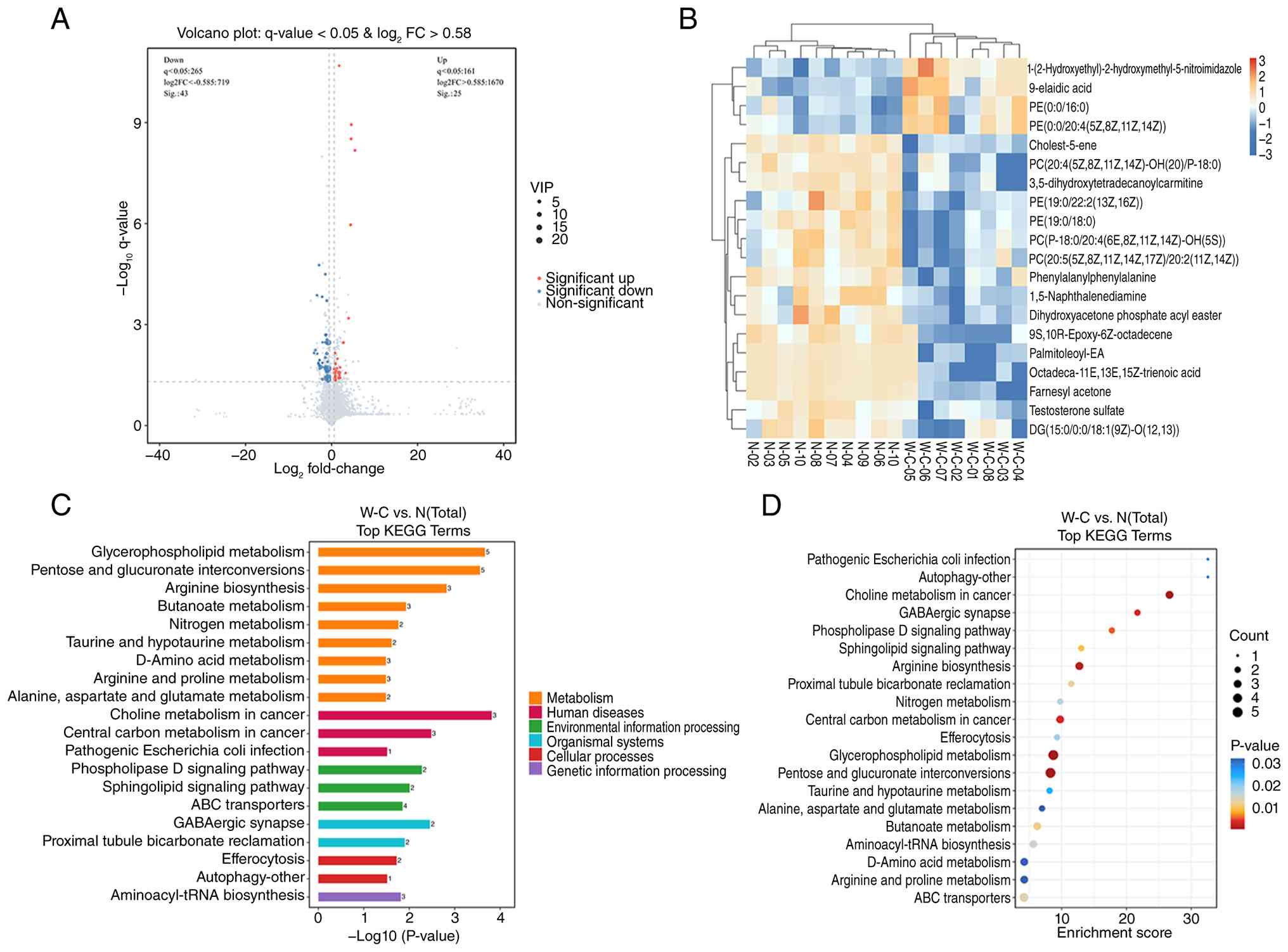
(q>0.05). Volcano plots and hierarchical clustering heatmaps
visually summarize the differential metabolic patterns between
groups (Fig. 2A and B).
| Table III.Differential metabolites between
patients with gastric signet-ring cell carcinoma and healthy
controls. |
Table III.
Differential metabolites between
patients with gastric signet-ring cell carcinoma and healthy
controls.
| No. | Metabolite | m/z | VIP | P-value | q-value | log2
FC | Trend |
|---|
| 1 |
PC(P-18:0/20:4(6E,8Z,11Z,14Z)-OH(5S)) | 810.60 | 23.98 | 0.0010 | 0.0204 | −0.79 | Down |
| 2 | PE(19:0/18:0) | 761.59 | 18.60 | <0.0001 | 0.0034 | −0.74 | Down |
| 3 |
PE(19:0/22:2(13Z,16Z)) | 813.62 | 8.75 | 0.0036 | 0.0431 | −0.59 | Down |
| 4 |
PC(20:5(5Z,8Z,11Z,14Z,17Z)/20:2(11Z,14Z)) | 832.58 | 8.09 | 0.0025 | 0.0354 | −0.60 | Down |
| 5 | Testosterone
sulfate | 367.16 | 6.74 | 0.0010 | 0.0204 | −1.37 | Down |
| 6 |
Phenylalanylphenylalanine | 313.15 | 6.47 | 0.0014 | 0.0247 | −1.12 | Down |
| 7 |
DG(15:0/0:0/18:1(9Z)-O(12,13)) | 617.48 | 5.99 | 0.0041 | 0.0477 | −0.88 | Down |
| 8 | 9-elaidic acid | 281.25 | 5.59 | 0.0016 | 0.0267 | 1.74 | Up |
| 9 | PE(0:0/16:0) | 454.29 | 4.06 | 0.0035 | 0.0428 | 0.76 | Up |
| 10 |
9S,10R-Epoxy-6Z-octadecene | 282.28 | 4.03 | 0.0008 | 0.0184 | −2.32 | Down |
| 11 |
PC(20:4(5Z,8Z,11Z,14Z)-OH(20)/P-18:0) | 854.59 | 3.72 | 0.0002 | 0.0073 | −1.13 | Down |
| 12 |
1-(2-Hydroxyethyl)-2-hydroxymethyl-5-nitroimidazole | 188.07 | 3.13 | 0.0029 | 0.0385 | 1.68 | Up |
| 13 |
3,5-Dihydroxytetradecanoylcarnitine | 805.58 | 3.04 | 0.0000 | 0.0021 | −1.34 | Down |
| 14 |
1,5-Naphthalenediamine | 159.09 | 2.83 | 0.0034 | 0.0417 | −2.12 | Down |
| 15 |
PE(0:0/20:4(5Z,8Z,11Z,14Z)) | 500.28 | 2.83 | 0.0018 | 0.0278 | 0.74 | Up |
| 16 | Cholest-5-ene | 388.39 | 2.60 | <0.0001 | 0.0002 | −1.09 | Down |
| 17 | Dihydroxyacetone
phosphate acyl ester | 197.99 | 2.35 | 0.0034 | 0.0422 | −1.36 | Down |
| 18 |
Octadeca-11E,13E,15Z-trienoic acid | 296.26 | 2.28 | 0.0010 | 0.0203 | −2.73 | Down |
| 19 |
Palmitoleoyl-EA | 298.27 | 2.25 | 0.0007 | 0.0164 | −2.81 | Down |
| 20 | Farnesyl
acetone | 280.26 | 2.22 | 0.0001 | 0.0058 | −3.75 | Down |
Metabolic pathway enrichment
analysis
KEGG pathway enrichment analysis identified 93
mapped pathways, with four pathways meeting significance criteria
(P<0.05, q<0.05): ‘Choline metabolism in cancer’,
‘glycerophospholipid metabolism’, ‘pentose and glucuronate
interconversions’ and ‘arginine biosynthesis’. The top 10 enriched
pathways are presented in Table IV
and presented as bar and bubble plots in Fig. 2C and D. Choline metabolism and
glycerophospholipid metabolism emerged as the most prominently
perturbed pathways, with their associated metabolites,
PC[20:5(5Z,8Z,11Z,14Z,17Z)/20:2(11Z,14Z)] and dihydroxyacetone
phosphate (DHAP) acyl ester showing marked alterations (Fig. 3A and B). ROC curve analysis
demonstrated the diagnostic performance of these metabolites.
PC[20:5(5Z,8Z,11Z,14Z,17Z)/20:2(11Z,14Z)] exhibited an area under
the curve (AUC) of 0.89 and DHAP acyl ester 0.99 (Fig. 3C and D). It should be noted that ROC
estimates derived from small discovery cohorts may overestimate
diagnostic performance.
 | Figure 3.Differential abundance and diagnostic
performance of key metabolites. Box plots of (A)
PC[20:5(5Z,8Z,11Z,14Z,17Z)/20:2(11Z,14Z)] and (B) DHAP acyl ester
in patients with GSRCC and healthy controls. Data are presented as
the mean ± SD. P<0.05 vs. healthy control. ROC curves for (C)
PC[20:5(5Z,8Z,11Z,14Z,17Z)/20:2(11Z,14Z)] and (D) DHAP acyl ester.
The AUC values are 0.89 and 0.99, respectively. **P<0.01. W-C,
GSRCC group; N, healthy control; ROC, receiver operating
characteristic; AUC, area under the curve; DHAP, dihydroxyacetone
phosphate. |
| Table IV.Significantly enriched metabolic
pathways in GSRCC. |
Table IV.
Significantly enriched metabolic
pathways in GSRCC.
| ID | Pathway name | Key
metabolites | P-value | q-value |
|---|
| hsa05231 | Choline metabolism
in cancer | Choline,
PC(16:0/0:0) and PC[20:5(5Z,8Z,11Z,14Z,17Z)/20:2(11Z,14Z)] | 0.0002 | 0.0086 |
| hsa00564 | Glycerophospholipid
metabolism | Choline,
PE[16:0/20:2(11Z,14Z)], DHAP,
PC[20:5(5Z,8Z,11Z,14Z,17Z)/20:2(11Z,14Z)] and PC(16:0/0:0) | 0.0002 | 0.0086 |
| hsa00040 | Pentose and
glucuronate interconversions | Oxoglutaric acid,
glycerol, adonitol, L-arabitol and 2,3-Diketo-L-gulonate | 0.0003 | 0.0086 |
| hsa00220 | Arginine
biosynthesis | L-glutamic acid,
oxoglutaric acid and citrulline | 0.0015 | 0.0350 |
| hsa05230 | Central carbon
metabolism in cancer | L-glutamic acid,
oxoglutaric acid and L-leucine | 0.0033 | 0.0542 |
| hsa04727 | GABAergic
synapse | L-glutamic acid and
oxoglutaric acid | 0.0035 | 0.0542 |
| hsa04072 | Phospholipase D
signaling pathway | L-glutamic acid and
sphingosine-1-phosphate | 0.0053 | 0.0701 |
| hsa04071 | Sphingolipid
signaling pathway | SM[d18:1/24:1(15Z)]
and sphingosine-1-phosphate | 0.0098 | 0.1141 |
| hsa00650 | Butanoate
metabolism | L-glutamic acid,
oxoglutaric acid and 3R-hydroxy-butanoic acid | 0.0117 | 0.1168 |
| hsa04964 | Proximal tubule
bicarbonate reclamation | L-glutamic acid and
oxoglutaric acid | 0.0126 | 0.1168 |
Clinical biochemistry parameters
To complement the metabolomic findings and assess
systemic metabolic status in GSRCC, routine clinical biochemistry
parameters were measured in both discovery and validation cohorts
(Table V). In both cohorts,
patients with GSRCC demonstrated significantly lower levels of
cholinesterase, total protein, albumin and hemoglobin compared with
healthy controls (P<0.01). However, no significant differences
were observed between stage I–II and stage III–IV patients for any
of these parameters (P>0.05), suggesting that these
abnormalities were not closely associated with tumor stage
progression. These findings indicate notable protein metabolic
dysregulation and compromised nutritional status in patients with
GSRCC, which aligns with the broader metabolic reprogramming
suggested by pathway enrichment.
| Table V.Clinical biochemistry parameters in
discovery and validation cohorts. |
Table V.
Clinical biochemistry parameters in
discovery and validation cohorts.
|
|
|
|
|
|
| P-value |
|---|
|
|
|
|
|
|
|
|
|---|
| Parameter | Cohort | Healthy
control | Total GSRCC | TNM stage I–II | TNM stage
III–IV | T vs. N | Stage I–II vs.
N | Stage III–IV vs.
N | Stage I–II vs.
III–IV |
|---|
| Cholinesterase,
U/l | Discovery | 297.80±73.59 | 200.63±48.98 | 203.25±41.10 | 198.00±62.37 | 0.006 | 0.034 | 0.035 | 0.893 |
|
| Validation | 317.49±61.20 | 246.63±60.23 | 236.89±45.95 | 258.19±73.66 | <0.001 | <0.001 | 0.004 | 0.304 |
| Total protein,
g/l | Discovery | 71.75±4.02 | 60.04±7.73 | 55.93±6.68 | 64.17±7.03 | 0.001 | <0.001 | 0.024 | 0.140 |
|
| Validation | 69.87±4.67 | 62.83±5.70 | 63.35±5.21 | 62.21±6.34 | <0.001 | <0.001 | <0.001 | 0.564 |
| Albumin, g/l | Discovery | 45.88±1.97 | 37.00±3.71 | 35.36±3.59 | 38.63±3.49 | <0.001 | <0.001 | <0.001 | 0.242 |
|
| Validation | 44.37±2.54 | 39.05±3.48 | 38.77±2.73 | 39.38±4.27 | <0.001 | <0.001 | <0.001 | 0.614 |
| Hemoglobin,
g/l | Discovery | 142.00 | 119.50 | 112.00±16.99 | 115.25±14.50 | 0.007 | 0.013 | 0.021 | 0.781 |
|
|
|
(124.75–149.50) | (99.50–124.00) |
|
|
|
|
|
|
|
| Validation | 142.11±11.85 | 127.46±19.86 | 128.68±22.37 | 132.00
(110.50–139.50) | <0.001 | 0.006 | 0.007 | 0.855 |
Discussion
GSRCC represents a biologically aggressive subtype
of gastric adenocarcinoma characterized by diffuse infiltration,
poor differentiation and high metastatic potential, frequently
presenting at advanced stages with unfavorable outcomes (15). The emergence of metabolomics has
provided unprecedented opportunities to elucidate cancer-associated
metabolic reprogramming and identify novel diagnostic biomarkers
(16,17). In the present study, a comprehensive
dual-platform metabolomics approach integrating LC-MS and GC-MS was
employed to characterize the serum metabolomic landscape of GSRCC,
an approach that offers broader metabolic coverage compared with
single-platform analyses commonly reported in gastric cancer
metabolomics literature (18,19).
The present analysis identified 68 differential
metabolites in patients with GSRCC, predominantly comprising
phospholipids (PC and PE) and amino acids. Pathway enrichment
analysis revealed notable perturbations in ‘choline metabolism in
cancer’, ‘glycerophospholipid metabolism’, ‘pentose and glucuronate
interconversions’ and ‘arginine biosynthesis’. Notably, choline
metabolism and glycerophospholipid metabolism emerged as the most
prominently dysregulated pathways, consistent with growing evidence
implicating lipid metabolic reprogramming in gastric carcinogenesis
(20,21). The underlying biological unit and
integrated functional context of the dysregulated metabolic system
were further emphasized. The perturbed metabolites were not
independent events but were functionally interconnected within core
biological modules, including choline metabolism,
glycerophospholipid metabolism, membrane turnover, energy
homeostasis and mitochondrial function, which collectively may
contribute to the malignant progression of GSRCC. Considering these
interconnected biological units strengthens the biological
importance and contributions of the present study.
Choline metabolism serves multifaceted roles in
cancer biology. As a precursor for acetylcholine neurotransmitter
synthesis, choline also participates in ketone body and fatty acid
metabolism via its metabolite betaine, which links methyl group
metabolism to mitochondrial function by providing methyl donors for
carnitine biosynthesis, thereby facilitating long-chain fatty acid
transport into mitochondria for β-oxidation and subsequent
acetyl-CoA production for ketogenesis (22). Choline is a component of cellular
and mitochondrial membranes and regulates lipid and energy
homeostasis (23).
Phosphatidylcholine and phosphatidylethanolamine, the major
phospholipid classes affected in the present study, collectively
account for ~50% of cellular membrane phospholipids (24). The observed downregulation of
PC[20:5(5Z,8Z,11Z,14Z,17Z)/20:2(11Z,14Z)] may reflect altered fatty
acid composition in malignant cell membranes associated with poor
differentiation and aggressive behavior. This PUFA-rich PC species
is critical for maintaining optimal membrane fluidity and lipid
raft integrity. Its downregulation may impair membrane biophysical
properties, leading to: i) Enhanced lateral mobility and clustering
of receptor tyrosine kinases, promoting sustained MAPK and PI3K/Akt
activation (25); ii) disrupted Ras
nanodomain organization, facilitating downstream oncogenic
signaling via flotillin-1-mediated Ras-SOS1 interactions (26); and iii) altered membrane curvature,
which enhances tumor cell migration and invasion (27). Collectively, these changes create a
permissive environment for cytoskeletal remodeling, ultimately
contributing to poor differentiation and dissemination in
GSRCC.
The association between choline metabolism and
metastatic potential has been previously recognized (28). GSRCC exhibits a unique
immunosuppressive tumor microenvironment that facilitates
peritoneal dissemination, with mucinous components infiltrating
surrounding stroma and promoting tumor cell spread to the
peritoneal cavity (29,30). Mechanistically, choline and
glycerophospholipid metabolites may influence tumor progression
through modulation of MAPK, PI3K/Akt/mTOR and Ras signaling
pathways, which are well-established drivers of gastric cancer
proliferation and metastasis (31,32).
Specifically, altered PC/PE ratios remodel membrane fluidity and
charge, affecting Ras nanoclustering and Raf-dependent MAPK
activation (31). Meanwhile,
glycerophospholipid turnover generates PA and LPA, which directly
activate mTORC1 and PI3K/Akt via allosteric binding and
receptor-mediated mechanisms (32).
PI3K/Akt pathway activation has been specifically associated with
lymph node metastasis and poor prognosis in gastric cancer
(33). Despite this, these
hypotheses require further experimental determination.
Notably, the marked downregulation of DHAP acyl
ester in GSRCC serum, with an AUC of 0.99, suggested superior
diagnostic potential. DHAP acyl ester serves as a key intermediate
in lipid metabolism, participating in glycerophospholipid and
glycerolipid biosynthesis (34). A
number of mechanisms may account for its reduced circulating levels
in GSRCC. Enhanced utilization of DHAP acyl ester as a precursor
for membrane phospholipid synthesis in rapidly proliferating tumor
cells could deplete circulating pools. In addition, the Warburg
effect, characterized by aerobic glycolysis in cancer cells, may
redirect DHAP flux toward glycolytic pathways rather than lipid
synthesis (35). Impaired
mitochondrial function in gastric cancer cells may further
compromise normal lipid metabolism, reducing DHAP acyl ester
production. These findings warrant further investigation into the
precise regulatory mechanisms governing DHAP acyl ester metabolism
in gastric cancer and its potential interplay with glucose and
amino acid metabolism.
To complement the metabolomic findings within
conventional clinical parameters, routine biochemistry markers
related to choline metabolism and nutritional status were assessed.
The marked reductions in cholinesterase, total protein, albumin and
hemoglobin in patients with GSRCC across both discovery and
validation cohorts provide complementary evidence of systemic
metabolic dysregulation. Cholinesterase, synthesized primarily in
the liver, serves as a marker of hepatic synthetic capacity; its
decline in patients with GSRCC suggests impaired liver function
secondary to cancer-associated cachexia or metabolic stress. The
concordant reductions in protein markers indicate profound protein
metabolic disturbances and compromised nutritional status,
consistent with the cachexia syndrome frequently accompanying
advanced malignancy (36). However,
BMI did not notably differ between patients with GSRCC and
controls, and no notable differences were observed across TNM
stages. The present findings suggest that GSRCC-associated
metabolic disturbances occur early in tumorigenesis, leading to
preferential depletion of protein reserves through systemic
inflammation and amino acid catabolism, with relative preservation
of adipose tissue. When interpreted alongside the pathway
enrichment findings (choline and glycerophospholipid metabolism),
these conventional parameters offer complementary information on
the systemic metabolic status of patients with GSRCC.
A number of limitations of the present study warrant
consideration. First, the modest sample size, particularly in the
discovery cohort, reflects the relative rarity of GSRCC and the
exploratory nature of the present investigation. Although strict
model validation was applied to avoid overfitting, the findings
should be regarded as exploratory rather than confirmatory. The
lack of disease control groups (namely, non-signet-ring gastric
adenocarcinoma and benign gastric diseases) prevents determination
of whether the observed metabolic signatures are specific to GSRCC
or reflect broader cancer-associated alterations. Third, the
present study performed non-targeted profiling without subsequent
targeted LC-MS/MS validation; therefore, candidate metabolites
should be considered exploratory signatures rather than fully
determined biomarkers. Fourth, functional validation is required to
elucidate mechanistic roles. Future studies with larger cohorts,
well-matched disease controls, targeted metabolomics and
experimental validation are needed to validate specificity,
robustness and clinical applicability.
In conclusion, the present study provides, to the
best of our knowledge, the first comprehensive metabolomic
characterization of GSRCC using a dual-platform mass spectrometry
approach. Notable perturbations in choline and glycerophospholipid
metabolism were identified, with
PC[20:5(5Z,8Z,11Z,14Z,17Z)/20:2(11Z,14Z)] and DHAP acyl ester
emerging as exploratory candidate metabolic signatures associated
with GSRCC, which may establish a foundation for future biomarker
discovery and contribute to the evolving understanding of GSRCC
biology.
Acknowledgements
Not applicable.
Funding
The present study was supported by the National Natural Science
Foundation of China (grant no. 81703830), the Natural Science
Foundation of Jiangsu Province (grant no. BK20241146), the Jiangsu
Provincial Traditional Chinese Medicine Science and Technology
Development Program (grant no. MS2025016) and the Affiliated
Hospital of Nanjing University of Chinese Medicine Program (grant
no. Y24042).
Availability of data and materials
The data generated in the present study may be found
in the OMIX database of the National Genomics Data Center under
accession number (OMIX017401) or at the following URL: (persistent,
http://ngdc.cncb.ac.cn/omix/release/OMIX017401).
Authors' contributions
HC, SG, JL and WG contributed to the present study
conception and design. HC, YB, JS, JW, FM, WS, QS and WG
contributed to material preparation, data collection and analysis.
HC, SG and JL wrote the first draft of the manuscript and all
authors commented on previous versions. All authors read and
approved the final version of the manuscript. HC, SG and WG confirm
the authenticity of all the raw data.
Ethics approval and consent to
participate
The present study was approved by the ethics
committee of Jiangsu Province Hospital of Chinese Medicine
(approval nos. 2021NL-187-02, 2021NL-187-03, 2021NL-187-04 and
2021NL-187-05). This ethics approval was issued for a broader
clinical cohort study, and the present metabolomics study was
conducted within the scope of this approved protocol. The ethics
approval has been extended to cover the entire study period,
including the serum sample collection period. Written informed
consent was obtained from each participant prior to enrollment and
their consent for the storage of serum samples for future secondary
analyses was also obtained.
Patient consent for publication
Not applicable.
Competing interests
The authors declare that they have no competing
interests.
Use of artificial intelligence tools
During the preparation of this work, artificial
intelligence tools were used to improve the readability and
language of the manuscript or to generate images, and subsequently,
the authors revised and edited the content produced by the
artificial intelligence tools as necessary, taking full
responsibility for the ultimate content of the present
manuscript.
Glossary
Abbreviations
Abbreviations:
|
GSRCC
|
gastric signet-ring cell carcinoma
|
|
LC-MS
|
liquid chromatography-mass
spectrometry
|
|
GC-MS
|
gas chromatography-mass
spectrometry
|
|
PCA
|
principal component analysis
|
|
PLS-DA
|
partial least squares discriminant
analysis
|
|
OPLS-DA
|
orthogonal partial least squares
discriminant analysis
|
|
VIP
|
variable importance in projection
|
|
KEGG
|
Kyoto Encyclopedia of Genes and
Genomes
|
|
PC
|
phosphatidylcholine
|
|
PE
|
phosphatidylethanolamine
|
|
DHAP
|
dihydroxyacetone phosphate
|
|
ROC
|
receiver operating characteristic
|
|
AUC
|
area under the curve
|
References
|
1
|
Chen Y, Wang B, Zhao Y, Shao J, Shen X,
Zhang L, Gu W, Xu J, Jiang J, Xu L, et al: Metabolomic machine
learning predictor for diagnosis and prognosis of gastric cancer.
Nat Commun. 15:16572024. View Article : Google Scholar : PubMed/NCBI
|
|
2
|
Yang WJ, Zhao HP, Yu Y, Wang JH, Guo L,
Liu JY, Pu J and Lv J: Updates on global epidemiology, risk and
prognostic factors of gastric cancer. World J Gastroenterol.
29:2452–2468. 2023. View Article : Google Scholar : PubMed/NCBI
|
|
3
|
Zhao W, Jia Y, Sun G, Yang H, Liu L, Qi X,
Ding J, Yu H, Xu B, Zhao S, et al: Single-cell analysis of gastric
signet ring cell carcinoma reveals cytological and immune
microenvironment features. Nat Commun. 14:29852023. View Article : Google Scholar : PubMed/NCBI
|
|
4
|
Kushima R: The updated who classification
of digestive system tumours-gastric adenocarcinoma and dysplasia.
Pathologe. 43:8–15. 2022. View Article : Google Scholar : PubMed/NCBI
|
|
5
|
Liang Z, Zheng L and Cao J: Endoscopic and
pathological examinations of early-signet-ring carcinoma in the
stomach. Healthcare (Basel). 13:26892025. View Article : Google Scholar : PubMed/NCBI
|
|
6
|
Yan J, Liu Y, Liu T and Zhu Q: A
predictive and prognostic model for metastasis risk and prognostic
factors in gastrointestinal signet ring cell carcinoma. Eur J Med
Res. 29:5452024. View Article : Google Scholar : PubMed/NCBI
|
|
7
|
Tang Y, Ren L and Mao T: Update on
diagnosis and treatment of early signet-ring cell gastric
carcinoma: A literature review. World J Gastrointest Endosc.
15:240–247. 2023. View Article : Google Scholar : PubMed/NCBI
|
|
8
|
Pappas A, Tan WK, Waldock W, Richardson S,
Tripathi M, Januszewicz W, Roberts G, O'Donovan M, Fitzgerald RC
and di Pietro M: Single-bite versus double-bite technique for
mapping biopsies during endoscopic surveillance for hereditary
diffuse gastric cancer: A single-center, randomized trial.
Endoscopy. 53:246–253. 2021. View Article : Google Scholar : PubMed/NCBI
|
|
9
|
Shi Y, Yang Q, Xu B, Yuan C, Qi K, Pan Y,
Wang Z, Chen Q, Sun D, Zhao W, et al: Key lipid reprogramming
revealed in gastric signet ring cell carcinoma by spatial mass
spectrometry metabolomics. J Am Soc Mass Spectrom. 36:1598–1608.
2025. View Article : Google Scholar : PubMed/NCBI
|
|
10
|
Fu L, Li M, Lv J, Yang C, Zhang Z, Qin S,
Li W, Wang X and Chen L: Deep neural network for discovering
metabolism-related biomarkers for lung adenocarcinoma. Front
Endocrinol (Lausanne). 14:12707722023. View Article : Google Scholar : PubMed/NCBI
|
|
11
|
Sun D, Zhang Y, Wang D, Zhao X, Han R, Li
N, Li X, Li T, Wang P, Jia Q, et al: Experimental study on changes
in metabolic mechanism of papillary thyroid carcinoma complicated
with hashimoto's thyroiditis. Heliyon. 9:e206612023. View Article : Google Scholar : PubMed/NCBI
|
|
12
|
Wiedmann L, De Angelis Rigotti F,
Vaquero-Siguero N, Donato E, Espinet E, Moll I, Alsina-Sanchis E,
Bohnenberger H, Fernandez-Florido E, Mülfarth R, et al: Hapln1
potentiates peritoneal metastasis in pancreatic cancer. Nat Commun.
14:23532023. View Article : Google Scholar : PubMed/NCBI
|
|
13
|
Sumner LW, Amberg A, Barrett D, Beale MH,
Beger R, Daykin CA, Fan TW, Fiehn O, Goodacre R, Griffin JL, et al:
Proposed minimum reporting standards for chemical analysis chemical
analysis working group (CAWG) metabolomics standards initiative
(MSI). Metabolomics. 3:211–221. 2007. View Article : Google Scholar : PubMed/NCBI
|
|
14
|
Amin MB, Greene FL, Edge SB, Compton CC,
Gershenwald JE, Brookland RK, Meyer L, Gress DM, Byrd DR and
Winchester DP: The eighth edition AJCC cancer staging manual:
Continuing to build a bridge from a population-based to a more
‘personalized’ approach to cancer staging. CA Cancer J Clin.
67:93–99. 2017.PubMed/NCBI
|
|
15
|
Raju D, Prabhu S, Maria A, Shirodkar SNS,
Damerla RR, Chakrabarty S, Shetty PS, Rao M and Kumar NAN: Gastric
signet ring cell carcinoma: Tumor microenvironment reprogramming
and novel therapeutic targets with emphasis on grin2d. Clin Transl
Sci. 18:e704242025. View Article : Google Scholar : PubMed/NCBI
|
|
16
|
Sun Y, Zhang X, Hang D, Lau HCH, Du J, Liu
C, Xie M, Pan Y, Wang L, Liang C, et al: Integrative plasma and
fecal metabolomics identify functional metabolites in
adenoma-colorectal cancer progression and as early diagnostic
biomarkers. Cancer Cell. 42:1386–1400.e8. 2024. View Article : Google Scholar : PubMed/NCBI
|
|
17
|
Xiao Y, Ma D, Yang YS, Yang F, Ding JH,
Gong Y, Jiang L, Ge LP, Wu SY, Yu Q, et al: Comprehensive
metabolomics expands precision medicine for triple-negative breast
cancer. Cell Res. 32:477–490. 2022. View Article : Google Scholar : PubMed/NCBI
|
|
18
|
Matsuoka T and Yashiro M: Bioinformatics
analysis and validation of potential markers associated with
prediction and prognosis of gastric cancer. Int J Mol Sci.
25:58802024. View Article : Google Scholar : PubMed/NCBI
|
|
19
|
Wang F, Pang R, Zhao X, Zhou B, Tian Y, Ma
Y and Rong L: Plasma metabolomics and lipidomics reveal potential
novel biomarkers in early gastric cancer: An explorative study. Int
J Biol Markers. 39:226–238. 2024. View Article : Google Scholar : PubMed/NCBI
|
|
20
|
Xiao J, Cao S, Wang J, Li P, Cheng Q, Zhou
X, Dong J, Li Y, Zhao X, Xu Z and Yang L: Leptin-mediated
suppression of lipoprotein lipase cleavage enhances lipid uptake
and facilitates lymph node metastasis in gastric cancer. Cancer
Commun (Lond). 44:855–878. 2024. View Article : Google Scholar : PubMed/NCBI
|
|
21
|
Chen S, Chen W, Xu T, Li J, Yu J, He Y and
Qiu S: The impact of aberrant lipid metabolism on the immune
microenvironment of gastric cancer: A mini review. Front Immunol.
16:16398232025. View Article : Google Scholar : PubMed/NCBI
|
|
22
|
Chen X, Qiu W, Ma X, Ren L, Feng M, Hu S,
Xue C and Chen R: Roles and mechanisms of choline metabolism in
nonalcoholic fatty liver disease and cancers. Front Biosci Landmark
Ed. 29:1822024. View Article : Google Scholar : PubMed/NCBI
|
|
23
|
Yao N, Li W, Xu G, Duan N, Yu G and Qu J:
Choline metabolism and its implications in cancer. Front Oncol.
13:12348872023. View Article : Google Scholar : PubMed/NCBI
|
|
24
|
Son Y, Kenny TC, Khan A, Birsoy K and Hite
RK: Structural basis of lipid head group entry to the kennedy
pathway by flvcr1. Nature. 629:710–716. 2024. View Article : Google Scholar : PubMed/NCBI
|
|
25
|
Mukerjee S, Saeedan AS, Ansari MN and
Singh M: Polyunsaturated fatty acids mediated regulation of
membrane biochemistry and tumor cell membrane integrity. Membranes
(Basel). 11:4792021. View Article : Google Scholar : PubMed/NCBI
|
|
26
|
Jin H, Koh M, Lim H, Yong HY, Kim ES, Kim
SY, Kim K, Jung J, Ryu WJ, Choi KY and Moon A: Lipid raft protein
flotillin-1 is important for the interaction between SOS1 and
H-Ras/K-Ras, leading to Ras activation. Int J Cancer. 52:1933–1946.
2023. View Article : Google Scholar
|
|
27
|
Damalas A, Kyriazis ID, Tutkus M,
Angelidis C and Trachana V: Membrane curvature and cancer:
Mechanisms, implications, and therapeutic perspectives. Cancers
(Basel). 18:10762026. View Article : Google Scholar : PubMed/NCBI
|
|
28
|
Pan J, Zhang L, Wang X, Li L, Yang C, Wang
Z, Su K, Hu X, Zhang Y, Ren G, et al: Chronic stress induces
pulmonary epithelial cells to produce acetylcholine that remodels
lung pre-metastatic niche of breast cancer by enhancing netosis. J
Exp Clin Cancer Res. 42:2552023. View Article : Google Scholar : PubMed/NCBI
|
|
29
|
Zhang J, Collingwood R, Al Diffalha S,
Manna DD, Paluri RK, Mejbel HA and Gbolahan O: Comprehensive
characterization of the immune microenvironment of colorectal and
gastric signet ring cell cancer. Cells. 15:302025. View Article : Google Scholar : PubMed/NCBI
|
|
30
|
Xie Y, Li C, Yu M and Cao J:
Immunosuppressive tumor microenvironment in gastric signet-ring
cell carcinoma. World J Clin Oncol. 15:1126–1131. 2024. View Article : Google Scholar : PubMed/NCBI
|
|
31
|
Zhou Y and Hancock JF: RAS nanoclusters
are cell surface transducers that convert extracellular stimuli to
intracellular signalling. FEBS Lett. 597:892–908. 2023. View Article : Google Scholar : PubMed/NCBI
|
|
32
|
Wang C, Yang Z, Xu E, Shen X, Wang X, Li
Z, Yu H, Chen K, Hu Q, Xia X, et al: Apolipoprotein C-II induces
EMT to promote gastric cancer peritoneal metastasis via
PI3K/AKT/mTOR pathway. Clin Transl Med. 11:e5222021. View Article : Google Scholar : PubMed/NCBI
|
|
33
|
Cao W, Zeng Z, Lan J, Yang Y, Lu M and Lei
S: Knockdown of fut11 inhibits the progression of gastric cancer
via the pi3k/akt pathway. Heliyon. 9:e176002023. View Article : Google Scholar : PubMed/NCBI
|
|
34
|
Oh S, Jo S, Bajzikova M, Kim HS, Dao TTP,
Rohlena J, Kim JM, Neuzil J and Park S: Non-bioenergetic roles of
mitochondrial gpd2 promote tumor progression. Theranostics.
13:438–457. 2023. View Article : Google Scholar : PubMed/NCBI
|
|
35
|
Singh J, Suh EH, Sharma G, Khemtong C,
Sherry AD and Kovacs Z: Probing carbohydrate metabolism using
hyperpolarized 13C-labeled molecules. NMR Biomed.
32:e40182019. View Article : Google Scholar : PubMed/NCBI
|
|
36
|
Tamayo-Torres E, Garrido A, de Cabo R,
Carretero J and Gómez-Cabrera MC: Molecular mechanisms of cancer
cachexia. Role of exercise training. Mol Aspects Med.
99:1012932024. View Article : Google Scholar : PubMed/NCBI
|