Introduction
Triple-negative breast cancer (TNBC) is a
biologically aggressive subtype of breast cancer defined by the
absence of estrogen receptor, progesterone receptor and human
epidermal growth factor receptor 2 (HER2) expression. It accounts
for 15–20% of all breast cancers worldwide and is
disproportionately diagnosed in younger women, with a median age at
diagnosis of ~53 years and a particularly high incidence among
premenopausal women and those under 40 years of age (1). Clinically, TNBC often presents as a
rapidly growing palpable mass and, due to its high proliferative
rate, it is more likely to manifest as an interval cancer between
screening mammograms (1). The
disease is characterized by substantial biological heterogeneity,
with the basal-like molecular subtype predominating, and is
associated with poor histological differentiation, aggressive
clinical behavior, and an elevated risk of early recurrence and
visceral metastasis (1,2). Unlike hormone receptor-positive or
HER2-positive breast cancers, TNBC lacks established molecular
targets, and until recently, cytotoxic chemotherapy remained the
only systemic treatment option. This therapeutic limitation,
combined with its aggressive natural history, has historically
resulted in a poorer prognosis, with a 5-year overall survival rate
of 60–65% for patients with locally advanced disease (1).
The treatment landscape for early-stage TNBC has
been transformed by the introduction of immune checkpoint
inhibitors (ICIs) targeting the programmed death-1
(PD-1)/programmed death-ligand 1 (PD-L1) axis. The landmark
KEYNOTE-522 phase III trial demonstrated that the addition of
pembrolizumab to standard neoadjuvant chemotherapy significantly
improved the pathological complete response (pCR) rate from 51.2 to
64.8% and prolonged event-free survival (3). Based on these results, neoadjuvant
pembrolizumab plus chemotherapy followed by adjuvant pembrolizumab
has become the standard of care for high-risk, early-stage TNBC. In
patients who achieve pCR after neoadjuvant therapy, the decision to
administer postoperative radiotherapy (RT) is typically guided by
the initial clinical stage and other risk factors, including tumor
size and lymph node involvement at presentation (4). For patients with residual disease
after neoadjuvant therapy, postoperative RT is routinely indicated.
Consequently, an increasing number of patients with TNBC are
receiving adjuvant ICIs concurrently with postoperative RT, raising
important clinical questions regarding the safety of this combined
approach.
Radiotherapy exerts its antitumor effects not only
through direct DNA damage but also through the induction of
immunogenic cell death, which can prime systemic antitumor immune
responses (5). Preclinical studies
have demonstrated that RT upregulates PD-L1 expression on tumor
cells and enhances the presentation of tumor-associated antigens,
thereby providing a mechanistic rationale for combining RT with
ICIs (6,7). This synergy has been shown to enhance
local tumor control and, in some instances, mediate abscopal
effects, where distant, non-irradiated tumors also regress
(7). However, this
immunostimulatory activity also raises concerns that the
combination of RT and ICIs could potentiate immune-mediated injury
in normal tissues within the radiation field. Evidence from
multiple studies in lung cancer has shown that the addition of ICIs
to thoracic RT is associated with an increased risk of serious
adverse events, particularly radiation pneumonitis (8,9). In
the PACIFIC trial of durvalumab following chemoradiotherapy for
stage III non-small-cell lung cancer, the incidence of any-grade
pneumonitis was 33.9% in the durvalumab group compared with 24.8%
in the placebo group, and grade 3–4 pneumonitis occurred in 3.4% of
patients receiving durvalumab (9).
In severe cases, radiation pneumonitis can be life-threatening, and
this toxicity has become a major consideration in the clinical
implementation of combined RT-ICI regimens.
Despite these concerns, data on the safety of
concurrent RT and ICIs in patients with breast cancer, and
particularly in those with TNBC, remain limited. Compared with
thoracic RT for lung cancer, RT fields for breast cancer typically
involve smaller lung volumes and lower doses to critical organs.
Furthermore, the patient population differs substantially in terms
of baseline pulmonary function and comorbidities. A recent
real-world study specifically evaluating the safety of concurrent
pembrolizumab and RT in patients with TNBC reported that the
combination was generally well tolerated, but the evidence base
remains limited by small sample sizes and short follow-up (10). Therefore, the safety of concurrent
postoperative RT and maintenance ICIs in patients with TNBC remains
poorly understood.
To address this uncertainty, the present study
retrospectively evaluated the safety and tolerability of concurrent
postoperative RT and ICIs following neoadjuvant ICIs in patients
with TNBC treated at Hubei Cancer Hospital (Wuhan, China). The
findings of the present study may help guide the concurrent use of
ICIs and postoperative RT in this patient population.
Materials and methods
Patient population
This retrospective study included a total of 132
female patients with TNBC who received neoadjuvant
chemoimmunotherapy or chemotherapy alone at Hubei Cancer Hospital
between 2022 and 2024. All patients were female, with a median age
of 45 years (range, 33–69 years). The inclusion criteria were as
follows: i) Histologically confirmed TNBC; ii) prior treatment with
neoadjuvant chemotherapy or chemoimmunotherapy; and iii) receipt of
postoperative RT to the chest wall and/or regional lymph nodes.
Patients who underwent breast-conserving surgery received
whole-breast irradiation only. The exclusion criteria were as
follows: i) Presence of distant metastasis; ii) severe comorbid
medical conditions; and iii) intolerance to local RT. This study
was conducted in accordance with the requirements of the Ethics
Committee of Hubei Cancer Hospital (approval no. LLHBCH2026YN-106).
Written informed consent was obtained from all patients who
received off-label immunotherapy.
Treatment protocol
The treatment protocol for all enrolled patients
consisted of three phases: Neoadjuvant systemic therapy, surgery,
and postoperative RT with or without adjuvant ICI therapy. During
the neoadjuvant phase, patients received chemotherapy with either
an anthracycline-cyclophosphamide regimen followed by a taxane
(AC-T) or docetaxel plus carboplatin (TCb), with or without
concurrent ICIs (pembrolizumab, tislelizumab, camrelizumab or
toripalimab), at the discretion of the treating medical oncologist.
Following completion of neoadjuvant therapy, all patients underwent
either breast-conserving surgery or radical mastectomy with
axillary lymph node dissection or sentinel lymph node biopsy.
During the postoperative phase, patients who had received
neoadjuvant ICIs continued the same ICI concurrently with adjuvant
RT, and comprised the ICIs-RT group. Patients who had not received
neoadjuvant ICIs proceeded to adjuvant RT without immunotherapy and
comprised the RT-alone group. No postoperative adjuvant
chemotherapy was administered to any patient in either group.
Assessment of pathological
response
pCR was defined as the absence of residual invasive
carcinoma in the breast and axillary lymph nodes (ypT0/is ypN0)
upon histopathological examination of surgical specimens,
consistent with the criteria used in the KEYNOTE-522 trial
(3). All surgical specimens were
evaluated by dedicated breast pathologists at Hubei Cancer
Hospital. The pCR status was determined according to standardized
institutional protocols, consistent with current international
guidelines for the pathological assessment of breast cancer
specimens (3).
Methods of radiation therapy
All patients received postoperative RT. The RT
technique was based on previously published methods (11). Volumetric modulated arc therapy was
delivered using 6 MV photons from a linear accelerator (Elekta
Infinity or Elekta Axesse; Elekta Instrument AB). For patients who
underwent breast-conserving surgery, the prescribed dose was 40–42
Gy in 15–16 fractions to the whole breast, followed by a sequential
boost to the tumor bed of 8.7–10 Gy in 3–5 fractions. For patients
who underwent radical mastectomy, RT was delivered at a dose of 50
Gy in 25 fractions. Target volumes and organs at risk (OARs) were
contoured according to the Radiation Therapy Oncology Group breast
cancer atlas (12). Supraclavicular
fossa irradiation was administered to patients with positive
axillary lymph nodes, whereas no patient received intentional
internal mammary lymph node irradiation. The dose constraints were
as follow: i) Ipsilateral volume of lung receiving at least 20 Gy
(V20)<30% and V5<60%; ii) mean heart dose <8 Gy for
left-sided tumors and <4 Gy for right-sided tumors; and iii)
maximum dose to the spinal cord planning organ-at-risk volume (PRV)
<45 Gy.
Follow-up
Patients were categorized into two groups:
Postoperative RT with concurrent ICIs (ICIs-RT) and postoperative
RT alone (RT-alone group). RT planning parameters, including
planning target volume (PTV) and mean target dose, were collected.
To evaluate target dose homogeneity, the volume of the PTV
receiving ≥106% (V53) of the prescribed dose of 50 Gy was assessed.
Total lung volume was also recorded. Lung dosimetric parameters,
including V5, V20 and V30 and the mean lung dose were analyzed. The
mean heart dose and the maximum dose to the spinal cord PRV were
also recorded.
Follow-up chest CT imaging was performed every 3
months after RT. Radiation pneumonitis in the ipsilateral lung was
graded according to the Common Terminology Criteria for Adverse
Events (CTCAE) version 4.0 (13).
Radiation dermatitis was graded using the same criteria. Complete
blood counts and thyroid function test results obtained before and
after immunotherapy were documented. During follow-up, event-free
survival (EFS), local control and overall survival (OS) were
evaluated. Notably, grade 1 radiation pneumonitis was defined
according to CTCAE version 4.0, as asymptomatic radiological
changes were detected on chest CT without clinical symptoms.
Definition of events
For the evaluation of EFS, events were defined as
local recurrence (histologically confirmed or radiologically
evident disease in the ipsilateral chest wall, breast or regional
lymph nodes), distant metastasis (radiologically or histologically
confirmed tumor spread beyond the locoregional region) or death
from any cause; whichever occurred first. Patients who were alive
and free of any of these events at the last follow-up were
censored.
Statistical analysis
All statistical analyses were performed using
GraphPad Prism version 5.0 (Dotmatics). The normality of continuous
data was assessed using the Shapiro-Wilk test. As the dosimetric
parameters were normally distributed, parametric tests were
applied. Continuous variables were compared between the two groups
using the unpaired (independent samples) Student's t-test.
Categorical variables were compared using the chi-square test or
Fisher's exact test, as appropriate. P<0.05 was considered to
indicate a statistically significant difference. EFS was estimated
using the Kaplan-Meier method and between-group comparisons were
performed using the log-rank test. A univariate Cox proportional
hazards model was used to calculate hazard ratios (HRs) and 95%
confidence intervals. The proportional hazards assumption was
assessed using Schoenfeld residuals and no violation was
identified.
Results
Baseline patient characteristics
A total of 132 patients with TNBC were enrolled in
this study. Baseline patient characteristics are summarized in
Table I. Among these patients, 30
were assigned to the ICIs-RT group and 102 to the RT-alone group.
Age, T stage, N stage, histological subtype and surgical procedure
were well balanced between the two groups, with no statistically
significant differences. The proportion of patients receiving
platinum-based regimens (TCb) was significantly higher in the
ICIs-RT group than in the RT-alone group (90.0 vs. 56.8%;
P<0.001). In addition, patients in the ICIs-RT group had a
significantly higher pCR rate after neoadjuvant therapy than those
in the RT-alone group (46.7 vs. 22.5%; P=0.01). Regarding
immunotherapy agents, 22 of the 30 patients (73.3%) in the ICIs-RT
group received tislelizumab.
 | Table I.Baseline characteristics of patients
receiving concurrent radiotherapy and immunotherapy for
triple-negative breast cancer. |
Table I.
Baseline characteristics of patients
receiving concurrent radiotherapy and immunotherapy for
triple-negative breast cancer.
| Factors | ICIs-RT group
(n=30) | RT-alone group
(n=102) | P-value |
|---|
| Age |
|
|
|
| Mean age,
years (range) | 45 (33–68) | 46.5 (42–69) | 0.91 |
| <65
years | 28 (93.3%) | 99 (97.1%) | 0.96 |
| T Stage |
|
| 0.90 |
|
T1/T2 | 12 (40%) | 42 (41.2%) |
|
|
T3/T4 | 18 (60%) | 60 (58.8%) |
|
| N Stage |
|
| 0.55 |
|
Positive | 27 (90%) | 97 (95.1%) |
|
|
Negative | 3 (10%) | 5 (4.9%) |
|
| Pathological
type |
|
| 0.54 |
|
Infiltrating ductal
carcinoma | 29 (96.7%) | 100 (98%) |
|
|
Micropapillary carcinoma | 1 (3.3%) | 2 (2%) |
|
| Neoadjuvant
chemotherapy drugs |
|
| 0.01a |
| AC-T | 3 (10%) | 44 (43.2%) |
|
| TCb | 27 (90%) | 58 (56.8%) |
|
| Immune checkpoint
inhibitors |
|
| - |
|
Pembrolizumab | 5 (16.7%) |
|
|
|
Tislelizumab | 22 (73.3%) |
|
|
|
Camrelizumab | 1 (3.3%) |
|
|
|
Toripalimab | 2 (6.7%) |
|
|
| Tumour
laterality |
|
| 0.80 |
| Left | 14 (46.7%) | 45 (44.1%) |
|
|
Right | 16 (53.3%) | 57 (55.9%) |
|
| Surgery approach |
|
| 0.94 |
|
Breast-conserving | 6 (20%) | 21 (20.6%) |
|
| Radical
mastectomy | 24 (80%) | 81 (79.4%) |
|
| Pathological
response |
|
| 0.01a |
| pCR | 14 (46.7%) | 23 (22.5%) |
|
|
NonPCR | 16 (53.3%) | 79 (77.5%) |
|
RT dosimetric parameters
Postoperative RT was administered to all patients in
both groups (Table II). RT dose
regimens were determined based on the surgical approach. Target
volumes were comparable between the two groups, with no
statistically significant differences observed. The mean target
dose and dosimetric parameters for the OARs were also similar
between the groups. Overall, the RT dosimetric parameters were well
balanced between the two groups.
| Table II.Radiotherapy dosimetric parameters of
patients receiving concurrent radiotherapy and immunotherapy for
triple-negative breast cancer. |
Table II.
Radiotherapy dosimetric parameters of
patients receiving concurrent radiotherapy and immunotherapy for
triple-negative breast cancer.
| Factors | ICIs-RT group
(n=30) | RT-alone group
(n=102) | P-value |
|---|
| Radiotherapy
dose |
|
|
|
|
Breast-conserving | 48-50.7 Gy in 18–21
fractions | 48-50.7 Gy in 18–21
fractions | - |
| Radical
mastectomy | 50 Gy in 25
fractions | 50 Gy in 25
fractions | - |
| Target volume,
cm3 |
|
|
|
|
Breast-conserving | 497.5±23.1 | 473.2±25.3 | 0.93 |
| Radical
mastectomy | 1241.2±156.3 | 1123.5±178.7 | 0.08 |
| Mean target dose,
Gy |
|
|
|
|
Breast-conserving | 43±2.1 | 42.9±3.1 | 0.97 |
| Radical
mastectomy | 51.5±0.8 | 51.9±1.1 | 0.99 |
| V53 of Target,
% |
|
|
|
|
Breast-conserving | - | - |
|
| Radical
mastectomy | 33±8.1 | 37±10.4 | 0.98 |
| Ipsilateral lung
V20, % |
|
|
|
|
Breast-conserving | 16.84±3.2 | 15.89±4.7 | 0.94 |
| Radical
mastectomy | 26.5±2.2 | 25.4±3.2 | 0.87 |
| Ipsilateral lung
V5, % |
|
|
|
|
Breast-conserving | 32.5±4.2 | 33.7±3.7 | 0.89 |
| Radical
mastectomy | 52.3±6.2 | 51.2±5.2 | 0.91 |
| Ipsilateral lung
V30, % |
|
|
|
|
Breast-conserving | 12.36±3.1 | 11.2±4.2 | 0.95 |
| Radical
mastectomy | 18.3±3.2 | 17.7±2.1 | 0.97 |
| Mean heart dose,
Gy |
|
|
|
| Left
breast tumor | 6.2±1.2 | 5.9±2.3 | 0.96 |
| Right
breast tumor | 2.3±1.2 | 2.1±2.4 | 0.93 |
| Maximum spinal cord
dose, Gy |
|
|
|
|
Breast-conserving | 1.91±1.2 | 2.4±1.4 | 0.99 |
| Radical
mastectomy | 19.3±3.2 | 18.6±3.7 | 0.95 |
Treatment toxicity
As shown in Table
III, no patient developed grade ≥2 radiation pneumonitis or
grade ≥3 radiation dermatitis. Grade 1 radiation pneumonitis
occurred in 63.3% (19/30) of patients in the ICIs-RT group and
46.1% (47/102) of patients in the RT-alone group, a difference that
was not statistically significant. Radiation dermatitis rates were
not statistically significant. Hypothyroidism was more frequent in
the ICIs-RT group than in the RT-alone group (33.3 vs. 11.8%;
P=0.01). All patients had normal baseline thyroid function before
ICI initiation. The proportion of patients receiving
supraclavicular fossa irradiation was comparable between the two
groups (86.7 vs. 85.3%); however, the thyroid dose was not
systematically analyzed as a dosimetric parameter in this study.
Hematological toxicities and lymphocyte parameters were similar
between the groups. Concurrent immunotherapy was not associated
with a statistically significant increase in radiation pneumonitis;
however, a clinically meaningful trend toward a higher incidence of
grade 1 radiation pneumonitis cannot be excluded. No increase in
radiation dermatitis was observed.
| Table III.Treatment toxicity of patients
receiving concurrent radiotherapy and immunotherapy for
triple-negative breast cancer. |
Table III.
Treatment toxicity of patients
receiving concurrent radiotherapy and immunotherapy for
triple-negative breast cancer.
| Toxicity | Grade | ICIs-RT group
(n=30) | RT-alone group
(n=102) | P-value |
|---|
| Radiation
pneumonitis |
|
|
| 0.10 |
|
| 1 | 19 (63.3%) | 47 (46.1%) |
|
|
| 2 | 0 | 0 |
|
|
| 3 | 0 | 0 |
|
|
| 4 | 0 | 0 |
|
|
| 5 | 0 | 0 |
|
| Radiation
dermatitis |
|
|
| 0.75 |
|
| 1 | 3 (10%) | 10 (9.8%) |
|
|
| 2 | 27 (90%) | 92 (90.2%) |
|
|
| 3 | 0 | 0 |
|
|
| 4 | 0 | 0 |
|
| Hypothyroidism | Any | 10 (33.3%) | 12 (11.8%) | 0.01a |
| Leukopenia |
|
|
| 0.85 |
|
| 1 | 17 (56.7%) | 58 (56.9%) |
|
|
| 2 | 10 (33.3%) | 41 (40.2%) |
|
|
| 3 | 1 (3.3%) | 3 (2.9%) |
|
|
| 4 | 0 | 0 |
|
| Anemia |
|
|
| 0.68 |
|
| 1 | 12 (40%) | 32 (31.4%) |
|
|
| 2 | 3 (10%) | 13 (12.7%) |
|
|
| 3 | 0 | 0 |
|
|
| 4 | 0 | 0 |
|
|
Thrombocytopenia |
|
|
| 0.99 |
|
| 0 | 29 (96.7%) | 99 (97.1%) |
|
|
| 1 | 1 (3.3%) | 3 (2.9%) |
|
|
| 2 | 0 | 0 |
|
|
| 3 | 0 | 0 |
|
|
| 4 | 0 | 0 |
|
| Lymphocyte
percentages, % |
| 35.8±5.2 | 37.7±8.2 | 0.99 |
| Lymphocyte count,
109/l |
| 2.54±1.2 | 2.86±1.1 | 0.98 |
Clinical outcomes
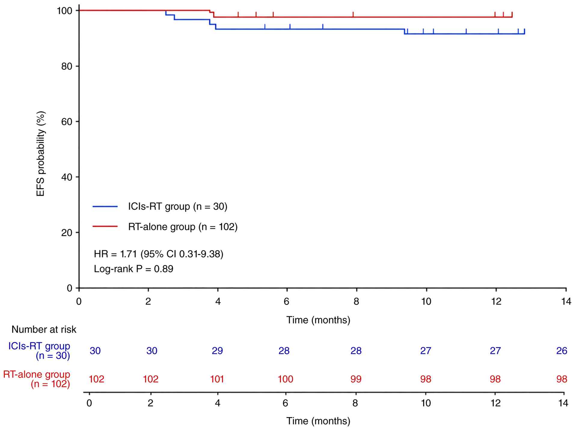
With a median follow-up of 12 months, events
occurred in 6.7% of patients in the ICIs-RT group and 3.9% of
patients in the RT-alone group (Table
IV; P>0.05). The 1-year EFS rates were 93.3 and 96.1%,
respectively (Fig. 1; HR=1.71; 95%
CI, 0.31–9.38; P=0.89). Local recurrence (3.3 vs. 1.0%) and distant
metastasis (3.3 vs. 2.9%) occurred at similar rates in the two
groups. No deaths occurred during follow-up, therefore the 1-year
OS rate was 100% in both groups and the median OS was not
estimable. The median EFS was not estimable because of the limited
number of events.
| Table IV.Clinical outcomes of patients
receiving concurrent radiotherapy and immunotherapy for
triple-negative breast cancer. |
Table IV.
Clinical outcomes of patients
receiving concurrent radiotherapy and immunotherapy for
triple-negative breast cancer.
| Event | ICIs-RT group
(n=30) | RT-alone group
(n=102) | P-value |
|---|
| Any recurrence or
metastasis | 2 (6.7%) | 4 (3.9%) | 0.89 |
| Local
recurrence | 1 (3.3%) | 1 (1.0%) | 0.41 |
| Distant
recurrence | 1 (3.3%) | 3 (2.9%) | 0.99 |
| Dead | 0 | 0 | - |
Discussion
The present study revealed that concurrent
immunotherapy was not associated with a statistically significant
increase in the risk of radiation pneumonitis during postoperative
RT for TNBC; however, a clinically meaningful trend toward a higher
incidence of grade 1 radiation pneumonitis cannot be excluded. This
finding is consistent with the results of the KEYNOTE-522 trial,
which reported a low incidence of radiation pneumonitis in patients
receiving immunotherapy (3).
However, hypothyroidism was observed more frequently in patients
treated with concurrent immunotherapy, likely attributable to
ICI-associated adverse events. Therefore, ICI-related immune
thyroiditis remains the most likely explanation for the increased
incidence of hypothyroidism.
RT has traditionally been considered a localized
cytotoxic treatment. However, emerging evidence suggests that RT
can induce immunogenic cell death, particularly with
hypofractionated regimens (14).
Irradiated tumor cells release damage-associated molecular patterns
(for example, high mobility group box 1, ATP and calreticulin),
which act as danger signals to promote dendritic cell maturation
and antigen presentation (15).
When combined with immune checkpoint blockade, these newly exposed
antigens stimulate tumor-specific T-cell responses, converting
bystander T cells into active effector cells and ultimately
triggering systemic antitumor immunity; the abscopal effect
(16). In postoperative breast
cancer RT, local elimination of microscopic residual disease may be
accompanied by enhanced systemic immune surveillance, which could
reduce the risk of recurrence and metastasis (17).
RT can reduce the infiltration of myeloid-derived
suppressor cells and M2-polarized tumor-associated macrophages
within the tumor microenvironment. Concurrently, RT upregulates
programmed death-ligand 1 (PD-L1) expression on tumor cells,
thereby providing a direct target for ICIs (18). RT also improves tumor vascular
structure and perfusion, facilitating the infiltration and
persistence of effector T cells within tumors. In the setting of
postoperative RT for breast cancer, in which treatment is typically
administered following chemotherapy and/or neoadjuvant
immunotherapy, immune memory may already be established.
Postoperative RT may further reshape the tumor microenvironment,
thereby enhancing the local execution of immune memory responses
(15).
Although the present study did not observe a
statistically significant increase in radiation pneumonitis, RT and
immunotherapy may exert additive toxic effects on normal tissues
from a mechanistic perspective. Radiation-induced lung injury is
mediated by inflammatory cytokines such as transforming growth
factor-β and tumor necrosis factor-α, whereas immune checkpoint
blockade may activate T cells which also target normal tissues
(5,19,20).
Clinically, the relatively low incidence of pneumonitis in patients
with breast cancer may be attributable to smaller radiation field
sizes and specific fractionation schedules. Notably, the relatively
high incidence of grade 1 radiation pneumonitis observed in the
present study (63.3% in the ICIs-RT group and 46.1% in the RT-alone
group) may partially reflect the sensitivity of routine
contrast-enhanced CT imaging performed every 3 months, which could
detect subtle, asymptomatic radiological changes that might
otherwise remain unnoticed. By contrast, the risk of pneumonitis is
substantially higher in patients with lung or esophageal cancer,
warranting careful monitoring in these settings. In patients with
left-sided breast cancer, postoperative RT may expose the heart to
radiation. Although immune-mediated myocarditis is rare, it can be
life-threatening; therefore, stricter cardiac dose constraints
should be considered when combining RT with immunotherapy (21).
The choice of PD-1 inhibitor (pembrolizumab,
tislelizumab, camrelizumab or toripalimab) was based on physician
preference and local drug availability, and was not determined by
patient or disease-specific factors. Given the small number of
patients receiving each agent, formal subgroup analyses were not
feasible. No obvious differences in toxicity profiles were observed
across agents; however, agent-specific effects cannot be excluded
and should be examined in larger cohorts.
The present study has several limitations. First, as
a single-center retrospective analysis, potential selection bias
cannot be excluded. Notably, the significantly higher proportion of
patients receiving platinum-based chemotherapy in the ICIs-RT group
(90.0 vs. 56.8%; P<0.001) reflects real-world clinical practice,
as ICIs in the neoadjuvant setting for TNBC are predominantly
combined with platinum-containing regimens according to the
KEYNOTE-522 protocol. This imbalance may have contributed to the
higher pCR rate observed in the ICIs-RT group, and the relative
contributions of platinum agents and ICIs cannot be disentangled
within the current retrospective study design. Due to the small
number of patients in the ICIs-RT group (n=30), propensity score
matching and multivariable regression analyses were not feasible
for adjusting baseline imbalances. Second, the follow-up duration
was limited (median, 12 months); therefore, the data from the
current study support conclusions regarding acute and early
subacute toxicities only. Late effects, such as radiation fibrosis,
cardiac toxicity and long-term survival outcomes, require extended
follow-up. Furthermore, the small sample size resulted in
insufficient statistical power. With only 30 patients in the
intervention group and a low event count, the study was
underpowered to detect uncommon but clinically meaningful
toxicities, such as grade ≥2 radiation pneumonitis. Therefore, the
absence of a statistically significant difference in grade 1
radiation pneumonitis should not be interpreted as evidence of
equivalent safety. Future prospective studies with larger sample
sizes and longer follow-up are warranted to validate these findings
and to employ appropriate methodologies for adjusting baseline
confounding. Multiple ongoing trials are currently evaluating
combined RT and immunotherapy strategies in the neoadjuvant,
adjuvant and metastatic settings. Radiation dose and fractionation
schedules may modulate immunologic synergy (22). Notably, hypofractionated RT may
enhance immune activation (23).
However, the optimal timing and sequencing; whether concurrent,
sequential or sandwich, remain undefined. Finally, the
identification of predictive biomarkers such as PD-L1 expression
and tumor-infiltrating lymphocyte levels, will be crucial for
identifying patients most likely to benefit from combined therapy
(24,25).
In summary, concurrent immunotherapy during
postoperative RT was not associated with a significant increase in
radiation dermatitis or a statistically significant increase in
radiation pneumonitis; however, a clinically meaningful trend
toward a higher incidence of grade 1 radiation pneumonitis cannot
be excluded. Concurrent immunotherapy did not appear to adversely
affect short-term clinical outcomes; however, the limited follow-up
duration and small number of events preclude definitive
conclusions. These findings support the safety of the combined
approach but highlight the need for continued vigilance.
Acknowledgements
Not applicable.
Funding
This work was supported by the China Health and Medical
Development Foundation (grant no. CHMDF2025-XRKT12-04).
Availability of data and materials
The data generated in the present study may be
requested from the corresponding author.
Authors' contributions
GL conceived and designed the study, performed the
investigation, prepared the figure and tables, and drafted and
revised the manuscript. QZ provided study materials and patient
resources, supervised the formal statistical analysis, and
contributed to data interpretation. MY collected and managed the
clinical data, and assisted with data visualization. GY collected
and curated the data, and participated in the investigation. FY
obtained funding, contributed to the conception and design of the
study, supervised the project and critically revised the manuscript
for important intellectual content. GL and FY confirm the
authenticity of all the raw data. All authors read and approved the
final version of the manuscript.
Ethical approval and consent to
participate
This retrospective study was approved by the Ethics
Committee of Hubei Cancer Hospital, Tongji Medical College,
Huazhong University of Science and Technology (approval no.
LLHBCH2026YN-106). All procedures involving human participants were
performed in accordance with the ethical standards of the
institutional research committee and the 1964 Declaration of
Helsinki and its subsequent amendments. Written informed consent
for participation in the study was obtained from all living
patients who were able to provide consent. For patients who were
deceased or lost to follow-up, the requirement for informed consent
was waived by the Ethics Committee, and all data were anonymized to
protect patient privacy. Written informed consent for the off-label
use of immunotherapy was also obtained from all patients who
received such treatment, in accordance with institutional and
regulatory requirements.
Patient consent for publication
Not applicable.
Competing interests
The authors declare that they have no competing
interests.
References
|
1
|
Foulkes WD, Smith IE and Reis-Filho JS:
Triple-negative breast cancer. N Engl J Med. 363:1938–1948. 2010.
View Article : Google Scholar : PubMed/NCBI
|
|
2
|
Kundu S and Suresh PK: Triple negative
breast cancer heterogeneity and tumour microenvironment-based model
systems' Focus on druggable targets. Curr Mol Med. 25:1450–1483.
2025. View Article : Google Scholar : PubMed/NCBI
|
|
3
|
Schmid P, Cortes J, Dent R, McArthur H,
Pusztai L, Kümmel S, Denkert C, Park YH, Hui R, Harbeck N, et al:
Overall survival with pembrolizumab in Early-stage triple-negative
breast cancer. N Engl J Med. 391:1981–1991. 2024. View Article : Google Scholar : PubMed/NCBI
|
|
4
|
Mamounas EP, Bandos H, White JR, Julian
TB, Khan AJ, Shaitelman SF, Torres MA, Vicini FA, Ganz PA,
McCloskey SA, et al: Omitting regional nodal irradiation after
response to neoadjuvant chemotherapy. N Engl J Med. 392:2113–2124.
2025. View Article : Google Scholar : PubMed/NCBI
|
|
5
|
Golden EB and Apetoh L: Radiotherapy and
immunogenic cell death. Semin Radiat Oncol. 25:11–17. 2015.
View Article : Google Scholar : PubMed/NCBI
|
|
6
|
Deng L, Liang H, Burnette B, Beckett M,
Darga T, Weichselbaum RR and Fu YX: Irradiation and anti-PD-L1
treatment synergistically promote antitumor immunity in mice. J
Clin Invest. 124:687–695. 2014. View
Article : Google Scholar : PubMed/NCBI
|
|
7
|
Dewan MZ, Galloway AE, Kawashima N,
Dewyngaert JK, Babb JS, Formenti SC and Demaria S: Fractionated but
not single-dose radiotherapy induces an immune-mediated abscopal
effect when combined with anti-CTLA-4 antibody. Clin Cancer Res.
15:5379–5388. 2009. View Article : Google Scholar : PubMed/NCBI
|
|
8
|
Senan S and Bahce I: Pulmonary toxicity in
patients treated with immune checkpoint inhibitors and radiation.
Ann Oncol. 31:1597–1598. 2020. View Article : Google Scholar : PubMed/NCBI
|
|
9
|
Antonia SJ, Villegas A, Daniel D, Vicente
D, Murakami S, Hui R, Yokoi T, Chiappori A, Lee KH, de Wit M, et
al: Durvalumab after chemoradiotherapy in stage III non-small-cell
lung cancer. N Engl J Med. 377:1919–1929. 2017. View Article : Google Scholar : PubMed/NCBI
|
|
10
|
El Fakih A, Loap P, Cabel L, Allali S, Cao
K, Mirabel M, Pierga JY and Kirova Y: Real-world safety of
concurrent pembrolizumab and radiotherapy in triple-negative breast
cancer. Am J Clin Oncol. 49:62–68. 2026. View Article : Google Scholar : PubMed/NCBI
|
|
11
|
Liang G, Chang R, Zhang Q, Luo Y, Peng Y
and Luo B: Segmental bronchi radiation dose affected the
progression of radiation pneumonitis in lung cancer patients.
Discov Oncol. 16:7932025. View Article : Google Scholar : PubMed/NCBI
|
|
12
|
Offersen BV, Boersma LJ, Kirkove C, Hol S,
Aznar MC, Biete Sola A, Kirova YM, Pignol JP, Remouchamps V,
Verhoeven K, et al: ESTRO consensus guideline on target volume
delineation for elective radiation therapy of early stage breast
cancer. Radiother Oncol. 114:3–10. 2015. View Article : Google Scholar : PubMed/NCBI
|
|
13
|
National Cancer Institute (NCI), . Common
Terminology Criteria for Adverse Events (CTCAE) version 4.0.
Published. May 28–2009.Updated June 14, 2010. August
9–2026https://ctep.cancer.gov/protocoldevelopment/electronic_applications/ctc.htm
|
|
14
|
Bertho A, Iturri L and Prezado Y:
Radiation-induced immune response in novel radiotherapy approaches
FLASH and spatially fractionated radiotherapies. Int Rev Cell Mol
Biol. 376:37–68. 2023. View Article : Google Scholar : PubMed/NCBI
|
|
15
|
Fenton M, Yoneyama M, Wennerberg E, Lund
T, Tutt A, Melcher A, Demaria S and Somaiah N: The untapped
potential of radiation and immunotherapy for hormone
receptor-positive breast cancer. NPJ Breast Cancer. 11:772025.
View Article : Google Scholar : PubMed/NCBI
|
|
16
|
Cho EJ, Ki MK, Baek HJ, Shin DH, Park EJ,
Kim TH, Deng CX, Choi BK and Kim SS: Synergistic effects of
radiotherapy and PD-1 blockade in a human-mimetic BRCAness model of
triple-negative breast cancer. Int J Biol Sci. 21:6270–6284. 2025.
View Article : Google Scholar : PubMed/NCBI
|
|
17
|
Panse D, Hsieh K, Arons D, Runnels J,
Wassel M, Shah A, Patel R, Tiersten A, Nehlsen AD, Marshall D, et
al: The role of immunotherapy and radiation therapy in the
treatment of breast cancer. Biomedicines. 13:22092025. View Article : Google Scholar : PubMed/NCBI
|
|
18
|
Li J, Wen L, Guo Y, Yao D, Sun B, Mou H,
Chen N, Zhou K, Wang Y and Chen W: Combination of low-intensity
pulsed ultrasound irradiating immune organs with immune checkpoint
blockade augments systemic anti-tumor immunity on low tumor burden
4T-1 breast cancer. Cancer Immunol Immunother. 74:2812025.
View Article : Google Scholar : PubMed/NCBI
|
|
19
|
Liu X, Song Y, Hu S, Bai Y, Zhang J, Tai
G, Shao C and Pan Y: Serum amyloid A contributes to
radiation-induced lung injury by activating macrophages through
FPR2/Rac1/NF-κB pathway. Int J Biol Sci. 20:4941–4956. 2024.
View Article : Google Scholar : PubMed/NCBI
|
|
20
|
Mahiat C, Cadranel J, Fallet V, Meteye C,
Gendarme S, Slomka J and Canellas A: Pulmonary toxicity of
immune-checkpoint inhibitors. Presse Med. 55:1043292025. View Article : Google Scholar : PubMed/NCBI
|
|
21
|
Gumusay O, Callan J and Rugo HS:
Immunotherapy toxicity: Identification and management. Breast
Cancer Res Treat. 192:1–17. 2022. View Article : Google Scholar : PubMed/NCBI
|
|
22
|
Zhou H, Wang S, Hu C and Kong FM:
Sequential versus concurrent strategy of immunotherapy and
radiotherapy in advanced non-small-cell lung cancer: A
territory-wide multicenter study (OCEANUS study). J Clin Oncol. 43
(16_suppl):S80732025. View Article : Google Scholar
|
|
23
|
Xiao J, Shao S, Deng Y, Wang D, Liu Y, He
S, Zhao Y, Liao W, Zhang J, Yang M and Zhang S: Prolonged interval
hypofractionated radiotherapy facilitates better antitumor
immunity. Radiother Oncol. 203:1106642025. View Article : Google Scholar : PubMed/NCBI
|
|
24
|
Froelich W: Radiotherapy boosts
immunotherapy in HR+/HER2-breast cancer. Oncol Times. 48:15–16.
2026. View Article : Google Scholar
|
|
25
|
Uprety D, Roden AC and Peters S: Adjuvant
immunotherapy should be used in patients with non-Small cell
carcinoma with a pathologic complete response to neoadjuvant
immunotherapy. J Thorac Oncol. 20:34–38. 2025. View Article : Google Scholar : PubMed/NCBI
|