Introduction
Nasopharyngeal carcinoma (NPC) has a distinct
geographic distribution, with a higher incidence in southern China
and Southeast Asia than in most other regions worldwide (1). With the use of intensity-modulated
radiotherapy (IMRT), platinum-based concurrent chemotherapy and
induction chemotherapy, locoregionally advanced NPC has achieved
improved locoregional control and long-term survival (1–3).
Nevertheless, patients with T3-4 disease still have risks of
recurrence, distant metastasis and death. Further identification of
high-risk patients within conventional staging categories may help
refine prognostic assessment and support clinical trial
stratification (4).
The tumor-node-metastasis (TNM) staging system
remains the main framework for treatment decision-making and
prognostic evaluation in NPC. It is largely based on the anatomic
extent of primary tumor invasion and regional nodal involvement
(4,5). However, the T stage is a categorical
variable and does not fully reflect continuous differences in tumor
burden within the same T-stage category (6,7). Among
patients with T3-4 disease, small-volume tumors may be classified
as advanced T stage due to the involvement of critical anatomic
structures, whereas other tumors may show extensive infiltration
and a much larger primary volume (6). These differences may be associated
with variations in tumor cell burden, hypoxia, local treatment
complexity and potential treatment resistance (8).
Gross tumor volume of the primary nasopharyngeal
tumor (GTVnx) is a routinely available volumetric parameter derived
from radiotherapy planning systems and may serve as a quantitative
measure of primary tumor burden (9). Previous studies have suggested that a
larger primary tumor volume is associated with poorer local
control, increased risk of distant metastasis and unfavorable
overall survival (OS) (7,8). GTVnx has also been incorporated with
Epstein-Barr virus (EBV) DNA, clinical stage or imaging features
for risk stratification, while radiomic and molecular biomarkers
have been increasingly investigated for prognostic assessment in
NPC (10–13). However, most previous studies
evaluated patients across all T stages or the broader
locoregionally advanced population (8,10).
Whether GTVnx can further identify patients with poor OS within the
anatomically high-risk T3-4 subgroup has remained elusive.
Beyond statistical significance, the clinical value
of a prognostic marker also depends on its incremental value beyond
established clinical factors (14,15).
Time-dependent receiver operating characteristic (ROC) analysis
(14), the concordance index
(C-index) (16) and Akaike
information criterion (AIC) (17)
can assess model performance from complementary perspectives,
including discrimination at a specific time point, overall
discriminative ability and model fit, while accounting for
complexity. Therefore, the present retrospective study evaluated
the association between GTVnx and OS in patients with T3-4 NPC and
explored whether GTVnx provides incremental prognostic information
beyond conventional clinical factors.
Patients and methods
Patients
The present single-center retrospective cohort study
included 118 patients with T3-4 NPC who received definitive IMRT at
Guilin People's Hospital (Guilin, China) between March 2020 and
June 2025. All patients had pathologically confirmed NPC and
underwent pretreatment staging examinations, including
contrast-enhanced magnetic resonance imaging (MRI) of the
nasopharynx and neck, chest imaging, abdominal imaging and bone
assessment using bone scintigraphy, skeletal MRI or positron
emission tomography/computed tomography (CT), as clinically
indicated. As the present study period spanned different TNM
classification periods, all patients were retrospectively restaged
according to the 9th edition of the American Joint Committee on
Cancer/Union for International Cancer Control TNM classification
system before statistical analysis using available pretreatment
clinical, imaging and pathological information.
The inclusion criteria were as follows: i)
Pathologically confirmed NPC; ii) T3-4 disease; iii) receipt of
definitive IMRT; iv) availability of complete pretreatment imaging
and radiotherapy planning data for GTVnx assessment; and v)
available follow-up data for OS. The exclusion criteria were as
follows: i) Distant metastasis at initial diagnosis; ii) previous
head and neck radiotherapy or treatment for another malignancy;
iii) concurrent active malignancy; and iv) missing key clinical,
radiotherapy planning or follow-up data. The study protocol was
approved by the Ethics Committee of Guilin People's Hospital
(Guilin, China; approval no. 2025-316KY) and written informed
consent for the use of medical data for research purposes was
obtained from all patients at admission.
Treatment and target delineation
All patients received definitive IMRT. GTVnx was
manually delineated by radiation oncologists based on pretreatment
contrast-enhanced MRI, simulation CT images and clinical
examination findings. The delineation included all radiologically
and clinically visible primary nasopharyngeal lesions and was
performed according to institutional target delineation protocols
for NPC. All GTVnx contours were independently reviewed and
confirmed by two senior radiation oncologists before radiotherapy
planning to minimize inter-observer variability. The GTVnx volume
was automatically calculated by the radiotherapy planning system
and recorded in cm3. Cervical nodal target volumes,
clinical target volumes and organs at risk were delineated
according to the institutional head and neck radiotherapy protocol.
All patients received platinum-based combined treatment, including
induction chemotherapy, concurrent chemoradiotherapy, concurrent
targeted therapy, adjuvant chemotherapy and maintenance
chemotherapy when indicated. The specific treatment regimen was
determined by the attending physician according to disease stage,
performance status and organ function. None of the patients
received immunotherapy during the study period.
Variables and outcomes
Data on age, sex, histological subtype, T stage, N
stage, clinical stage, pretreatment EBV DNA level, radiotherapy
dose, GTVnx and follow-up outcomes were collected. The pretreatment
plasma EBV DNA level was dichotomized using a cut-off value of 400
copies/ml, consistent with previous studies and commonly used
clinical thresholds for risk stratification in NPC (18). OS was defined as the time from the
start of radiotherapy to death from any cause. Patients who were
alive at the last follow-up were censored.
Statistical analysis
All statistical analyses were performed using R
software (version 4.5.0; R Foundation for Statistical Computing)
and SPSS (version 25.0; IBM Corp.). Continuous variables were first
assessed for normality. Normally distributed variables were
presented as the mean ± standard deviation and were compared using
the independent-samples t-test. Non-normally distributed variables
were presented as the median (first quartile, third quartile) and
were compared using the Mann-Whitney U-test. Categorical variables
were summarized as counts and percentages and were compared using
the χ2 test or Fisher's exact test.
A 36-month time-dependent ROC curve was used to
evaluate the ability of GTVnx to discriminate patients according to
their OS outcomes. The 36-month time-point was selected based on
the maturity of follow-up data as it approximated the median
follow-up duration of the cohort (34.1 months) and represented an
intermediate-term survival endpoint. The variability of the cut-off
value was further assessed using bootstrap resampling with 1,000
repetitions, and the median bootstrap-derived cut-off value and 95%
percentile interval were calculated. Patients were then divided
into low- and high-GTVnx groups based on this cut-off. OS curves
were generated using the Kaplan-Meier method and differences
between groups were compared using the log-rank test. Cox
proportional hazards models were used to estimate hazard ratios
(HRs) and 95% confidence intervals (CIs).
Given the limited number of OS events (n=18), the
events-per-variable ratio was below the commonly recommended
threshold of 10 events per variable. Therefore, multivariable Cox
regression models and incremental model performance analyses were
considered exploratory and hypothesis-generating. Variables
included in the multivariable models were selected a priori based
on established clinical relevance and prognostic significance
rather than solely on univariable statistical significance. The
clinical model included T stage and pretreatment EBV DNA, and the
incremental prognostic value of GTVnx was subsequently evaluated by
adding GTVnx to this model. Considering the risk of model
overfitting caused by limited outcome events, the N stage was not
incorporated into the primary model and was examined separately in
sensitivity analyses. Model performance was compared using the
C-index, AIC and the 36-month time-dependent area under the curve
(AUC). In addition, bootstrap resampling was used to estimate the
CI of the difference in C-index between the clinical and extended
models.
To assess the robustness of the findings, several
sensitivity analyses were performed as follows: i) Further
adjustment for N stage based on the main model; ii) replacement of
T stage with overall clinical stage; iii) inclusion of GTVnx as a
continuous variable in the model, with the HR calculated per 10
cm3 increase; and iv) use of a conventional ROC-derived
alternative cut-off to repeat the Kaplan-Meier survival analysis. A
two-sided P<0.05 was considered statistically
significant.
Results
Patient baseline characteristics
A total of 118 patients with T3-4 NPC were included
in the present study. The median age was 53 years (range, 23–75
years), and the cohort included 84 male and 34 female patients
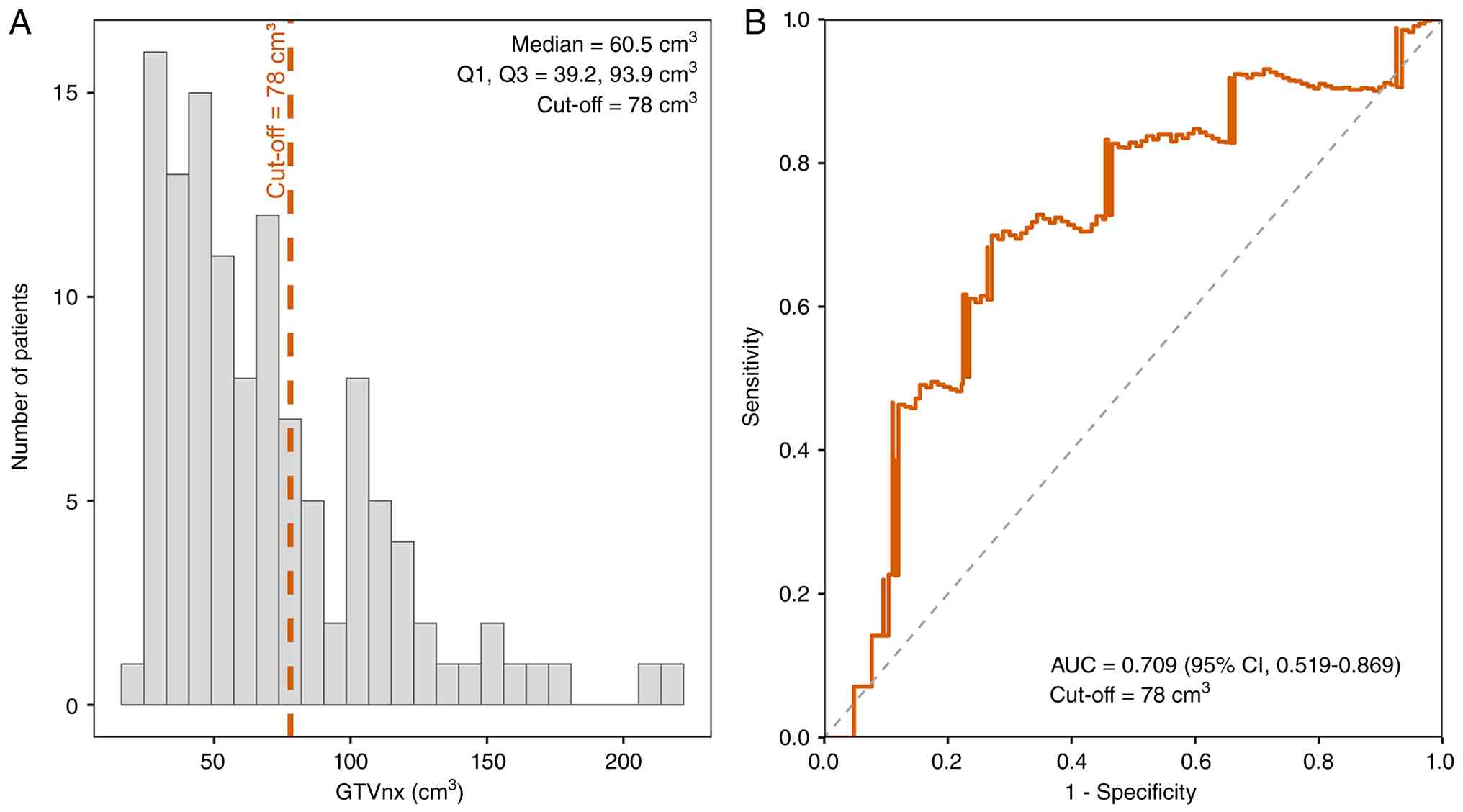
(Table I). Based on the 78
cm3 cut-off determined by the 36-month time-dependent
ROC analysis (Fig. 1), patients
were divided into the low-GTVnx group (GTVnx ≤78 cm3;
n=80) and the high-GTVnx group (GTVnx >78 cm3; n=38).
Compared with the low-GTVnx group, patients in the high-GTVnx group
had significantly higher proportions of male patients, T4 disease,
stage III disease and pretreatment EBV DNA ≥400 copies/ml. There
were no statistically significant differences between the two
groups in terms of age, age category, N stage, histological
subtype, follow-up duration, loss to follow-up rate, concurrent
targeted therapy, adjuvant chemotherapy or capecitabine/S-1
maintenance chemotherapy (all P>0.05). The baseline
characteristics are shown in Table
I.
 | Table I.Baseline characteristics of patients
with T3-4 nasopharyngeal carcinoma according to GTVnx group. |
Table I.
Baseline characteristics of patients
with T3-4 nasopharyngeal carcinoma according to GTVnx group.
| Characteristic | Total (n=118) | GTVnx ≤78
cm3 (n=80) | GTVnx >78
cm3 (n=38) | P-value |
|---|
| Age, years | 52.4 ± 11.9 | 51.6 ± 11.3 | 54.2 ± 10.0 | 0.234 |
| Age group,
years |
|
|
| 0.103 |
|
≤40 | 18 (15.3) | 16 (20.0) | 2 (5.3) |
|
| >40,
≤60 | 78 (66.1) | 49 (61.3) | 29 (76.3) |
|
|
>60 | 22 (18.6) | 15 (18.8) | 7 (18.4) |
|
| Sex |
|
|
| 0.031 |
|
Male | 84 (71.2) | 52 (65.0) | 32 (84.2) |
|
|
Female | 34 (28.8) | 28 (35.0) | 6 (15.8) |
|
| T stage |
|
|
| <0.001 |
| T3 | 57 (48.3) | 50 (62.5) | 7 (18.4) |
|
| T4 | 61 (51.7) | 30 (37.5) | 31 (81.6) |
|
| N stage |
|
|
| 0.360 |
| N1 | 9 (7.6) | 8 (10.0) | 1 (2.6) |
|
| N2 | 75 (63.6) | 49 (61.3) | 26 (68.4) |
|
| N3 | 34 (28.8) | 23 (28.7) | 11 (28.9) |
|
| Clinical stage |
|
|
| 0.001 |
| Stage
II | 45 (38.1) | 39 (48.8) | 6 (15.8) |
|
| Stage
III | 73 (61.9) | 41 (51.2) | 32 (84.2) |
|
| Histological
subtype |
|
|
| 0.663 |
|
Non-keratinizing
undifferentiated carcinoma | 112 (94.9) | 75 (93.8) | 37 (97.4) |
|
|
Non-keratinizing
differentiated carcinoma | 6 (5.1) | 5 (6.3) | 1 (2.6) |
|
| EBV DNA,
copies/ml |
|
|
| 0.033 |
|
<400 | 54 (45.8) | 42 (52.5) | 12 (31.6) |
|
|
≥400 | 64 (54.2) | 38 (47.5) | 26 (68.4) |
|
| GTVnx,
cm3 | 60.5 (39.2,
93.9) | 46.6
(33.9,61.0) | 109.0 (95.9,
131.0) | <0.001 |
| Induction
chemotherapy | 118 (100) | 80 (100) | 38 (100) | - |
| Concurrent
chemoradiotherapy | 118 (100) | 80 (100) | 38 (100) | - |
| Concurrent targeted
therapy | 63 (53.4) | 41 (51.2) | 22 (57.9) | 0.499 |
| Adjuvant
chemotherapy | 31 (26.3) | 22 (27.5) | 9 (23.7) | 0.660 |
| Capecitabine/S-1
maintenance chemotherapy | 63 (53.4) | 42 (52.5) | 21 (55.3) | 0.779 |
Follow-up and survival outcomes
The median (25th, 75th percentile) follow-up time
for the overall cohort was 34.1 (20.4, 49.3) months. There was no
statistically significant difference in median follow-up time
between the low- and high-GTVnx groups. During follow-up, 18 deaths
occurred (15.3%). The proportion of OS events was significantly
higher in the high-GTVnx group than in the low-GTVnx group (31.6
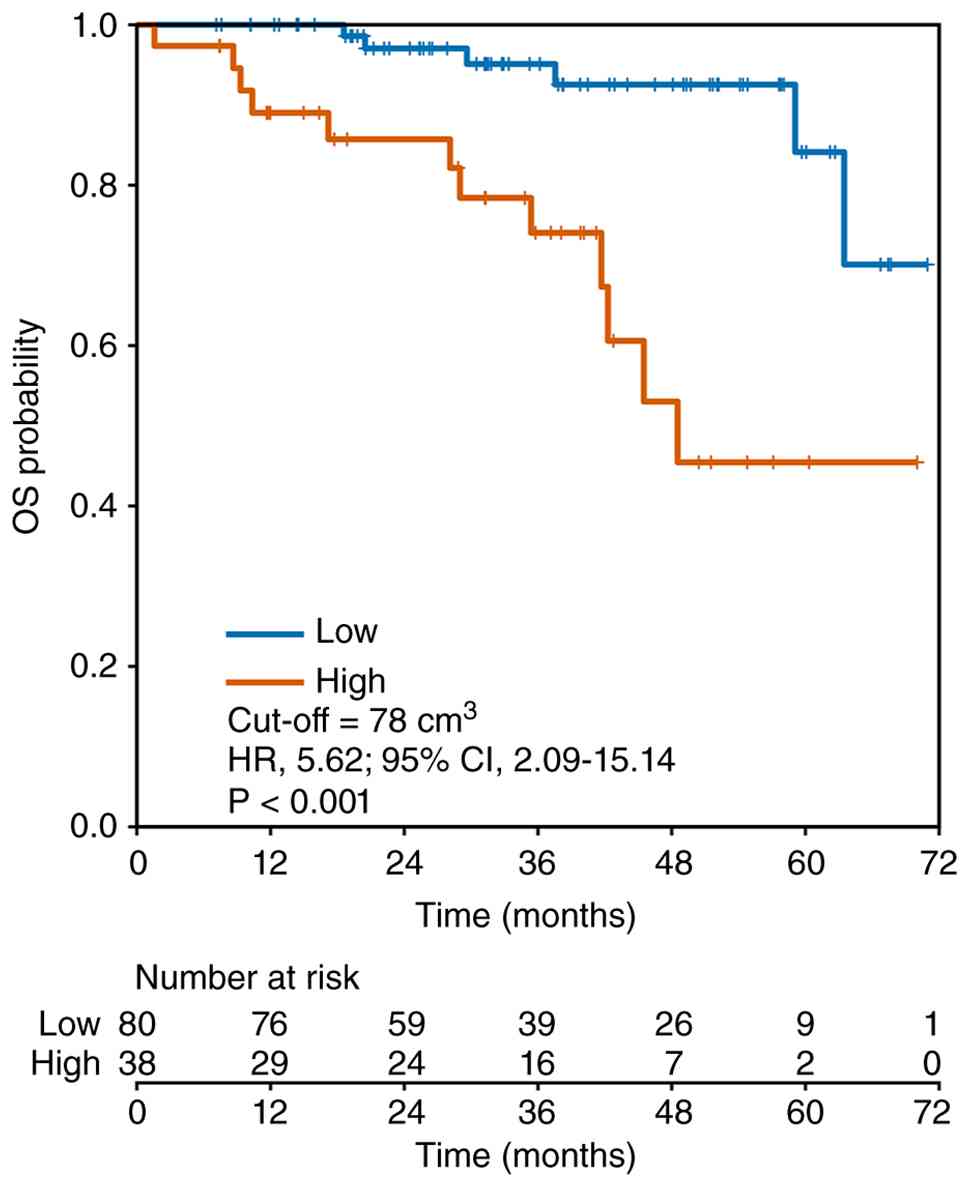
vs. 7.5%; P=0.002). The 3-year OS rate was lower in the
high-GTVnx group than in the low-GTVnx group (74.1 vs. 95.1%;
P<0.001).
GTVnx distribution and cut-off
determination
GTVnx showed a right-skewed distribution in the
overall cohort, with a median value of 60.5 cm3 and
first and third quartiles of 39.2 and 93.9 cm3,
respectively. Time-dependent ROC analysis for 3-year OS showed that
GTVnx had discriminatory ability for predicting 3-year OS, with an
AUC of 0.709. Based on the Youden index, the optimal cut-off value
for GTVnx was 78 cm3, with a sensitivity of 69.9% and a
specificity of 72.9%. The distribution of GTVnx and the
time-dependent ROC curve are shown in Fig. 1. The bootstrap assessment of cut-off
variability is presented in Table
SI.
Prognostic value of GTVnx
Kaplan-Meier survival analysis
Kaplan-Meier survival analysis showed that, when
patients were stratified according to the 78 cm3
cut-off, the high-GTVnx group had significantly poorer OS than the
low-GTVnx group (log-rank P<0.001; Fig. 2). Cox regression analysis showed
that, compared with patients with GTVnx ≤78 cm3, those
with GTVnx >78 cm3 had a significantly increased risk
of death (HR, 5.62; 95% CI, 2.09–15.14; P<0.001).
Cox survival analysis
Univariable Cox regression analysis showed that
stage III disease, EBV DNA ≥400 copies/ml and high GTVnx were
significantly associated with poorer OS. Multivariable Cox
regression analysis further showed that, after adjustment for T
stage and EBV DNA, high GTVnx remained independently associated
with unfavorable OS (HR, 4.375; 95% CI, 1.474–12.986; P=0.008). The
results of the univariable and multivariable Cox analyses are shown
in Table II.
| Table II.Univariable and multivariable Cox
regression analyses for overall survival in patients with T3-4
nasopharyngeal carcinoma. |
Table II.
Univariable and multivariable Cox
regression analyses for overall survival in patients with T3-4
nasopharyngeal carcinoma.
| Variable | Univariable HR (95%
CI) | P-value | Multivariable HR
(95% CI) | P-value |
|---|
| Age, years | 1.042
(0.993–1.094) | 0.091 | - | - |
| Sex, female vs.
male | 1.354
(0.502–3.650) | 0.549 | - | - |
| T stage, T4 vs.
T3 | 2.330
(0.828–6.556) | 0.109 | 1.089
(0.359–3.303) | 0.881 |
| N stage, N3 vs.
N0-2 | 2.115
(0.810–5.518) | 0.126 | - | - |
| Clinical stage, III
vs. II | 4.042
(1.153–14.171) | 0.029 | - | - |
| EBV DNA, ≥400 vs.
<400 copies/ml | 6.223
(1.972–19.636) | 0.002 | 5.041
(1.516–16.763) | 0.008 |
| GTVnx, >78 vs.
≤78 cm3 | 5.623
(2.088–15.143) | 0.001 | 4.375
(1.474–12.986) | 0.008 |
Incremental prognostic value of GTVnx
beyond the clinical model
In the same 118 patients with 18 deaths, the
extended model incorporating GTVnx showed improved predictive
performance compared with the clinical model including only T stage
and EBV DNA. The C-index increased from 0.759 to 0.817, with a
bootstrap-estimated difference in C-index of 0.052 (95% CI,
−0.009–0.127). The 36-month AUC increased from 0.748 to 0.787, and
the AIC decreased from 135.151 to 133.085 after adding GTVnx, with
a ΔAIC of −2.066 (Table III).
| Table III.Comparison of predictive performance
between the clinical and extended models. |
Table III.
Comparison of predictive performance
between the clinical and extended models.
| Metric | Clinical model | Extended model |
|---|
| Variables
included | T stage + EBV
DNA | T stage + EBV DNA +
GTVnx |
| C-index | 0.759 | 0.817 |
| Bootstrap
difference in C-index | - | 0.052 (95% CI,
−0.009–0.127) |
| 36-month AUC | 0.748 | 0.787 |
| AIC | 135.151 | 133.085 |
| ΔAIC | - | −2.066 |
Sensitivity analysis
After further adjustment for N stage based on the
main model, the association between GTVnx and poorer OS remained
consistent. Replacing T stage with overall clinical stage did not
materially change the results. When GTVnx was included as a
continuous variable, each 10 cm3 increase in GTVnx
remained associated with an increased risk of death. In addition,
after using a conventional ROC-derived cut-off of 106
cm3 as an alternative threshold for regrouping, the
high-GTVnx group still showed a trend toward poorer OS. Detailed
results are provided in Table SII
and Fig. S1. Overall, the
sensitivity analyses supported the robustness of the main
findings.
Discussion
The present study examined whether GTVnx
could provide additional prognostic information in patients with
T3-4 NPC. The main finding was that patients with a larger GTVnx
had poorer OS, and this association persisted after adjustment for
T stage and pretreatment EBV DNA. When GTVnx was added to a
clinical model based on these conventional factors, the C-index,
AIC and time-dependent AUC all changed in a favorable direction,
although the absolute improvement was modest. Similar results were
obtained across sensitivity analyses using alternative model
specifications and variable definitions. These findings suggest
that GTVnx may help refine prognostic assessment within the T3-4
subgroup, although further validation is required before clinical
application.
TNM staging remains the foundation for prognostic
assessment and treatment decision-making in NPC (4). However, T stage is primarily based on
the anatomic extent of tumor invasion and does not directly
quantify tumor burden (19). In
clinical practice, patients classified as T3-4 may have
substantially different primary tumor volumes. Certain tumors are
assigned to an advanced T category due to the involvement of
critical anatomic structures, even when the measurable primary
volume is relatively limited. By contrast, other tumors within the
same T category may show more extensive infiltration and a much
larger volume (19,20). As a continuous volumetric parameter
routinely derived from radiotherapy planning systems, GTVnx may
capture part of this heterogeneity (19). The association between larger GTVnx
and poorer OS suggests that tumor volume provides additional
information regarding disease burden.
Previous studies have linked primary tumor volume
with locoregional control, distant metastasis and survival outcomes
in NPC (9,21–23).
Most of these studies evaluated mixed-stage cohorts or the broader
locoregionally advanced population (9,23). The
present study stands out by focusing specifically on patients with
T3-4 disease, a subgroup already considered anatomically high-risk.
The present findings indicate that tumor volume may still
distinguish prognostic differences within this advanced T-stage
category (24,25). It may serve as a simple quantitative
adjunct to conventional staging, particularly when patients with
the same T stage show markedly different primary tumor burdens.
The incremental predictive value of GTVnx should
also be interpreted with caution (26–28).
Although adding GTVnx to the clinical model resulted in an
improvement in model performance, the magnitude of incremental
improvement was modest. The bootstrap CI for the difference in
C-index included zero, suggesting uncertainty regarding the
magnitude of improvement. This may be partly because the clinical
model already included established prognostic factors, such as T
stage and pretreatment EBV DNA (18). Although N stage showed a clinically
meaningful HR, it was not included in the primary model due to the
limited number of OS events and the potential risk of overfitting.
However, sensitivity analysis incorporating N stage demonstrated
consistent results, suggesting that the prognostic association of
GTVnx remained consistent. In addition, GTVnx and T stage are not
completely independent, as larger tumors are more likely to involve
adjacent anatomic structures. These overlapping features may limit
the additional contribution of GTVnx when it is combined with
established clinical variables. Therefore, GTVnx should be viewed
as supplementary risk information rather than as a stand-alone
basis for treatment selection. For patients with a larger GTVnx,
clinical interpretation should still integrate T stage, N stage,
EBV DNA, treatment response and the general condition of the
patient (29,30).
Several limitations should be acknowledged. First,
this was a single-center retrospective study, and selection bias
and unmeasured confounding cannot be excluded. Second, the
relatively small number of OS events may have limited the
statistical power and stability of multivariable modeling.
Therefore, the prognostic value of GTVnx requires further
evaluation. Third, the 78 cm3 cut-off identified by
time-dependent ROC analysis was derived from the present cohort and
should be considered an exploratory threshold rather than a
generally applicable clinical threshold. Although bootstrap
analysis showed good agreement between the original cut-off and the
median bootstrap-derived cut-off, the relatively wide percentile
interval indicated uncertainty regarding the precision of the
cut-off estimate. Therefore, this cut-off requires validation in
larger independent cohorts before clinical application. Fourth, the
present study focused on OS and did not systematically assess
progression-free survival, locoregional recurrence-free survival or
distant metastasis-free survival. Finally, dynamic EBV DNA,
response to induction chemotherapy, radiomic features and molecular
biomarkers were not included. Larger multicenter studies with
external validation are needed to confirm the stability of GTVnx
and to determine how it can be integrated with other prognostic
factors.
In summary, in patients with T3-4 NPC, GTVnx was
associated with poorer OS and may provide supplementary prognostic
information beyond T stage and pretreatment EBV DNA. The 78
cm3 cut-off derived from time-dependent ROC analysis
helped identify patients at higher risk, and adding GTVnx to the
clinical model modestly improved model performance. GTVnx should be
regarded as an auxiliary quantitative parameter rather than an
alternative to TNM staging. Further multicenter studies with larger
sample sizes and external validation are needed to confirm these
findings.
Supplementary Material
Supporting Data
Supporting Data
Acknowledgements
Not applicable.
Funding
This study was supported by The Guilin Science Research and
Technology Development Program (grant no. 20220139-9-8).
Availability of data and materials
The data generated in the present study may be
requested from the corresponding author.
Authors' contributions
HH and QQC conceived and designed the study; HH,
YSL, YJO, MP, XYT, TFZ and QG collected the data; HH and HRH
performed the statistical analyses; HH, YSL, CQC and QQC analyzed
and interpreted the data; HH and YSL drafted the manuscript; CQC
and QQC supervised the study and critically revised the manuscript;
HH and QQC confirm the authenticity of all the raw data. All
authors read and approved the final version of the manuscript.
Ethics approval and consent to
participate
This study was approved by the Ethics Committee of
Guilin People's Hospital (Guilin, China; approval no. 2025-316KY)
and was conducted in accordance with the Declaration of Helsinki.
Written informed consent for the use of medical data for research
purposes was obtained from all patients at admission.
Patient consent for publication
Not applicable.
Competing interests
The authors declare that they have no competing
interests.
Use of artificial intelligence tools
During the preparation of this work, artificial
intelligence tools were used to improve the readability and
language of the manuscript, and subsequently, the authors revised
and edited the content produced by the artificial intelligence
tools as necessary, taking full responsibility for the ultimate
content of the present manuscript.
Glossary
Abbreviations
Abbreviations:
|
AIC
|
Akaike information criterion
|
|
AUC
|
area under the curve
|
|
CI
|
confidence interval
|
|
EBV DNA
|
Epstein-Barr virus DNA
|
|
GTVnx
|
gross tumor volume of the primary
nasopharyngeal tumor
|
|
HR
|
hazard ratio
|
|
IMRT
|
intensity-modulated radiotherapy
|
|
NPC
|
nasopharyngeal carcinoma
|
|
OS
|
overall survival
|
|
ROC
|
receiver operating characteristic
|
|
TNM
|
tumor-node-metastasis
|
References
|
1
|
Chen YP, Chan ATC, Le QT, Blanchard P, Sun
Y and Ma J: Nasopharyngeal carcinoma. Lancet. 394:64–80. 2019.
View Article : Google Scholar : PubMed/NCBI
|
|
2
|
Bossi P, Chan AT, Licitra L, Trama A,
Orlandi E, Hui EP, Halámková J, Mattheis S, Baujat B, Hardillo J,
et al: Nasopharyngeal carcinoma: ESMO-EURACAN Clinical Practice
Guidelines for diagnosis, treatment and follow-up†. Ann Oncol.
32:452–465. 2021. View Article : Google Scholar : PubMed/NCBI
|
|
3
|
Sun Y, Li WF, Chen NY, Zhang N, Hu GQ, Xie
FY, Sun Y, Chen XZ, Li JG, Zhu XD, et al: Induction chemotherapy
plus concurrent chemoradiotherapy versus concurrent
chemoradiotherapy alone in locoregionally advanced nasopharyngeal
carcinoma: A phase 3, multicentre, randomised controlled trial.
Lancet Oncol. 17:1509–1520. 2016. View Article : Google Scholar : PubMed/NCBI
|
|
4
|
Pan JJ, Mai HQ, Ng WT, Hu CS, Li JG, Chen
XZ, Chow JCH, Wong E, Lee V, Ma LY, et al: Ninth version of the
AJCC and UICC nasopharyngeal cancer TNM staging classification.
JAMA Oncol. 10:1627–1635. 2024. View Article : Google Scholar : PubMed/NCBI
|
|
5
|
Du XJ, Wang GY, Zhu XD, Han YQ, Lei F,
Shen LF, Yang KY, Chen L, Mao YP, Tang LL, et al: Refining the 8th
edition TNM classification for EBV related nasopharyngeal
carcinoma. Cancer Cell. 42:464–473.e3. 2024. View Article : Google Scholar : PubMed/NCBI
|
|
6
|
King AD and Bhatia KS: Magnetic resonance
imaging staging of nasopharyngeal carcinoma in the head and neck.
World J Radiol. 2:159–165. 2010. View Article : Google Scholar : PubMed/NCBI
|
|
7
|
Chen C, Fei Z, Pan J, Bai P and Chen L:
Significance of primary tumor volume and T-stage on prognosis in
nasopharyngeal carcinoma treated with intensity-modulated radiation
therapy. Jpn J Clin Oncol. 41:537–542. 2011. View Article : Google Scholar : PubMed/NCBI
|
|
8
|
Wu Z, Su Y, Zeng RF, Gu MF and Huang SM:
Prognostic value of tumor volume for patients with nasopharyngeal
carcinoma treated with concurrent chemotherapy and
intensity-modulated radiotherapy. J Cancer Res Clin Oncol.
140:69–76. 2014. View Article : Google Scholar : PubMed/NCBI
|
|
9
|
Yang LN, Song YR, Zhang H, Tu HL, Lu MY,
Liu DQ, Sui JD, Li D, Xie Y and Wang Y: Monocyte count combined
with GTVnx is an independent prognostic factor in non-metastatic
nasopharyngeal carcinoma receiving radiotherapy. Front Oncol.
15:15412122025. View Article : Google Scholar : PubMed/NCBI
|
|
10
|
Chen QY, Guo SY, Tang LQ, Lu TY, Chen BL,
Zhong QY, Zou MS, Tang QN, Chen WH, Guo SS, et al: Combination of
tumor volume and Epstein-Barr virus DNA improved prognostic
stratification of stage II nasopharyngeal carcinoma in the
intensity modulated radiotherapy era: A Large-scale cohort study.
Cancer Res Treat. 50:861–871. 2018. View Article : Google Scholar : PubMed/NCBI
|
|
11
|
Liu L, Pei W, Liao H, Wang Q, Gu D, Liu L,
Su D and Jin G: A Clinical-radiomics nomogram based on magnetic
resonance imaging for predicting Progression-free survival after
induction chemotherapy in nasopharyngeal carcinoma. Front Oncol.
12:7925352022. View Article : Google Scholar : PubMed/NCBI
|
|
12
|
Khongwirotphan S, Oonsiri S, Kitpanit S,
Prayongrat A, Kannarunimit D, Chakkabat C, Lertbutsayanukul C,
Sriswasdi S and Rakvongthai Y: Multimodality radiomics for tumor
prognosis in nasopharyngeal carcinoma. PLoS One. 19:e02981112024.
View Article : Google Scholar : PubMed/NCBI
|
|
13
|
Liu HT, Li M, Qu SH, Cheng JJ, Liang P, Lu
JL, Deng S, Gu JZ, Luo YQ, Gui Z, et al: Overexpression of STOML1
is associated with good prognosis in nasopharyngeal carcinoma.
Oncol Lett. 30:4612025. View Article : Google Scholar : PubMed/NCBI
|
|
14
|
Park SY, Park JE, Kim H and Park SH:
Review of statistical methods for evaluating the performance of
survival or other Time-to-Event prediction models (from
Conventional to Deep Learning Approaches). Korean J Radiol.
22:1697–1707. 2021. View Article : Google Scholar : PubMed/NCBI
|
|
15
|
Collins GS, Moons KGM, Dhiman P, Riley RD,
Beam AL, Van Calster B, Ghassemi M, Liu X, Reitsma JB, van Smeden
M, et al: TRIPOD+AI statement: Updated guidance for reporting
clinical prediction models that use regression or machine learning
methods. BMJ. 385:e0783782024. View Article : Google Scholar : PubMed/NCBI
|
|
16
|
Hartman N, Kim S, He K and Kalbfleisch JD:
Pitfalls of the concordance index for survival outcomes. Stat Med.
42:2179–2190. 2023. View
Article : Google Scholar : PubMed/NCBI
|
|
17
|
Akaike H: A new look at the statistical
model identification. IEEE Trans Autom Control. 19:716–723. 1974.
View Article : Google Scholar
|
|
18
|
Zhang L, Tang LQ, Chen QY, Liu H, Guo SS,
Liu LT, Guo L, Mo HY, Zhao C, Guo X, et al: Plasma Epstein-Barr
viral DNA complements TNM classification of nasopharyngeal
carcinoma in the era of intensity-modulated radiotherapy.
Oncotarget. 7:6221–6230. 2016. View Article : Google Scholar : PubMed/NCBI
|
|
19
|
Lin SJ, Guo QJ, Liu Q, Ng WT, Ahn YC,
AlHussain H, Chan AW, Chow J, Chua MLK, Corry J, et al:
International consensus guideline on delineation of the clinical
target volumes at different dose levels for nasopharyngeal
carcinoma (2024 version). Int J Radiat Oncol Biol Phys.
123:415–431. 2025. View Article : Google Scholar : PubMed/NCBI
|
|
20
|
Luo X, Fu J, Zhong Y, Liu S, Han B,
Astaraki M, Bendazzoli S, Toma-Dasu I, Ye Y, Chen Z, et al:
SegRap2023: A benchmark of organs-at-risk and gross tumor volume
segmentation for radiotherapy planning of nasopharyngeal carcinoma.
Med Image Anal. 101:1034472025. View Article : Google Scholar : PubMed/NCBI
|
|
21
|
Chiang CL, Guo Q, Ng WT, Lin S, Ma TSW, Xu
Z, Xiao Y, Li J, Lu T, Choi HCW, et al: Prognostic factors for
overall survival in nasopharyngeal cancer and implication for TNM
staging by UICC: A systematic review of the literature. Front
Oncol. 11:7039952021. View Article : Google Scholar : PubMed/NCBI
|
|
22
|
Ni W, Qi W, Xu F, Cao W, Xu C, Chen J and
Gao Y: Prognostic value of nasopharynx tumour volume in
local-regional advanced nasopharyngeal carcinoma. Ann Transl Med.
8:2232020. View Article : Google Scholar : PubMed/NCBI
|
|
23
|
He YX, Wang Y, Cao PF, Shen L, Zhao YJ,
Zhang ZJ, Chen DM, Yang TB, Huang XQ, Qin Z, et al: Prognostic
value and predictive threshold of tumor volume for patients with
locally advanced nasopharyngeal carcinoma receiving
intensity-modulated radiotherapy. Chin J Cancer. 35:962016.
View Article : Google Scholar : PubMed/NCBI
|
|
24
|
Yan G, Feng Y, Wu M, Li C, Wei Y, Hua L,
Zhao G, Hu Z, Yao S, Hou L, et al: Prognostic significance of
MRI-based late-course tumor volume in locoregionally advanced
nasopharyngeal carcinoma. Radiat Oncol. 17:1112022. View Article : Google Scholar : PubMed/NCBI
|
|
25
|
Shen C, Lu JJ, Gu Y, Zhu G, Hu C and He S:
Prognostic impact of primary tumor volume in patients with
nasopharyngeal carcinoma treated by definitive radiation therapy.
Laryngoscope. 118:1206–1210. 2008. View Article : Google Scholar : PubMed/NCBI
|
|
26
|
Jiang S, Rosner BA and Colditz GA:
Statistically significant association does not imply improvement in
prediction of clinical outcomes. Cancer Prev Res (Phila).
18:727–733. 2025. View Article : Google Scholar : PubMed/NCBI
|
|
27
|
Steyerberg EW, Pencina MJ, Lingsma HF,
Kattan MW, Vickers AJ and Van Calster B: Assessing the incremental
value of diagnostic and prognostic markers: A review and
illustration. Eur J Clin Invest. 42:216–228. 2012. View Article : Google Scholar : PubMed/NCBI
|
|
28
|
Cook NR: Quantifying the added value of
new biomarkers: How and how not. Diagn Progn Res. 2:142018.
View Article : Google Scholar : PubMed/NCBI
|
|
29
|
Peng H, Chen BB, Wang XH, Mo YX and Han F:
Prognostic value of regression rate of plasma EBV DNA after
induction chemotherapy for stage II–IVA nasopharyngeal carcinoma.
Front Oncol. 11:6895932021. View Article : Google Scholar : PubMed/NCBI
|
|
30
|
Zheng H, Zhou P, Wang J, Yu YF, Zhou R,
Lin Q and Wu SG: Prognostic effect of residual plasma Epstein-Barr
viral DNA after induction chemotherapy for locoregionally advanced
nasopharyngeal carcinoma. Cancer Med. 12:14979–14987. 2023.
View Article : Google Scholar : PubMed/NCBI
|