Introduction
Non-small cell lung cancer (NSCLC) accounts for 85%
of all confirmed lung cancer cases in the United States (1). NSCLC is difficult to detect in the
early stages and has a high degree of malignancy and invasiveness.
Therefore, the majority of patients with lung cancer are diagnosed
with an already locally advanced stage or metastatic lesions at
first presentation. Although surgery, radiotherapy (RT),
chemotherapy, and other treatments can prolong the survival time of
patients with late-stage disease, the overall survival rate of
NSCLC is still poor. For patients with stage IV NSCLC in the United
States, the 1-year survival rate was 25.1%, with a median overall
survival of only 8.2 months (2).
EGFR mutation is one of the most common types of
gene mutations in NSCLC. Patients with advanced or metastatic NSCLC
carrying EGFR gene mutations (such as exon 19 deletion or exon 21
point mutation) exhibit high sensitivity to EGFR tyrosine kinase
inhibitors (TKIs) (3). Osimertinib
is a representative third-generation EGFR-TKI that can exert marked
inhibitory effects on tumor cell proliferation, invasion, and
metastasis by effectively inhibiting mutant EGFR activation
(3,4). Osimertinib irreversibly binds to
cysteine residue 797 at the ATP binding site of EGFR. Compared with
gefitinib and erlotinib, osimertinib exhibits marked antitumor
activity (4) and has the ability to
overcome T790M mutation-mediated drug resistance, whilst moderately
inhibiting the abnormal activation state of human epidermal growth
factor receptor 2 (5). In addition,
osimertinib addresses the toxic side effects of second-generation
EGFR-TKIs.
Thoracic RT (TRT) is an indispensable component of
multimodal therapy for patients with advanced NSCLC, particularly
for achieving local tumor control. Accumulating evidence suggests
that the combination of local TRT with systemic therapy may be a
promising strategy for patients with oligo-metastatic or
oligo-progressive disease (6,7), as it
may eliminate resistant tumor cells or control dominant local
lesions, thereby prolonging the time to systemic treatment failure.
Pre-emptive TRT before the onset of disease progression has also
been proposed as an effective strategy to counteract resistance,
especially that mediated by the T790M mutation (8). Furthermore, TKIs possess a
radiosensitizing effect, which can enhance the antitumor efficacy
of TRT and mitigate secondary drug resistance (9). However, the optimal timing and the
sequencing of TRT combined with EGFR-TKIs remain subjects of
considerable debate. Whilst concurrent administration may offer
enhanced antitumor effects, it also carries an increased risk of
severe adverse events, such as radiation pneumonitis or
esophagitis, which can lead to treatment interruption. By contrast,
a sequential approach may reduce toxicity, but risks compromising
local control by delaying intervention (10). In addition, the majority of existing
studies on this combined modality have focused on first- or
second-generation EGFR-TKIs (11).
Despite the existence of studies reporting improvements in
progression-free survival (PFS) and overall survival (OS) as a
result of the combination of TRT and earlier-generation EGFR-TKI
(12), there remains a distinct
lack of evidence for guiding the optimal timing of TRT in the
context of third-generation osimertinib therapy. This knowledge gap
highlights the need for a comprehensive investigation into the
relationship between different TRT intervals and the prognosis of
patients receiving osimertinib.
The aim of the present study was to investigate the
relationship between different TRT intervals and the prognosis of
patients with advanced lung adenocarcinoma receiving osimertinib,
while exploring the optimal treatment intervals and beneficiary
population for TRT.
Patients and methods
Study design
The present study was a single-center, retrospective
study performed at The Fourth Affiliated Hospital of Guangxi
Medical University (Liuzhou, China). The study protocol was
approved by the Institutional Ethics Committee of The Fourth
Affiliated Hospital of Guangxi Medical University (approval no.
KY2024162). All analyses were conducted after patient
de-identification, ensuring that there was no interference with
therapeutic management. In the present study, patients with stage
IIIB and IV NSCLC who received osimertinib treatment between
January 2018 and December 2022 were included. All these patients
had pathologically confirmed EGFR gene mutations (exon 19 deletions
or exon 21 L858R point mutations) and were evaluated as
unresectable by a multidisciplinary team. All patients received
osimertinib (80 mg once daily) as a first-line therapy until
disease progression or treatment intolerance necessitated a regimen
change.
Inclusion criteria were as follows: i) Age >18
years; ii) Eastern Cooperative Oncology Group (ECOG) performance
status of ≤2 (13); iii) receipt of
TRT for consolidation therapy; iv) stage IIIB and IV NSCLC
according to the American Joint Committee on Cancer 8th edition
staging system (14); v) presence
of measurable lesions and assessable disease status based on
Response Evaluation Criteria in Solid Tumors (RECIST) version 1.1
(15); and vi) complete follow-up
data.
Exclusion criteria were as follows: i) Missing
clinical or imaging data; ii) presence of other primary
malignancies; iii) pathologically or cytologically confirmed
non-adenocarcinoma histology; iv) lack of an EGFR mutation; v)
patients who did not receive both targeted therapy and RT; vi)
succumbing to a non-oncological cause; vii) brain metastasis at
initial diagnosis; viii) performing radiation therapy first,
followed by drug therapy; and ix) patients with an interruption of
osimertinib treatment for >7 days prior to stereotactic RT.
Clinical and follow-up data were collected from electronic medical
records and telephone interviews. All patients included in the
present study underwent RECIST 1.1 assessment by chest CT imaging
within 7 days prior to the initiation of TRT, to ensure the
homogeneity of the study cohort and to ensure that all patients
received appropriate treatment.
RT protocols
All patients received intensity-modulated conformal
RT. The tumor target volumes and doses were defined as the primary
thoracic lesion and metastatic mediastinal lymph nodes.
Extrathoracic metastatic sites were not included. Palliative RT for
thoracic lesions was delivered at a total dose of 50–60 Gy in 25–30
fractions, with a single fraction dose of 1.8–2.0 Gy. Organ-at-risk
(OAR) dose constraints were as follows: i) Spinal cord, maximum
dose <45 Gy; ii) ipsilateral lung, volume of the OAR receiving
at least 5 Gy (V5) ≤65%, V20 ≤30%, and V30 ≤20%; iii) bilateral
lungs, V20 ≤28%; iv) heart, V30 ≤30% and mean dose <35 Gy; v)
esophagus, V50 ≤50% and mean dose <34 Gy; vi) liver, V30
<30%; vii) kidney, V20 <30%; and viii) trachea, maximum dose
<60 Gy. All patient target volumes were delineated by the same
RT team.
The follow-up period for each patient began on the
first day of osimertinib administration and concluded on December
31, 2023, or the date of mortality. No further radiological data on
target lesions were collected after disease progression. The status
of initial lesions, treatment response, and disease progression for
all evaluation scans were assessed by an experienced radiologist
according to RECIST v1.1 criteria.
Study endpoints
The primary study endpoints were PFS, local PFS
(LPFS), and OS. PFS was defined as the time from the first day of
targeted therapy administration until the date of radiologically
confirmed disease progression or the date of last follow-up. LPFS
was defined as the time from the first day of targeted therapy
until radiologically confirmed progression of the intrathoracic
lesion or the date of last follow-up. OS was defined as the time
from the first day of targeted therapy until mortality from any
cause or the date of last follow-up.
The treatment interval was defined as the number of
days between the date of the first osimertinib administration and
the date of the first fraction of chest RT. Oligometastasis was
defined as having a total of <5 metastatic lesions in ≤3
affected organs, excluding the primary intrapulmonary lesion.
Extensive metastasis was defined as having a total of >6
metastatic lesions and/or >4 affected organs. Radiation-related
toxicities included radiation pneumonitis and radiation
esophagitis.
Statistical analysis
Clinical and follow-up data of the patients were
retrospectively extracted from the hospital's electronic medical
record system between January 2025 and March 2025, and all
retrospective statistical analyses were completed within this
timeframe. The primary outcome examined was the relationship
between RT intervals and prognosis. Demographic and pathological
factors were evaluated as potential influencing factors. Data that
followed a normal distribution are presented as mean ± standard
deviation, whilst skewed data are expressed as median
[interquartile range (IQR)]. Categorical data are described using
counts and percentages. Incomplete data were excluded to ensure
that only complete data were used for further analysis. Univariate
and multivariate Cox proportional hazards regression models were
used to identify potential influencing factors that may affect
prognosis. Baseline characteristics were compared between groups
using the χ2 test for categorical variables and the
Mann-Whitney U test for continuous variables. The association
between RT intervals and the prognosis was described using
univariate and multivariable Cox regression with the visual 4-knots
restricted cubic splines (RCS) model. Variables with P<0.1 in
univariate Cox analyses were adjusted in the multivariate model,
which is consistent with standard epidemiological practice. This
approach avoids over-restrictive P<0.05 thresholds that can
introduce model bias by excluding important prognostic variables,
and adheres to the 10 events per variable rule. If non-linearity
was detected, the inflection point was first calculated using a
recursive algorithm, before a three-piecewise Cox
proportional-hazards regression model was then constructed on both
sides of the inflection point. The cumulative rates of mortality
were compared using the Kaplan-Meier curves. For sensitivity
analysis, intervals were categorized into tertiles and quartiles
using a recursive algorithm before the associations between the
quartiles of intervals and prognosis were calculated. Stratified
analysis was performed by mutation type. The response to
osimertinib was considered as a covariate in the additional
multivariate analyses for confounding factor adjustment. To address
immortal time bias, left-truncated time-dependent Cox regression
and RCS models were used. In these models, patients entered the
risk set only from TRT initiation rather than from osimertinib
commencement. Landmark analyses at 3, 6, and 12 months were further
conducted by excluding patients with short follow-up before each
landmark cut-off. Multivariable Cox models and Kaplan-Meier curves
were repeated, with log-rank tests applied to compare inter-group
survival differences. Landmark Kaplan-Meier curves stratified
according to the median treatment interval were plotted to compare
survival outcomes across the two subgroups. Splitting at the median
treatment interval in Landmark Kaplan-Meier curves was performed to
compare markedly distinct survival. To balance baseline
characteristics, inverse probability of treatment weighting (IPTW),
which is based on the generalized propensity score, was performed
to balance baseline characteristics across different interval
groups. Pearson's correlation was used to evaluate covariate
balance, with a Pearson's correlation coefficient <0.1 defined
as adequate balance. Weighted Cox regression was conducted with the
multivariable-adjusted primary model.
All statistical analyses were conducted in R 4.3.1
utilizing four CRAN-hosted packages: Survival (https://cran.r-project.org/web/packages/survival),
rms (https://cran.r-project.org/web/packages/rms), dplyr
(https://cran.r-project.org/web/packages/dplyr) and
survminer (https://cran.r-project.org/web/packages/survminer).
Two-sided P<0.05 was considered to indicate a statistically
significant difference.
Results
Patient characteristics
A total of 179 patients with advanced NSCLC
initially received first-line osimertinib at The Fourth Affiliated
Hospital of Guangxi Medical University. From this cohort, 175 met
the inclusion criteria. Due to the use of RT before drug treatment,
4 patients were excluded from the cohort study (Fig. S1). Amongst the remaining patients,
there were no cases of missing data. The cohort included 110 males
and 65 females. According to tumor, node, and metastasis (TNM)
classification distribution (16),
there were 29 patients with stage IIIB and 146 with stage IV
disease. The median age was 61 years [95% confidence interval (CI),
29–88]. A total of 55 patients (31.4%) were older than 65 years,
and 39 (22.3%) had a history of smoking. Among the cohort, 40
patients had oligometastasis, whilst 135 had extensive metastasis.
By ECOG performance status, there were 142 patients in the 0–1
points category and 33 patients in the 2 points category. In total,
96 patients had an EGFR exon 19 deletion and 79 had an EGFR exon 21
L858R point mutation. The demographic and oncological
characteristics of the patients are summarized in Table I. The median treatment interval was
67 days (IQR, 72.50 days), with a mean of 71.66 days (95% CI,
64.98–78.35). The 29 patients with stage IIIB disease included in
the present study showed no significant differences in baseline
characteristics compared with those with stage IV disease (Table SI). The responses to osimertinib
treatment before TRT were as follows: 116 cases (66.3%) with a
partial response and 59 cases (33.7%) with stable disease (Table SII).
 | Table I.Characteristics of the 175
patients. |
Table I.
Characteristics of the 175
patients.
|
Characteristics | Value |
|---|
| Age, n (%) |
|
| <65
years | 120 (68.57) |
| ≥65
years | 55 (31.43) |
| Sex, n (%) |
|
|
Male | 110 (62.86) |
|
Female | 65 (37.14) |
| Smoking history, n
(%) |
|
| No | 136 (77.71) |
|
Yes | 39 (22.29) |
| Family history of
malignant tumors, n (%) |
|
| No | 153 (87.43) |
|
Yes | 22 (12.57) |
| TMN stage, n
(%) |
|
|
IIIB | 29 (16.57) |
| IV | 146 (83.43) |
| Distant metastasis,
n (%) |
|
|
Oligometastasis | 40 (22.86) |
|
Extensive metastasis | 135 (77.14) |
| PS score/ECOG
score, n (%) |
|
|
0-1 | 142 (81.14) |
| 2 | 33 (18.86) |
| Genetic typing, n
(%) |
|
| EGFR 19
exon mutation | 96 (54.86) |
| EGFR
exon 21 L858R point mutation | 79 (45.14) |
| Treatment interval,
days |
|
|
Median | 67 |
|
Range | 38-111 |
Treatment outcomes and toxicities
As of the final follow-up, the median follow-up time
was 25.3 months (IQR, 16.50 months). Events were observed in 155
patients (88.6%) for LPFS (local progression), in 160 patients
(91.4%) for PFS (disease progression) and in 118 patients (67.4%)
for OS (death from any cause). The median LPFS time was 17.50
months (IQR, 10.40 months), the median PFS time was 14.40 months
(IQR, 8.20 months), and the median OS time was 25.60 months (IQR,
16.75 months). During the study period, 66 patients were diagnosed
with radiation pneumonitis and 33 with radiation esophagitis. For
patients who developed metastases in extra-regional sites,
radiation therapy was administered to the corresponding locations.
When patients developed drug resistance or experienced severe
adverse reactions, the treatment was switched to a second-line or
subsequent-line therapy.
Analysis
Univariate Cox regression analysis indicated that
treatment interval was a candidate prognostic factor for LPFS, PFS
and OS (all in P<0.001), whereas age was only predictive of LPFS
(P=0.002) and PFS (P<0.001) (Table
II, Table III, Table IV). As illustrated in Table IV, stage IV disease (P=0.056) and
EGFR exon 21 L858R mutation (P=0.045) yielded significant
associations with OS. Multivariate regression analysis revealed
that both treatment interval [hazard ratio (HR), 1.02; 95% CI,
1.01–1.02; P<0.001] and age ≥65 years (HR, 0.57; 95% CI,
0.39–0.81; P=0.002) were significantly associated with LPFS
(Table II). For PFS, multivariate
regression showed that treatment interval (HR, 1.01; 95% CI,
1.01–1.02; P<0.001) and age ≥65 years (HR, 0.51; 95% CI,
0.36–0.73; P<0.001) were significantly associated with outcome
(Table III). In terms of OS,
univariate Cox regression analysis showed that TNM stage, EGFR
mutation subtype, and treatment interval were potential influencing
factors. Multivariate regression analysis further demonstrated that
EGFR mutation subtype (HR, 1.47; 95% CI, 1.01–2.13; P=0.045) and
treatment interval (HR, 1.02; 95% CI, 1.01–1.02; P<0.001) were
significantly associated with OS (Table IV). In the sensitivity analysis,
the risk estimates of the treatment interval for prognosis remained
significantly associated with LPFS, PFS and OS across the tertile
groups (Table SIII, Table SIV, Table SV).
| Table II.Univariate and multivariate cox
regression for predictors of local progression-free survival. |
Table II.
Univariate and multivariate cox
regression for predictors of local progression-free survival.
|
| Univariable | Multivariable |
|---|
|
|
|
|
|---|
| Characteristic | HR | 95% CI | P-value | HR | 95% CI | P-value |
|---|
| Age ≥65 years | 0.61 | 0.43–0.88 | 0.007 | 0.57 | 0.39–0.81 | 0.002 |
| Female | 1.04 | 0.75–1.44 | 0.82 |
|
|
|
| Smoking
history | 1.06 | 0.73–1.53 | 0.779 |
|
|
|
| Family history of
malignant tumors | 0.80 | 0.48–1.33 | 0.388 |
|
|
|
| TMN stage IV | 0.98 | 0.64–1.51 | 0.937 |
|
|
|
| Extensive
metastasis | 0.85 | 0.59–1.22 | 0.378 |
|
|
|
| PS score/ECOG
score | 1.01 | 0.67–1.51 | 0.977 |
|
|
|
| EGFR exon 21 L858R
point mutation | 1.02 | 0.74–1.40 | 0.910 |
|
|
|
| Treatment interval,
days | 1.02 | 1.01–1.02 | <0.001 | 1.02 | 1.01–1.02 | <0.001 |
| Radiation-induced
lung injury | 1.06 | 0.77–1.47 | 0.719 |
|
|
|
| Radiation-induced
esophagitis | 1.16 | 0.78–1.74 | 0.465 |
|
|
|
| Table III.Univariate and multivariate cox
regression for predictors of progression-free survival. |
Table III.
Univariate and multivariate cox
regression for predictors of progression-free survival.
|
| Univariable | Multivariable |
|---|
|
|
|
|
|---|
| Characteristic | HR | 95% CI | P-value | HR | 95% CI | P-value |
|---|
| Age ≥65 years | 0.55 | 0.38–0.78 | <0.001 | 0.51 | 0.36–0.73 | <0.001 |
| Female | 1.04 | 0.75–1.43 | 0.821 |
|
|
|
| Smoking
history | 1.02 | 0.71–1.48 | 0.903 |
|
|
|
| Family history of
malignant tumors | 0.71 | 0.43–1.18 | 0.188 |
|
|
|
| TMN stage IV | 1.06 | 0.69–1.63 | 0.782 |
|
|
|
| Extensive
metastasis | 0.77 | 0.54–1.11 | 0.157 |
|
|
|
| PS score/ECOG
score | 1.02 | 0.68–1.53 | 0.910 |
|
|
|
| EGFR exon 21 L858R
point mutation | 0.98 | 0.71–1.34 | 0.887 |
|
|
|
| Treatment interval,
days | 1.01 | 1.01–1.02 | <0.001 | 1.01 | 1.01–1.02 | <0.001 |
| Radiation-induced
lung injury | 1.10 | 0.80–1.52 | 0.558 |
|
|
|
| Radiation-induced
esophagitis | 1.16 | 0.78–1.74 | 0.46 |
|
|
|
| Table IV.Univariate and multivariate cox
regression for predictors of overall survival. |
Table IV.
Univariate and multivariate cox
regression for predictors of overall survival.
|
| Univariable | Multivariable |
|---|
|
|
|
|
|---|
| Characteristic | HR | 95% CI | P-value | HR | 95% CI | P-value |
|---|
| Age ≥65 years | 1.10 | 0.74–1.62 | 0.641 |
|
|
|
| Female | 1.01 | 0.69–1.46 | 0.975 |
|
|
|
| Smoking
history | 0.83 | 0.52–1.30 | 0.411 |
|
|
|
| Family history of
malignant tumors | 1.01 | 0.59–1.75 | 0.965 |
|
|
|
| TNM stage IV | 2.07 | 1.14–3.77 | 0.017 | 1.79 | 0.98–3.27 | 0.056 |
| Extensive
metastasis | 1.05 | 0.68–1.62 | 0.842 |
|
|
|
| PS score/ECOG
score | 1.03 | 0.64–1.63 | 0.914 |
|
|
|
| EGFR exon 21 L858R
point mutation | 1.68 | 1.16–2.43 | 0.006 | 1.47 | 1.01–2.13 | 0.045 |
| Treatment
intervals, days | 1.02 | 1.01–1.02 | <0.001 | 1.02 | 1.01–1.02 | <0.001 |
| Radiation-induced
lung injury | 1.22 | 0.84–1.77 | 0.301 |
|
|
|
| Radiation-induced
esophagitis | 1.34 | 0.85–2.13 | 0.206 |
|
|
|
After controlling for covariates, longer treatment
intervals remained non-linearly correlated with survival endpoints,
with significant non-linear relationships observed for LPFS
(non-linear P=0.003), PFS (non-linear P=0.010) and OS (non-linear
P=0.004) (Fig. 1). Using
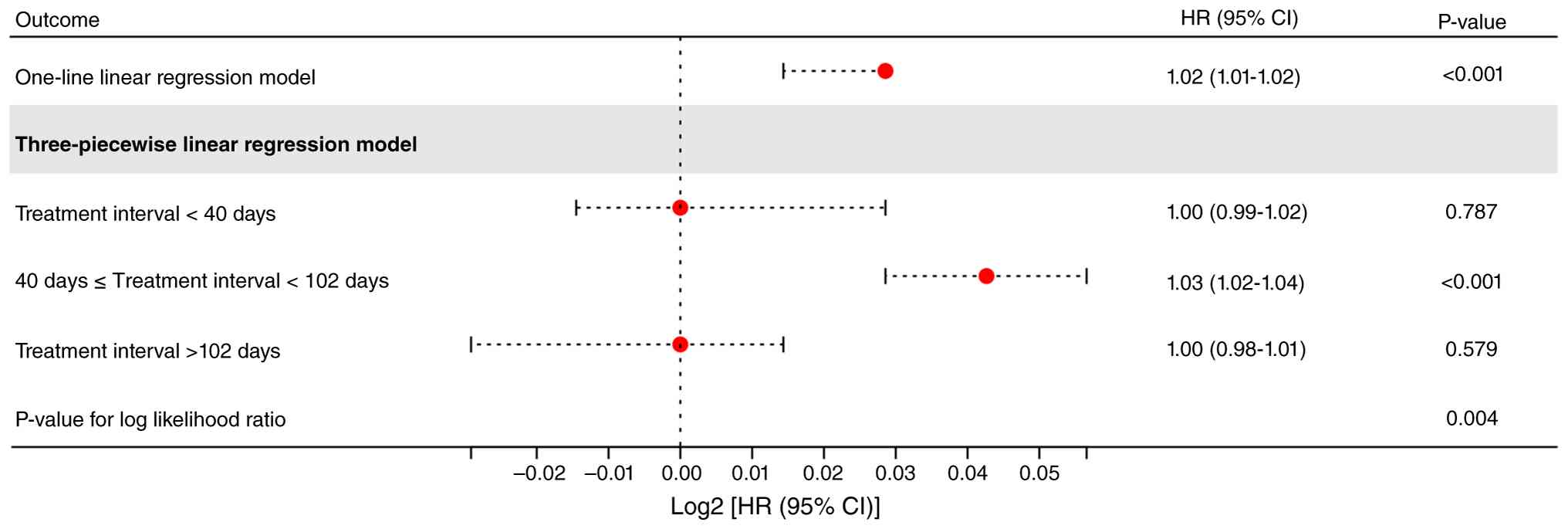
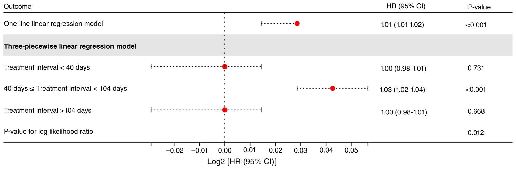
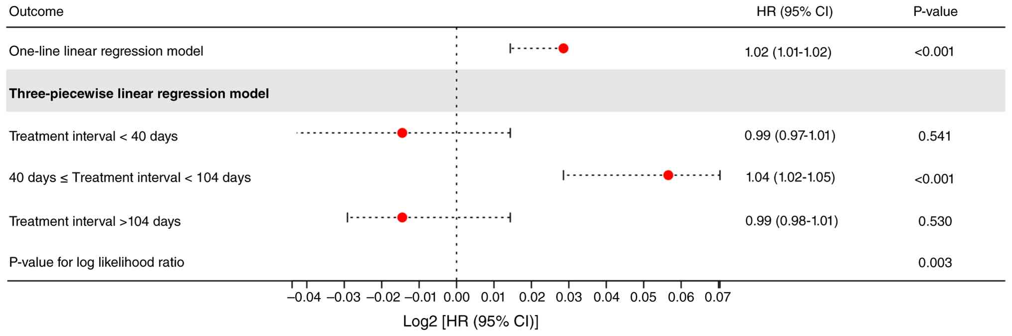
three-piecewise linear regression models, it was determined that 40
and 102 days were the inflection points for LPFS (Fig. 2), whilst 40 and 104 days were the
inflection points for PFS and OS (Figs.
3 and 4). For LPFS, the risk
rapidly increased between 40 and 102 days (HR, 1.03; 95% CI,
1.02–1.04; P<0.001). A highly similar pattern was observed for
PFS (HR, 1.03; 95% CI, 1.02–1.04; P<0.001) and OS (HR, 1.04; 95%
CI, 1.02–1.05; P<0.001). When outside of the threshold (such as
before 40 days or after the upper threshold), the prognostic risk
of LPFS, PFS, and OS did not change significantly.
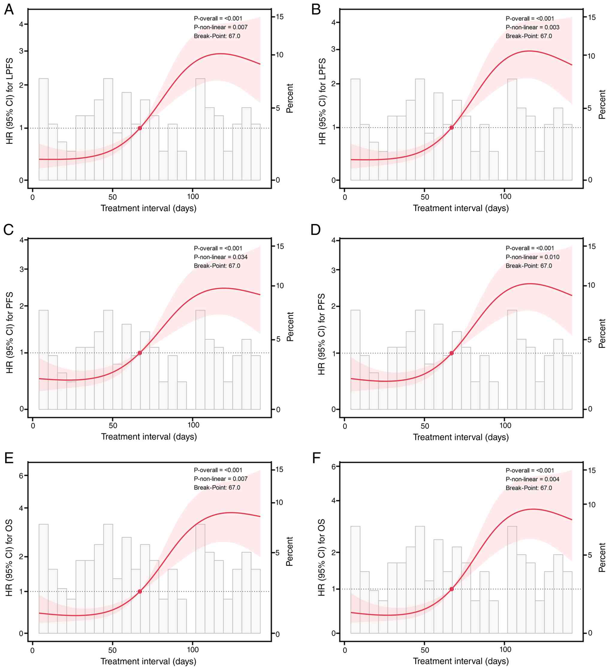 | Figure 1.Restricted cubic spline curves
between osimertinib-to-thoracic radiotherapy interval and risk of
LPFS, PFS and OS. Solid lines represent hazard ratios, shaded areas
represent 95% confidence intervals, and dashed lines indicate HR=1.
(A and B) LPFS, (C and D) PFS and (E and F) OS curves. (A, C and E)
Unadjusted and (B, D and F) multivariable-adjusted models [(B) and
(D) are adjusted for age; (F) is adjusted for Tumor-Node-Metastasis
stage and EGFR mutation subtype]. PFS, progression-free survival;
LPFS, local PFS; OS, overall survival; HR, hazard ratio; CI,
confidence interval. |
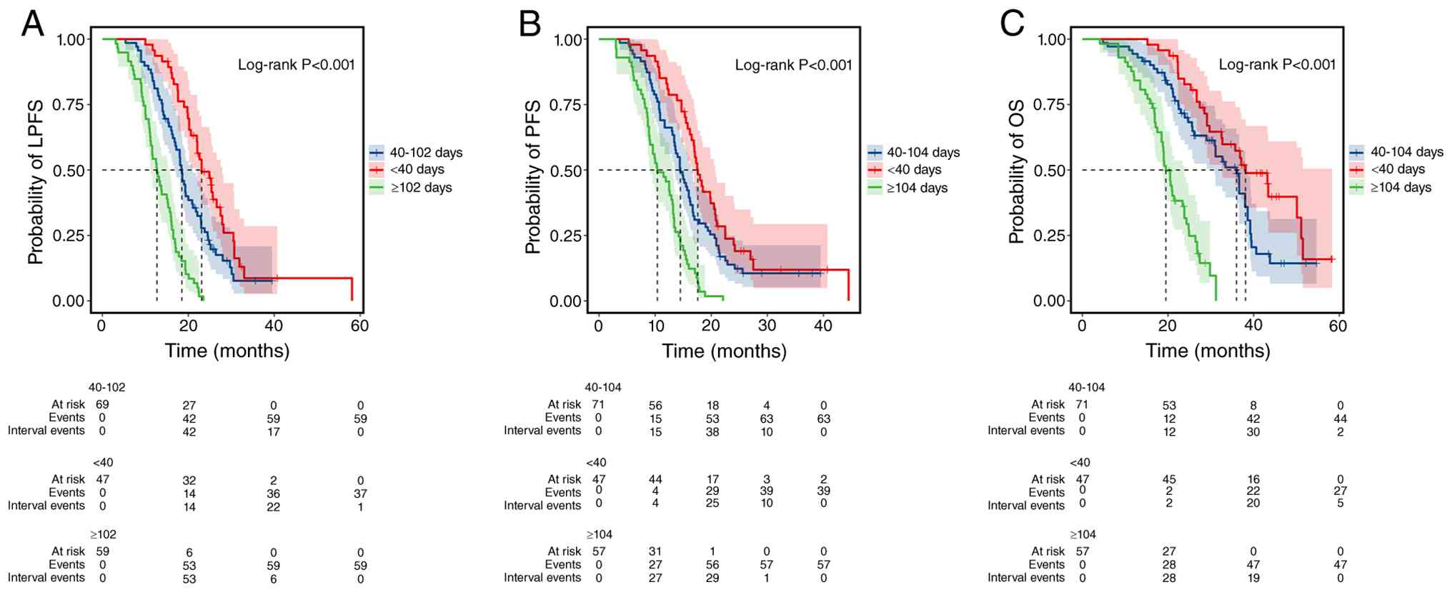
Patients were then divided into three groups. In the
Kaplan-Meier curve, longer LPFS, PFS, and OS times were observed in
patients in the early-treatment group (<40 days; log-rank test,
P<0.001; Fig. 5). In the
sensitivity analysis, after adjusting for the response to
osimertinib, the treatment interval remained an independent
prognostic factor for LPFS (HR, 1.02; 95% CI, 1.01–1.02;
P<0.001), PFS (HR, 1.01; 95% CI, 1.01–1.02; P<0.001), and OS
(HR, 1.02; 95% CI, 1.01–1.02; P<0.001) (Table SVI, Table SVII, Table SVIII). Furthermore, after
adjustment, the original non-linear association remained
statistically significant (Fig.
S2). In left-truncated time-dependent Cox regression, a longer
RT interval remained significantly associated with worse LPFS and
PFS, whereas no meaningful association with OS was detected
(Table SIX). Left-truncated RCS
models showed significant non-linear relationships between the RT
interval and OS, PFS, and LPFS (Fig.
S3). In landmark analyses at 3, 6, and 12 months, for each
1-day increase in the interval, the rate of adverse survival risks
increased significantly for OS, PFS, and LPFS across all landmark
time points (Fig. S4).
Kaplan-Meier curves in the landmark analysis stratified by the
median 67-day treatment interval revealed significant differences
in LPFS, PFS and OS between subgroups (all P<0.0001). Patients
with treatment intervals <67 days achieved longer survival than
those with intervals of ≥67 days (Fig.
S5, Fig. S6, Fig. S7). In the four-piecewise Cox
regression model, when the treatment interval exceeded the
threshold of 67 days, longer intervals were closely associated with
poor prognosis for LPFS, PFS, and OS (Fig. S8; Table SX, Table SXI, Table SXII). Subgroup analysis showed that
prolonged RT interval independently predicted poorer prognosis
across both EGFR subtypes (Table
SXIII). After adjustment for confounding covariates, a
significant interaction was identified between EGFR genotype and
treatment interval with respect to OS. No comparable significant
interaction was found for PFS or LPFS (Table SXIII). After IPTW, all baseline
covariates were well-balanced (Table
SXIV). IPTW-weighted Cox regression confirmed that prolonged RT
interval remained significantly associated with inferior OS, PFS,
and LPFS (Table SXV). IPTW-RCS
showed significant non-linear trends (Fig. S9).
Discussion
To the best of our knowledge, the relationship
between the treatment interval for chest RT and prognosis in
patients with advanced NSCLC treated with first-line osimertinib
has not been sufficiently analyzed. A key novel finding of the
present study is the significant impact of RT interval on prognosis
and their non-linear relationship. Treatment interval was
significantly associated with LPFS, PFS, and OS. In the middle
intervals (40–102 days/40-104 days), the risk rapidly increased for
LPFS, PFS, and OS. However, outside this threshold range, no
significant change in prognostic risk was observed. In the
Kaplan-Meier curve, patients treated early had a superior prognosis
in terms of LPFS, PFS, and OS. These findings indicate that TRT
initiation within 40 days of osimertinib commencement is associated
with maximal survival benefit, whilst delays between 40 and 104
days confer a significantly elevated risk of disease progression
and mortality.
The sensitivity of NSCLC to RT is low, and the
timing of radiation therapy initiation remains a controversial
issue. Previous studies have shown that the benefit of EGFR-TKI
combined with RT in terms of PFS is superior compared with that of
combined anti-angiogenesis therapy, chemotherapy, and any other
treatment (17), in addition to
being superior to EGFR-TKI alone (18). In terms of mechanism, there is
evidence that the combination of EGFR-TKI and RT can significantly
reduce the proportion of S-phase cells, induce apoptosis, inhibit
the autophosphorylation of epidermal EGFR and downregulate the
expression of Rad51 recombinase to enhance radiosensitivity
(19). Similarly, induction
chemotherapy before RT has a benefit on the OS of patients with
NSCLC receiving RT and chemotherapy (20), which indicates the rationality of
EGFR-TKI therapy combined with consolidation RT. However, Xu et
al (21) enrolled 45 patients
with stage III NSCLC treated with concurrent radiotherapy plus
EGFR-TKIs, 20 of whom (44.4%) harbored EGFR mutations. The cohort
achieved a median PFS time of 27.9 months (95% CI, 18.7–37.2) and a
median OS time of 49.7 months (95% CI, 27.7–71.8). Different from
this concurrent treatment strategy, the present study adopted
sequential EGFR-TKI consolidation following radiotherapy and
included patients with stage IV disease, who generally have poorer
baseline prognostic outcomes. Despite this disadvantage at
baseline, the patients still achieved promising survival outcomes,
which may be explained by the longer median duration of EGFR-TKI
treatment in the present cohort (28.4 vs. 14.4 months) compared
with that of the cohort in the study by Xu et al (21). Previous studies found that the
treatment time was positively associated with OS (21), which was also the reason for the
shorter median OS time observed in the present cohort. Hu et
al (22) adopted a similar
treatment strategy to the present study, combining EGFR-TKIs with
local consolidative radiotherapy. Nevertheless, the cohort achieved
a longer median OS time than in the present study (38.0 vs. 25.6
months). This survival discrepancy was mainly attributed to
differences in baseline patient characteristics, as the study only
enrolled elderly patients with oligometastatic disease who carried
more favorable prognostic profiles (22). Importantly, while baseline patient
characteristics and EGFR-TKI treatment duration have been
established as prognostic predictors for EGFR-mutant NSCLC
(23), few studies have
specifically investigated how the time interval between EGFR-TKI
initiation and thoracic radiotherapy affects long-term survival.
Although this temporal parameter has not yet been fully
characterized, it bears important clinical implications for guiding
the optimal sequencing of targeted therapy and radiotherapy.
The present study shows that the intervals of 40 and
102/104 days are the inflection points for the therapeutic effect
of the time interval between EGFR-TKI initiation and thoracic
radiotherapy. Within 40–102/104 days, the longer interval is
associated with a higher probability of adverse prognostic risk.
EGFR-TKI can inhibit the proliferation and survival of tumor cells
while altering the immune environment (22). When the interval between EGFR-TKI
and RT is short, EGFR-TKI and RT can exert synergistic effects,
making the killing effect of RT on tumor cells more potent
(24,25). This can explain the time trend of
the RT effect observed in the present study. An interval of <30
days may be too brief to permit full TKI-mediated
radiosensitization, attenuating clinical benefit. Li et al
(26) observed that primary tumor
radiotherapy initiated 30 days after EGFR-TKI yielded better
overall survival than either earlier treatment or post-progression
radiotherapy, but did not evaluate non-linear interval effects.
Several lines of evidence support the rationality of the upper
limit of 102/104 days for radiotherapy initiation identified. Tang
et al (27) found that
patients receiving single-agent EGFR-TKIs achieved a median time to
therapeutic response of 2.00 months (95% CI, 1.28 to 2.92 months).
For patients with stable disease, the median time to maximum tumor
shrinkage was also 2.00 months, corresponding to a 95% CI of
1.42–2.58 months. These clinical observations align with another
independent study confirming that effective antitumor responses can
be achieved within 8 weeks after EGFR-TKI treatment initiation
(27). Therefore, these published
data presented above reveal that EGFR-TKIs mostly induce maximal
tumor regression within the first 2 months of treatment. This
temporal characteristic matches the 67-day breakpoint observed in
the present restricted cubic spline analysis. Radiotherapy delayed
beyond this time point correlates with an elevated risk of local
disease progression. Such temporal consistency further supports the
biological rationality of the present findings. Clinically,
administering thoracic radiotherapy after EGFR-TKI-induced tumor
shrinkage, and before the development of drug resistance and tumor
regrowth, constitutes an optimal treatment window to maximize local
tumor control. This appropriate time window opens 6 to 8 weeks
after TKI administration and closes no later than 102 to 104 days
after TKI initiation.
In the present study, restricted cubic spline and
piecewise regression analyses confirmed a non-linear association
between the osimertinib-to-TRT interval and survival outcomes. In
the present results, the mortality risk remained stable for
intervals of <40 days and rose significantly for intervals
between 40 and 102 days, before plateauing again for intervals
>102 days. This trend may be attributed to the dynamic changes
in tumor cell cycle status, immune microenvironment evolution, and
clinical confounding factors. This interval-specific elevation in
risk may be primarily driven by the waning of osimertinib-induced
radiosensitization. Preclinical studies in EGFR-mutant NSCLC models
have demonstrated that EGFR-TKI induces G1 cell cycle
arrest and exerts transient radiosensitizing effects (28,29).
This radiosensitization gradually diminishes with prolonged drug
exposure, as surviving tumor cells restore proliferative capacity
via upregulation of cyclin D1 and CDK4/6 signaling (30–32).
By 40 days post-initiation, the majority of surviving tumor cells
have exited G1 arrest. The upregulation of cyclin D1 and
CDK4/6 signaling restores their proliferative capacity and reverses
intrinsic radiosensitivity by a 2-times decrease compared with
those in the first 2 weeks of the treatment period. Furthermore,
this 40–102-day window corresponds to a transient immunosuppressive
phase under continuous EGFR inhibition. In a phase III FLAURA
trial, the proportion of immunosuppressive M2-polarised
tumor-associated macrophages and regulatory T cells was found to be
notably increased between 6 and 14 weeks post-osimertinib
initiation (33). A 2.1-times rise
in tumor cell programmed death ligand 1 (PD-L1) expression is
observed at 12 weeks. Consistent with a prior finding (26), long-term single-agent EGFR-TKI
exposure can progressively consolidate the tumor microenvironment
into a deeply immunosuppressive state, promoting drug resistance
and immune escape. Whilst RT can induce immunogenic cell death and
promote antitumor immune responses (34,35),
it also instantaneously upregulates immunosuppressive molecules,
such as PD-L1, indoleamine 2,3-dioxygenase 1, and TGF-β, while
activating regulatory T cell and myeloid-derived suppressor cell
function (36). Regardless of the
local radiation dose, within the 40- to 102-day window, the
pre-existing immunosuppressive milieu may blunt the RT-induced
abscopal effect, eliminating the synergistic benefit of combined
treatment. A previous retrospective cohort study demonstrated that
RT initiated within this interval was associated with a 1.8-times
higher risk of early local progression relative to intervals
greater than or less than this threshold, with no notable
difference in grade ≥2 radiation toxicity between groups (37). The observed plateau in mortality
risk for intervals >102 days likely reflects clinical
confounding. Patients requiring delayed RT beyond this point
typically have rapidly progressive disease or severe local
symptoms. In such cases, RT is delivered for palliative symptom
control rather than with curative intent, leading to an inherently
poor prognosis independent of timing. Although further mechanistic
validation with longitudinal immune microenvironment profiling is
warranted, this interval-specific risk likely reflects the
convergence of lost cell cycle-mediated radiosensitivity and
transient immunosuppression. Future studies establishing multi-omic
prediction models for optimal EGFR-TKI-RT interval will enable
real-time individualized treatment timing to improve patient
outcomes.
In the present study, the EGFR exon 21 L858R point
mutation was identified as an independent prognostic factor
significantly associated with OS. This finding is consistent with
the general understanding that patients harboring EGFR mutations
exhibit enhanced sensitivity to EGFR-TKIs (37), consequently leading to prolonged OS
times compared with patients harboring wild-type EGFR (38). However, within the spectrum of
common activating mutations, exon 19 deletions are generally
considered to confer a superior median OS time over the L858R
mutation in the setting of first-line EGFR-TKI treatment (39,40).
This difference is typically mechanistically ascribed to distinct
structural alterations present within the EGFR-TKI binding domain,
which confer greater TKI sensitivity to the exon 19 deletion
subtype (41,42).
A notable discrepancy observed in the present cohort
was the lack of a significant prognostic effect of the mutation
subtype on PFS, despite the strong association with OS. This
difference in outcomes (PFS vs. OS) may be attributable to the
established association of the L858R subtype with high-risk
oncogenic features, such as a greater propensity for liver/brain
metastases or concomitant TP53 co-mutation (43,44),
factors that are more robustly associated with compromised OS
compared with initial PFS.
Furthermore, the mechanistic rationale for combining
osimertinib with ionizing radiation (IR) is robust. In vitro
evidence has shown that osimertinib can synergistically enhance the
antitumor activity of IR by inhibiting cellular proliferation and
clonal survival, reducing IR-induced G2/M cell cycle
arrest and delaying DNA damage repair in a concentration- and
time-dependent manner. This synergy is also mediated by effectively
blocking the phosphorylation of EGFR (Tyr1068/Tyr1173) and key
downstream signaling pathways, including AKT and ERK (45). The present study established a
critical time threshold for the TKI-TRT interval in the overall
cohort. The observed differential impact of the L858R subtype on OS
highlights the need for tailored strategies. Future large-scale,
prospective investigations are therefore warranted to explore the
optimal TRT initiation timing specific to individual EGFR mutation
subtypes, ensuring maximized long-term clinical benefit.
The present study has several limitations. The
retrospective design, single-center and relatively small sample
size introduce the potential for selection bias. Although the
present findings strongly suggest an optimal treatment window, the
definitive establishment of the ideal combination strategy
necessitates rigorous validation through larger, prospective,
multi-center trials. Furthermore, despite multivariable adjustment,
IPTW weighting and rigorous immortal time bias elimination, the
present study remains susceptible to unmeasured confounding, among
which tumor molecular heterogeneity is particularly relevant. The
clonal heterogeneity of the T790M mutation and co-existing gene
mutations, such as those in TP53, can accelerate the
development of osimertinib resistance (46). As such molecular profiles may prompt
clinicians to initiate TRT earlier while independently worsening
prognosis, they act as unmeasured confounders that could create a
spurious association of ‘benefits of early TRT’. Another potential
source of unmeasured confounding is molecular residual disease
(MRD) status, typically detected through circulating tumor DNA
(ctDNA) sequencing. As MRD/ctDNA data were unavailable in the
present retrospective cohort, adjustments could not be made for it.
However, external evidence shows that patients with undetectable
MRD have a superior prognosis (47). Such patients may be preferentially
assigned to delayed TRT by their physicians. This would concentrate
biologically favorable cases in the delayed-TRT arm, artificially
inflating its apparent survival benefit and biasing the estimated
effect of TRT timing. In addition, differences in treatment
regimens that patients receive subsequently can directly affect
survival, which can in turn inversely influence clinician decisions
regarding the timing of TRT (48).
In summary, the interval between osimertinib
initiation and TRT was an independent prognostic factor with a
non-linear association with clinical outcomes. TRT delivered within
40 days was associated with optimal LPFS, PFS and OS. Between 40
and 104 days, longer intervals correlated with progressively worse
outcomes. Beyond 104 days, further delay did not significantly
alter prognostic risk. These findings support the integration of
TRT within 40 days of osimertinib initiation.
Supplementary Material
Supporting Data
Supporting Data
Acknowledgements
Not applicable.
Funding
This research was funded by grants from the Guangxi Zhuang
Autonomous Region Health Commission (grant nos. Z-B20241428,
Z-B20241429, and Z-B20231444) and the National Natural Science
Foundation of China (grant no. 82560001).
Availability of data and materials
The data generated in the present study are not
publicly available due to privacy and ethical restrictions, but may
be requested from the corresponding author.
Authors' contribution
ML and YZ were responsible for the study analysis,
developing the methodology, writing the original draft,
visualization, and revision of the manuscript. ZC and XY
contributed to methodology, data curation, acquisition of clinical
data, and reviewing and editing of the manuscript. WY and JH
contributed to validation, data verification, endpoint
adjudication, and reviewing and editing of the manuscript. DH and
LQ contributed to conceptualization, supervision, project
administration, resources, data interpretation, and critically
revising the manuscript for important intellectual content. ML and
YZ confirm the authenticity of all the raw data. All authors read
and approved the final manuscript.
Ethics approval and consent to
participate
The study protocol was approved by the Institutional
Ethics Committee of The Fourth Affiliated Hospital of Guangxi
Medical University (approval no. KY2024162). Due to the
retrospective observational nature of the study and the use of
fully de-identified patient data, the requirement for written
informed consent was formally waived by the Institutional Ethics
Committee.
Patient consent for publication
Not applicable.
Competing interests
The authors declare that they have no competing
interests.
References
|
1
|
Jia B, Zheng Q, Wang J, Sun H, Zhao J, Wu
M, An T, Wang Y, Zhuo M, Li J, et al: A nomogram model to predict
death rate among non-small cell lung cancer (NSCLC) patients with
surgery in surveillance, epidemiology, and end results (SEER)
database. BMC Cancer. 20:6662020. View Article : Google Scholar : PubMed/NCBI
|
|
2
|
Snider M, Salama JK and Boyer M: Survival
and recurrence rates following SBRT or surgery in medically
operable stage I NSCLC. Lung Cancer. 197:1079622024. View Article : Google Scholar : PubMed/NCBI
|
|
3
|
Wu YL, Planchard D, Lu S, Sun H, Yamamoto
N, Kim DW, Tan DSW, Yang JC, Azrif M, Mitsudomi T, et al: Pan-Asian
adapted clinical practice guidelines for the management of patients
with metastatic non-small-cell lung cancer: A CSCO-ESMO initiative
endorsed by JSMO, KSMO, MOS, SSO and TOS. Ann Oncol. 30:171–210.
2019. View Article : Google Scholar : PubMed/NCBI
|
|
4
|
Soria JC, Ohe Y, Vansteenkiste J,
Reungwetwattana T, Chewaskulyong B, Lee KH, Dechaphunkul A, Imamura
F, Nogami N, Kurata T, et al: Osimertinib in untreated EGFR-mutated
advanced non-small-cell lung cancer. N Engl J Med. 378:113–125.
2018. View Article : Google Scholar : PubMed/NCBI
|
|
5
|
Nagasaka M, Zhu VW, Lim SM, Greco M, Wu F
and Ou SI: Beyond osimertinib: The development of third-generation
EGFR tyrosine kinase inhibitors for advanced EGFR+ NSCLC. J Thorac
Oncol. 16:740–763. 2021. View Article : Google Scholar : PubMed/NCBI
|
|
6
|
Cho JH, Lim SH, An HJ, Kim KH, Park KU,
Kang EJ, Choi YH, Ahn MS, Lee MH, Sun JM, et al: Osimertinib
combined with gefitinib in patients with EGFR-mutated
non-small-cell lung cancer: Open-label, phase II trial
(KCSG-LU15-09). J Clin Oncol. 38:488–495. 2020. View Article : Google Scholar : PubMed/NCBI
|
|
7
|
Gomez DR, Blumenschein GR Jr, Lee JJ,
Hernandez M, Ye R, Camidge DR, Doebele RC, Skoulidis F, Gaspar LE,
Gibbons DL, et al: Local consolidative therapy versus maintenance
therapy or observation for patients with oligometastatic
non-small-cell lung cancer without progression after first-line
systemic therapy: A multicentre, randomised, controlled, phase 2
study. Lancet Oncol. 17:1672–1682. 2016. View Article : Google Scholar : PubMed/NCBI
|
|
8
|
Iyengar P, Wardak Z, Gerber DE, Tumati V,
Ahn C, Hughes RS, Dowell JE, Cheedella N, Nedzi L, Westover KD, et
al: Consolidative radiotherapy for limited metastatic
non-small-cell lung cancer: A phase 2 randomized clinical trial.
JAMA Oncol. 4:e1735012018. View Article : Google Scholar : PubMed/NCBI
|
|
9
|
Xing L, Wu G, Wang L, Li J, Wang J, Yuan
Z, Chen M, Xu Y, Fu X, Zhu Z, et al: Erlotinib versus
etoposide/cisplatin with radiation therapy in unresectable stage
III epidermal growth factor receptor mutation-positive non-small
cell lung cancer: A multicenter, randomized, open-label, phase 2
trial. Int J Radiat Oncol Biol Phys. 109:1349–1358. 2021.
View Article : Google Scholar : PubMed/NCBI
|
|
10
|
Liu R, Wei S, Zhang Q, Zhang X, Luo H,
Tian J, Li Y, Ge L and Wang X: Epidermal growth factor receptor
tyrosine kinase inhibitors combined with thoracic radiotherapy or
chemoradiotherapy for advanced or metastatic non-small cell lung
cancer: A systematic review and meta-analysis of single-arm trials.
Medicine. 98:e164272019. View Article : Google Scholar : PubMed/NCBI
|
|
11
|
Zhong W, Wu L, Huang L, Wang J, Shi H and
Wu S: Double-dose osimertinib combined with intrathecal injection
of pemetrexed improves the efficacy of EGFR-mutated non-small cell
lung cancer and leptomeningeal metastasis: Case report and
literature review. Front Oncol. 14:13774512024. View Article : Google Scholar : PubMed/NCBI
|
|
12
|
Xu J and Wang Z: Recent advances progress
of targeted drugs combined with radiotherapy for advanced non-small
cell lung cancer: A review. Front Oncol. 13:12855932023. View Article : Google Scholar : PubMed/NCBI
|
|
13
|
Oken MM, Creech RH, Tormey DC, Horton J,
Davis TE, McFadden ET and Carbone PP: Toxicity and response
criteria of the Eastern Cooperative Oncology Group. Am J Clin
Oncol. 5:649–655. 1982. View Article : Google Scholar : PubMed/NCBI
|
|
14
|
Ramón-Porta R, Asamura H, Travis WD and
Rusch VW: Lung cancer - major changes in the American Joint
Committee on Cancer eighth edition cancer staging manual. CA Cancer
J Clin. 67:138–155. 2017.
|
|
15
|
Eisenhauer EA, Therasse P, Bogaerts J,
Schwartz LH, Sargent D, Ford R, Dancey J, Arbuck S, Gwyther S,
Mooney M, et al: New response evaluation criteria in solid tumours:
Revised RECIST guideline (version 1.1). Eur J Cancer. 45:228–247.
2009. View Article : Google Scholar : PubMed/NCBI
|
|
16
|
Goldstraw P, Chansky K, Crowley J,
Rami-Porta R, Asamura H, Eberhardt WE, Nicholson AG, Groome P,
Mitchell A, Bolejack V, et al: The IASLC lung cancer staging
project: Proposals for revision of the TNM stage groupings in the
forthcoming (eighth) edition of the TNM classification for lung
cancer. J Thorac Oncol. 11:39–51. 2016. View Article : Google Scholar : PubMed/NCBI
|
|
17
|
Xue J, Li B, Wang Y, Huang Z, Liu X, Guo
C, Zheng Z, Liang N, Le X and Li S: Efficacy and safety of
epidermal growth factor receptor (EGFR)-tyrosine kinase inhibitor
combination therapy as first-line treatment for patients with
advanced EGFR-mutated, non-small cell lung cancer: A systematic
review and Bayesian network meta-analysis. Cancers (Basel).
14:48942022. View Article : Google Scholar : PubMed/NCBI
|
|
18
|
Zhou Y, Peng L, Liang F, Chu L, Chu X,
Yang X, Zhang J, Guo T, Jiang S, Pang Y, et al: Safety and efficacy
of consolidative stereotactic radiotherapy for oligo-residual
EGFR-mutant non-small cell lung cancer after first-line
third-generation EGFR-tyrosine kinase inhibitors: A single-arm,
phase 2 trial. EClinicalMedicine. 76:1028532024. View Article : Google Scholar : PubMed/NCBI
|
|
19
|
Tsai YC, Ho PY, Tzen KY, Tuan TF, Liu WL,
Cheng AL, Pu YS and Cheng JC: Synergistic blockade of EGFR and HER2
by new-generation EGFR tyrosine kinase inhibitor enhances radiation
effect in bladder cancer cells. Mol Cancer Ther. 14:810–820. 2015.
View Article : Google Scholar : PubMed/NCBI
|
|
20
|
Luo H, Yu X, Liang N, Xie J, Deng G, Liu
Q, Zhang J, Zhang J and Ge H: The effect of induction chemotherapy
in patients with locally advanced non-small cell lung cancer who
received chemoradiotherapy: A systematic review and meta-analysis.
Medicine. 96:e61652017. View Article : Google Scholar : PubMed/NCBI
|
|
21
|
Xu K, Liang J, Zhang T, Zhou Z, Chen D,
Feng Q, Xiao Z, Hui Z, Lu J, Wang X, et al: Clinical outcomes and
radiation pneumonitis after concurrent EGFR-tyrosine kinase
inhibitors and radiotherapy for unresectable stage III non-small
cell lung cancer. Thorac Cancer. 12:814–823. 2021. View Article : Google Scholar : PubMed/NCBI
|
|
22
|
Hu X, Li H, Kang X, Wang X, Pang H, Liu C,
Zhang J and Wang Y: First-line tyrosine kinase inhibitors combined
with local consolidative radiation therapy for elderly patients
with oligometastatic non-small cell lung cancer harboring EGFR
activating mutations. Front Oncol. 12:7660662022. View Article : Google Scholar : PubMed/NCBI
|
|
23
|
Liu JF, Sun XS, Yin JH and Xu XE: Adjuvant
EGFR-TKI therapy in resected EGFR-mutation positive non-small cell
lung cancer: A real-world study. Front Oncol. 13:11328542023.
View Article : Google Scholar : PubMed/NCBI
|
|
24
|
Georgakopoulos I, Kouloulias V, Ntoumas G,
Desse D, Koukourakis I, Kougioumtzopoulou A, Charpidou A, Syrigos
KN and Zygogianni A: Combined use of radiotherapy and tyrosine
kinase inhibitors in the management of metastatic non-small cell
lung cancer: A literature review. Crit Rev Oncol Hematol.
204:1045202024. View Article : Google Scholar : PubMed/NCBI
|
|
25
|
Zhao X, Zhang S, Sun X, Lin Y, Capone L,
Ko EC, Kann BH, Li Y and Wang X: Narrative review of stereotactic
body radiation therapy combined with tyrosine kinase inhibitors for
oligometastatic EGFR-mutated non-small cell lung cancer: Present
and future developments. Transl Lung Cancer Res. 13:1383–1395.
2024. View Article : Google Scholar : PubMed/NCBI
|
|
26
|
Li Q, Liang N, Ouyang W, Su S, Ma Z, Geng
Y, Hu Y, Li H and Lu B: Appropriate delay of primary tumour
radiotherapy may lead to better long-term overall survival for
non-small cell lung cancer treated with EGFR-TKIs. BMC Cancer.
24:10532024. View Article : Google Scholar : PubMed/NCBI
|
|
27
|
Tang Y, Xia B, Xie R, Xu X, Zhang M, Wu K,
Wang B and Ma S: Timing in combination with radiotherapy and
patterns of disease progression in non-small cell lung cancer
treated with EGFR-TKI. Lung Cancer. 140:65–70. 2020. View Article : Google Scholar : PubMed/NCBI
|
|
28
|
Wu TH, Hsiue EH, Lee J H, Lin CC, Liao WY,
Ho CC, Shih JY, Yu CJ and Yang JC: Best response according to
RECIST during first-line EGFR-TKI treatment predicts survival in
EGFR mutation-positive non-small-cell lung cancer patients. Clin
Lung Cancer. 19:e361–e372. 2018. View Article : Google Scholar : PubMed/NCBI
|
|
29
|
Wang N, Wang L, Meng X, Wang J, Zhu L, Liu
C, Li S, Zheng L, Yang Z, Xing L and Yu J: Osimertinib (AZD9291)
increases radio-sensitivity in EGFR T790M non-small cell lung
cancer. Oncol Rep. 41:77–86. 2019.PubMed/NCBI
|
|
30
|
Kriegs M, Gurtner K, Can Y, Brammer I,
Rieckmann T, Oertel R, Wysocki M, Dorniok F, Gal A, Grob TJ, et al:
Radiosensitization of NSCLC cells by EGFR inhibition is the result
of an enhanced p53-dependent G1 arrest. Radiother Oncol.
115:120–127. 2015. View Article : Google Scholar : PubMed/NCBI
|
|
31
|
Starrett JH, Guernet AA, Cuomo ME, Poels
KE, van Alderwerelt van Rosenburgh IK, Nagelberg A, Farnsworth D,
Price KS, Khan H, Ashtekar KD, et al: Drug sensitivity and allele
specificity of first-line osimertinib resistance EGFR mutations.
Cancer Res. 80:2017–2028. 2020. View Article : Google Scholar : PubMed/NCBI
|
|
32
|
Ahsan A, Hiniker SM, Davis MA, Lawrence TS
and Nyati MK: Role of cell cycle in epidermal growth factor
receptor inhibitor-mediated radiosensitization. Cancer Res.
69:5108–5114. 2009. View Article : Google Scholar : PubMed/NCBI
|
|
33
|
Hara N, Ichihara E, Kano H, Ando C, Morita
A, Nishi T, Okawa S, Nakasuka T, Hirabae A, Abe M, et al: CDK4/6
signaling attenuates the effect of epidermal growth factor receptor
tyrosine kinase inhibitors in EGFR-mutant non-small cell lung
cancer. Transl Lung Cancer Res. 9:2132–2145. 2020.
|
|
34
|
Chmielecki J, Gray JE, Cheng Y, Ohe Y,
Imamura F, Cho BC, Lin MC, Majem M, Shah R, Rukazenkov Y, et al:
Candidate mechanisms of acquired resistance to first-line
osimertinib in EGFR-mutated advanced non-small cell lung cancer.
Nat Commun. 14:3212023. View Article : Google Scholar
|
|
35
|
Galluzzi L, Vitale I, Warren S, Adjemian
S, Agostinis P, Martinez AB, Chan TA, Coukos G, Demaria S, Deutsch
E, et al: Consensus guidelines for the definition and detection of
immunogenic cell death. J Immunother Cancer. 8:e0003372020.
View Article : Google Scholar : PubMed/NCBI
|
|
36
|
Fucikova J, Kepp O, Kasikova L, Petroni G,
Yamazaki T, Liu P, Zhao L, Spisek R, Kroemer G and Galluzzi L:
Mechanisms and detection of immunogenic cell death in cancer
therapy. Cell Death Dis. 11:10132020. View Article : Google Scholar : PubMed/NCBI
|
|
37
|
Weichselbaum RR, Liang H, Deng L and Fu
YX: Radiotherapy and immunotherapy: A beneficial liaison? Nat Rev
Clin Oncol. 14:365–379. 2017. View Article : Google Scholar : PubMed/NCBI
|
|
38
|
Maemondo M, Inoue A, Kobayashi K, Sugawara
S, Oizumi S, Isobe H, Gemma A, Harada M, Yoshizawa H, Kinoshita I,
et al: Gefitinib or chemotherapy for non-small-cell lung cancer
with mutated EGFR. N Engl J Med. 362:2380–2388. 2010. View Article : Google Scholar : PubMed/NCBI
|
|
39
|
De B, Farooqi AS, Mitchell KG, Ludmir EB,
Lewis J, Rinsurongkawong W, Rinsurongkawong V, Lee JJ, Swisher SG,
Gibbons DL, et al: Benchmarking outcomes for molecularly
characterized synchronous oligometastatic non-small-cell lung
cancer reveals EGFR mutations to be associated with longer overall
survival. JCO Precis Oncol. 7:e22005402023. View Article : Google Scholar : PubMed/NCBI
|
|
40
|
Choi YJ, Lee DH, Choi CM, Lee JS, Lee SJ,
Ahn JH and Kim SW: Randomized phase II study of
paclitaxel/carboplatin intercalated with gefitinib compared to
paclitaxel/carboplatin alone for chemotherapy-naïve non-small cell
lung cancer in a clinically selected population excluding patients
with non-smoking adenocarcinoma or mutated EGFR. BMC Cancer.
15:7632015. View Article : Google Scholar : PubMed/NCBI
|
|
41
|
Yang JC, Wu YL, Schuler M, Sebastian M,
Popat S, Yamamoto N, Zhou C, Hu CP, O'Byrne K, Feng J, et al:
Afatinib versus cisplatin-based chemotherapy for EGFR
mutation-positive lung adenocarcinoma (LUX-Lung 3 and LUX-Lung 6):
Analysis of overall survival data from two randomised, phase 3
trials. Lancet Oncol. 16:141–151. 2015. View Article : Google Scholar : PubMed/NCBI
|
|
42
|
Ramalingam SS, Vansteenkiste J, Planchard
D, Cho BC, Gray JE, Ohe Y, Zhou C, Reungwetwattana T, Cheng Y,
Chewaskulyong B, et al: Overall survival with osimertinib in
untreated, EGFR-mutated advanced NSCLC. N Engl J Med. 382:41–50.
2020. View Article : Google Scholar : PubMed/NCBI
|
|
43
|
Yu HA, Arcila ME, Rekhtman N, Sima CS,
Zakowski MF, Pao W, Kris MG, Miller VA, Ladanyi M and Riely GJ:
Analysis of tumor specimens at the time of acquired resistance to
EGFR-TKI therapy in 155 patients with EGFR-mutant lung cancers.
Clin Cancer Res. 19:2240–2247. 2013. View Article : Google Scholar : PubMed/NCBI
|
|
44
|
Le X, Molife C, Leusch MS, Rizzo MT,
Peterson PM, Caria N, Chen Y, Gugel EG and Visseren-Grul C: TP53
co-mutation status association with clinical outcomes in patients
with EGFR-mutated non-small cell lung cancer. Cancers. 14:61272022.
View Article : Google Scholar : PubMed/NCBI
|
|
45
|
Vokes NI, Galan Cobo A, Fernandez-Chas M,
Molkentine D, Treviño S III, Druker V, Qian Y, Patel S, Schmidt S,
Hong L, et al: ATM mutations associate with distinct co-mutational
patterns and therapeutic vulnerabilities in NSCLC. Clin Cancer Res.
29:4958–4972. 2023. View Article : Google Scholar : PubMed/NCBI
|
|
46
|
Vokes NI, Chambers ES, Nguyen T, Coolidge
A, Lydon CA, Le X, Sholl L, Heymach JV, Nishino M, Van Allen EM and
Jänne PA: Concurrent TP53 mutations facilitate resistance evolution
in EGFR-mutant lung adenocarcinoma. J Thorac Oncol. 17:779–792.
2022. View Article : Google Scholar : PubMed/NCBI
|
|
47
|
Qiu B, Guo W, Zhang F, Lv F, Ji Y, Peng Y,
Chen X, Bao H, Xu Y, Shao Y, et al: Dynamic recurrence risk and
adjuvant chemotherapy benefit prediction by ctDNA in resected
NSCLC. Nat Commun. 12:67702021. View Article : Google Scholar : PubMed/NCBI
|
|
48
|
Shahi J, Wright JR, Gabos Z and Swaminath
A: Management of small-cell lung cancer with radiotherapy-a
pan-Canadian survey of radiation oncologists. Curr Oncol.
23:184–195. 2016. View Article : Google Scholar : PubMed/NCBI
|