Introduction
Among all gynecological malignancies, epithelial
ovarian cancer (EOC) is associated with the highest mortality rate
worldwide. The annual ovarian cancer case and associated mortality
rate are 324,000 and 207,000, respectively, of which 80–90% are EOC
(1). EOC is not detected at onset
owing to the lack of efficient early screening strategies. In
addition, ~70% of patients are diagnosed with advanced-stage
ovarian cancer [International Federation of Gynecology and
Obstetrics (FIGO) stages III/IV] with extensive peritoneal
dissemination (2,3). Patients with EOC are responsive to
standard-of-care treatments, including platinum-based chemotherapy
and cytoreductive surgery. However, the majority of patients
develop tumor recurrence and chemoresistance. Consequently, the
5-year survival rate of patients with EOC is only reported to be
30–40% (4,5). The clinical outcomes of EOC are highly
heterogeneous, even across close clinicopathological stages. Thus,
the need to identify novel and efficient molecular biomarkers for
improving risk stratification and guiding individualized treatments
is prominent (6–8).
At present, the clinical decision-making for EOC is
dependent on conventional parameters, such as FIGO stage, serum
CA-125 levels and residual disease volume. However, the prognostic
value of these parameters is limited. For instance, CA-125 has a
short preclinical detectable phase relative to in vivo
tumour doubling time, leaving a narrow early-detection window so
that many tumours are already advanced when CA-125 crosses the
threshold (9). Similarly, residual
disease volume is unavailable pre-operatively and leaves notable
unexplained survival variance within a given stage (10). Similarly, FIGO staging exhibits
limited discrimination, whereby C-index values in low-grade serous
ovarian carcinoma are only reported to be 0.689 [disease-free
survival (DFS)] and 0.679 [overall survival (OS)] (11). FIGO staging also fails to capture
heterogeneity, with marked prognostic differences exhibited among
patients sharing the same FIGO stage (12). Recently, molecular classifications,
including homologous recombination deficiency (HRD) and BRCA1/BRCA2
mutation status, have guided poly (ADP-ribose) polymerase (PARP)
inhibitor application in ovarian cancer, but their benefits are
confined to a minority of patients: Despite tumors with HRD/BRCA
alterations exhibiting marked progression-free survival (PFS)
improvement with PARP inhibition, only ~50% of high-grade serous
ovarian cancers are HRD-positive (with 15–20% harboring BRCA
mutations), whereas homologous recombination-proficient tumors
derive minimal gain (13–15). In addition to malignant epithelial
cells, tumor progression is dependent on the tumor microenvironment
(TME) (16–18). Thus, biomarkers must be identified
for tumor aggressiveness and the stromal environment in EOC.
Kazal type serine peptidase inhibitor domain 1
[KAZALD1; also known as insulin-like growth factor binding protein
(IGFBP)-rP10] is a secreted glycoprotein belonging to the IGFBP
superfamily (19). KAZALD1
comprises a characteristic kazal-type serine protease inhibitor
domain with a high conservation degree. Previous studies have
reported that KAZALD1 is involved in development, especially in
regulating tissue remodeling, cell-matrix interactions and
fibroblast proliferation (19,20).
KAZALD1 is able to modulate the interaction of cells with the
stromal environment owing to its active secretion in the
extracellular space and structural features (20,21).
Previous studies have reported that KAZALD1 is
associated with tumor development (19,22,23).
The expression of KAZALD1 is aberrantly upregulated in a number of
solid tumors, such as high-grade gliomas, small bowel
adenocarcinoma and osteosarcoma, leading to aggressive tumor
phenotypes (24–26). In addition, KAZALD1 upregulation is
associated with advanced tumor stages, drug resistance and
unfavorable prognosis of patients with cancer (27,28).
At the mechanistic level, studies regarding KAZALD1 still remain
limited. Despite this, its family members, such as IGFBP7, are
established regulators of the TME and extracellular matrix (ECM)
(29,30). However, to the best of our
knowledge, the expression profiles, functions and prognostic value
of KAZALD1 in the EOC have not been previously reported.
Accordingly, the present study was designed to
investigate three questions: i) Is KAZALD1 upregulated in EOC
relative to non-carcinoma ovarian tissues; ii) does its expression
independently predict OS and DFS once established
clinicopathological variables are taken into account; and iii) is
it associated with immune-cell infiltration and stromal signaling
within the TME. The present working hypotheses were that KAZALD1 is
upregulated in EOC, that higher expression predicts worse survival
independently of conventional risk factors and that its expression
aligns with ECM-associated transcriptional programs and reduced
infiltration of antigen-presenting cells (APCs). To test these
hypotheses, a multi-cohort design was used: GSE66957 for
differential-expression analysis, GSE140082 for independent
validation of survival associations at the mRNA level, an in-house
cohort of 136 EOC specimens for protein-level validation by IHC and
The Cancer Genome Atlas Ovarian Serous Cystadenocarcinoma (TCGA-OV)
cohort for exploratory analyses of immune infiltration and pathway
activity. Limited by the lack of an immunotherapy-treated cohort,
the present study failed to directly test whether KAZALD1 could
predict immunotherapy response.
Materials and methods
Public dataset pre-processing
By combining the clinical and transcriptome data
from numerous datasets, the molecular landscape of ovarian cancer
was examined. Datasets were obtained through the Gene Expression
Omnibus (GEO; http://www.ncbi.nlm.nih.gov/geo/). The GSE66957
dataset (GPL15048 platform; Rosetta/Merck Human Rosetta Standard
Transcriptome Array Custom Affymetrix 2.0 microarray) comprises 57
ovarian cancer and 12 normal ovarian surface epithelium samples
(31) and was selected for
differential-expression analysis as it is one of the few publicly
available datasets containing both EOC and non-carcinoma ovarian
tissues with adequate sample sizes. The GSE140082 dataset (GPL14951
platform; Illumina HumanHT-12 Whole Genome-cDNA-mediated Annealing,
Selection, Extension and Ligation V4.0) comprises 380 ovarian
cancer samples annotated with OS and PFS data (32) and was therefore selected as an
independent cohort for survival validation. The two datasets were
generated on different platforms; accordingly, they were normalized
and analyzed independently and no cross-platform data merging was
performed, thereby avoiding the introduction of batch effects from
direct data integration. Probes were mapped to gene symbols using
the corresponding platform annotation files; when multiple probes
mapped to the same gene, the mean expression across those probes
was used.
RNA-sequencing (RNA-seq) data and the corresponding
clinicopathological annotations for TCGA-OV (https://portal.gdc.cancer.gov/projects/TCGA-OV)
were obtained from the NCI Genomic Data Commons (https://portal.gdc.cancer.gov) and accessed through
the cBioPortal for Cancer Genomics Application Programming
Interface (https://www.cbioportal.org) in April
2026. Gene-level expression was retrieved as RNA-Seq V2 RNA-seq by
expectation-maximization (RSEM) values from the TCGA-OV RNA-seq
profile (ov_tcga_rna_seq_v2_mrna) and transformed as
log2 (RSEM + 1) before downstream analysis. Only primary
tumor specimens with available KAZALD1 expression data and the
transcriptomic features required for downstream analysis were
retained, yielding 307 cases for expression profiling. Within this
cohort, 29 cases with complete immune-cell marker gene expression
profiles (including CD19, CD4, CD8A, Fc γ-receptor IIIb, CD68 and
integrin subunit α-X) were used for the immune-infiltration
correlation analysis and 303 cases with complete survival follow-up
were included in the present survival analysis. The TCGA-OV cohort
was not merged with the GEO datasets.
Patient recruitment and clinical
specimen collection
To validate the in-silico findings in a
real-world setting, 136 EOC specimens were obtained from 136 female
patients (median age: 51 years; range: 20–75 years) who underwent
surgical resection between January 2008 and December 2013 at the
Obstetrics and Gynecology Hospital of Tongji University (Shanghai,
China). All specimens were enrolled according to pre-defined
inclusion and exclusion criteria. The inclusion criteria were as
follows: i) Histologically determined primary EOC; ii) receipt of
primary cytoreductive surgery without prior neoadjuvant
chemotherapy, radiotherapy or targeted therapy; iii) availability
of adequate formalin-fixed, paraffin-embedded tumor tissues for
immunohistochemical staining; and iv) complete clinicopathological
and follow-up data for OS and DFS analysis. Patients with
non-epithelial ovarian tumors, borderline tumors, ovarian
metastatic tumors, insufficient or necrotic non-representative
tissues or incomplete clinical and survival information were
excluded. The clinicopathological data of patients, including age,
FIGO stage, tumor size and TNM classification, were retrieved from
medical archives. The OS was calculated from the date of initial
surgery to the date of mortality or the final follow-up. The
present study was approved by the Ethics Committee of the
Obstetrics and Gynecology Hospital of Tongji University (Shanghai,
China; approval no. KS25118) with individual patient informed
consent exempted.
IHC analysis
All ovarian tumor tissues were fixed in 4%
paraformaldehyde at room temperature for 24 h, routinely
dehydrated, paraffin-embedded and sectioned into 4-µm-thick
continuous slices. The sections were dewaxed in xylene and
rehydrated through a descending alcohol series. Antigen retrieval
was performed using citrate buffer (pH 6.0) at 121°C for 15 min
using a pressure cooker. After cooling and washing with PBS, tissue
sections were incubated with anti-KAZALD1 primary antibodies (cat.
no. 20815-1-AP; 1:100; Proteintech Group, Inc.) overnight at 4°C,
followed by incubation with the secondary antibodies
[HRP-conjugated Affinipure Goat Anti-Rabbit IgG (H+L); cat. no.
SA00001-2; 1:200; Proteintech Group, Inc.] for 2 h at 37°C. The
sections were counterstained with hematoxylin for 10 min at room
temperature. Immunoreactive signals were developed using DAB
solution (cat. no. P0202, Beyotime Biotechnology), according to the
manufacturer's instructions. The samples were observed under an
Olympus BX53 upright light microscope (Olympus Corporation)
equipped with a digital camera.
A total of two independent researchers blinded to
the clinical data scored the staining intensity as follows: 0
(negative), 1 (weak), 2 (moderate) and 3 (strong). Furthermore, the
positive cell percentage was scored as follows: 1 (1–24%), 2
(25–49%), 3 (50–74%) and 4 (75–100%). The staining intensity and
positive cell percentage scores were multiplied to obtain the final
score, which was in the range of 0–12. Scores of >6 indicated
upregulation, while those of ≤6 indicated downregulation.
Functional enrichment and immune
infiltration analysis
Biological functions of KAZALD1 in the TME of EOC
were examined using integrated bioinformatic analyses. The present
study examined the functional enrichment of the 100 most
significant genes co-expressed with KAZALD1 identified in the
TCGA-OV cohort (Spearman's r >0.25; P<0.05) using the
Metascape platform (https://metascape.org), Gene Ontology biological
processes, Kyoto Encyclopedia of Genes and Genomes pathways and
Reactome pathways. The hub genes in these enriched clusters were
identified by constructing a gene-pathway association matrix.
The correlation of KAZALD1 expression with
tumor-infiltrating immune cells (TIICs) was assessed using the
Tumor Immune Estimation Resource 2.0 (TIMER 2.0; http://timer.comp-genomics.org/) algorithm.
Immune-cell infiltration was first estimated with a
marker-gene-based approach within the TIMER 2.0 framework and the
results were cross-validated using five complementary deconvolution
algorithms, namely Microenvironment Cell Populations-counter
(MCP-counter; http://github.com/cit-bioinfo/MCP-counter), EPIC
(http://epic.gfellerlab.org/), Cell-type
Identification By Estimating Relative Subsets of RNA Transcripts
(CIBERSORT; http://cibersort.stanford.edu/), xCell (http://xcell.ucsf.edu/) and quanTIseq (http://icbi.at/quantiseq). Spearman correlation
analysis was performed to examine the correlation of KAZALD1
expression with infiltration scores of six main types of immune
cells, including APCs. Python (version 3.10; Python Software
Foundation) was used for visualization using the ‘matplotlib’ and
‘seaborn’ libraries.
Statistical analysis
All statistical analyses were performed using SPSS
Statistics (version 25.0; IBM Corp.), R software (version 4.2;
Posit Software, PBC) and Python (version 3.10; Python Software
Foundation). Continuous data were compared using the Mann-Whitney U
test. Prior to this, the Shapiro-Wilk test determined non-normal
distribution of continuous variables. For categorical data, binary
variables including lymph node metastasis (N classification) and
recurrence were analyzed using χ2 tests, while distant
metastasis (M classification) was assessed using Fisher's exact
test. Ordered categorical variables (T classification and FIGO
stage) were analyzed with Mann-Whitney U tests. The Kaplan-Meier
curves for OS and DFS were plotted and compared using the log-rank
test. The correlation of KAZALD1 expression with survival was
examined using Spearman's rank correlation. The hazard ratio (HR)
and 95% CI values were determined by constructing univariate and
multivariate Cox proportional hazards models. The clinical
variables were screened to reduce multicollinearity and
consequently ensure the construction of a robust prognostic model.
Two-sided P<0.05 was considered to indicate a statistically
significant difference.
Results
KAZALD1 upregulation in epithelial
ovarian cancer is associated with poor prognosis
GEO transcriptomic data (GSE66957) was first
compared between EOC and non-carcinoma ovarian tissues to find
genes implicated in EOC development, before the field was narrowed
to those meeting strict criteria for both differential expression
[log2 fold change (FC) >1.5; adjusted P<0.001] and
survival impact (HR>1.5; P<0.05). In this comparison, KAZALD1
ranked 845th among 2680 significantly upregulated genes by adjusted
P-value, falling markedly outside the top tier (Table SI). From this shortlist, KAZALD1
emerged as the primary candidate within the present study, chosen
for its established roles in ECM remodeling, EMT and poor prognosis
in other cancer types (25,27,28),
rather than for topping the differential-expression list.
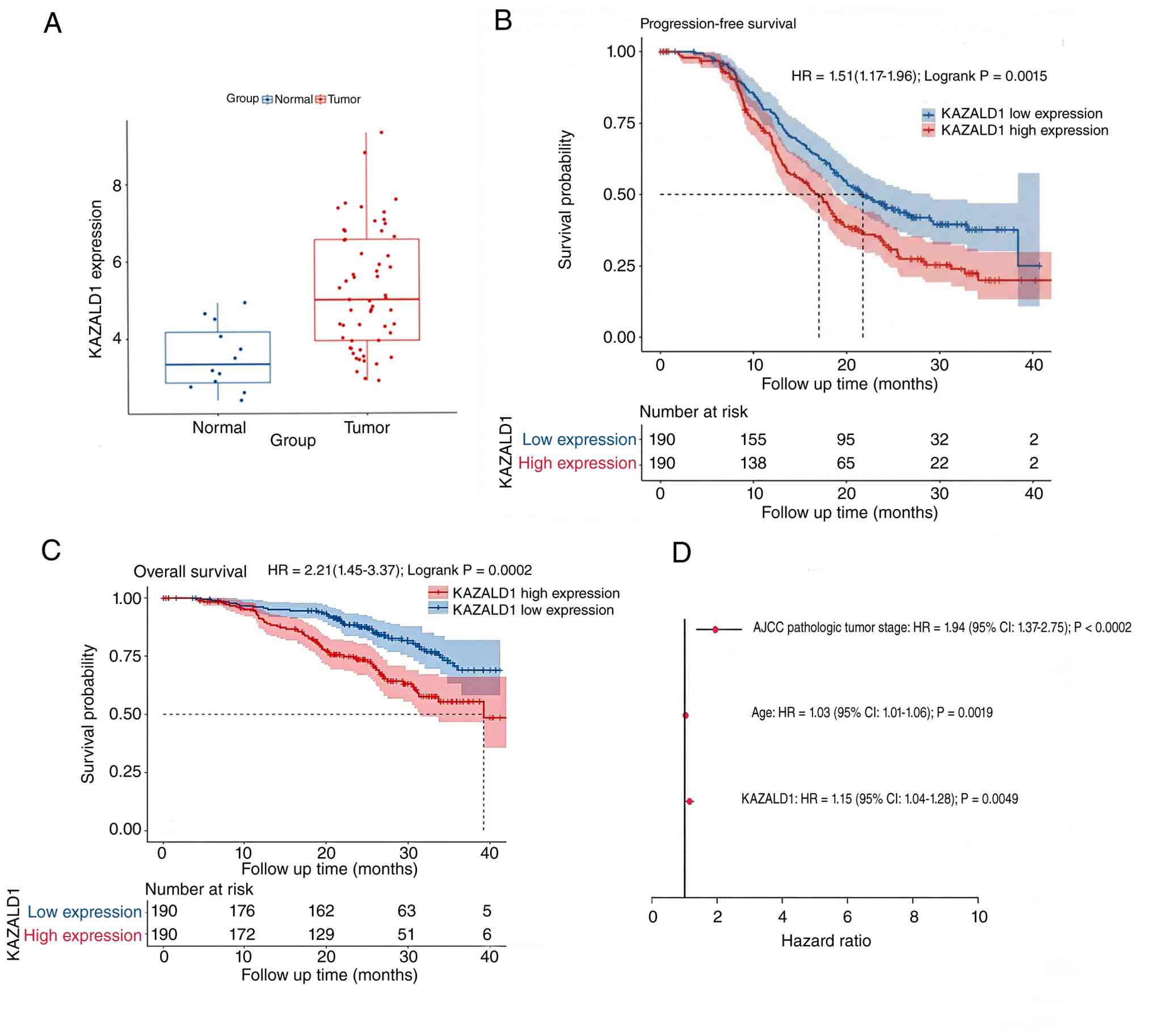
The KAZALD1 mRNA levels in the GSE66957 dataset were
compared between the ovarian cancer and non-carcinoma tissue
samples. Compared with those in the non-carcinoma samples, the
KAZALD1 mRNA levels were significantly upregulated in the
ovarian cancer samples (log2 FC=1.73; adjusted
P=5.42×10⁻⁴; Fig. 1A).
This indicates the oncogenic role of KAZALD1.
Subsequently, the correlation between KAZALD1
upregulation and prognosis was evaluated. Patients in the
independent GSE140082 cohort were categorized into the high-KAZALD1
and low-KAZALD1 groups based on the median KAZALD1 level.
Kaplan-Meir analysis revealed that patients in the high-KAZALD1
group exhibited poor PFS (HR: 1.51; 95% CI: 1.17–1.96; log-rank
P=0.0015; Fig. 1B) and OS (HR:
2.21; 95% CI: 1.45–3.37; log-rank P=0.0002; Fig. 1C). Multivariate Cox regression
analysis revealed that KAZALD1 was a significant independent
predictor of mortality in patients with EOC even after controlling
for age and American Joint Committee on Cancer stage (HR: 1.15; 95%
CI: 1.04–1.28; P=0.0049; Fig.
1D).
KAZALD1 is associated with aggressive
clinicopathological characteristics
The present study examined the KAZALD1 levels in 136
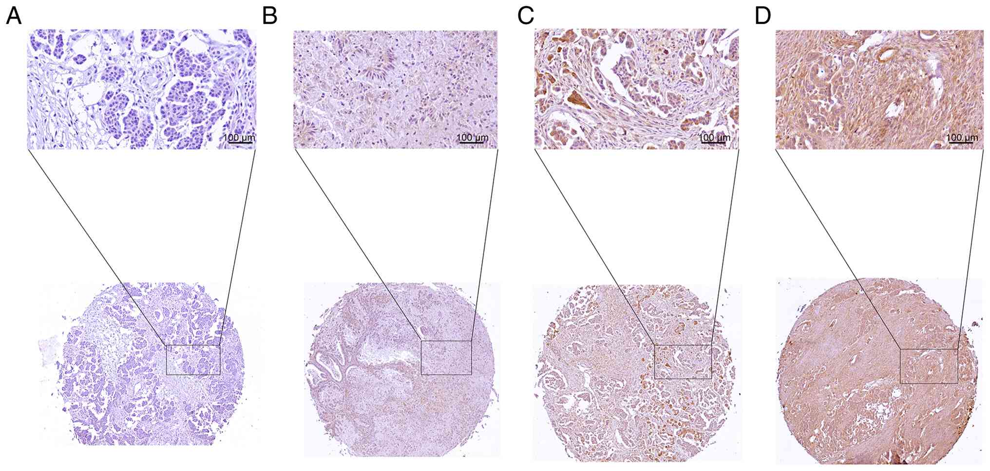
EOC specimens through IHC analysis. The representative images
across the staining intensity range are shown in Fig. 2A-D. The expression of KAZALD1 was
primarily localized to the cytoplasm of tumor cells. Based on the
0–12 composite scoring system, 42 (30.9%) and 94 (69.1%) patients
exhibited IHC scores of >6 (high expression) and ≤6 (low
expression), respectively (Fig.
3A). As shown in Table I, the
upregulation of KAZALD1 was associated with advanced FIGO stage
(P=0.011; Mann-Whitney U test), distant metastasis (P=0.002;
Fisher's exact test) and lymph node metastasis (P=0.025;
χ2 test). Spearman correlation analysis revealed that
KAZALD1 expression was correlated with distant metastasis
(rs=0.224; P=0.009) and tumor size (rs=0.208;
P=0.015; Table II). These findings
suggest that KAZALD1 upregulation increases the aggressiveness of
EOC.
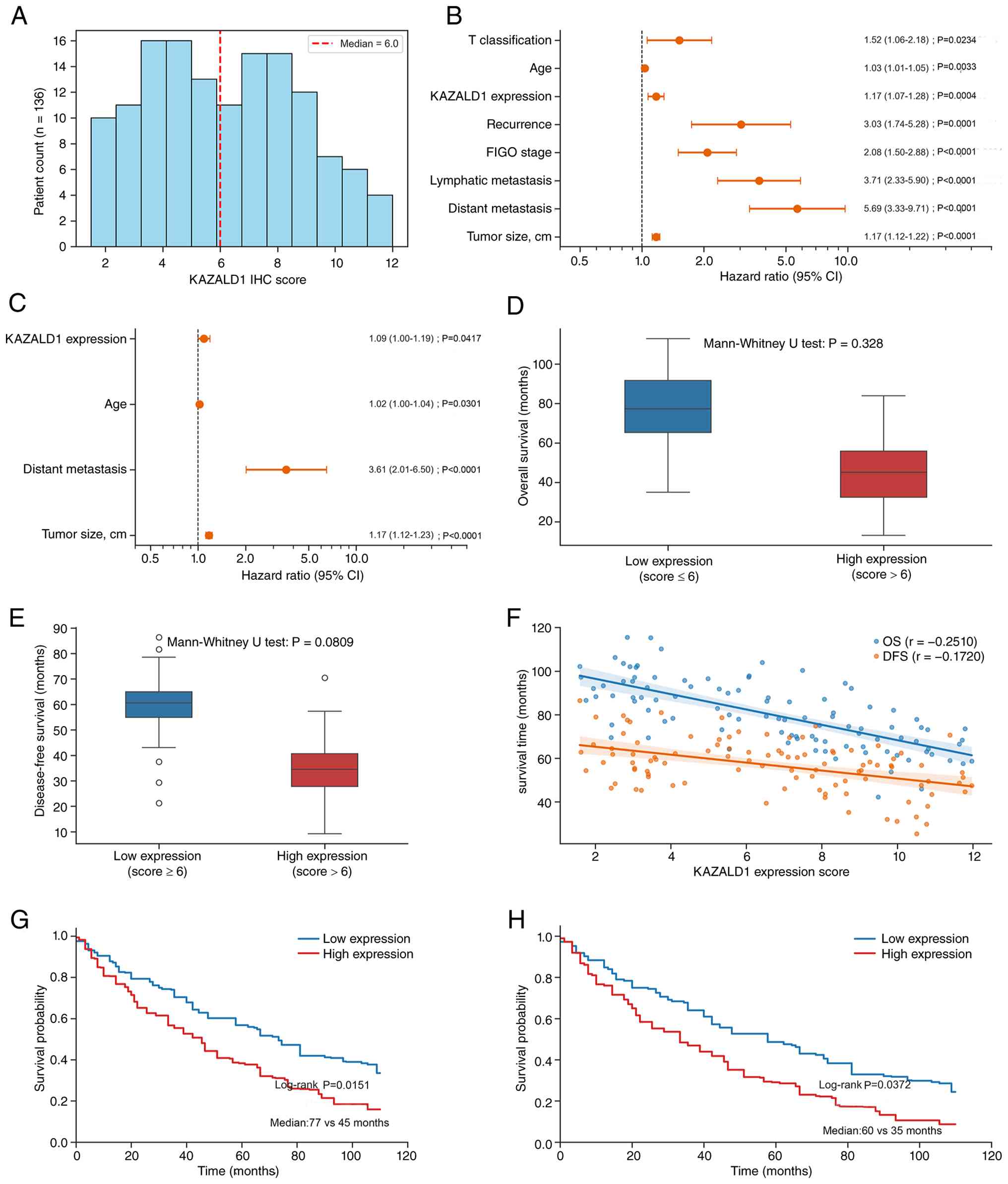 | Figure 3.Prognostic evaluation of KAZALD1
expression in patients with ovarian cancer. (A) Histogram
illustrating the distribution of KAZALD1 IHC scores in the clinical
cohort (n=136). The vertical dashed line indicates the median score
of 6.0, which was used as the cut-off for patient stratification.
(B) Forest plot of univariate Cox regression analysis results for
the prognostic significance of KAZALD1 expression along with
clinicopathological variables (T classification, age, recurrence,
FIGO stage, lymph node metastasis, distant metastasis and tumor
size) for OS. (C) Forest plot of multivariate Cox regression
analysis results. KAZALD1 expression is an independent predictor of
poor OS after adjusting for age, distant metastasis and tumor size.
Box plots comparing (D) OS and (E) DFS durations between the
low-KAZALD1 (score ≤6) and high-KAZALD1 (score >6) groups.
P-values were calculated using the Mann-Whitney U test. (F) Scatter
plots showing the inverse correlation between KAZALD1 expression
score and survival duration (blue indicates OS, r=−0.2510; orange
indicates DFS, r=−0.1720), which was determined using Spearman's
rank correlation. Kaplan-Meier survival curves for (G) OS and (H)
DFS stratified according to KAZALD1 expression. Significance was
determined using the log-rank test. Median survival durations are
annotated. Orange circles in the forest plots represent the hazard
ratio and horizontal lines indicate 95% CI. The vertical dashed
line denotes a hazard ratio of 1.0. The x-axis is displayed on a
logarithmic scale. Detailed survival statistics are provided in
Table III. KAZALD1, kazal type
serine peptidase inhibitor domain 1; IHC, immunohistochemical;
FIGO: International Federation of Gynecology and Obstetrics; OS,
overall survival; DFS, disease-free survival. |
 | Table I.Association between KAZALD1
expression and tumor characteristics in patients with ovarian
cancer. |
Table I.
Association between KAZALD1
expression and tumor characteristics in patients with ovarian
cancer.
| Feature | No. of
patients | KAZALD1
low-expression group | KAZALD1
high-expression group | P-value |
|---|
| All patients | 136 | 94 | 42 |
|
| Age | 136 | 50.1±11.8
(20.0–75.0) | 50.9±11.6
(23.0–73.0) | 0.708 |
| FIGO Stage |
|
|
| 0.011 |
| I | 11 (8.09%) | 8 (8.50%) | 3 (7.10%) |
|
| II | 33 (24.26%) | 26 (27.70%) | 7 (16.70%) |
|
|
III | 66 (48.53%) | 49 (52.10%) | 17 (40.50%) |
|
| IV | 26 (19.12%) | 11 (11.70%) | 15 (35.70%) |
|
| Tumor size, cm | 137 | 11.2±5.8
(1.9–24.4) | 12.4±5.5
(1.9–21.0) | 0.211 |
| T
classification |
|
|
| 0.338 |
| T1 | 11 (8.09%) | 8 (8.50%) | 3 (7.10%) |
|
| T2 | 33 (24.26%) | 26 (27.70%) | 7 (16.70%) |
|
| T3 | 92 (67.65%) | 60 (63.80%) | 32 (76.20%) |
|
| Lymphatic
metastasis |
|
|
| 0.025 |
| N0 | 104 (76.47%) | 77 (81.90%) | 27 (64.30%) |
|
| N1 | 32 (23.53%) | 17 (18.10%) | 15 (35.70%) |
|
| Distant
metastasis |
|
|
| 0.002 |
| M0 | 110 (80.88%) | 83 (88.30%) | 27 (64.30%) |
|
| M1 | 26 (19.12%) | 11 (11.70%) | 15 (35.70%) |
|
| Recurrence |
|
|
| 0.376 |
| No | 29 (21.43%) | 22 (21.30%) | 7 (16.70%) |
|
|
Yes | 107 (78.57%) | 72 (78.70%) | 35 (83.30%) |
|
| Table II.Spearman correlation analysis between
kazal type serine peptidase inhibitor domain 1 expression and
significant clinicopathological features in patients with ovarian
cancer (n=140). |
Table II.
Spearman correlation analysis between
kazal type serine peptidase inhibitor domain 1 expression and
significant clinicopathological features in patients with ovarian
cancer (n=140).
| Clinicopathological
feature | Spearman
correlation coefficient (rs) | P-value |
|---|
| Tumor size | 0.208 | 0.015 |
| Distant
metastasis | 0.224 | 0.009 |
KAZALD1 is an independent prognostic
biomarker for epithelial ovarian cancer
As KAZALD1 expression was associated with tumor
aggressiveness, the present study further assessed the independent
prognostic value of KAZALD1. Based on the median IHC score (6.0),
patients were categorized into the low-KAZALD1 (n=94) and
high-KAZALD1 (n=42) groups. Univariate Cox regression analysis
revealed that KAZALD1 expression (HR: 1.17; 95% CI: 1.07–1.28;
P=0.0004), FIGO stage (HR: 2.08; 95% CI: 1.50–2.88; P<0.0001),
tumor size (HR: 1.17; 95% CI: 1.12–1.22; P<0.0001) and distant
metastasis (HR: 5.69; 95% CI: 3.33–9.71; P<0.0001) were
associated with poor OS (Fig. 3B).
KAZALD1 remained an independent prognostic biomarker for EOC even
after adjusting for the confounders age, tumor size and distant
metastasis in the multivariate model for limiting collinearity (HR:
1.09; 95% CI: 1.00–1.19; P=0.0417; Fig.
3C).
Subsequently, the clinical outcomes of the two
groups were compared. Patients in the high-KAZALD1 group exhibited
decreased OS (P=0.0328; Fig. 3D)
and DFS (P=0.0809; Fig. 3E) when
compared with those in the low-KAZALD1 group. Spearman correlation
analysis revealed that KAZALD1 expression was inversely correlated
with OS (r=−0.2510) and DFS (r=−0.1720; Fig. 3F). The two subgroups exhibited
differential survival outcomes. Kaplan-Meier analysis revealed that
patients in the high-KAZALD1 group exhibited poor median OS (45 vs.
77 months; log-rank P=0.0151; Fig.
3G) and median DFS (35 vs. 60 months; log-rank P=0.0372;
Fig. 3H). The median OS and DFS of
patients in the high-KAZALD1 group were 41.6 and 36.0% lower
compared with those of patients in the low-KAZALD1 expression
subgroup (Table III). In the
univariate hazard model, every single-point increase in IHC score
increased the mortality and progression risks by 17.3% (HR: 1.173,
95% CI: 1.068–1.288) and 11.8% (HR=1.118; 95% CI: 1.002–1.248),
respectively.
| Table III.Summary of survival statistics
according to kazal type serine peptidase inhibitor domain 1
expression levels. |
Table III.
Summary of survival statistics
according to kazal type serine peptidase inhibitor domain 1
expression levels.
| Survival
indices | Low expression
(n=94) | High expression
(n=42) |
Difference/change | Statistical test
(P-value) | Univariate HR (95%
CI) |
|---|
| Overall
survival |
|
|
|
|
|
| Median
survival, months | 77.0 | 45.0 | −41.6% | 0.0151a | 1.173
(1.068–1.288) |
|
Survival correlation |
|
| −0.251 | 0.0328b |
|
| Disease-free
survival |
|
|
|
|
|
| Median
survival, months | 44.5 | 28.5 | −36.0% | 0.0372a | 1.118
(1.002–1.248)c |
|
Survival correlation |
|
| −0.172 | 0.0809b |
|
KAZALD1 expression is associated with
ECM/TGF-β pathway enrichment and reduced estimated APC
infiltration
Subsequently, the importance of KAZALD1 expression
was determined. KAZALD1 upregulation was associated with disease
aggressiveness and poor survival. Thus, the molecular mechanisms
underlying these effects were explored. Functional enrichment
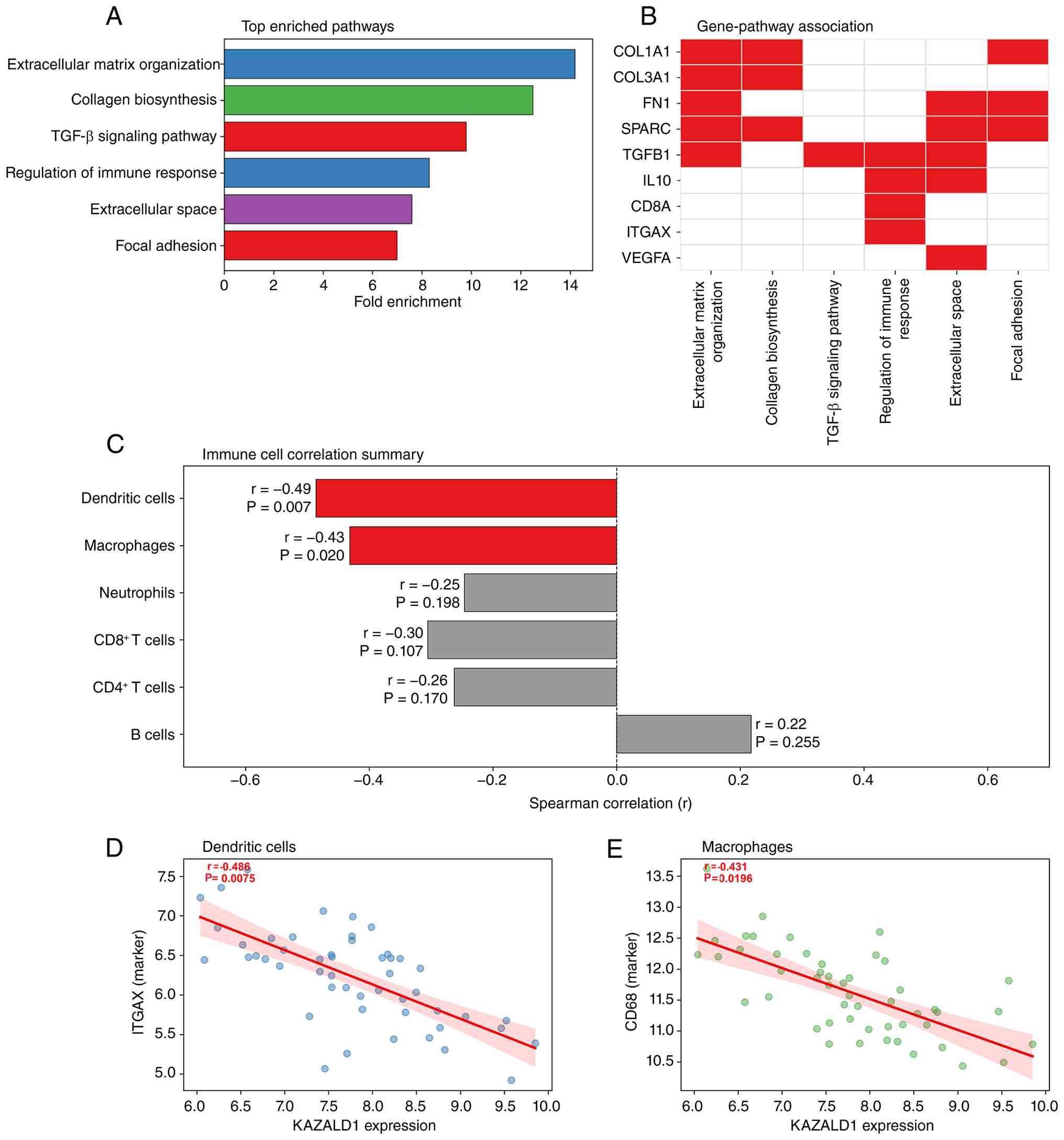
analysis of the 100 most significant genes co-expressed with
KAZALD1 revealed the following two key pathways: ‘Extracellular
matrix organization’ (P=1.23×10⁻⁹; 14.2-fold enrichment)
and ‘TGF-β signaling’ (P=3.45×10⁻⁶; 9.8-fold enrichment;
Fig. 4A). In the gene-pathway map,
TGFB1 and IL10 were found to be central components of the ECM/TGF-β
pathway for immunomodulation (Fig.
4B). Therefore, KAZALD1 was hypothesized to aid in the
establishment of an immunosuppressive TME.
To test this hypothesis, the correlation of KAZALD1
expression with TIICs was examined using the TIMER 2.0 algorithm.
KAZALD1 expression was found to be negatively correlated with the
infiltration of APCs, especially dendritic cells (r=−0.486;
P=0.0075; Fig. 4D) and macrophages
(r=−0.431; P=0.0196; Fig. 4E) but
not with that of adaptive immune cells, such as B cells or CD8+ T
cells (Fig. 4C). Collectively,
these correlative findings suggest an association between high
KAZALD1 expression and a TME characterized by ECM/TGF-β pathway
enrichment and reduced estimated infiltration of dendritic cells
and macrophages.
Discussion
To the best of our knowledge, the present study was
the first to comprehensively validate KAZALD1 as a novel prognostic
biomarker for EOC. Integrated multi-cohort analyses revealed that
KAZALD1 expression in ovarian cancer tissues was significantly
upregulated when compared with that in non-carcinoma tissues.
KAZALD1 upregulation was correlated with aggressive
clinicopathological characteristics and independently predicted
unfavorable OS and DFS. Exploratory bioinformatic analyses revealed
that KAZALD1 expression was correlated with ECM remodeling and the
immune-excluded microenvironment.
A major strength of the present study was the
rigorous validation of the clinical importance of KAZALD1. Genomic
information is provided in publicly available transcriptomic
datasets (such as the GSE140082 dataset used in the present study).
However, the mRNA expression level may not always indicate the
active protein level owing to complex post-transcriptional
modifications and differential translation rates (15,33).
Analysis of a cohort of 136 clinical cases demonstrated that
KAZALD1 is upregulated in ~33% of patients with EOC. Multivariate
Cox regression analysis revealed that KAZALD1 independently
predicted prognosis even after controlling for key confounders
(age, tumor size and distant metastasis). IHC analysis is a cheap
and easily accessible clinical technique. Based on the IHC scoring
system established for KAZALD1 in the present study, KAZALD1 has
potential application in stratifying the postoperative risk of
EOC.
KAZALD1 upregulation could predict advanced FIGO
stage and lymphatic and distant metastases. Previous studies have
reported that KAZALD1 mediates tissue remodeling (19–21,34),
altering stromal physical properties to promote tumor
dissemination. This biomechanical process is considered to drive
tumor metastasis (35–37). Exploratory bioinformatic analyses
with TCGA-OV cohort supported this hypothesis. In particular, the
KAZALD1 co-expression network was highly enriched in ‘Collagen
biosynthesis’, ‘Extracellular matrix organization’ and ‘TGF-β
signaling pathway’. Aberrant ECM accumulation and TGF-β activation
are reported to promote EMT and peritoneal metastasis of EOC
(38–40).
In addition to the structural significance, the
present study provided useful insights into the correlation of
KAZALD1 with the tumor immune microenvironment. KAZALD1 exerts
immunomodulatory effects by promoting dynamic shifts in certain
immune populations. For example, KAZALD1 expression is positively
correlated with M1 macrophages and naive CD8+ T cells and
negatively correlated with B cells and total CD8+ T cells (21). These findings suggest that KAZALD1
expression is correlated with the infiltration levels of APCs,
especially macrophages and dendritic cells. APCs exhibit key roles
in tumor antigen capture and T-cell-mediated anti-tumor immune
activation in immunological paradigms (41,42).
The depletion of APCs results in an ‘immune-excluded’ phenotype,
which is characterized by high resistance to immune checkpoint
inhibitors (43–45).
Previous studies have reported that TGF-β is a
master immunosuppressor driving stromal fibrosis and physically
trapping immune cells within the peritumoral stroma (46,47).
Therefore, the present study hypothesizes that KAZALD1 may promote
a dense, TGF-β-abundant fibrotic matrix to establish a physical and
biochemical barrier for preventing APC recruitment in the tumor
bed. The identification of tumors with KAZALD1 upregulation may aid
in determining patients who do not benefit from standard
immunotherapy and require new stromal barrier-targeting strategies.
However, because these analyses were derived from bulk
transcriptomic deconvolution, they identify statistical
associations only and do not establish a causal role of KAZALD1 in
impeding APC recruitment or mediating immune evasion. Functional
validation and prospective clinical studies are required.
The present study also exhibits a number of
limitations. The clinical validation cohort was retrospective and
single-center, so selection bias and unmeasured confounders,
including histological subtype, residual disease, platinum
sensitivity and BRCA/HRD status, cannot be excluded. Furthermore,
the independent prognostic value of KAZALD1 holds only within the
covariates available to the present model. In addition, the
transcriptomic analyses integrated datasets from different
platforms; although each was normalized and analyzed independently
rather than merged, residual platform heterogeneity and batch
effects may limit cross-cohort comparability. All mechanistic
inferences, including the associations of KAZALD1 with ECM/TGF-β
pathways and APC infiltration, were derived from correlational
analyses of bulk transcriptomic data and do not establish
causality; in addition, bulk-tissue deconvolution cannot resolve
the spatial localization and polarization states of infiltrating
macrophages and dendritic cells within the TME and no
immunotherapy-treated cohort was analyzed, leaving the predictive
value of KAZALD1 for immunotherapy response untested. Future
studies using multicenter prospective cohorts with standardized IHC
scoring, spatial transcriptomics with multiplex immunofluorescence
and gain- and loss-of-function experiments in EOC cell lines,
co-culture systems and animal models are therefore needed to
validate these findings and define the causal role of KAZALD1 in
ECM remodeling, TGF-β signaling and APC recruitment.
In conclusion, KAZALD1 was upregulated in EOC and
elevated KAZALD1 protein expression was associated with aggressive
clinicopathological features and independently predicts unfavorable
OS and DFS in the present cohort. Exploratory bioinformatic
analyses further revealed correlative associations between KAZALD1
expression, ECM/TGF-β-associated transcriptional programs and
reduced estimated APC infiltration. These findings support KAZALD1
as a candidate prognostic biomarker for EOC, whereas its potential
role in shaping the immune microenvironment and its relevance to
immunotherapy stratification remain hypotheses that require further
confirmation through functional experiments and prospective
clinical studies.
Supplementary Material
Supporting Data
Acknowledgements
Not applicable.
Funding
Funding: No funding was received.
Availability of data and materials
The data generated in the present study may be
requested from the corresponding author.
Authors' contributions
RZ made notable contributions to the conception of
the present study, acquisition and bioinformatic analysis of GEO
and TCGA datasets and participated in drafting the manuscript. YH
made notable contributions to the design of the present study and
collection of clinical samples, whilst also performing IHC
experiments and survival analysis, participating in drafting and
revising the manuscript. YH and RZ confirm the authenticity of all
the raw data. JW and JL conducted experiments and participated in
data verification. GD (corresponding author) made notable
contributions to the conception and design of the present study and
participated in revising the manuscript critically for important
intellectual content. All authors read and approved the final
version of the manuscript.
Ethics approval and consent to
participate
The Ethics Committee of the Obstetrics and
Gynecology Hospital of Tongji University approved the analysis of
patient samples (approval no. KS25118). Individual patient informed
consent was exempted for the present research.
Patient consent for publication
Not applicable.
Competing interests
The authors declare that they have no competing
interests.
References
|
1
|
Caruso G, Weroha SJ and Cliby W: Ovarian
cancer: A review. JAMA. 334:1278–1291. 2025. View Article : Google Scholar : PubMed/NCBI
|
|
2
|
Nougaret S, Lakhman Y, Suarez-Weiss KE,
Patel-Lippman K, Sadowski E and Shinagare AB: Imaging in advanced
epithelial ovarian cancer: Assessment of peritoneal spread.
Radiology. 318:e2501162026. View Article : Google Scholar : PubMed/NCBI
|
|
3
|
Xie W, Tang S, Tang R, Li L and Liu X:
Global, regional, and National burden of ovarian cancer, 1990–2021,
and projections to 2050: A cross-sectional analysis of the global
burden of disease study 2021. Int J Surg. 112:226–238. 2026.
View Article : Google Scholar : PubMed/NCBI
|
|
4
|
Li H, Sheng J, Zheng S, Liu PW, Wu N, Zeng
WJ, Li YH and Wang J: Platinum-resistant ovarian cancer: From
mechanisms to treatment strategies. Genes Dis. 13:1018012025.
View Article : Google Scholar : PubMed/NCBI
|
|
5
|
Richardson DL, Eskander RN and O'Malley
DM: Advances in ovarian cancer care and unmet treatment needs for
patients with platinum resistance: A narrative review. JAMA Oncol.
9:851–859. 2023. View Article : Google Scholar : PubMed/NCBI
|
|
6
|
Veneziani AC, Gonzalez-Ochoa E, Alqaisi H,
Madariaga A, Bhat G, Rouzbahman M, Sneha S and Oza AM:
Heterogeneity and treatment landscape of ovarian carcinoma. Nat Rev
Clin Oncol. 20:820–842. 2023. View Article : Google Scholar : PubMed/NCBI
|
|
7
|
To SKY, Yang Y, Shi Z, Fung KSW, Lai SL,
Li JJX, Ip PPC and Wong AST: Subclonal β-catenin/YAP signaling
heterogeneity accelerates ovarian cancer metastasis through a
senescence-associated secretory phenotype. Cell Death Dis.
23:5392026. View Article : Google Scholar
|
|
8
|
Sun T, Zhang Z, Tian L, Zheng Y, Wu L, Guo
Y, Li X, Li Y, Shen H, Lai Y, et al: Dualistic classification of
high grade serous ovarian carcinoma has its root in spatial
heterogeneity. J Adv Res. 48:213–225. 2023. View Article : Google Scholar : PubMed/NCBI
|
|
9
|
Bedia JS, Jacobs IJ, Ryan A,
Gentry-Maharaj A, Burnell M, Singh N, Manchanda R, Kalsi JK, Dawnay
A, Fallowfield L, et al: Estimating the ovarian cancer CA-125
preclinical detectable phase, in-vivo tumour doubling time, and
window for detection in early stage: An exploratory analysis of
UKCTOCS. EBioMedicine. 112:1055542025. View Article : Google Scholar : PubMed/NCBI
|
|
10
|
Boehm KM, Aherne EA, Ellenson L,
Nikolovski I, Alghamdi M, Vázquez-García I, Zamarin D, Long Roche
K, Liu Y, Patel D, et al: Multimodal data integration using machine
learning improves risk stratification of high-grade serous ovarian
cancer. Nat Cancer. 3:723–733. 2022. View Article : Google Scholar : PubMed/NCBI
|
|
11
|
Xue Y, Chen Z, Fang X, Chu R, Li M, Shen
Y, Yao Q, Fu B, Qin T, Li L and Qiao X: Risk prediction model of
survival in patients with low-grade serous ovarian cancer: A
multicenter Cohort study. J Ovarian Res. 19:1992026. View Article : Google Scholar
|
|
12
|
Kawakami E, Tabata J, Yanaihara N,
Ishikawa T, Koseki K, Iida Y, Saito M, Komazaki H, Shapiro JS, Goto
C, et al: Application of artificial intelligence for preoperative
diagnostic and prognostic prediction in epithelial ovarian cancer
based on blood biomarkers. Clin Cancer Res. 25:3006–3015. 2019.
View Article : Google Scholar : PubMed/NCBI
|
|
13
|
Dibitetto D, Widmer CA and Rottenberg S:
PARPi, BRCA, and gaps: Controversies and future research. Trends
Cancer. 10:857–869. 2024. View Article : Google Scholar : PubMed/NCBI
|
|
14
|
Herzog TJ, Vergote I, Gomella LG,
Milenkova T, French T, Tonikian R, Poehlein C and Hussain M:
Testing for homologous recombination repair or homologous
recombination deficiency for poly (ADP-ribose) polymerase
inhibitors: A current perspective. Eur J Cancer. 179:136–146. 2023.
View Article : Google Scholar
|
|
15
|
Witz A, Dardare J, Betz M, Michel C,
Husson M, Gilson P, Merlin JL and Harlé A: Homologous recombination
deficiency (HRD) testing landscape: Clinical applications and
technical validation for routine diagnostics. Biomark Res.
13:312025. View Article : Google Scholar : PubMed/NCBI
|
|
16
|
Yin Z, Li C, Wang J and Xue L:
Myeloid-derived suppressor cells: Roles in the tumor
microenvironment and tumor radiotherapy. Int J Cancer. 144:933–946.
2019. View Article : Google Scholar : PubMed/NCBI
|
|
17
|
Qiu L, Yue J, Ding L, Yin Z, Zhang K and
Zhang H: Cancer-associated fibroblasts: An emerging target against
esophageal squamous cell carcinoma. Cancer Lett. 546:2158602022.
View Article : Google Scholar : PubMed/NCBI
|
|
18
|
Xie Y, Wang X, Wang W, Pu N and Liu L:
Epithelial-mesenchymal transition orchestrates tumor
microenvironment: Current perceptions and challenges. J Transl Med.
23:3862025. View Article : Google Scholar : PubMed/NCBI
|
|
19
|
Shibata Y, Tsukazaki T, Hirata K, Xin C
and Yamaguchi A: Role of a new member of IGFBP superfamily,
IGFBP-rP10, in proliferation and differentiation of osteoblastic
cells. Biochem Biophys Res Commun. 325:1194–1200. 2004. View Article : Google Scholar : PubMed/NCBI
|
|
20
|
Zhu Y, Liu H, Zhang Y, Zheng D, Li Y, Chen
J, Xu J and Zhang W: Kazald1 attenuates chondrocyte fibrosis to
potentiate hyaline cartilage regeneration by interfering with the
pro-fibrotic TGF-β signaling. Theranostics. 15:9885–9910. 2025.
View Article : Google Scholar : PubMed/NCBI
|
|
21
|
Zhou S, An H, Wei H, Wu R, Wang Z, Liu M,
Liu T and Li K: Identification and verification of biomarkers
related to polyamine metabolism in diabetic nephropathy. J Diabetes
Res. 2025:95397342025. View Article : Google Scholar : PubMed/NCBI
|
|
22
|
Goto Y, Shinjo K, Kondo Y, Shen L, Toyota
M, Suzuki H, Gao W, An B, Fujii M, Murakami H, et al: Epigenetic
profiles distinguish malignant pleural mesothelioma from lung
adenocarcinoma. Cancer Res. 69:9073–9082. 2009. View Article : Google Scholar : PubMed/NCBI
|
|
23
|
Shen Q, Polom K, Williams C, de Oliveira
FMS, Guergova-Kuras M, Lisacek F, Karlsson NG, Roviello F and
Kamali-Moghaddam M: A targeted proteomics approach reveals a serum
protein signature as diagnostic biomarker for resectable gastric
cancer. EBioMedicine. 44:322–333. 2019. View Article : Google Scholar : PubMed/NCBI
|
|
24
|
Devall MA, Eaton S, Ali MW, Dampier CH,
Weisenberger D, Powell SM, Li L and Casey G: DNA methylation
analysis of normal colon organoids from familial adenomatous
polyposis patients reveals novel insight into colon cancer
development. Clin Epigenetics. 14:1042022. View Article : Google Scholar : PubMed/NCBI
|
|
25
|
Wang H, Feng Y, Bao Z and Jiang C, Yan W,
Wang Y, Zhang C, Liu Y, Zhang Q, Zhang W and Jiang C: Epigenetic
silencing of KAZALD1 confers a better prognosis and is associated
with malignant transformation/progression in glioma. Oncol Rep.
30:2089–2096. 2013. View Article : Google Scholar : PubMed/NCBI
|
|
26
|
Rothzerg E, Xu J, Wood D and Kõks S: 12
Survival-related differentially expressed genes based on the
TARGET-osteosarcoma database. Exp Biol Med (Maywood).
246:2072–2081. 2021. View Article : Google Scholar : PubMed/NCBI
|
|
27
|
Feleke M, Feng W, Rothzerg E, Song D, Wei
Q, Kõks S, Wood D, Liu Y and Xu J: Single-cell RNA-seq
identification of four differentially expressed survival-related
genes by a TARGET: Osteosarcoma database analysis. Exp Biol Med
(Maywood). 247:921–930. 2022. View Article : Google Scholar : PubMed/NCBI
|
|
28
|
Asmar C, Awad G, Boutros M, Daccache S,
Chebly A, Alix-Panabières C and Kourié HR: Single-cell RNA
sequencing in osteosarcoma: Applications in diagnosis, prognosis,
and treatment. Med Oncol. 42:5512025. View Article : Google Scholar : PubMed/NCBI
|
|
29
|
Li X, Zhang J, Wu Y, Ma C, Wei D, Pan L
and Cai L: IGFBP7 remodels the tumor microenvironment of esophageal
squamous cell carcinoma by activating the TGFβ1/SMAD signaling
pathway. Oncol Lett. 24:2512022. View Article : Google Scholar : PubMed/NCBI
|
|
30
|
Hong Z, Xie W, Zhuo H, Wei X, Wang K,
Cheng J, Lin L, Hou J, Chen X and Cai J: Crosstalk between cancer
cells and Cancer-Associated fibroblasts mediated by TGF-β1-IGFBP7
signaling promotes the progression of infiltrative gastric cancer.
Cancers (Basel). 15:39652023. View Article : Google Scholar
|
|
31
|
Cheng Y: Gene expression profiling of
high-grade serous ovarian carcinoma. NCBI Gene Expression Omnibus;
2015
|
|
32
|
Kommoss S, Winterhoff B, Oberg AL, Konecny
GE, Wang C, Riska SM, Fan JB, Maurer MJ, April C, Shridhar V, et
al: Bevacizumab may differentially improve ovarian cancer outcome
in patients with proliferative and mesenchymal molecular subtypes.
Clin Cancer Res. 23:3794–3801. 2017. View Article : Google Scholar : PubMed/NCBI
|
|
33
|
Lahtvee PJ, Sánchez BJ, Smialowska A,
Kasvandik S, Elsemman IE, Gatto F and Nielsen J: Absolute
quantification of protein and mRNA abundances demonstrate
variability in gene-specific translation efficiency in yeast. Cell
Syst. 4:495–504. 2017. View Article : Google Scholar : PubMed/NCBI
|
|
34
|
Bryant DM, Johnson K, DiTommaso T, Tickle
T, Couger MB, Payzin-Dogru D, Lee TJ, Leigh ND, Kuo TH, Davis FG,
et al: A Tissue-mapped axolotl de novo transcriptome enables
identification of limb regeneration factors. Cell Rep. 18:762–776.
2017. View Article : Google Scholar : PubMed/NCBI
|
|
35
|
Mohan V, Das A and Sagi I: Emerging roles
of ECM remodeling processes in cancer. Elsevier. 192–200. 2020.
|
|
36
|
Mai Z, Lin Y, Lin P, Zhao X and Cui L:
Modulating extracellular matrix stiffness: A strategic approach to
boost cancer immunotherapy. Cell Death Dis. 15:3072024. View Article : Google Scholar : PubMed/NCBI
|
|
37
|
Stylianou A, Mpekris F, Voutouri C, Papoui
A, Constantinidou A, Kitiris E, Kailides M and Stylianopoulos T:
Nanomechanical properties of solid tumors as treatment monitoring
biomarkers. Acta Biomater. 154:324–334. 2022. View Article : Google Scholar : PubMed/NCBI
|
|
38
|
Tie Y, Tang F, Peng D, Zhang Y and Shi H:
TGF-beta signal transduction: Biology, function and therapy for
diseases. Mol Biomed. 3:452022. View Article : Google Scholar : PubMed/NCBI
|
|
39
|
Chen Y, Wang DD, Wu YP, Su D, Zhou TY, Gai
RH, Fu YY, Zheng L, He QJ, Zhu H and Yang B: MDM2 promotes
epithelial-mesenchymal transition and metastasis of ovarian cancer
SKOV3 cells. Br J Cancer. 117:1192–1201. 2017. View Article : Google Scholar
|
|
40
|
Gjerde CH, Kleinmanns K, Langer A, Ruiz de
Garibay Ponce G, Rozmus E, Stangeland GN, Leitch C, Elnour R,
Dongre HN, Berger C, et al: A 3D ovarian cancer metastasis model
using a decellularised peritoneal matrix to study therapy response.
EBioMedicine. 124:1061352026. View Article : Google Scholar : PubMed/NCBI
|
|
41
|
Sánchez-Paulete AR, Teijeira A, Cueto FJ,
Garasa S, Pérez-Gracia JL, Sánchez-Arráez A, Sancho D and Melero I:
Antigen cross-presentation and T-cell cross-priming in cancer
immunology and immunotherapy. Ann Oncol. 28 (Suppl_12):xii44–xii55.
2017. View Article : Google Scholar : PubMed/NCBI
|
|
42
|
Kim J, Song SG, Park S, Ko YG, Ham J, Kim
T, Ha SJ, Bae YS, Chung DH and Kim HY: Antigen cross-presentation
by Type-2 Innate Lymphoid Cells facilitates the activation of
antitumor CD8+ T cells. Cancer Res. 85:2659–2678. 2025. View Article : Google Scholar : PubMed/NCBI
|
|
43
|
Yu J, Wu X, Song J, Zhao Y, Li H, Luo M
and Liu X: Loss of MHC-I antigen presentation correlated with
immune checkpoint blockade tolerance in MAPK inhibitor-resistant
melanoma. Front Pharmacol. 13:9282262022. View Article : Google Scholar : PubMed/NCBI
|
|
44
|
Nguyen KB and Spranger S: Modulation of
the immune microenvironment by tumor-intrinsic oncogenic signaling.
J Cell Biol. 219:e2019082242019. View Article : Google Scholar
|
|
45
|
Wang R, Zhang Y, Meng X, Zhao J, Xing Y,
Ouyang Q, Zhang N, Chen H, Miao N, Song E and Huang D:
Tumor-derived CDC37 inhibits antigen cross-presentation in
dendritic cells and impairs anti-tumor immunity in breast cancer.
Adv Sci. 13:e065182026. View Article : Google Scholar : PubMed/NCBI
|
|
46
|
Derynck R, Turley SJ and Akhurst RJ: TGFβ
biology in cancer progression and immunotherapy. Nat Rev Clin
Oncol. 18:9–34. 2021. View Article : Google Scholar
|
|
47
|
Cheng JY, Tsai HH, Hung JT, Hung TH, Lin
CC, Lee CW, Lo ZC, Huang JR, Chiou SP, Huang Y, et al:
Tumor-associated glycan exploits adenosine receptor 2A signaling to
facilitate immune evasion. Adv Sci. 12:24165012025. View Article : Google Scholar
|