Introduction
Breast cancer continues to pose a notable global
health burden, representing one of the most prevalent malignancies
affecting women worldwide. According to GLOBOCAN 2024, female
breast cancer accounted for an ~2.43 million new cases and 693,660
mortalities worldwide in 2024, and was the most commonly diagnosed
cancer and the leading cause of cancer-associated mortality among
women globally (1). For patients
with lymph node metastasis or other high-risk factors,
post-operative radiotherapy is important for reducing local
recurrence and breast cancer-specific mortality (2,3). The
increasing adoption of regional nodal irradiation (RNI) has further
improved oncological outcomes (4);
however, it also elevates the risk of developing toxicities to
adjacent organs.
The esophagus, due to its anatomical proximity to
the supraclavicular and internal mammary nodal regions, often
receives incidental radiation during RNI, resulting in acute
radiation esophagitis (ARE). It has been reported that symptomatic
ARE occurs in 30–50% of patients with breast cancer receiving RNI
(5). ARE typically manifests as
odynophagia, dysphagia and retrosternal pain, which, in severe
cases, may lead to malnutrition, dehydration and treatment
interruptions, thereby substantially impairing the quality of life
of patients. In the era of precision radiotherapy, the early
identification of high-risk patients is key for improving quality
of life, maintaining treatment efficacy and reducing
treatment-related morbidity (6).
Previous studies have identified numerous factors
associated with ARE, including esophageal dose-volume parameters,
treatment techniques and patient characteristics. Mean esophageal
dose (Dmean), V10, V20, and absolute volumes (AV) of an organ at
risk receiving 30 or 35 Gy (AV30 and AV35) have been shown to be
associated with the incidence and severity of esophagitis (7–9).
Treatment techniques may also influence esophageal exposure during
breast RNI (8,10), and tumor laterality has been found
to be associated with the risk of developing ARE (11). However, conventional prediction
approaches may not adequately capture interactions among clinical
and dosimetric variables, and clinically interpretable risk tools
remain limited.
Cancer is biologically heterogeneous, with recent
studies identifying prognostic molecular markers, including
endoplasmic reticulum stress-associated proteins, AIMP2 and
transcription factors associated with disease-free survival in
triple-negative breast cancer (12–14).
Although these molecular studies did not evaluate radiotherapy
toxicity, they reinforce the broader rationale for individualized
risk assessment. In parallel, artificial intelligence (AI) is
increasingly being explored in radiotherapy and immunotherapy, with
emphasis on clinically validated and interpretable models (15).
Machine-learning (ML) provides a framework for
analyzing complex, high-dimensional data and may capture non-linear
associations that are difficult to represent with conventional
approaches (16,17). This flexibility has motivated the
use of ML for clinical outcome prediction (18,19).
Interpretability methods, such as SHapley Additive exPlanations
(SHAP) can further decompose model predictions into feature-level
contributions and may improve transparency in clinical decision
support (20).
Therefore, the present study aimed to develop and
evaluate an interpretable ML-based model for predicting grade ≥2
ARE in patients with breast cancer receiving RNI. In the present
study, univariable and multivariable analysis, least absolute
shrinkage and selection operator (LASSO) regression and the Boruta
algorithm for feature selection were used, followed by the
comparison of 10 ML algorithms. Bootstrap resampling was used to
quantify optimism and model stability. The final model was selected
by considering internal validation, held-out performance,
calibration, parsimony and clinical interpretability. The present
study was reported in accordance with the transparent reporting of
a multivariable prediction model for individual prognosis or
diagnosis (TRIPOD)+AI statement (21).
Materials and methods
Study design
The present single-center retrospective
observational study included patients with breast cancer who
received post-operative RNI at The General Hospital of Western
Theater Command of the Chinese People's Liberation Army (Chengdu,
China) between January 2016 and June 2025. Patient data were
retrieved between July and September 2025, and statistical analyses
were conducted between September and December 2025. The study
protocol received approval from the Institutional Review Board of
The General Hospital of Western Theater Command of the Chinese
People's Liberation Army (approval no. 2025EC11-ky019) and was
conducted in accordance with the principles of the amended
Declaration of Helsinki. Due to the retrospective design, the
Institutional Review Board granted a waiver for written informed
consent. All patient identifiers were anonymized during the
analysis.
Patient selection
A total of 324 patients with breast cancer who
received adjuvant RNI between January 2016 and June 2025 were
retrospectively enrolled. The inclusion criteria were as follows:
i) Female patients with pathologically confirmed breast cancer; ii)
those who underwent mastectomy or breast-conserving surgery; iii)
those whose post-operative radiotherapy target volume included the
supraclavicular/infraclavicular and/or internal mammary lymph node
regions; iv) those whose radiotherapy technique was
intensity-modulated radiotherapy (IMRT) alone or IMRT combined with
electron beams; and v) those with an availability of complete
clinical, laboratory and radiotherapy planning data. The key
exclusion criteria included the following: i) The receipt of
concurrent chemotherapy; ii) a prior history of thoracic
radiotherapy; iii) pre-existing esophageal disease or swallowing
dysfunction; and iv) a diagnosis of other active malignancies or
notable missing key data (>10% for any variable).
Treatment and dosimetric
parameters
All patients received radiotherapy within 8 weeks
post-operatively (if adjuvant chemotherapy was not planned) or upon
the completion of adjuvant chemotherapy. Patients were positioned
using computed tomography (CT) simulation scans and immobilized
with thermoplastic body masks. For patients who underwent
mastectomy, a 5-mm-thick silicone sheet was placed on the chest
wall surface during radiotherapy. The irradiation fields
encompassed the whole breast/chest wall,
supraclavicular/infraclavicular fossa and level II axillary lymph
nodes.
For the present study, the esophagus was contoured
from the inferior border of the cricoid cartilage to the inferior
border of the aortic arch (Fig.
S1). A representative intensity-modulated radiotherapy plan and
dose distribution is presented in Fig.
S2. Other organs at risk, including the spinal cord, thyroid
gland, contralateral breast, lungs and heart, were also delineated.
Patients received IMRT with conventional or hypofractionated
regimens. As the total dose varied with fractionation and the use
of a tumor-bed boost following breast-conserving surgery, the total
dose was analyzed as a continuous variable. Dose-volume histograms
were used to extract the Dmean, maximum dose (Dmax), total
esophageal volume, relative volumes receiving 10–40 Gy
(RV_V10-RV_V40) and AV receiving 10–40 Gy (AV_V10-AV_V40). Relative
dose-volume metrics are expressed as the percentage of the
contoured esophageal volume receiving at least the specified dose.
Concurrent targeted therapy was permitted for patients with
HER2-positive disease, whereas concurrent chemotherapy was not
permitted. Baseline pruritus was defined as pruritus documented
prior to radiotherapy initiation, either at CT simulation or at the
pretreatment visit within 7 days and was recorded as present or
absent. Pruritus arising during radiotherapy was not included in
the prediction model to avoid post-baseline information
leakage.
Missing data
Missing data was assessed for all candidate
predictors and was found to account for <5%. Little's test was
used to assess whether the missingness pattern was compatible with
missing completely at random (MCAR). The null hypothesis was that
the data were MCAR, and a two-sided P>0.05 was interpreted as
insufficient evidence to reject this hypothesis. Given the low
overall missingness and the MCAR result (P>0.05), complete-case
analysis was used for model development and evaluation.
Follow-up and toxicity evaluation
ARE was graded according to the Radiation Therapy
Oncology Group acute radiation morbidity scoring criteria (22). Assessments were performed during
radiotherapy and at 1 week, 2 weeks and 3 months following the
completion of treatment. At least two radiation oncologists
reviewed the ARE status. Disagreements were adjudicated by a senior
oncologist. The highest recorded ARE grade during the assessment
period was used for analysis. The primary endpoint was the
occurrence of grade ≥2 ARE at any assessment during this predefined
period.
Feature selection and model
construction
Patients were divided into a training set (70%) and
a held-out test set (30%) by outcome-stratified random sampling
using a fixed seed of 52. Feature selection and model development
were performed primarily in the training set. No post-enrollment
imputation was required for the complete-case dataset used for
model development. For algorithms requiring scaled inputs,
centering and scaling parameters were estimated from the training
data and then applied unchanged to the held-out data.
Candidate predictors were screened in the training
set using three complementary methods: Univariable analysis
followed by multivariable logistic regression (LR), LASSO
regression and the Boruta algorithm (23–25).
For LASSO, a fixed random seed of 43 was used with a binomial
model, α=1, a maximum of 100,000 iterations and 10-fold
cross-validation based on binomial deviance; λ1 standard error
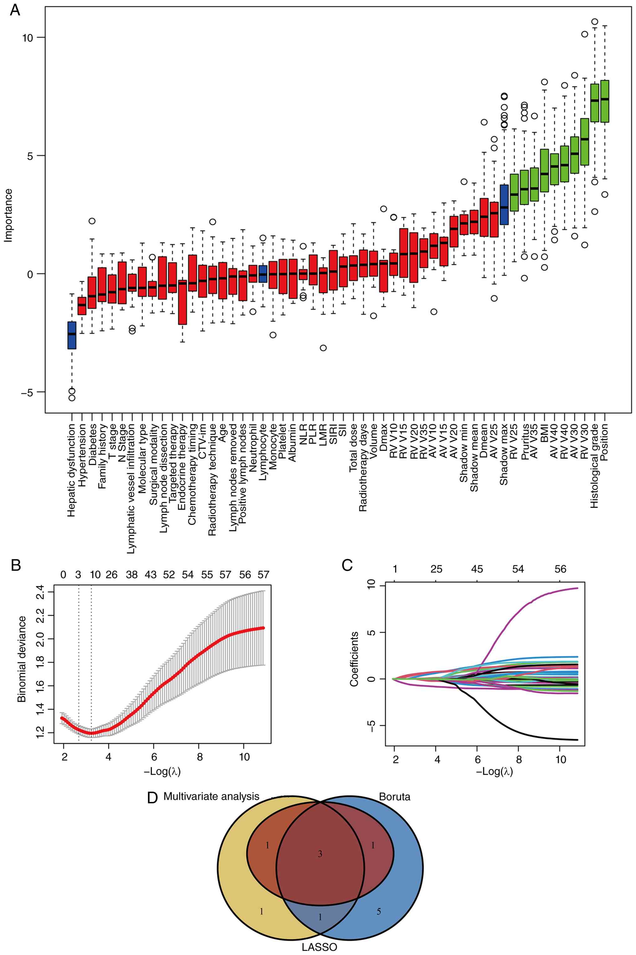
(λ1SE) was selected as the penalty parameter. For Boruta, a fixed
random seed of 123 and maxRuns=500 were used, followed by
TentativeRoughFix for tentative attributes. The final consensus
feature set comprised variables retained by >2 of the three
selection approaches: Pre-existing pruritus, histological grade,
BMI, diabetes, tumor laterality and esophageal RV_V30.
In total, 10 supervised ML algorithms were
evaluated: LR, support vector machine (SVM), gradient boosting
machine (GBM), neural network (NN), random forest (RF), extreme
gradient boosting (XGBoost), K-Nearest Neighbors (KNN), AdaBoost,
light gradient boosting machine (LightGBM) and categorical boosting
(CatBoost). For models implemented through the caret framework, a
fixed set of outcome-stratified five-fold cross-validation folds
(seed=520) was used for tuning and comparison. XGBoost was
evaluated using its native R interface with fixed hyperparameters
over the same training folds, whereas LightGBM and CatBoost were
fitted using their native R interfaces. Model-specific tuning
grids, fixed settings, resampling details and available random-seed
information are reported in Table
SI.
Internal validation was used to assess model
stability and identify preferred candidate models. For the three
pre-selected models, bootstrap resampling (B=1,000) was performed
within the training set to quantify optimism. In each bootstrap
sample, the apparent area under the curve (AUC) was compared with
performance on the original training cohort, and the mean optimism
was subtracted from the apparent AUC of the full training-set model
to obtain the optimism-corrected AUC. Final model selection
considered bootstrap optimism and corrected performance together
with held-out test-set discrimination and calibration, model
complexity and clinical interpretability. For threshold-dependent
classification metrics, the probability threshold was determined in
the training set using the maximum Youden index and then fixed for
application to the held-out test set; no threshold optimization was
performed in the test set. Held-out discrimination was summarized
by AUC with DeLong 95% confidence intervals (CIs). Calibration was
assessed using the Brier score, calibration slope and
calibration-in-the-large (CITL), and clinical utility was evaluated
by decision curve analysis. Pairwise AUC comparisons with LR used
DeLong's test.
Model interpretation was performed
using SHAP
Training data were used as the SHAP background
distribution, and global importance, beeswarm and dependence plots
were generated for the final LR model. Dependence plots were
prespecified for RV_V30 and BMI to illustrate the direction of the
two continuous predictors with high global importance. Continuous
predictor values in the dependence plots are displayed on the
standardized model-input scale. For clinical implementation, a
static nomogram was constructed from the final LR coefficients
using the rms package (https://cran.r-project.org/package=rms), and a
web-based dynamic calculator was generated using DynNom (https://cran.r-project.org/package=DynNom) and shiny
(https://cran.r-project.org/package=shiny). The
nomogram and web calculator were based on the same final LR
model.
Statistical analysis
Continuous variables are presented as the mean ±
standard deviation when normally distributed and as median
(interquartile range) otherwise; categorical variables are
presented as frequencies and percentages. Binary variables were
coded according to their prespecified categories, and multilevel
categorical variables were represented using indicator variables as
required. Between-group comparisons were performed using the
independent-samples t-test for normally distributed continuous
variables and the Mann-Whitney U test for non-normally distributed
continuous variables. χ2 or Fisher's exact tests were
used for categorical variables. All tests were two-sided, and
P<0.05 was considered to indicate a statistically significant
difference.
Statistical analyses were performed using R software
(version 4.5.2; Posit Software, PBC). The glmnet and Boruta
packages were used for LASSO and Boruta feature selection,
respectively (23–25). The caret framework was used for LR,
SVM, GBM, NN, RF, KNN and AdaBoost. XGBoost, LightGBM and CatBoost
were fitted with their native R interfaces in the final analysis.
Model-specific tuning grids, fixed settings, resampling details and
available random-seed information are summarized in Table SI.
Results
Baseline characteristics
A total of 324 patients with breast cancer who
received post-operative RNI were included. Grade ≥2 ARE occurred in
35.8% (116/324) of the patients. The cohort was divided by
outcome-stratified random sampling into a training set (n=228; 70%)
and a held-out test set (n=96; 30%). The study workflow is
illustrated in Fig. 1.
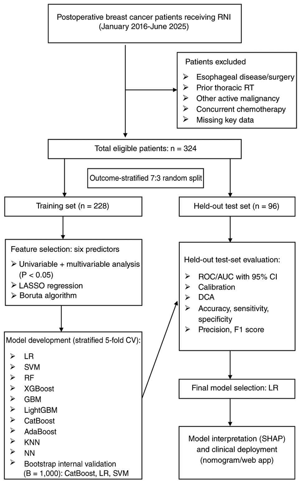 | Figure 1.Study flowchart for development and
evaluation of the prediction model for grade ≥2 acute radiation
esophagitis. Eligible patients were divided by outcome-stratified
7:3 random sampling into a training set (n=228) and a held-out test
set (n=96). Feature selection, five-fold CV and bootstrap internal
validation were performed in the model-development workflow.
Held-out performance was assessed using discrimination,
calibration, threshold-dependent classification metrics and DCA.
RNI, regional nodal irradiation; CV, cross-validation; DCA;
decision curve analysis; RT, radiotherapy; LASSO, least absolute
shrinkage and selection operator; ROC, receiver operating
characteristic; AUC, area under the curve; LR, logistic regression;
SVM, support vector machine; GBM, gradient boosting machine; NN,
neural network; RF, random forest; XGBoost, extreme gradient
boosting; KNN, K-Nearest Neighbors; LightGBM, light gradient
boosting machine; CatBoost, categorical boosting; SHAP, SHapley
Additive exPlanations. |
In accordance with TRIPOD+AI (21), baseline characteristics are reported
for both the training and held-out test sets (Table I). The two subsets were generally
comparable for the majority of the variables. However, internal
mammary node irradiation and several low-dose esophageal
dose-volume histogram (DVH) parameters, including RV_V10, RV_V15,
AV_V10 and AV_V15, exhibited significant imbalances. None of these
imbalanced variables was retained among the six consensus
predictors.
 | Table I.Baseline characteristics of the
training and held-out test sets. |
Table I.
Baseline characteristics of the
training and held-out test sets.
|
Characteristics | Training
(n=228) | Held-out test set
(n=96) | P-value |
|---|
| Hepatic
dysfunction |
|
| 0.617 |
| No | 198 (86.84) | 86 (89.58) |
|
|
Yes | 30 (13.16) | 10 (10.42) |
|
| Pruritus |
|
| 0.200 |
| No | 154 (67.54) | 57 (59.38) |
|
|
Yes | 74 (32.46) | 39 (40.62) |
|
| Hypertension |
|
| >0.999 |
| No | 191 (83.77) | 81 (84.38) |
|
|
Yes | 37 (16.23) | 15 (15.62) |
|
| Diabetes |
|
| >0.999 |
| No | 210 (92.11) | 89 (92.71) |
|
|
Yes | 18 (7.89) | 7 (7.29) |
|
| Family history |
|
| 0.919 |
| No | 223 (97.81) | 93 (96.88) |
|
|
Yes | 5 (2.19) | 3 (3.12) |
|
| Tumor
laterality |
|
| 0.504 |
|
Left | 132 (57.89) | 51 (53.12) |
|
|
Right | 96 (42.11) | 45 (46.88) |
|
| T stage |
|
| 0.232 |
| T1 | 49 (21.49) | 17 (17.71) |
|
| T2 | 147 (64.47) | 61 (63.54) |
|
| T3 | 22 (9.65) | 16 (16.67) |
|
| T4 | 10 (4.39) | 2 (2.08) |
|
| N stage |
|
| 0.416 |
| N0 | 21 (9.21) | 5 (5.21) |
|
| N1 | 97 (42.54) | 49 (51.04) |
|
| N2 | 56 (24.56) | 20 (20.83) |
|
| N3 | 54 (23.68) | 22 (22.92) |
|
| Lymphatic vessel
infiltration |
|
| 0.241 |
| No | 96 (42.11) | 33 (34.38) |
|
|
Yes | 132 (57.89) | 63 (65.62) |
|
| Histological
grade |
|
| 0.294 |
| Grade
1–2 | 151 (66.23) | 70 (72.92) |
|
| Grade
3 | 77 (33.77) | 26 (27.08) |
|
| Molecular type |
|
| 0.502 |
| Luminal
A | 26 (11.40) | 14 (14.58) |
|
| Luminal
B | 102 (44.74) | 48 (50.00) |
|
| HER-2
positive | 55 (24.12) | 17 (17.71) |
|
| Triple
negative | 45 (19.74) | 17 (17.71) |
|
| Surgical
modality |
|
| 0.776 |
|
Modified radical
mastectomy | 210 (92.11) | 90 (93.75) |
|
|
Breast-conserving surgery | 18 (7.89) | 6 (6.25) |
|
| Axillary lymph node
dissection |
|
| 0.827 |
| No | 10 (4.39) | 3 (3.12) |
|
|
Yes | 218 (95.61) | 93 (96.88) |
|
| Targeted
therapy |
|
| 0.598 |
| No | 153 (67.11) | 68 (70.83) |
|
|
Yes | 75 (32.89) | 28 (29.17) |
|
| Endocrine
therapy |
|
| 0.689 |
| No | 83 (36.40) | 32 (33.33) |
|
|
Yes | 145 (63.60) | 64 (66.67) |
|
| Chemotherapy
timing |
|
| 0.430 |
|
Adjuvant chemotherapy | 136 (59.65) | 52 (54.17) |
|
|
Neoadjuvant therapy | 92 (40.35) | 44 (45.83) |
|
| Tumor bed
boost |
|
| 0.881 |
| No | 211 (92.54) | 90 (93.75) |
|
|
Yes | 17 (7.46) | 6 (6.25) |
|
| Internal mammary
lymph nodes irradiation |
|
| 0.009 |
| No | 223 (97.81) | 87 (90.62) |
|
|
Yes | 5 (2.19) | 9 (9.38) |
|
| Radiotherapy
technique |
|
| 0.359 |
| Hybrid
IMRT and electron | 81 (35.53) | 40 (41.67) |
|
|
IMRT | 147 (64.47) | 56 (58.33) |
|
| Age, years | 54.21 (9.81) | 53.34 (9.41) | 0.461 |
| Body mass index,
kg/m2 | 24.16 (3.48) | 24.50 (3.33) | 0.411 |
| Lymph nodes, n
removed | 13.09 (7.31) | 12.89 (6.69) | 0.812 |
| Positive lymph
nodes, n | 4.57 (6.64) | 4.39 (4.98) | 0.802 |
| Neutrophil,
×109/l | 3.20 (1.51) | 2.97 (1.42) | 0.207 |
| Lymphocyte,
×109/l | 1.28 (0.49) | 1.27 (0.47) | 0.830 |
| Monocyte,
×109/l | 0.38 (0.22) | 0.36 (0.15) | 0.465 |
| Platelet,
×109/l | 192.66 | 186.44 (63.12) | 0.412 |
|
| (62.01) | (62.01) |
|
| Albumin, g/l | 44.19 (3.66) | 43.61 (3.43) | 0.180 |
|
Neutrophil-to-lymphocyte ratio | 2.74 (1.42) | 2.62 (1.65) | 0.516 |
|
Platelet-to-lymphocyte ratio | 168.50 (78.72) | 168.92 (88.75) | 0.966 |
|
Lymphocyte-to-monocyte ratio | 4.31 (6.12) | 4.66 (5.16) | 0.624 |
| Systemic
inflammation response index | 1.09 (1.01) | 0.99 (0.93) | 0.441 |
| Systemic
immune-inflammation index | 526.13
(302.12) | 500.42
(360.49) | 0.510 |
| Total dose, Gy | 50.34 (3.06) | 50.24 (2.61) | 0.761 |
| Radiotherapy days,
days | 24.61 (2.77) | 24.61 (2.30) | 0.999 |
| Esophageal volume,
cm3 | 12.50 (6.39) | 12.43 (6.30) | 0.931 |
| Maximum esophageal
dose, Gy | 49.87 (6.80) | 49.98 (6.94) | 0.892 |
| Mean esophageal
dose, Gy | 16.22 (7.40) | 17.40 (7.46) | 0.190 |
| RV_V10, % | 46.79 (21.74) | 53.33 (24.52) | 0.018 |
| RV_V15, % | 39.42 (19.97) | 44.76 (22.18) | 0.034 |
| RV_V20, % | 33.89 (18.77) | 37.63 (20.29) | 0.111 |
| RV_V25, % | 29.03 (17.38) | 31.36 (18.89) | 0.284 |
| RV_V30, % | 24.14 (16.36) | 25.82 (17.87) | 0.413 |
| RV_V35, % | 18.93 (15.17) | 19.77 (15.59) | 0.651 |
| RV_V40, % | 13.95 (13.35) | 14.36 (13.33) | 0.798 |
| AV_V10,
cm3 | 5.70 (3.94) | 6.74 (5.12) | 0.049 |
| AV_V15,
cm3 | 4.69 (2.97) | 5.51 (3.79) | 0.038 |
| AV_V20,
cm3 | 3.96 (2.51) | 4.46 (2.76) | 0.109 |
| AV_V25,
cm3 | 3.33 (2.19) | 3.59 (2.18) | 0.335 |
| AV_V30,
cm3 | 2.73 (1.96) | 2.88 (1.93) | 0.530 |
| AV_V35,
cm3 | 2.10 (1.69) | 2.18 (1.69) | 0.708 |
| AV_V40,
cm3 | 1.55 (1.46) | 1.56 (1.40) | 0.948 |
Feature selection and predictor
identification
In total, three feature-selection approaches were
applied in the training set: Univariable analysis followed by
multivariable logistic regression, LASSO with 10-fold
cross-validation (λ1SE=0.069) and Boruta. The corresponding
reproducibility settings are described in the aforementioned
section in materials and methods.
Multivariable LR was used for association screening
in the training set; the resulting coefficients were interpreted as
predictive associations rather than causal effects. LASSO retained
five variables with non-zero coefficients: Pre-existing pruritus,
tumor laterality, histological grade, BMI and diabetes. Boruta
retained 10 features: Pre-existing pruritus, tumor laterality,
histological grade, BMI, RV_V25, RV_V30, RV_V40, AV_V30, AV_V35 and
AV_V40. The results of univariable and multivariable LR analyses
for the candidate predictors are presented in Table II.
| Table II.Univariable and multivariable
logistic regression analyses of candidate predictors in the
training set. |
Table II.
Univariable and multivariable
logistic regression analyses of candidate predictors in the
training set.
| Variable | No (n=146) | Yes (n=82) | OR
(univariable) | OR
(multivariable) |
|---|
| Hepatic
dysfunction |
|
|
|
|
| No | 127 (87.0%) | 71 (86.6%) |
|
|
|
Yes | 19 (13.0%) | 11 (13.4%) | 1.04 (0.47–2.30;
P=0.931) |
|
| Pruritus |
|
|
|
|
| No | 107 (73.3%) | 47 (57.3%) |
|
|
|
Yes | 39 (26.7%) | 35 (42.7%) | 2.04 (1.15–3.62;
P=0.014) | 2.02 (1.03–3.98;
P=0.042) |
| Hypertension |
|
|
|
|
| No | 123 (84.2%) | 68 (82.9%) |
|
|
|
Yes | 23 (15.8%) | 14 (17.1%) | 1.10 (0.53–2.28;
P=0.795) |
|
| Diabetes |
|
|
|
|
| No | 140 (95.9) | 70 (85.4%) |
|
|
|
Yes | 6 (4.1 %) | 12 (14.6%) | 4.00 (1.44–11.11;
P=0.008) | 3.80 (1.14–12.61;
P=0.029) |
| Family history |
|
|
|
|
| No | 142 (97.3%) | 81 (98.8%) |
|
|
|
Yes | 4 (2.7%) | 1 (1.2%) | 0.44 (0.05–3.99;
P=0.464) |
|
| Tumor
laterality |
|
|
|
|
|
Left | 73 (50.0%) | 59 (72.0%) |
|
|
|
Right | 73 (50.0%) | 23 (28.0%) | 0.39 (0.22–0.70;
P=0.002) | 0.64 (0.31–1.32;
P=0.228) |
| T stage |
|
|
|
|
| T1 | 33 (22.6%) | 16 (19.5%) |
|
|
| T2 | 94 (64.4%) | 53 (64.6%) | 1.16 (0.59–2.31;
P=0.666) |
|
| T3 | 14 (9.6%) | 8 (9.8%) | 1.18 (0.41–3.38;
P=0.760) |
|
| T4 | 5 (3.4%) | 5 (6.1%) | 2.06 (0.52–8.16;
P=0.302) |
|
| N stage |
|
|
|
|
| N0 | 15 (10.3%) | 6 (7.3%) |
|
|
| N1 | 65 (44.5%) | 32 (39.0%) | 1.23 (0.44–3.47;
P=0.695) |
|
| N2 | 35 (24%) | 21 (25.6%) | 1.50 (0.50–4.46;
P=0.466) |
|
| N3 | 31 (21.2%) | 23 (28.0%) | 1.85 (0.62–5.51;
P=0.267) |
|
| Lymphatic vessel
infiltration |
|
|
|
|
| No | 66 (45.2%) | 30 (36.6%) |
|
|
|
Yes | 80 (54.8%) | 52 (63.4%) | 1.43 (0.82–2.49;
P=0.207) |
|
| Histological
grade |
|
|
|
|
| Grade
1–2 | 111 (76.0%) | 40 (48.8%) |
|
|
| Grade
3 | 35 (24.0%) | 42 (51.2%) | 3.33 (1.87–5.92;
P<0.001) | 3.49 (1.79–6.83;
P<0.001) |
| Molecular type |
|
|
|
|
| Luminal
A | 19 (13.0%) | 7 (8.5%) |
|
|
| Luminal
B | 60 (41.1%) | 42 (51.2%) | 1.90 (0.73–4.92;
P=0.186) |
|
| HER-2
positive | 39 (26.7%) | 16 (19.5%) | 1.11 (0.39–3.16;
P=0.840) |
|
| Triple
negative | 28 (19.2%) | 17 (20.7%) | 1.65 (0.57–4.74;
P=0.354) |
|
| Surgical
modality |
|
|
|
|
|
Modified radical
mastectomy | 135 (92.5%) | 75 (91.5%) |
|
|
|
Breast-conserving surgery | 11 (7.5%) | 7 (8.5%) | 1.15 (0.43–3.08;
P=0.788) |
|
| Axillary lymph node
dissection |
|
|
|
|
| No | 6 (4.1 %) | 4 (4.9%) |
|
|
|
Yes | 140 (95.9%) | 78 (95.1%) | 0.84 (0.23–3.05;
P=0.786) |
|
| Targeted
therapy |
|
|
|
|
| No | 93 (63.7%) | 60 (73.2%) |
|
|
|
Yes | 53 (36.3%) | 22 (26.8%) | 0.64 (0.36–1.16;
P=0.145) |
|
| Endocrine
therapy |
|
|
|
|
| No | 56 (38.4%) | 27 (32.9%) |
|
|
|
Yes | 90 (61.6%) | 55 (67.1%) | 1.27 (0.72–2.24;
P=0.414) |
|
| Chemotherapy
timing |
|
|
|
|
|
Adjuvant chemotherapy | 83 (56.8%) | 53 (64.6%) |
|
|
|
Neoadjuvant therapy | 63 (43.2)% | 29 (35.4%) | 0.72 (0.41–1.26;
P=0.251) |
|
| Tumor bed
boost |
|
|
|
|
| No | 136 (93.2%) | 75 (91.5%) |
|
|
|
Yes | 10 (6.8%) | 7 (8.5%) | 1.27 (0.46–3.47;
P=0.642) |
|
| Internal mammary
lymph node irradiation |
|
|
|
|
| No | 143 (97.9%) | 80 (97.6) |
|
|
|
Yes | 3 (2.1%) | 2 (2.4%) | 1.19 (0.20–7.28;
P=0.849) |
|
| Radiotherapy
technique |
|
|
|
|
| Hybrid
IMRT and electron | 49 (33.6%) | 32 (39%) |
|
|
|
IMRT | 97 (66.4%) | 50 (61%) | 0.79 (0.45–1.38;
P=0.409) |
|
| Age, years | 53.6±9.9 | 55.2±9.6 | 1.02 (0.99–1.05;
P=0.241) |
|
| BMI, kg/m2 | 23.7±3.5 | 24.9±3.3 | 1.10 (1.02–1.19;
P=0.017) | 1.12 (1.02–1.23;
P=0.014) |
| Lymph nodes
removed, n | 13.2±8.1 | 12.9±5.7 | 0.99 (0.96–1.03;
P=0.769) |
|
| Positive lymph
nodes, n | 4.4±7.2 | 4.8±5.5 | 1.01 (0.97–1.05;
P=0.649) |
|
| Neutrophil,
×109/l | 3.1±1.3 | 3.3±1.9 | 1.10 (0.92–1.31;
P=0.307) |
|
| Lymphocyte,
×109/l | 1.3±0.5 | 1.3±0.5 | 1.11 (0.64–1.92;
P=0.720) |
|
| Monocyte,
×109/l | 0.4±0.2 | 0.4±0.2 | 0.52 (0.13–2.10;
P=0.362) |
|
| Platelet,
×109/l | 195.3±62.4 | 187.9±61.4 | 1.00 (0.99–1.00;
P=0.387) |
|
| Albumin, g/l | 44.3±3.9 | 44.0±3.3 | 0.97 (0.90–1.05;
P=0.506) |
|
|
Neutrophil-to-lymphocyte ratio | 2.7±1.4 | 2.7±1.4 | 0.99 (0.82–1.20;
P=0.939) |
|
|
Platelet-to-lymphocyte ratio | 173.1±81.5 | 160.3±73.2 | 1.00 (0.99–1.00;
P=0.242) |
|
|
Lymphocyte-to-monocyte ratio | 3.9±1.9 | 5.1±9.9 | 1.05 (0.95–1.17;
P=0.352) |
|
| Systemic
inflammation response index | 1.1±0.9 | 1.1±1.2 | 1.01 (0.78–1.32;
P=0.929) |
|
| Systemic
immune-inflammation index | 532.8±304.6 | 514.3±299.1 | 1.00 (1.00–1.00;
P=0.658) |
|
| Total dose, Gy | 50.3±3.0 | 50.4±3.1 | 1.02 (0.93–1.11;
P=0.712) |
|
| Radiotherapy days,
days | 24.5±2.9 | 24.9±2.5 | 1.05 (0.95–1.17;
P=0.330) |
|
| Esophageal volume,
cm3 | 12.8±6.8 | 12.0±5.5 | 0.98 (0.94–1.03;
P=0.401) |
|
| Maximum esophageal
dose, Gy | 49.0±7.1 | 51.4±5.9 | 1.06 (1.01–1.11;
P=0.014) | 1.07 (0.99–1.15;
P=0.074) |
| Mean esophageal
dose, Gy | 15.3±7.2 | 17.9±7.5 | 1.05 (1.01–1.09;
P=0.013) | 0.91 (0.75–1.11;
P=0.367) |
| RV_V10, % | 45.4±21.1 | 49.2±22.7 | 1.01 (1.00–1.02;
P=0.209) |
|
| RV_V15, % | 37.3±18.9 | 43.2±21.4 | 1.01 (1.00–1.03;
P=0.034) | 1.01 (0.92–1.11;
P=0.812) |
| RV_V20, % | 31.5±17.9 | 38.2±19.6 | 1.02 (1.00–1.03;
P=0.011) | 1.04 (0.89–1.22;
P=0.584) |
| RV_V25, % | 26.6±16.9 | 33.4±17.5 | 1.02 (1.01–1.04;
P=0.005) | 0.92 (0.75–1.13;
P=0.423) |
| RV_V30, % | 21.7±16.1 | 28.5±16.0 | 1.03 (1.01–1.04;
P=0.003) | 1.36 (1.06–1.74;
P=0.016) |
| RV_V35, % | 16.8±14.7 | 22.7±15.4 | 1.03 (1.01–1.04;
P=0.006) | 0.73 (0.55–0.96;
P=0.024) |
| RV_V40, % | 12.2±12.5 | 17.1±14.2 | 1.03 (1.01–1.05;
P=0.008) | 1.09 (0.91–1.29;
P=0.343) |
| AV_V10,
cm3 | 5.7±4.3 | 5.6±3.3 | 0.99 (0.93–1.07;
P=0.859) |
|
| AV_V15,
cm3 | 4.6±3.1 | 4.9±2.7 | 1.04 (0.95–1.13;
P=0.444) |
|
| AV_V20,
cm3 | 3.8±2.6 | 4.3±2.3 | 1.08 (0.97–1.20;
P=0.172) |
|
| AV_V25,
cm3 | 3.1±2.3 | 3.7±1.9 | 1.12 (0.99–1.27;
P=0.077) |
|
| AV_V30,
cm3 | 2.5±2.1 | 3.1±1.7 | 1.17 (1.02–1.35;
P=0.028) | 0.29 (0.07–1.19;
P=0.086) |
| AV_V35,
cm3 | 1.9±1.7 | 2.5±1.7 | 1.23 (1.05–1.44;
P=0.013) | 3.93 (0.29–52.43;
P=0.300) |
| AV_V40,
cm3 | 1.4±1.4 | 1.9±1.5 | 1.27 (1.06–1.53;
P=0.012) | 0.90 (0.15–5.59;
P=0.914) |
After integrating the three selection approaches and
accounting for collinearity among related DVH and inflammatory
variables, six consensus predictors were retained: Pre-existing
pruritus, histological grade, BMI, diabetes, tumor laterality and
esophageal RV_V30. In the present study, RV_V30 denotes the
percentage of the contoured esophageal volume receiving ≥30 Gy; it
emerged from the multi-strategy feature-selection process among the
candidate DVH metrics. The selection process is illustrated in
Fig. 2, and univariable
discrimination and correlation analyses are provided in Fig. S3.
Model development and performance
evaluation
Using the six consensus predictors, 10 ML models
were developed. The models implemented in caret were evaluated
using the same outcome-stratified five-fold cross-validation folds
within the training set. Based on internal performance, stability
considerations and parsimony, CatBoost, LR and SVM were carried
forward for 1,000-resample bootstrap internal validation.
The bootstrap internal-validation results are
presented in Table III. The
optimism-corrected AUC values were 0.804 for CatBoost, 0.744 for
SVM and 0.715 for logistic regression. The corresponding mean
optimism values were 0.113, 0.089 and 0.027, respectively. Thus, LR
exhibited substantially lower bootstrap optimism than the two more
complex pre-selected models, although its optimism-corrected AUC
was not the highest.
| Table III.Bootstrap internal validation
(B=1,000) for the three pre-selected models in the training
cohort. |
Table III.
Bootstrap internal validation
(B=1,000) for the three pre-selected models in the training
cohort.
| Model | Apparent AUC | Mean optimism
(SD) | Optimism-corrected
AUC (95% CI) | 95% percentile
interval for bootstrap-refit AUC on original training set |
|---|
| Categorical
boosting | 0.917 | 0.113 (0.018) | 0.804
(0.764–0.838) | 0.822–0.886 |
| Support vector
machine | 0.833 | 0.089 (0.025) | 0.744
(0.698–0.795) | 0.745–0.815 |
| Logistic
regression | 0.741 | 0.027 (0.034) | 0.715
(0.650–0.778) | 0.708–0.741 |
Threshold-dependent held-out test-set metrics are
summarized in Table IV. Using the
threshold determined in the training set (0.396), LR achieved an
accuracy of 0.729, sensitivity of 0.735, a specificity of 0.726, a
precision of 0.595 and a F1 score of 0.658. Performance profiles
varied across the other algorithms, and no single model was
uniformly superior across all threshold-dependent metrics.
| Table IV.Threshold-dependent classification
metrics for the 10 machine-learning models in the held-out test
set. |
Table IV.
Threshold-dependent classification
metrics for the 10 machine-learning models in the held-out test
set.
| Model | Threshold | Accuracy | Sensitivity | Specificity | Precision | F1 score |
|---|
| Logistic
regression | 0.396 | 0.729 | 0.735 | 0.726 | 0.595 | 0.658 |
| Support vector
machine | 0.331 | 0.729 | 0.794 | 0.694 | 0.587 | 0.675 |
| Gradient boosting
machine | 0.346 | 0.688 | 0.676 | 0.694 | 0.548 | 0.605 |
| Neural network | 0.386 | 0.677 | 0.735 | 0.645 | 0.532 | 0.617 |
| Random forest | 0.301 | 0.708 | 0.794 | 0.661 | 0.562 | 0.659 |
| Extreme gradient
boosting | 0.370 | 0.719 | 0.794 | 0.677 | 0.574 | 0.667 |
| K-nearest
neighbors | 0.441 | 0.646 | 0.559 | 0.694 | 0.500 | 0.528 |
| AdaBoost | 0.437 | 0.719 | 0.765 | 0.694 | 0.578 | 0.658 |
| Light gradient
boosting machine | 0.366 | 0.719 | 0.824 | 0.661 | 0.571 | 0.675 |
| Categorical
boosting | 0.361 | 0.719 | 0.853 | 0.645 | 0.569 | 0.682 |
The discrimination and calibration results for all
10 models are reported in Table V.
LR achieved the highest held-out AUC value of 0.797 (95% CI,
0.706–0.889), followed by SVM (0.791; 95% CI, 0.699–0.883) and RF
(0.786; 95% CI, 0.693–0.878). CatBoost achieved a held-out AUC
value of 0.762 (95% CI, 0.659–0.865), as shown in Fig. 3A. In the final 10-model comparison,
CatBoost exhibited a training-cohort AUC of 0.923 (95% CI,
0.887–0.959; Fig. S4A). The
held-out set was inspected during candidate-model evaluation and
served as the early-stopping monitor for LightGBM. Accordingly,
these estimates are internal test-set results and should not be
interpreted as independent validation performance. For LR, the
Brier score was 0.179, the calibration slope was 1.258 and CITL was
−0.056. These values indicate generally acceptable calibration with
a modest deviation from the ideal slope of 1. Decision curve
analysis revealed that LR provided greater net benefit than both
treat-all and treat-none strategies across threshold probabilities
of 10–66% (Fig. 3B). Considering
bootstrap stability, held-out discrimination and calibration,
parsimony and clinical interpretability, LR was selected as the
final model. The test-set receiver operating characteristic,
decision-curve and calibration results are illustrated in Fig. 3, and the corresponding
training-cohort plots are provided in Fig. S4.
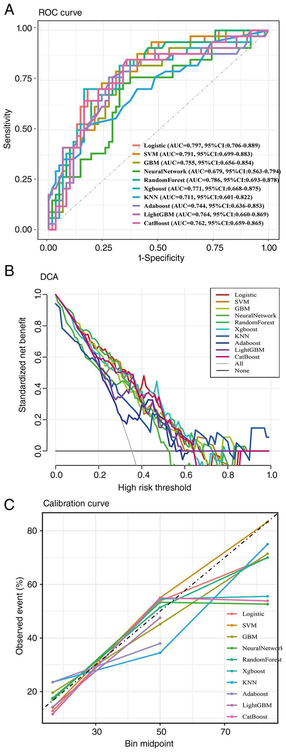 | Figure 3.Performance of the prediction models
in the held-out test set. (A) ROC curves for the 10
machine-learning algorithms; LR achieved an AUC of 0.797 (95% CI,
0.706–0.889). CatBoost achieved an AUC of 0.762 (95% CI,
0.659–0.865). (B) DCA calculated from the established predicted
risks. LR showed greater net benefit than treat-all and treat-none
strategies across threshold probabilities of 10–66%. (C)
Calibration curves for the 10 models. For LR, the Brier score was
0.179, calibration slope was 1.258, and calibration-in-the-large
was −0.056. ROC, receiver operating characteristic; AUC, area under
the curve; CI, confidence interval; LR, logistic regression; DCA,
decision curve analysis; LR, logistic regression; SVM, support
vector machine; GBM, gradient boosting machine; XGBoost, extreme
gradient boosting; KNN, K-Nearest Neighbors; LightGBM, light
gradient boosting machine; CatBoost, categorical boosting; CI,
confidence interval. |
| Table V.Discrimination and calibration
performance of the 10 machine-learning models in the held-out test
set. |
Table V.
Discrimination and calibration
performance of the 10 machine-learning models in the held-out test
set.
| Model | AUC (95% CI,
DeLong) | Brier score | Calibration
slope | CITL |
|---|
| Logistic
regression | 0.797
(0.706–0.889) | 0.179 | 1.258 | −0.056 |
| Support vector
machine | 0.791
(0.699–0.883) | 0.188 | 1.530 | −0.114 |
| Random forest | 0.786
(0.693–0.878) | 0.181 | 0.936 | 0.153 |
| Extreme gradient
boosting | 0.771
(0.668–0.875) | 0.184 | 1.044 | −0.090 |
| Light gradient
boosting machine | 0.764
(0.660–0.869) | 0.199 | 2.773 | −0.068 |
| Categorical
boosting | 0.762
(0.659–0.865) | 0.189 | 0.894 | −0.184 |
| Gradient boosting
machine | 0.755
(0.656–0.854) | 0.189 | 0.898 | −0.021 |
| AdaBoost | 0.744
(0.636–0.853) | 0.210 | 1.676 | −0.285 |
| K-nearest
neighbors | 0.711
(0.601–0.822) | 0.211 | 0.165 | 0.086 |
| Neural network | 0.679
(0.563–0.794) | 0.232 | 0.216 | −0.313 |
Model interpretation
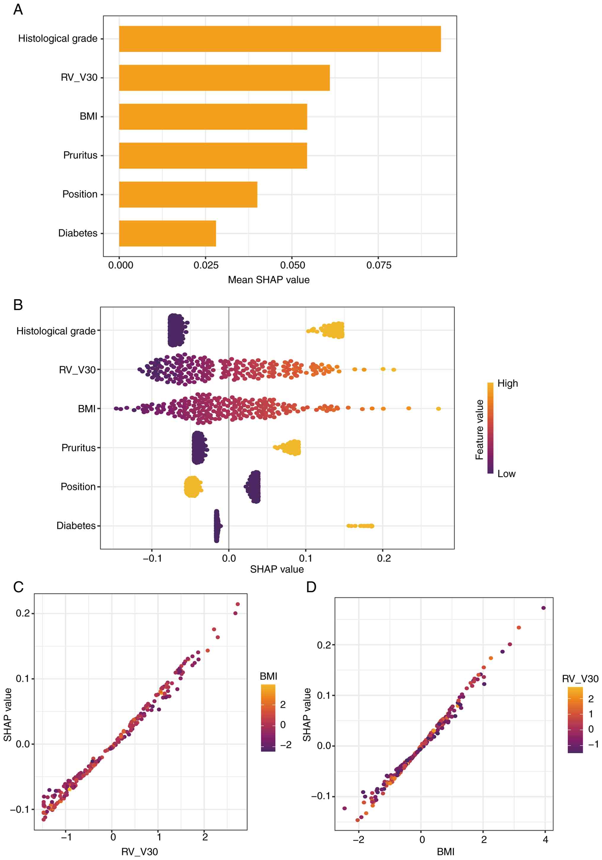
SHAP analysis was used to interpret the final LR
model (Fig. 4A-D). Histological
grade exhibited the highest global importance, followed by RV_V30
and BMI; pre-existing pruritus, tumor laterality and diabetes also
contributed to model predictions (Fig.
4A). The beeswarm plot exhibited positive SHAP contributions
for a higher histological grade, higher RV_V30 and a higher BMI
(Fig. 4B). Left-sided laterality
also tended to contribute toward a higher predicted risk, whereas
the encoded higher value for laterality (right) tended to
contribute negatively. Dependence plots for RV_V30 and BMI further
illustrated the direction and distribution of these continuous
feature effects (Fig. 4C and
D).
Visual nomogram and clinical
deployment
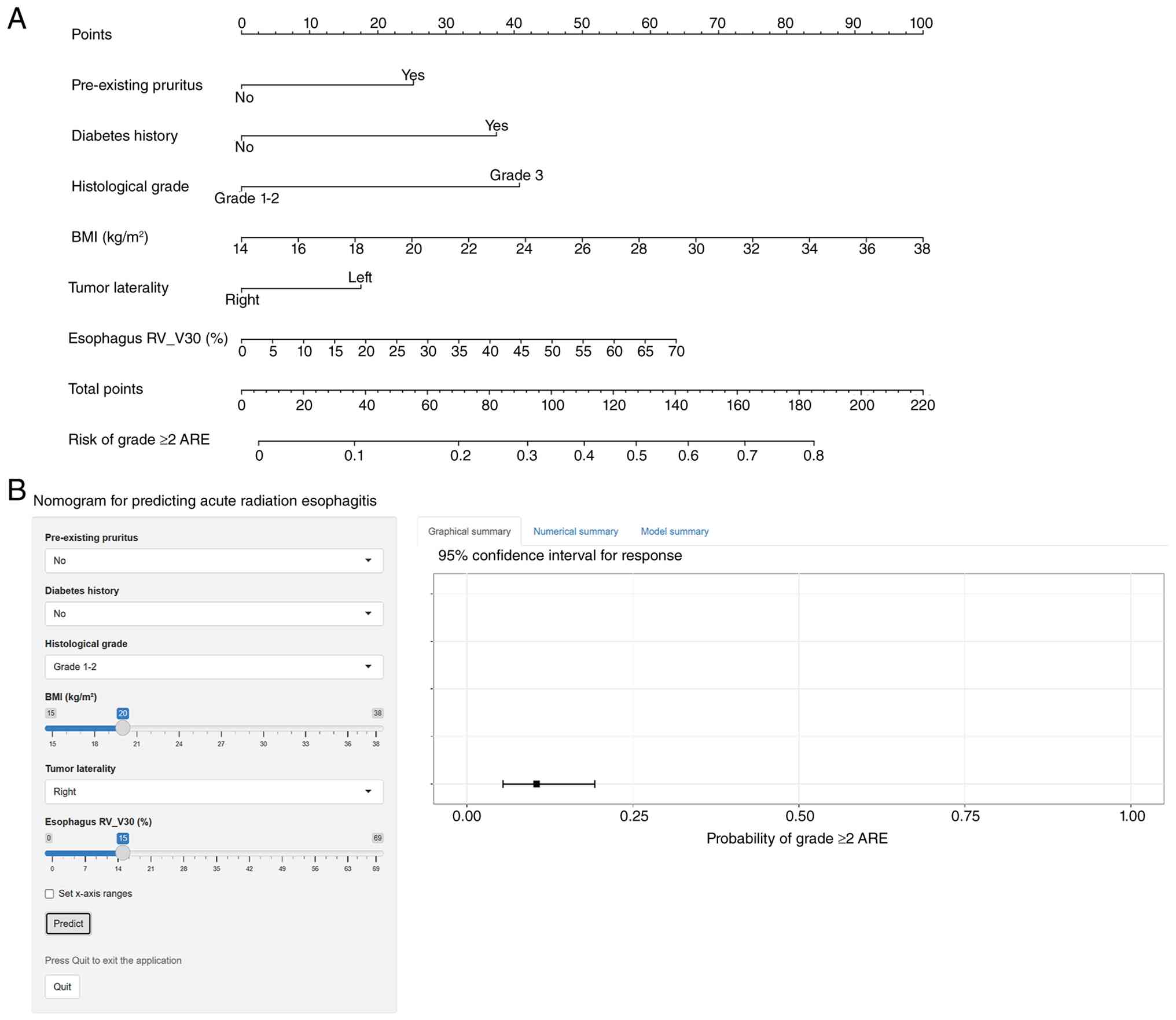
To support clinical interpretation, the final LR
model was presented as a static nomogram (Fig. 5A). The nomogram incorporates the six
consensus predictors: Pre-existing pruritus, histological grade,
BMI, diabetes, tumor laterality and esophageal RV_V30. Each
predictor contributes points according to its regression
coefficient, and the summed score is mapped to the predicted
probability of grade ≥2 ARE.
An open-access web-based calculator was also
developed for point-of-care risk estimation (Fig. 5B). The calculator uses the same
final LR model as the static nomogram. A numerical consistency
check confirmed concordant predicted probabilities between the
nomogram implementation and the final LR model (correlation=1.000;
maximum absolute difference, 1.16×10⁻⁸). The web
application is accessible at: https://hebin7290.shinyapps.io/Radiation-esophagitis/.
As an illustrative example, a hypothetical patient with
pre-existing pruritus, grade 3 histology, BMI 25 kg/m2,
no diabetes, a left-sided tumor and RV_V30 of 20% would have a
predicted probability of grade ≥2 ARE of ~0.62.
Discussion
The present study developed an interpretable
clinical-dosimetric model for grade ≥2 ARE following RNI in
patients with breast cancer. A total of six predictors captured
baseline symptoms, tumor characteristics, metabolic status, anatomy
and esophageal dose exposure. Bootstrap internal validation
revealed that the more flexible pre-selected models had greater
optimism, whereas LR had the lowest mean optimism. Final model
selection therefore considered stability together with held-out
discrimination and calibration, model complexity and
interpretability rather than relying on a single performance
metric. The held-out AUC value of LR was 0.797, with a Brier score
of 0.179 and modest calibration deviation. These findings support
LR as a clinically transparent candidate for further external
validation. The selected variables should be interpreted as
predictive markers rather than causal determinants of ARE.
Histological grade exhibited the strongest global
contribution in the SHAP analysis and was associated with the risk
of developing ARE in the predictive model. This association should
not be interpreted as evidence that tumor grade directly alters
esophageal radiosensitivity. High-grade breast cancers can exhibit
hypoxia-related and inflammatory phenotypes (26), and hypoxia and inflammatory
signaling can interact through pathways, including
hypoxia-inducible factor and NF-κB (27). Systemic inflammatory indices have
also been evaluated in radiation-esophagitis prediction in other
thoracic malignancies (28–30). These observations provide a possible
biological context; however, any association between tumor grade
and host normal-tissue susceptibility remains hypothesis-generating
and requires dedicated biological and prospective validation.
RV_V30 was retained as the principal dosimetric
predictor, supporting the relevance of intermediate-dose esophageal
exposure. Prospective evidence in breast RNI has also identified
clinically relevant esophageal dose-volume constraints. For
example, Wang et al (11)
reported RV_V30 and AV_V30 constraints in conventionally
fractionated RNI, whereas Ji et al (9) reported associations for AV30 and AV35
in a smaller cohort. In the present study, RV_V30 was selected from
a broader candidate set that included Dmean, Dmax and multiple
relative and absolute dose-volume metrics. As RV_V30 is modifiable
during planning, reducing intermediate-dose esophageal exposure
while maintaining target coverage is a plausible optimization
strategy. Since no universal RV_V30 cut-off was derived in the
present study, this finding should be interpreted as supporting
dose minimization rather than a fixed planning constraint.
BMI and diabetes were both retained in the final
prediction model. Their contributions may reflect aspects of
metabolic status, inflammation, vascular health or treatment
geometry rather than a single causal pathway. Obesity is associated
with chronic inflammation and oxidative stress (31), and diabetes can impair microvascular
function (32). A higher BMI can
also affect radiotherapy geometry and planning complexity (33). These mechanisms provide plausible
context for the observed predictive associations but were not
directly measured in the present study.
Pre-existing pruritus was retained as a predictive
marker. By contrast to a formal patient-reported outcome measure,
pruritus was retrospectively documented as a binary present/absent
variable before radiotherapy. Previous research has linked
normal-tissue reactions to inter-individual susceptibility
(34), while taxane-related skin
toxicity and neuroimmune mechanisms may contribute to persistent
pruritus (35–37). A recent breast-radiotherapy study
also identified pruritus as a contributor to esophagitis prediction
using SHAP (38). Although these
observations support further investigation, they do not establish
pruritus as a mechanistic marker of radiosensitivity.
A possible explanation is that residual
treatment-related symptoms may reflect a host state that also
modifies tolerance to subsequent irradiation. Taxane-related
cutaneous toxicity and radiation-induced mucosal injury involve
inflammatory and epithelial-injury pathways (35,39–41);
however, the data of the present study do not establish a shared
mechanism or a causal association between pre-existing pruritus and
esophageal radiosensitivity. This interpretation is therefore
hypothesis-generating and requires prospective validation. The
binary ascertainment used in the present retrospective cohort also
did not capture pruritus severity, duration or patient-reported
burden. Future studies using standardized grading and
patient-reported assessment rather than a simple present/absent
variable are warranted.
Tumor laterality was retained as a model predictor.
In the current coding, right was the higher numeric category and
had a negative model coefficient relative to left, consistent with
a greater contribution from left-sided laterality in the SHAP
analysis. This association may partly reflect treatment geometry,
particularly when internal mammary nodal irradiation places beam
paths closer to the esophagus. Techniques, such as deep-inspiration
breath-hold can modify organ-at-risk geometry and dose exposure
(42). Laterality should therefore
be regarded as an anatomical prediction marker rather than a causal
determinant of ARE.
Published breast cancer RNI studies have largely
emphasized dosimetric predictors, with heterogeneous sample sizes,
fractionation schedules and validation strategies. Calibration and
decision-analytic measures have not been reported consistently,
which can make apparent model performance difficult to compare
across cohorts.
Yaney et al (8) analyzed 531 conventionally fractionated
RNI cases and proposed practical dose cut-points, including mean
esophageal dose, V10 and V20. Wang et al (43) prospectively evaluated esophageal
constraints in hypofractionated RNI and reported moderate
discrimination for parsimonious models. Ji et al (9) examined a smaller hypofractionated
cohort and reported high apparent discrimination for AV30 and AV35.
These studies illustrate both the clinical relevance of esophageal
dosimetry and the sensitivity of reported performance to cohort
size and validation design.
The results of the present study add a mixed
clinical-dosimetric model that incorporates RV_V30, BMI, diabetes,
laterality and a baseline symptom. The comparison of multiple
algorithms also illustrates the potential for optimism in flexible
learners when the sample size is modest. CatBoost, for example, had
a high apparent AUC, but substantially greater bootstrap optimism
than LR. A similar observation has been reported in
radiotherapy-toxicity modeling outside the breast-RNI setting: In
an externally validated head-and-neck dysphagia study, penalized LR
using dose-volume metrics performed as well as support vector and
RF models (external-validation AUC of 0.82), demonstrating that
additional model complexity does not necessarily improve
transportable performance (44).
This does not imply that ensemble methods are intrinsically
inferior; rather, higher-capacity models may be more vulnerable to
optimism and calibration instability in limited datasets. Bootstrap
methods are useful for quantifying this optimism, although internal
validation does not replace external evaluation (45,46).
When held-out discrimination is similar, calibration, stability,
parsimony and interpretability are therefore key complementary
selection criteria.
Internal validation was used to identify preferred
candidates and quantify model optimism. Final model selection then
considered bootstrap-corrected performance together with held-out
test-set discrimination and calibration, model complexity and
clinical interpretability. LR was selected as it provided a
favorable balance across these criteria rather than because it was
uniformly superior on every metric. The held-out test results
should therefore be interpreted as internal held-out evaluation,
not as external validation.
The model is intended to support pre-radiotherapy
risk stratification rather than dictate treatment. Decision curve
analysis revealed positive net benefit for LR over treat-all and
treat-none strategies across threshold probabilities of 10–66%. In
practice, in the event that the predicted risk of a patient exceeds
a threshold judged clinically meaningful by the treating team, the
plan could be reviewed for opportunities to reduce RV_V30, while
maintaining target coverage, and the patient could receive closer
symptom monitoring and earlier supportive care. The static nomogram
and web-based calculator provide transparent tools for this risk
estimate (Fig. 5).
The present study has several limitations which
should be mentioned. First, its retrospective design may have
introduced selection bias through eligibility criteria,
complete-case inclusion and the availability of clinical and
dosimetric records. Second, the single-center cohort and modest
sample size may limit generalizability. Institutional differences
in esophageal contouring, RNI technique, fractionation,
treatment-planning systems, accelerators and supportive care may
alter predictor distributions and model calibration. Third,
calibration estimates from the held-out test set (n=96) remain
imprecise. The calibration slope of 1.258 and CITL of −0.056
indicate modest deviation from ideal calibration. Fourth,
pre-existing pruritus was recorded only as present or absent and
did not capture severity, duration or standardized patient-reported
burden. Fifth, the held-out set was inspected during
candidate-model evaluation and served as the early-stopping monitor
for LightGBM. It therefore provided internal test-set evaluation
rather than independent or external validation. Sixth, the present
study did not formally compare the final clinical-dosimetric model
with a clinical-only baseline model. The incremental value of
RV_V30 therefore remains to be quantified. Prospective multicenter
studies are thus required to validate the model across different
patient populations, RNI techniques, fractionation schedules,
planning systems and accelerators. Such studies should also
evaluate recalibration and compare clinical-only and
clinical-dosimetric models. This comparison should assess
discrimination, calibration and decision-curve performance to
quantify the incremental value of RV_V30.
In conclusion, an interpretable six-feature
clinical-dosimetric model was developed and internally evaluated
for grade ≥2 ARE after breast cancer RNI. The final LR model
incorporates pre-existing pruritus, histological grade, BMI,
diabetes, tumor laterality and esophageal RV_V30. It provided a
favorable balance of discrimination, calibration, stability,
parsimony and interpretability in the present cohort and was
implemented as a nomogram and web-based calculator. Prospective
multicenter external validation and, where necessary, recalibration
are required before routine clinical implementation.
Supplementary Material
Supporting Data
Supporting Data
Acknowledgements
The authors wish to thank Dr Yong Diao (Department
of Oncology, The General Hospital of Western Theater Command of the
Chinese People's Liberation Army, Chengdu, China) for sharing
clinical insights regarding patient management and toxicity
assessment in radiotherapy. Their expertise contributed to the
clinical perspective of the present study.
Funding
Funding: No funding was received.
Availability of data and materials
The data generated in the present study may be
requested from the corresponding author.
Authors' contributions
BH and ZQ contributed equally to this work. BH and
LZ conceived the study and designed the analytical framework. BH
drafted the manuscript and coordinated the project. ZQ, LL and HL
contributed to patient data collection, clinical variable
assessment and model validation. XZ and YT performed the formal
data analysis, developed the machine-learning models and software
tools, and created the visualizations. HZ contributed to the
acquisition and interpretation of clinical and radiotherapy data
and critically revised the manuscript. FK contributed to study
conception and design, interpretation of the results, critical
revision of the manuscript and supervision of the study. LZ
supervised the study and provided critical methodological and
clinical guidance. BH and ZQ confirm the authenticity of all the
raw data. All authors read and approved the final version of the
manuscript.
Ethics approval and consent to
participate
The present study was approved by the Ethics
Committee of The General Hospital of Western Theater Command
(approval no. 2025EC11-ky019) and was performed in accordance with
the ethical standards of the Declaration of Helsinki. Due to the
retrospective nature of the study, the requirement for written
informed consent was waived by the Ethics Committee of The General
Hospital of Western Theater Command.
Patient consent for publication
Not applicable.
Competing interests
The authors declare that they have no competing
interests.
Use of artificial intelligence tools
During the preparation of this work, artificial
intelligence tools were used to improve the readability and
language of the manuscript or to generate images, and subsequently,
the authors revised and edited the content produced by the
artificial intelligence tools as necessary, taking full
responsibility for the ultimate content of the present
manuscript.
References
|
1
|
Sung H, Filho AM, Laversanne M, Ferlay J,
Siegel RL, Soerjomataram I, Jemal A and Bray F: Global cancer
statistics 2024: GLOBOCAN estimates of incidence and mortality
worldwide for 34 cancers in 186 countries. CA Cancer J Clin.
76:e700902026.PubMed/NCBI
|
|
2
|
EBCTCG (Early Breast Cancer Trialists'
Collaborative Group), . McGale P, Taylor C, Correa C, Cutter D,
Duane F, Ewertz M, Gray R, Mannu G, Peto R, et al: Effect of
radiotherapy after mastectomy and axillary surgery on 10-year
recurrence and 20-year breast cancer mortality: Meta-analysis of
individual patient data for 8135 women in 22 randomised trials.
Lancet. 383:2127–2135. 2014. View Article : Google Scholar : PubMed/NCBI
|
|
3
|
Poortmans PM, Weltens C, Fortpied C,
Kirkove C, Peignaux-Casasnovas K, Budach V, van der Leij F, Vonk E,
Weidner N, Rivera S, et al: Internal mammary and medial
supraclavicular lymph node chain irradiation in stage I–III breast
cancer (EORTC 22922/10925): 15-year results of a randomised, phase
3 trial. Lancet Oncol. 21:1602–1610. 2020. View Article : Google Scholar : PubMed/NCBI
|
|
4
|
Poortmans PM, Collette S, Kirkove C, Van
Limbergen E, Budach V, Struikmans H, Collette L, Fourquet A,
Maingon P, Valli M, et al: Internal mammary and medial
supraclavicular irradiation in breast cancer. N Engl J Med.
373:317–327. 2015. View Article : Google Scholar : PubMed/NCBI
|
|
5
|
West K, Schneider M, Wright C,
Beldham-Collins R, Coburn N, Tiver K, Gebski V and Stuart KE:
Radiation-induced oesophagitis in breast cancer: Factors
influencing onset and severity for patients receiving
supraclavicular nodal irradiation. J Med Imaging Radiat Oncol.
64:113–119. 2020. View Article : Google Scholar : PubMed/NCBI
|
|
6
|
Baker S and Fairchild A: Radiation-induced
esophagitis in lung cancer. Lung Cancer (Auckl). 7:119–127.
2016.PubMed/NCBI
|
|
7
|
Wang DQ, Zhang N, Dong LH, Zhong YH, Wu
HF, Zhong QZ, Jin J, Hou XR, Jing H, Tang Y, et al: Dose-volume
predictors for radiation esophagitis in patients with breast cancer
undergoing hypofractionated regional nodal radiation therapy. Int J
Radiat Oncol Biol Phys. 117:186–197. 2023. View Article : Google Scholar : PubMed/NCBI
|
|
8
|
Yaney A, Ayan AS, Pan X, Jhawar S, Healy
E, Beyer S, Lindsey K, Kuhn K, Tedrick K, White JR and Bazan JG:
Dosimetric parameters associated with radiation-induced esophagitis
in breast cancer patients undergoing regional nodal irradiation.
Radiother Oncol. 155:167–173. 2021. View Article : Google Scholar : PubMed/NCBI
|
|
9
|
Ji MC, Li ZJ, Li K, Wang YX, Yang B, Lv
LL, Su Y, Zhang ZW, Huo ZC, Qi Q, et al: Dosimetric risk factors
for radiation esophagitis in patients with breast cancer following
regional nodal radiation. World J Clin Cases. 12:2995–3003. 2024.
View Article : Google Scholar : PubMed/NCBI
|
|
10
|
Bazan JG, Healy E, Beyer S, Kuhn K,
DiCostanzo D, Smith TL, Jhawar S and White JR: Clinical
effectiveness of an adaptive treatment planning algorithm for
intensity modulated radiation therapy versus 3D conformal radiation
therapy for node-positive breast cancer patients undergoing
regional nodal irradiation/postmastectomy radiation therapy. Int J
Radiat Oncol Biol Phys. 108:1159–1171. 2020. View Article : Google Scholar : PubMed/NCBI
|
|
11
|
Wang DQ, Shen DX, Han N, Lu YF, Yang WF,
Tie J, Hou XR, Wang XH, Zhong YH, Yu XL, et al: Dosimetric and
clinical predictors for radiation esophagitis in patients with
breast cancer undergoing conventional fractionated regional nodal
irradiation: A prospective study. Int J Cancer. 158:1055–1066.
2026. View Article : Google Scholar : PubMed/NCBI
|
|
12
|
Shreya S, Pandey S, Ghosh DK, Puneet
Prasad SB, Grosset CF and Jain BP: Analysis of endoplasmic
reticulum stress-associated proteins as prognostic markers in
breast cancer. Curr Genomics. 26:621–632. 2025. View Article : Google Scholar : PubMed/NCBI
|
|
13
|
Qiu J, Zhou T, Wang D, Hong W, Qian D,
Meng X and Liu X: Pan-cancer analysis identifies AIMP2 as a
potential biomarker for breast cancer. Curr Genomics. 24:307–329.
2023. View Article : Google Scholar : PubMed/NCBI
|
|
14
|
Wang H, Hao R, Liu W, Zhang Y, Ma S, Lu Y,
Hu J and Qi Y: Identification of transcription factors associated
with the disease-free survival of triple-negative breast cancer
through weighted gene co-expression network analysis. CytoJournal.
21:712024. View Article : Google Scholar : PubMed/NCBI
|
|
15
|
Shi Q, Xuan M, Lv C, Zhang Z, Geng X,
Huang D and Hu X: Recent applications of artificial intelligence in
cancer radiotherapy and immunotherapy: Current status and future
directions. Front Immunol. 17:17764722026. View Article : Google Scholar : PubMed/NCBI
|
|
16
|
Shamout F, Zhu T and Clifton DA: Machine
learning for clinical outcome prediction. IEEE Rev Biomed Eng.
14:116–126. 2021. View Article : Google Scholar : PubMed/NCBI
|
|
17
|
Greener JG, Kandathil SM, Moffat L and
Jones DT: A guide to machine learning for biologists. Nat Rev Mol
Cell Biol. 23:40–55. 2022. View Article : Google Scholar : PubMed/NCBI
|
|
18
|
Khene ZE, Bigot P, Doumerc N, Ouzaid I,
Boissier R, Nouhaud FX, Albiges L, Bernhard JC, Ingels A,
Borchiellini D, et al: Application of machine learning models to
predict recurrence after surgical resection of nonmetastatic renal
cell carcinoma. Eur Urol Oncol. 6:323–330. 2023. View Article : Google Scholar : PubMed/NCBI
|
|
19
|
Yu YD, Lee KS, Kim JM, Ryu JH, Lee JG, Lee
KW, Kim BW and Kim DS; Korean Organ Transplantation Registry Study
Group, : Artificial intelligence for predicting survival following
deceased donor liver transplantation: Retrospective multi-center
study. Int J Surg. 105:1068382022. View Article : Google Scholar : PubMed/NCBI
|
|
20
|
Amann J, Vetter D, Blomberg SN,
Christensen HC, Coffee M, Gerke S, Gilbert TK, Hagendorff T, Holm
S, Livne M, et al: To explain or not to explain?-Artificial
intelligence explainability in clinical decision support systems.
PLoS Digit Health. 1:e00000162022. View Article : Google Scholar : PubMed/NCBI
|
|
21
|
Collins GS, Moons KGM, Dhiman P, Riley RD,
Beam AL, Van Calster B, Ghassemi M, Liu X, Reitsma JB, van Smeden
M, et al: TRIPOD+AI statement: Updated guidance for reporting
clinical prediction models that use regression or machine learning
methods. BMJ. 385:e0783782024. View Article : Google Scholar : PubMed/NCBI
|
|
22
|
Cox JD, Stetz J and Pajak TF: Toxicity
criteria of the radiation therapy oncology group (RTOG) and the
European organization for research and treatment of cancer (EORTC).
Int J Radiat Oncol Biol Phys. 31:1341–1346. 1995. View Article : Google Scholar : PubMed/NCBI
|
|
23
|
Kursa MB and Rudnicki WR: Feature
selection with the Boruta package. J Stat Softw. 36:1–13. 2010.
View Article : Google Scholar
|
|
24
|
Engebretsen S and Bohlin J: Statistical
predictions with glmnet. Clin Epigenetics. 11:1232019. View Article : Google Scholar : PubMed/NCBI
|
|
25
|
Friedman J, Hastie T and Tibshirani R:
Regularization paths for generalized linear models via coordinate
descent. J Stat Softw. 33:1–22. 2010. View Article : Google Scholar : PubMed/NCBI
|
|
26
|
Bos R, Zhong H, Hanrahan CF, Mommers EC,
Semenza GL, Pinedo HM, Abeloff MD, Simons JW, van Diest PJ and van
der Wall E: Levels of hypoxia-inducible factor-1 alpha during
breast carcinogenesis. J Natl Cancer Inst. 93:309–314. 2001.
View Article : Google Scholar : PubMed/NCBI
|
|
27
|
D'Ignazio L, Batie M and Rocha S: Hypoxia
and inflammation in cancer, focus on HIF and NF-κB. Biomedicines.
5:212017. View Article : Google Scholar : PubMed/NCBI
|
|
28
|
Zeng X, Ye L, Luo M, Zeng D and Chen Y:
Prognostic value of pretreatment systemic immune-inflammation index
in Chinese esophageal squamous cell carcinoma patients receiving
radical radiotherapy: A meta-analysis. Medicine (Baltimore).
102:e341172023. View Article : Google Scholar : PubMed/NCBI
|
|
29
|
Yu Y, Zheng H, Liu L, Li H, Zheng Q, Wang
Z, Wu Y and Li J: Predicting severe radiation esophagitis in
patients with locally advanced esophageal squamous cell carcinoma
receiving definitive chemoradiotherapy: Construction and validation
of a model based in the clinical and dosimetric parameters as well
as inflammatory indexes. Front Oncol. 11:6870352021. View Article : Google Scholar : PubMed/NCBI
|
|
30
|
Qiu J, Ke D, Lin H, Yu Y, Zheng Q, Li H,
Zheng H, Liu L and Li J: Using inflammatory indexes and clinical
parameters to predict radiation esophagitis in patients with
small-cell lung cancer undergoing chemoradiotherapy. Front Oncol.
12:8986532022. View Article : Google Scholar : PubMed/NCBI
|
|
31
|
Iyengar NM, Gucalp A, Dannenberg AJ and
Hudis CA: Obesity and cancer mechanisms: Tumor microenvironment and
inflammation. J Clin Oncol. 34:4270–4276. 2016. View Article : Google Scholar : PubMed/NCBI
|
|
32
|
Mehnati P, Baradaran B, Vahidian F and
Nadiriazam S: Functional response difference between
diabetic/normal cancerous patients to inflammatory cytokines and
oxidative stresses after radiotherapy. Rep Pract Oncol Radiother.
25:730–737. 2020. View Article : Google Scholar : PubMed/NCBI
|
|
33
|
Winters E and Poole C: Challenges and
impact of patient obesity in radiation therapy practice.
Radiography (Lond). 26:e158–e163. 2020. View Article : Google Scholar : PubMed/NCBI
|
|
34
|
Barnett GC, West CML, Dunning AM, Elliott
RM, Coles CE, Pharoah PDP and Burnet NG: Normal tissue reactions to
radiotherapy: Towards tailoring treatment dose by genotype. Nat Rev
Cancer. 9:134–142. 2009. View Article : Google Scholar : PubMed/NCBI
|
|
35
|
Sibaud V, Lebœuf NR, Roche H, Belum VR,
Gladieff L, Deslandres M, Montastruc M, Eche A, Vigarios E, Dalenc
F and Lacouture ME: Dermatological adverse events with taxane
chemotherapy. Eur J Dermatol. 26:427–443. 2016. View Article : Google Scholar : PubMed/NCBI
|
|
36
|
Yosipovitch G and Bernhard JD: Clinical
practice. Chronic pruritus. N Engl J Med. 368:1625–1634. 2013.
View Article : Google Scholar : PubMed/NCBI
|
|
37
|
Agelopoulos K, Pereira MP, Wiegmann H and
Ständer S: Cutaneous neuroimmune crosstalk in pruritus. Trends Mol
Med. 28:452–462. 2022. View Article : Google Scholar : PubMed/NCBI
|
|
38
|
Zhang HW, Wang YR, Li J, Huang W, Xu B,
Pang HW and Jiang CL: Predicting radiation esophagitis in patients
undergoing synchronous boost radiotherapy post-breast-conserving
surgery. Dose Response. 23:155932582513358022025. View Article : Google Scholar : PubMed/NCBI
|
|
39
|
Sonis ST: The pathobiology of mucositis.
Nat Rev Cancer. 4:277–284. 2004. View Article : Google Scholar : PubMed/NCBI
|
|
40
|
Bentzen SM: Preventing or reducing late
side effects of radiation therapy: Radiobiology meets molecular
pathology. Nat Rev Cancer. 6:702–713. 2006. View Article : Google Scholar : PubMed/NCBI
|
|
41
|
Koch A, Reinhardt P, Elicin O, Aebersold
DM and Schanne DH: Predictive biomarkers of radiotherapy-related
dermatitis, xerostomia, mucositis and dysphagia in head and neck
cancer: A systematic review. Radiother Oncol. 203:1106892025.
View Article : Google Scholar : PubMed/NCBI
|
|
42
|
Pandeli C, Smyth LML, David S and See AW:
Dose reduction to organs at risk with deep-inspiration breath-hold
during right breast radiotherapy: A treatment planning study.
Radiat Oncol. 14:2232019. View Article : Google Scholar : PubMed/NCBI
|
|
43
|
Wang DQ, Yan L, Wu HF, Zhong YH, Huang XB,
Jin J, Zhong QZ, Zhao LN, Wang XH, Jing H, et al: Prospective
cohort study to validate esophageal dose constraints and predictive
models for esophagitis in patients with breast cancer undergoing
hypofractionated regional nodal radiotherapy. Radiother Oncol.
212:1111202025. View Article : Google Scholar : PubMed/NCBI
|
|
44
|
Dean JA, Wong KH, Gay H, Welsh LC, Jones
AB, Schick U, Oh JH, Apte A, Newbold K, Bhide S, et al:
Incorporating spatial dose metrics in machine learning-based normal
tissue complication probability (NTCP) models of severe acute
dysphagia resulting from head and neck radiotherapy. Clin Transl
Radiat Oncol. 8:27–39. 2018.PubMed/NCBI
|
|
45
|
Steyerberg EW and Harrell FE Jr:
Prediction models need appropriate internal, internal-external, and
external validation. J Clin Epidemiol. 69:245–247. 2016. View Article : Google Scholar : PubMed/NCBI
|
|
46
|
Efron B and Tibshirani R: Improvements on
cross-validation: The 632+ bootstrap method. J Am Stat Assoc.
92:548–560. 1997. View Article : Google Scholar
|